Cryopreservation impairs 3-D migration and cytotoxicity of natural killer cells - FAU
←
→
Page content transcription
If your browser does not render page correctly, please read the page content below
ARTICLE
                  https://doi.org/10.1038/s41467-020-19094-0                OPEN

                  Cryopreservation impairs 3-D migration and
                  cytotoxicity of natural killer cells
                  Christoph Mark1,6, Tina Czerwinski1,6, Susanne Roessner2,3,4, Astrid Mainka1, Franziska Hörsch 1,
                  Lucas Heublein1, Alexander Winterl 1, Sebastian Sanokowski 1, Sebastian Richter 1, Nina Bauer1,
                  Thomas E. Angelini5, Gerold Schuler2,3,4, Ben Fabry 1 ✉ & Caroline J. Voskens 2,3,4
1234567890():,;

                  Natural killer (NK) cells are important effector cells in the immune response to cancer.
                  Clinical trials on adoptively transferred NK cells in patients with solid tumors, however, have
                  thus far been unsuccessful. As NK cells need to pass stringent safety evaluation tests before
                  clinical use, the cells are cryopreserved to bridge the necessary evaluation time. Standard
                  degranulation and chromium release cytotoxicity assays confirm the ability of cryopreserved
                  NK cells to kill target cells. Here, we report that tumor cells embedded in a 3-dimensional
                  collagen gel, however, are killed by cryopreserved NK cells at a 5.6-fold lower rate compared
                  to fresh NK cells. This difference is mainly caused by a 6-fold decrease in the fraction of
                  motile NK cells after cryopreservation. These findings may explain the persistent failure of NK
                  cell therapy in patients with solid tumors and highlight the crucial role of a 3-D environment
                  for testing NK cell function.

                  1 Friedrich-Alexander University Erlangen-Nürnberg, Department of Physics, Erlangen, Germany. 2 Friedrich-Alexander University Erlangen-Nürnberg and

                  University Hospital Erlangen, Department of Dermatology, Erlangen, Germany. 3 Comprehensive Cancer Center Erlangen-European Metropolitan Area of
                  Nürnberg (CCC ER-EMN), Erlangen, Germany. 4 Deutsches Zentrum für Immuntherapie (DZI), Erlangen, Germany. 5 University of Florida, Department of
                  Mechanical and Aerospace Engineering, Gainesville, FL, USA. 6These authors contributed equally: Christoph Mark, Tina Czerwinski. ✉email: ben.fabry@fau.de

                  NATURE COMMUNICATIONS | (2020)11:5224 | https://doi.org/10.1038/s41467-020-19094-0 | www.nature.com/naturecommunications                               1
ARTICLE                                                                 NATURE COMMUNICATIONS | https://doi.org/10.1038/s41467-020-19094-0

N
         atural killer (NK) cells are important effector cells in the        induce target cell death. We find no significant difference in the
         early innate immune response to various pathogens,                  expression of CD107a between fresh and cryopreserved NK cells,
         including cancer. For the elimination of target cells, NK           indicating that cryopreserved NK cells retain the ability to induce
cells form a temporary immune synapse with the target cell and               target cell death (Fig. 1c).
secrete granules containing cell-toxic granzymes and perforin. To
reach target cells outside the blood stream, NK cells can extra-
vasate and migrate through the connective tissue of numerous                 Cryopreservation reduces number of motile NK cells. Next, we
organs1. Adoptive transfer of human NK cells in mice has been                evaluate the target cell death directly in a coculture of NK cells
shown to suppress the development of primary tumors and                      and K562 leukemia cells across a range of NK-to-target cell ratios,
metastases2–4, and clinical studies have shown encouraging                   using a chromium-release cytotoxicity assay. After 4 h of incu-
results in patients with hematological malignancies5,6. Studies in           bation time, we find a statistically significant (p < 0.05; Spear-
patients with solid tumors, however, have thus far failed to                 man’s rank-order correlation) decrease in target cell death after
demonstrate antitumor responses7–11, and although the trans-                 cryopreservation compared to fresh NK cells (Fig. 1d). This
ferred NK cells remain viable in the peripheral circulation, they            decrease in cytotoxicity may result from two different mechan-
seem to lose their cytotoxic function in vivo10.                             isms: first, cryopreservation may affect NK cell activation and
   The cytotoxicity of NK cells is typically evaluated in CD107a             induce subsequent cleavage of CD16 by the activation of matrix
degranulation and chromium-release assays12–14. The degranu-                 metalloproteinases19, as indicated by the reduced number of
lation assay detects CD107a proteins from cytolytic granule                  CD16+ cells. Second, cryopreservation may reduce NK cell
membranes that are transported to the surface of NK cells upon               motility such that they cannot come in contact with target cells
the formation of an immune synapse. The chromium-release                     beyond their immediate neighbors.
assay measures the actual number of tumor cells that are lysed by               In support of the second mechanism, we find that the decrease
NK cells. In both assays, the NK cells are in close contact with the         in cytotoxicity after cryopreservation is more pronounced for
tumor cells and do not need to migrate far to reach tumor cells. In          smaller NK-to-target cell ratios (for the same number of K562
vivo, however, the ability to infiltrate a three-dimensional (3-D)            cells). Assuming that only a fraction of NK cells mediates the
environment is crucial for NK cells to reach the tumor cells.                majority of target cell deaths20, those NK cells would need to kill
   Clinical application of NK cells demands stringent evaluation             a larger number of K562 cells to retain the same overall
regarding sterility, purity, and function. This requires freezing            cytotoxicity in cell populations that contain fewer NK cells. At the
and thawing of the cells to bridge the necessary time for passing
predefined lot-release criteria. Therefore, clinical studies mainly
rely on the use of cryopreserved cells. Reports on the effect of                                                     C
cryopreservation on the cytotoxicity of ex vivo-expanded NK cells
are mixed, ranging from no significant effects15 to a more than
twofold reduction in cytotoxicity16.                                           a                       b
   In this work, we confirm that NK cells retain their ability to
induce target cell death in a degranulation assay, but we find a
decrease of cytotoxic function in a chromium-release assay after
cryopreservation, following a good manufacturing practice con-
form protocol, which was developed and approved for the cryo-
preservation of T cells intended for clinical use17,18. To
investigate the origin of this decreased cytotoxicity, we perform
time-lapse imaging, and tracking of NK cells and target cells
embedded in 3-D collagen gels that serve as a model system for
the extracellular matrix of connective tissue. We find that the
                                                                                     c
fraction of motile NK cells in 3-D collagen gels is decreased by
sixfold after cryopreservation, while the small remaining popu-
lation of motile cells retains its cytotoxic function in a 3-D
environment.

Results
NK cells are viable and active after cryopreservation. We con-
firm by live–dead staining and flow cytometry that cell viability is
not affected by the process of freezing and thawing (Fig. 1a). The
14-day expansion protocol of NK cells from peripheral blood
mononuclear cell (PBMC) significantly increases the fraction of
NK cells (identified as CD56+ CD3−) by a factor of 4, which                          d
remains unchanged after cryopreservation (Fig. 1b). However,
cryopreservation results in a significant decrease of the CD16+
subpopulation of NK cells (Fig. 1b). This subpopulation is also
referred to as activated or cytotoxic NK cells, as the CD16 surface
receptor is a known mediator of NK cell cytotoxicity.
   To assess whether the relative decrease in the CD16+
subpopulation of NK cells affects the potential antitumor activity
of the whole expanded NK cell population, we perform a CD107a
degranulation assay. This assay tests the ability of NK cells to fuse
lytic granules with their membrane, which is an essential step to

2                NATURE COMMUNICATIONS | (2020)11:5224 | https://doi.org/10.1038/s41467-020-19094-0 | www.nature.com/naturecommunications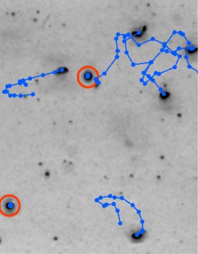
NATURE COMMUNICATIONS | https://doi.org/10.1038/s41467-020-19094-0                                                                      ARTICLE

Fig. 1 NK cell expansion and NK cell function in standard assays. a NK cell       embedding in collagen) to 20% (after 48 h; Supplementary
viability of fresh (blue) and cryopreserved (red) NK cells as measured by         Fig. 3a). These data demonstrate that the fraction of motile
flow cytometry (n = 9; p = 0.36 by two-sided nonparametric Wilcoxon                cryopreserved NK cells does not fully recover even after 48 h, and
signed-rank test for paired data). b Fraction of NK cells (single-hatched)        remains approximately threefold lower compared to fresh cells.
and cytotoxic NK cells (double-hatched) within PBMCs before expansion                To explore if the negative effect of cryopreservation on 3-D cell
(Day 0; white bars), and after expansion (Day 14) for fresh (blue) and            migration is specific for the expansion protocol used in this study,
cryopreserved (red) samples (n = 10). The increase in the fraction of NK          we measure the migration of the NK cell line NK92, instead of
cells and cytotoxic NK cell after expansion is significant (p = 0.002 for both     NK cells expanded from primary PBMCs. NK92 cells migrate in
conditions by two-sided nonparametric Wilcoxon signed-rank test for               collagen at a similar speed, but slightly less persistent compared
paired data). Cryopreservation does not decrease the fraction of NK cells         to expanded NK cells. Importantly, we find that the motile
(p = 0.49), but decreases the fraction of cytotoxic NK cells (p = 0.004 by        fraction of NK92 cells decreases from 31 to 6% after
two-sided nonparametric Wilcoxon signed-rank test for paired data). c             cryopreservation, similar to our findings for expanded NK cells
Percentage of CD107a-expressing NK cells for different NK to target cell          (Supplementary Fig. 4a).
ratios, as measured in a degranulation assay (n = 8). Control                        Moreover, to explore if the negative effect of cryopreservation
measurements are conducted in the absence of target cells. PMA/Iono is            on 3-D migration is specific for a 3-D collagen network, we
added for positive control experiments. Differences in the degranulation          suspend fresh and cryopreserved NK cells in carbomer, a
between fresh and cryopreserved NK cells do not reach statistical                 hydrogel-forming polymer based on acrylic acid. Carbomer
significance (−Ctrl: p = 0.055, +Ctrl: p = 0.74, 20:1: p = 0.74, 5:1:              forms ~10 µm colloidal particles that are jammed to a viscoelastic
p = 0.38; two-sided nonparametric Wilcoxon signed-rank test for paired            hydrogel (yield stress 10 Pa) reminiscent of a cell pellet. In
data). d NK cell cytotoxicity as measured in a chromium-release assay for         contrast to collagen, carbomer does not support the adhesion of
different NK to target cell ratios (n = 10). Differences between fresh to         cells to the matrix and only allows the cells to migrate in an
cryopreserved NK cells reach statistical significance for all conditions (20:1:    ameboid mode. Despite the pronounced mechanical and
p = 0.014, 10:1: p = 0.006, 5:1: p = 0.004, 2.5:1: p = 0.002; Spearman’s          structural differences between both hydrogels, the motile fraction,
rank-order correlation). For fresh NK cells, there is no significant correlation   migration speed, and persistence of fresh NK cells is only slightly
between NK-to-target cell ratio and cytotoxicity (correlation coefficient          lower in carbomer compared to collagen (Supplementary Fig. 5).
ρ = 0.14, p = 0.38; Spearman’s rank-order correlation), but for                   Importantly, the motile fraction after cryopreservation is strongly
cryopreserved NK cells, cytotoxicity decreases for lower NK-to-target cell        reduced in carbomer, to a similar degree as in collagen,
ratios (correlation coefficient ρ = 0.41, p = 0.009; Spearman’s rank-order         demonstrating that the negative effect of cryopreservation on 3-
correlation). Error bars denote 1 sem. Source data are provided as a Source       D migration is of a more general nature. This finding is also
data file.                                                                         consistent with the above argument that the reduced cytotoxicity
                                                                                  after cryopreservation, as measured in a chromium-release assay
                                                                                  is largely attributable to a reduced motile fraction of the NK cells
                                                                                  in a cell pellet.
same time, those NK cells would need to migrate over larger                          As chromium-release assays and motility assays are performed
distances to reach target cells. For fresh NK cells, by contrast, we              pair wise, we can quantify the relationship between cytotoxicity
find no statistically significant correlation between cytotoxicity                  and motile cell fraction in 3-D collagen gels for individual
and NK-to-target ratio (Fig. 1d), indicating that fresh NK cells                  samples (specified by donor and expansion). We find that
can compensate for a decreasing NK-to-target cell ratio, but                      cytotoxicity increases with motile fraction according to a power-
cryopreserved NK cells cannot. By this rationale, it would be                     law relationship with exponents ranging from 0.2 to 0.4. This
expected that at earlier time points after mixing NK cells and                    relationship reaches statistical significance, with a coefficient of
target cells in a cell pellet, serial killing and migration are less              determination of R2 ≥ 0.42 for all NK-to-target cell ratios between
important, and the chromium-release cytotoxicity of fresh and                     20:1 and 2.5:1 (Fig. 3b). This finding adds further support to the
cryopreserved cells equalizes cytotoxicity similar to the low NK-                 notion that NK cells are able to kill target cells beyond their
to-target ratios. We find this to be true for up to 1 h after cell                 immediate neighbors in a cell pellet (e.g., in a chromium-release
mixing (Supplementary Fig. 1).                                                    assay), for which the ability to migrate is of advantage.
   Since the chromium-release assay only reports the decreased
cytotoxicity, but cannot measure NK cell motility directly, we
perform additional assays for which NK cells are embedded in 3-                   Motile NK cell fraction retains killing efficiency. We next
D reconstituted collagen gels (Fig. 2). To assess the fraction of NK              investigate whether the small remaining population of motile NK
cells that are motile in this 3-D environment, we perform z-scans                 cells after cryopreservation retains full cell function. First, we
through a 1 mm thick gel every 30 s, and track NK cell positions                  characterize cell speed and directional persistence of fresh and
by their appearance in minimum intensity projections (Fig. 2a–c,                  cryopreserved motile NK cells within 3-D collagen gels, and find no
see “Methods” section). We find that NK cell motility after                        significant difference (Fig. 4a, b, and Supplementary Figs. 2b, c, 3b, c
cryopreservation in a 3-D environment is dramatically impaired                    and 5b, c). Thus, NK cells that remain motile after cryopreservation
after cryopreservation. Specifically, we find that 29.2% of fresh                   retain their normal exploration behavior within tissue.
NK cells are motile, in line with reported findings for 2-D                           Second, we evaluate the cytotoxic function of the motile NK
migration21, while only 4.9% of cryopreserved NK cells are                        cell fraction in 3-D collagen gels by identifying individual K562
motile, corresponding to a sixfold decrease in motility (Fig. 3a                  target cell killing events in time-lapse image series (Fig. 2d, e,
and Supplementary Fig. 2a).                                                       Supplementary Movies 1 and 3, and see “Methods” section). We
   To more closely mimic the situation in the human body after                    confirm by microscopy inspection that each killed K562 cell was
adoptive transfer of expanded NK cells, we monitor the migration                  attacked by an NK cell during the last 60 min before cell death.
behavior of fresh and cryopreserved NK cells from three subjects                  The number of live target cells decreases exponentially over time,
over 48 h in the absence of IL-2. The motile fraction of                          which can be modeled by a first-order differential equation (see
cryopreserved NK cells increases from 2 (6 h after thawing, 4 h                   Eq. (1) in “Methods” section). The characteristic time constant of
after embedding in collagen) to 7% (after 48 h), whereas the                      the exponential decay, which reports the average killing rate and
motile fraction of fresh cells decreases from 36 (4 h after                       hence the cytotoxicity of the motile NK cells, can be estimated

NATURE COMMUNICATIONS | (2020)11:5224 | https://doi.org/10.1038/s41467-020-19094-0 | www.nature.com/naturecommunications                               3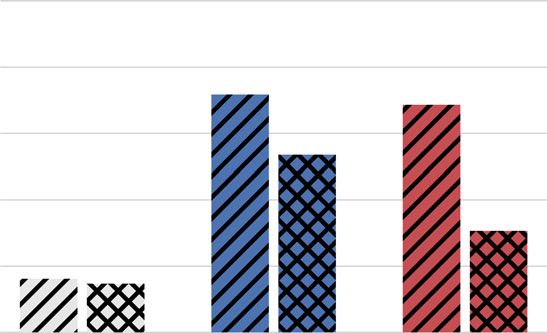
ARTICLE                                                                   NATURE COMMUNICATIONS | https://doi.org/10.1038/s41467-020-19094-0

                        a                                         b                                c

                        d

                        e

Fig. 2 NK cell motility assay and cytotoxicity assay in a 3-D collagen gel. a Minimum intensity projections along the x/y/z-axes of a bright-field image
stack of NK cells migrating through a 3-D collagen gel. In the projections, the NK cells appear as dark spots. b Exemplary minimum intensity projection
along the z-axis, taken from one out of ten similar, independent experiments. Cell trajectories over 30 min are indicated in blue, nonmotile cells are marked
red (scale bar: 50 µm). c False-color representation of the z-position of every pixel in the minimum projection. This information is used to estimate the
z-position of motile NK cells (cell outlines are indicated in black). d Time-lapse image sequence of an NK cell-mediated killing of a K562 tumor cell, taken
from one out of six similar, independent experiments. e Same as d, with colored cell morphologies as a guide for the eye.

                             Fresh
                             Cryopreserved
                                              b
                       a

Fig. 3 Influence of 3-D NK cell motility on cytotoxicity. a Motile fraction of fresh (blue) and cryopreserved (red) NK cells from n = 10 independent
experiments from five subjects and five different expansions in 1.2 mg/ml collagen gel (p = 0.002; two-sided nonparametric Wilcoxon signed-rank test for
paired data). Each symbol represents mean ± se from cells measured in five field of view, with ~80 cells (motile plus nonmotile) in each field of view. In
total, n = 1248 fresh motile cells and n = 122 cryopreserved motile NK cells were measured. Paired (fresh vs. cryopreserved) data from each subject and
expansion are connected by lines. b NK cell cytotoxicity (as measured in a chromium-release assay) as a function of the motile NK cell fraction for fresh
(blue) and cryopreserved (red) NK cells. Different NK-to-target cell ratios are noted above each graph. Dashed lines indicate a power-law fit of the form f
(x) = a·xb. R2 values are computed in log–log space. Statistical significance assuming a power-law exponent of b = 0 as null hypothesis: 20:1: p = 0.0013;
10:1: p = 0.0028; 5:1: p = 0.0013; 2.5:1: p = 0.00045 by two-sided nonparametric Wilcoxon signed-rank test for paired data. Source data are provided as a
Source data file.

from the number of live target cells at the beginning and the end               (Fig. 4d). Therefore, our data demonstrate that NK cells that
of the experiment (see Eq. (2) in “Methods” section and Fig. 4c).               remain motile after cryopreservation retain their cytotoxic cell
Note that the killing rate of an individual NK cell cannot be                   function. However, if we compute a composite killing efficiency
determined in this assay as the cells are free to migrate into and              that considers both motile and nonmotile NK cells, we find a
out of the microscope’s field of view during the observation                     significant decrease of effective cytotoxicity in a 3-D environment
period. Normalizing this exponential decay rate to 1 h, as well as              by a factor of 5.6 (Fig. 4e), which directly reflects the sixfold
to one motile NK cell and one living target cell per 106 µm3                    decrease in NK cell motility.
volume of tissue (corresponding to a cube of 100 µm length), we
arrive at a measure of killing efficiency that is not biased by
varying cell concentrations or fractions of motile cells (see Eq. (3)           Discussion
in “Methods” section and Supplementary Fig. 6). We find a 26%                    In this study, we report that ex vivo-expanded NK cells retain
decrease of cytotoxicity in a 3-D environment after cryopreserva-               their ability to induce target cell death after cryopreservation (as
tion, but this difference is smaller than the difference detected in            measured in a degranulation assay), but suffer a significant
the classic cytotoxicity assays and is not statistically significant             decrease of their cytotoxic function (as measured in a chromium-

4                  NATURE COMMUNICATIONS | (2020)11:5224 | https://doi.org/10.1038/s41467-020-19094-0 | www.nature.com/naturecommunications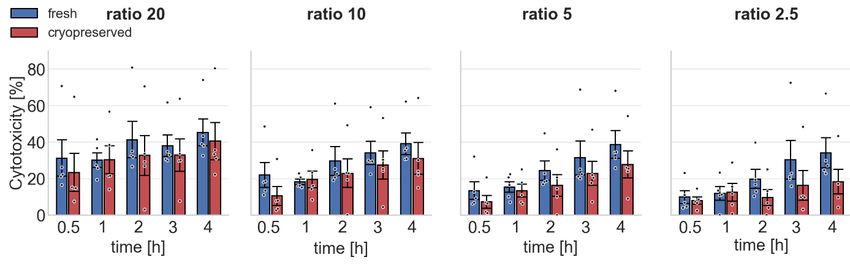
NATURE COMMUNICATIONS | https://doi.org/10.1038/s41467-020-19094-0                                                                                ARTICLE

              a                       b                       c                                       d                          e

Fig. 4 NK cell migration and cytotoxicity in 3-D collagen gels. a Migration cell speed of fresh (blue) and cryopreserved (red) NK cells from n = 10
independent experiments from five subjects and five different expansions in a 3-D collagen gel. Each symbol represents mean ± se (measured in the x–y-
imaging plane) from cells measured in five field of view, with ~80 cells (motile plus nonmotile) in each field of view. Migration cell speed and persistence is
computed from on average n = 125 motile fresh cells and n = 12 motile cryopreserved cells for each subject and expansion (each data point). In total,
n = 1248 motile fresh and n = 122 motile cryopreserved NK cells were measured. Paired (fresh vs. cryopreserved) data from each subject and expansion
are connected by lines. a Cell speed (p = 0.076; two-sided nonparametric Wilcoxon signed-rank test for paired data). b Migration directional persistence
(p = 1.0; two-sided nonparametric Wilcoxon signed-rank test for paired data). c Number of live K562 target cells (black line) in a representative
experiment as a function of measurement time. Killing events are marked by green circles. The dashed green line indicates an exponential fit of the
individual killing events, the black dashed line indicates an approximation using an exponential curve based only on the number of live targets at the
beginning and the end of the measurement time. d Estimated killing efficiency (weighted mean ± se determined by bootstrapping) of fresh and
cryopreserved motile NK cells against K562 target cells embedded in a 3-D collagen gel (p = 0.56; two-sided bootstrapping; n = 6 independent
experiments from four subjects and three different expansions). e Estimated composite killing efficiency (weighted mean ± se determined by
bootstrapping) of fresh and cryopreserved NK cells (considering both motile and nonmotile NK cells) against K562 target cells embedded in a 3-D collagen
gel (p = 0.025; two-sided bootstrapping; n = 6). Note that the killing efficiency of the individual experiments is not shown in d, e as the reported mean
values are weighted by the respective cell concentrations (see “Methods” section). Individual values are provided as a Source data file.

release assay). By complementing standard assays of NK cell                    of NK cells and tumor cell killing events in a 3-D matrix may
function with measurements of NK cell motility and cytotoxicity                serve as a better predictor of NK cell function compared to
in a 3-D environment, we are able to attribute this decrease of                standard cytotoxicity assays.
cytotoxic function to an impairment of NK cell motility. Speci-
fically, we find a dramatic sixfold decrease in the fraction of                  Methods
motile NK cells after cryopreservation. We further show that the               PBMC isolation and storage. NK cells are generated from PBMCs from 11
small remaining population of motile NK cells retains its cyto-                healthy donors (Department of Transfusion Medicine, University Hospital Erlan-
toxicity after cryopreservation.                                               gen, Germany; IRB approval number 147_13B). We have obtained informed
                                                                               consent from all participants. Specifically, leukocyte reduction system chambers are
   NK cells are generally cryopreserved using 10–20% dimethyl                  obtained from the department of transfusion medicine, from which PBMCs are
sulfoxide (DMSO) solution as a cryoprotectant at a slow freezing               isolated. PBMCs are resuspended in human serum albumin (Baxter) at a con-
rate of −1 °C/min (ref. 22). Several groups have reported changes              centration of 5–10 × 106 cells/ml. A total of 500 µl of cell suspension is filled into a
in cell viability and/or function in non-expanded (primary) NK                 1 ml cryovial (Nalge, Nunc), and 500 µl of freezing medium (55.5 vol% human
                                                                               serum albumin, 25.0 vol% DMSO, 8% (w/v) glucose) is added. After gently mixing,
cells after cryopreservation. Specifically, the NK cell compartment             the vials are transferred into a freeze container (Mr. Frosty, Thermo Scientific,
in PBMC loses cytotoxic function after thawing, while retaining                which allow for a cooling rate of −1 °C/min) and stored at −80 °C. Cells are used
cell viability23,24. Likewise, purified primary NK cells also show              within 65 days after cryopreservation.
reduced cytotoxicity after cryopreservation, although cytotoxicity
is significantly improved after an overnight recovery culture in                NK cell expansion. After thawing the PBMC samples (which includes NK, NKT,
the presence of IL-2 (refs. 16,25). Treatment of patients with IL-2            and T cells), cells are expanded in the presence of irradiated K562-mbIL15-41BBL
                                                                               feeder cells (gift from Prof. D. Campana, Department of Pediatrics, University
after adoptive transfer of NK cells, however, did not translate into           Hospital Singapore; formerly St. Jude Children’s Research Hospital, Memphis, TN,
clinical success beyond the effect of IL-2 alone7,10. In addition,             USA) for 14 days in RPMI 1640 medium supplemented with 10% fetal bovine
non-expanded NK cells may be susceptible to cryopreservation-                  serum, 20 µg/ml gentamycin and 1% L-glutamine (hereafter called cRPMI), and
induced phenotype changes, since cryopreservation induced a                    200 IU/ml human IL-2 cytokine28. K562 feeder cells are confirmed negative for
transient increase in CD56+ CD16− NK cells in peripheral blood                 mycoplasma contamination using the Venor GeM Classic detection kit (Minerva
                                                                               Biolabs). This expansion process is performed five times in the case of 1 donor,
from hematopoietic stem cell transplantation recipients26. This                three times in the case of 3 donors, two times in the case of 3 donors, and one time
phenotypic change is similar to the CD16 decline we noted after                in the case of the remaining 4 donors, resulting in a total of 24 donor/expansion
freezing and thawing, which may be the net result of activation-               combinations.
induced cleavage of CD16 (ref. 19). Thus far, attempts to optimize
current NK cell cryopreservation protocols have had limited                    NK92 cell line. NK-92 cells (purchase from ATCC CRL-2407) are cultured for
success16,27.                                                                  3 weeks prior to measurements in Alpha-MEM medium (Stemcell Technologies)
                                                                               with 15% fetal calf serum, 15% horse serum, 500 IU/ml human IL2-cytokine, and
   Overall, the presented findings point to the cryopreservation                1% penicillin–streptomycin solution (10,000 units/ml penicillin, 10,000 µg/ml
step as a major cause for the current failure of NK cell therapy in            streptomcycin).
patients with solid tumors. In the future, this problem may be
solved by sterile expansion of NK cells in a closed bioreactor that            Cryopreservation and thawing. Expanded NK cell aliquots (5–10 × 106 cells/ml)
circumvents the cryopreservation step altogether. Moreover, our                are either directly measured (in the following referred to as “fresh”), or are frozen,
data show that the fraction of motile NK cells largely determines              thawed, and then measured (in the following referred to as “cryopreserved”). We
the effective cytotoxicity, and is therefore a crucial parameter               conduct paired experiments (fresh versus cryopreserved) for all assays, but due to
                                                                               technical difficulties in some of the assays, the number of paired experiments varied
when deciding on the number of cells to be administered to the                 as noted in the figure legends. Freezing of expanded NK cells is performed as
patient. Finally, this work demonstrates that time-lapse imaging               described above for PBMCs. On the following day, cryopreserved expanded NK

NATURE COMMUNICATIONS | (2020)11:5224 | https://doi.org/10.1038/s41467-020-19094-0 | www.nature.com/naturecommunications                                            5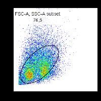
ARTICLE                                                                               NATURE COMMUNICATIONS | https://doi.org/10.1038/s41467-020-19094-0

cells are rapidly thawed in a 37 °C water bath until a small visible ice chip remains,      cross in the minimum intensity projection images. In a similar way, we compute
and then are dropwise transferred into 10 ml of cRPMI, which is preheated to                the maximum intensity projection.
37 °C. Cells are centrifuged at 300 × g for 5 min and resuspended in cRPMI.                     For analysis, we detect individual NK cells using a convolutional neural network
                                                                                            that is trained on 70 manually labeled minimum/maximum intensity projections
                                                                                            with ~100 cells in each projection. We assume a minimum diameter of nonmotile
Flow cytometry. Cell viability of fresh and cryopreserved NK cells is assessed by           NK cells of 4 µm to separate them from smaller cell fragments (Supplementary
staining with the Zombie NIR dye (dilution 1:1000; Biolegend). Fresh and cryo-              Fig. 9 and Supplementary Movie 3). In addition, we perform data augmentation by
preserved NK cells are phenotypically characterized as described in refs. 28,29 by          image flipping and zooming. The network is based on the U-Net architecture31.
staining with directly conjugated mouse anti-human antibodies against CD3 (clone            The labeling accuracy of the network is 94% (F1-score), which is comparable to the
UCHT1; dilution 1:50; Biolegend), CD56 (clone HCD56; dilution 1:50; Biolegend),             manual labeling accuracy of the investigators involved in this study
and CD16 (3G8; dilution 1:50; Biolegend). NK cells are defined as CD3− and                   (n = 4 subjects). We then connect the x,y positions of detected NK cells between
CD56+ cells (Supplementary Fig. 7). A minimum of 10,000 cells are analyzed using            subsequent images to obtain migration trajectories (Fig. 2b and Supplementary
a BD Canto II flow cytometer (BD Biosciences) and Flowjo Software (FLOWJO,                   Movie 2). An NK cell is classified as motile if it moves away from its starting point
LLC Data analysis software).                                                                by ≥13 µm within 5 min. The cell speed is determined as the diagonal of the
                                                                                            bounding box of each cell trajectory, divided by the measurement time of 5 min.
                                                                                            Directional persistence is determined as the average cosine of the turning angles
CD107a degranulation assay. A total of 1 × 10⁶ expanded NK cells are incubated              between consecutive cell movements. Zero persistence corresponds to random
for 6 h at 37 °C, 5% CO2, 95% RH with cells from the myeloid cell line K562 (gift           motion, whereas a persistence of unity corresponds to ballistic motion.
from Dr. J.J. Bosch, Department of Medicine 5, University Hospital Erlangen) at an              As the expanded cell populations contain small fractions of other cell types (as
NK-to-K562 cell ratio of 20:1 and 5:1 in a final volume of 500 μl cRPMI supple-              measured with flow cytometry, Fig. 1b), we perform confocal microscopy of an
mented with anti-CD107a antibody (clone H4A3, 10 µl/ml, BD Biosciences). K562               expanded cell population that is stained using CD56-APC and CD3-Alexa488
cells are confirmed negative for mycoplasma contamination. To prevent protein                antibodies, and is embedded in a 3-D collagen gel. We find that 82% of all motile
secretion and degradation of internalized CD107a, monensin (1 µM) and brefeldin             cells are NK cells (Supplementary Fig. 10).
A (10 ng/ml, both from Sigma) are added after 1 h of incubation. NK cells alone
serve as a negative control, and NK cells stimulated for 6 h with phorbol 12-
myristate 13-acetate (PMA, 50 ng/ml) and ionomycine (250 ng/ml, both from                   3-D cytotoxicity assay. We mix 600,000 fresh or cryopreserved NK cells together
Sigma) serve as a positive control for anti-CD107a antibody binding. After 6 h of           with 120,000 K562 cells in 2.5 ml of collagen (1.2 mg/ml), as described for the 3-D
incubation, cells are harvested, washed, resuspended in 50 μl PBS, and stained with         motility assay. z-stack time-lapse imaging is performed at four positions in each
live–dead Zombie NIR (BioLegend), anti-CD56 (clone CHD56, BioLegend), and                   well in parallel, at a rate of two frames per min for 15 h in total.
CD16 antibody (clone 3G8, BioLegend). Samples are analyzed using a Becton                       To automatically detect the number of motile NK cells, as well as the number of
Dickinson FACS CANTOII flow cytometer and Flowjo software.                                   live and dead K562 cells in each frame, we train a convolutional neural network
                                                                                            with 48 minimum and maximum intensity projection images, in which all motile
                                                                                            and nonmotile NK cells, as well as live and dead K562 cells are manually labeled.
Chromium-release assay. K562 cells are labeled with radioactive (150 μCi, 5.55              Living K562 cells are characterized by a round appearance and small (1–2 µm)
MBq) sodium chromate (20 µl/condition, 5 mCi/ml, Perkin Elmer) for 1 h. After               wiggling motion. When a K562 cell, after being contacted by an NK cell, starts to
incubation, cells are washed two times and incubated for an additional 30 min to            bleb, change shape, shrink, expand, stop wiggling, or change its bright-field
reduce spontaneous chromium release. Labeled cells are then plated at a density of          contrast due to refractive index changes, we consider the cell as dead or as
5000 cells/well in 100 µl cRPMI in a 96-well U-bottom plate. Fresh expanded or              undergoing lysis or apoptosis (Fig. 2d, e and Supplementary Movie 3). We
cryopreserved NK cells are added at NK-to-target cell ratios of 20:1, 10:1, 5:1, and        complement this manually labeled data set with 11 images in which K562 cells are
2.5:1 to give a final volume of 200 μl per well. After 0.5, 1, 2, 3, or 4 h of incubation,   fluorescently labeled using Hoechst 33342 dye (incubation with 2.5 µg/ml for
100 μl supernatant is mixed with 100 μl scintillation cocktail (Perkin Elmer) in a          20 min followed by two washing steps in PBS), 11 images in which NK cells are
96-well sample plate (Perkin Elmer). Release of radioactive chromium-51 is                  fluorescently labeled using Hoechst 33342 dye, and one image of dead K562 cells in
measured using a gamma-counter (Perkin Elmer), and the fraction of lysed target             which cell death is induced by adding 0.01% TritonX. The complete data set is used
cells is calculated as the ratio of (experimental release − spontaneous release)/           to train a convolutional neural network based on the U-Net architecture31. As
(maximum release − spontaneous release). Spontaneous release is measured from               nonmotile NK cells are smaller, but otherwise appear similar to K562 cells, we only
5000 labeled K562 cells without addition of NK cells, and maximum release is                consider K562 cells that are larger than 14 µm in diameter for the analysis
measured from 5000 labeled K562 cells that are lysed with 100 µl 1% Nonidet P-40            (Supplementary Fig. 9). An example demonstrating the performance of the
(Sigma). All experiments are performed in triplicates.                                      automated detection and labeling is shown in Supplementary Movie 3. For
                                                                                            evaluating NK cytotoxicity, each NK cell-mediated killing event identified by the
                                                                                            network is visually inspected and confirmed, using the image-annotation software
3-D cell motility assay. We suspend 150,000 fresh or cryopreserved NK cells in              ClickPoints32.
2.5 ml of a 1.2 mg/ml collagen solution or in 2.5 ml of 9 mg/ml carbomer hydrogel               To evaluate the accuracy of determining target cell death in bright field images,
(Ashland 980 Carbomer, Covington, USA) in each well of a tissue-culture treated             we add the dye NucRed Dead 647 (Ready Probes, Thermo Fisher) to the media
six-well plate (Corning). The collagen solution is prepared from a 2:1 mixture of rat       immediately after the polymerization of the collagen gel and also at the end of the
tail collagen (Collagen R, 2 mg/ml, Matrix Bioscience) and bovine skin collagen             measurement (Supplementary Fig. 11). At the beginning and at the end of the 15 h
(Collagen G, 4 mg/ml, Matrix Bioscience). We add 10% (vol/vol) sodium bicar-                measurement period, an image stack is recorded. All K562 cells that are classified as
bonate (23 mg/ml) and 10% (vol/vol) 10× RPMI (Gibco). For a final collagen                   living based on the bright-field criteria are stained negative. Therefore, the false-
concentration of 1.2 mg/ml, we dilute the solution before polymerization with a             negative error rate is 0% (none of the dead cells are labeled falsely as living).
mixture of one volume part NaHCO3, one part 10× cRPMI, and eight parts H2O                  However, from the K562 cells which are classified as dead based on the bright-field
(ref. 30) and adjust the solution to pH 9 with NaOH. After polymerization at 37 °C,         criteria listed above, 10.6% are stained negative, possibly because they still have an
5% CO2, and 95% RH for 1 h, 1.5 ml of RPMI medium (for primary NK cells) or                 intact cell membrane despite undergoing apoptosis. Therefore, we potentially
1.5 ml of Alpha-MEM medium (for NK92 cells) is added to each well of a six-                 overestimate the killing rate (see below) by up to 10%.
well plate.                                                                                     We describe the decline in the concentration of live K562 cells due to NK cell-
    Carbomer hydrogel is prepared by mixing carbomer powder with RPMI 1640                  mediated killing during the 15 h observation time as a first-order enzyme reaction
medium (9 mg/ml). The pH is titrated to a value of 7.4 with 10 M NaOH. After                of the form ½NK þ ½K562live  !k ½NK þ ½K562dead , whereby the NK cells serve as
mixing-in the cells, the migration assay is started without a waiting period. In the        the enzyme catalyzing this reaction, and k is the reaction rate (the killing
case of cells embedded in collagen, we add a waiting period of 3 h to ensure that the       efficiency). We assume that k remains constant and that the cytotoxicity of NK cells
NK cells have adapted to the collagen gel, attained their characteristic elongated          does not exhaust over time, which is an oversimplification, but justified by the high
shape and recovered their full migratory potential (Supplementary Fig. 8). For the          ratio of NK cells to target cells. Moreover, we consider the average concentration of
migration assay, the six-well plate is transferred to a motorized microscope                motile NK cells ([NK]) as being constant throughout the measurement. Finally, we
(Applied Scientific Instrumentation, USA, equipped with a 10× 0.3 NA objective               assume that the reverse reaction does not take place, implying that K562 cells do
(Olympus) and an Infinity III CCD camera, Lumenera) that is placed inside an                 not recover once they are undergoing apoptosis or lysis. Therefore, if a K562 cell
incubator, and time-lapse imaging is started. We perform z-scans (10 µm apart)              forms small temporary blebs that disappear after a while, so that the cell appears
through the 1 mm thick gel every 30 s for a duration of 5 min. Afterward, another           alive for the remainder of the experiment, this cell is counted as living.
randomly chosen position is selected, and time-lapse imaging continues. In total,               The dynamics of the above killing reaction can be described with a first-order
six positions are imaged in each well. Fast image acquisition of z-scans is achieved        differential equation, which has the solution
by taking images at 27.5 frames per second, while the focal plane of the microscope
is moving with a constant speed through the gel along the z-axis. We do not store                                       ln½K562live;t¼0   ln½K562live;t¼T
the entire z-stack of images, but instead store the lowest intensity value of each                                 k¼                                         :               ð1Þ
                                                                                                                                  ½NKmotile   T
pixel along the z-direction (minimum intensity projection, Fig. 2a, b), and the
z-position where the lowest intensity value is found (Fig. 2c). The z-position                 The only free parameter is the killing efficiency k that we compute from the
information aids in the tracking of two individual cells when their paths seem to           concentration (# of cells per (100 µm)3 of gel volume) of live K562 cells at the

6                     NATURE COMMUNICATIONS | (2020)11:5224 | https://doi.org/10.1038/s41467-020-19094-0 | www.nature.com/naturecommunications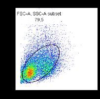
NATURE COMMUNICATIONS | https://doi.org/10.1038/s41467-020-19094-0                                                                                             ARTICLE

beginning (t = 0) and the end of the experiment (t = T), and the average                 12. Rubio, V. et al. Ex vivo identification, isolation and analysis of tumor-cytolytic
concentration of motile NK cells.                                                            T cells. Nat. Med. 9, 1377–1382 (2003).
   As individual experiments i have different cell concentrations, we compute            13. Li, Q. Natural killer (NK) cell assays in immunotoxicity testing. Methods Mol.
weighted mean killing efficiencies                                                            Biol. 1803, 231–241 (2018).
                           P                                                             14. Lin, W. et al. Fc-dependent expression of CD137 on human NK cells: insights
                              i ki  ½K562live;t¼0 i  ½NKmotile i
                    kavg ¼ P                                         ;        ð2Þ            into ‘agonistic’ effects of anti-CD137 monoclonal antibodies. Blood 112,
                                 i ½K562live;t¼0 i  ½NKmotile i                           699–707 (2008).
for both fresh and cryopreserved NK cells. Below, we also report an apparent or          15. Domogala, A., Alejandro Madrigal, J. & Saudemont, A. Cryopreservation has
composite killing efficiency for all NK cells regardless of their ability to move,            no effect on function of natural killer cells differentiated in vitro from
which is computed according to                                                               umbilical cord blood CD34+ cells. Cytotherapy 18, 754–759 (2016).
                               ln½K562live;t¼0   ln½K562live;t¼T                      16. Miller, JeffreyS., Rooney, ClionaM., Curtsinger, Julie & Expansion, R. M.
                   kcomposite ¼                                      :            ð3Þ        Expansion and homing of adoptively transferred human NK cells in
                                ð½NKmotile  þ ½NKnonmotile Þ  T
                                                                                             immunodeficient mice varies with product preparation and in vivo cytokine
    Since only motile NK cells can reach their target cell to kill, this composite           administration: implications for clinical therapy. Biol. Blood Marrow Transpl.
killing rate combines cytotoxicity and motility in one parameter.                            20, 1252–1257 (2014).
                                                                                         17. Wiesinger, M. et al. Clinical-scale production of car-t cells for the treatment of
Statistical tests. Unless noted otherwise, all statistical tests use the two-sided           melanoma patients by mrna transfection of a cspg4-specific car under full gmp
nonparametric Wilcoxon signed-rank test for paired data. The correlation between             compliance. Cancers 11, 1–18 (2019).
NK-to-target ratio and cytotoxicity reported in Fig. 1d is determined by Spear-          18. Wiesinger, M. et al. Good manufacturing practice-compliant production and
man’s rank-order correlation; the corresponding test statistic is based on a t-              lot-release of ex vivo expanded regulatory T cells as basis for treatment of
distribution to account for the small sample size and to avoid over-rejection.               patients with autoimmune and inflammatory disorders. Front. Immunol. 8,
Significant differences for the killing efficiency reported in Fig. 4d, e are determined       1371 (2017).
by bootstrapping (two-sided test). Differences are considered as statistically sig-      19. Grzywacz, B., Kataria, N. & Verneris, M. R. CD56dimCD16+ NK cells
nificant for p < 0.05.                                                                        downregulate CD16 following target cell induced activation of matrix
                                                                                             metalloproteinases [4]. Leukemia 21, 356–359 (2007).
Reporting summary. Further information on research design is available in the Nature     20. Vanherberghen, B. et al. Classification of human natural killer cells based on
Research Reporting Summary linked to this article.                                           migration behavior and cytotoxic response. Blood 121, 1326–1334 (2013).
                                                                                         21. Olofsson, P. E. et al. Distinct migration and contact dynamics of resting and
                                                                                             IL-2-activated human natural killer cells. Front. Immunol. 5, 1–10 (2014).
Data availability                                                                        22. El Assal, R. et al. Bioinspired preservation of natural killer cells for cancer
Data supporting the findings of this study are available within the article and               immunotherapy. Adv. Sci. 6, 1802045 (2019).
its Supplementary information files or from the corresponding author upon reasonable      23. Dominguez, E., Lowdell, M. W., Perez-Cruz, I., Madrigal, A. & Cohen, S. B. A.
request. Source data are provided with this paper.                                           Natural killer cell function is altered by freezing in DMSO. Biochem. Soc.
                                                                                             Trans. 25, 997 (1997).
Code availability                                                                        24. Mata, M. M., Mahmood, F., Sowell, R. T. & Baum, L. L. Effects of
The software code for data analysis is available from the corresponding author on            cryopreservation on effector cells for antibody dependent cell-mediated
request.                                                                                     cytotoxicity (ADCC) and natural killer (NK) cell activity in 51Cr-release and
                                                                                             CD107a assays. J. Immunol. Methods 406, 1–9 (2014).
                                                                                         25. Voshol, H., Dullens, H. F. J. & Otter, W. Den & Vliegenthart, J. F. G. Human
Received: 18 December 2019; Accepted: 24 September 2020;                                     natural killer cells: a convenient purification procedure and the influence of
                                                                                             cryopreservation on cytotoxic activity. J. Immunol. Methods 165, 21–30
                                                                                             (1993).
                                                                                         26. Lugthart, G. CD56dimCD162 NK cell phenotype can be induced by
                                                                                             cryopreservation natural. Blood 31, 254–256 (2015).
                                                                                         27. Pasley, S., Zylberberg, C. & Matosevic, S. Natural killer-92 cells maintain
References                                                                                   cytotoxic activity after long-term cryopreservation in novel DMSO-free media.
1.  Carrega, P. & Ferlazzo, G. Natural killer cell distribution and trafficking in            Immunol. Lett. 192, 35–41 (2017).
    human tissues. Front. Immunol. 3, 1–6 (2012).                                        28. Voskens, C. J. et al. Ex-vivo expanded human NK cells express activating
2. Multhoff, G. et al. Adoptive transfer of human natural killer cells in mice with          receptors that mediate cytotoxicity of allogeneic and autologous cancer cell
    severe combined immunodeficiency inhibits growth of Hsp70-expressing                      lines by direct recognition and antibody directed cellular cytotoxicity. J. Exp.
    tumors. Int. J. Cancer 88, 791–797 (2000).                                               Clin. Cancer Res. 29, 1–13 (2010).
3. Tong, A. A. et al. Adoptive natural killer cell therapy is effective in               29. Voskens, C. J. et al. Characterization and expansion of autologous GMP-ready
    reducing pulmonary metastasis of Ewing sarcoma. Oncoimmunology 6, 1–8                    regulatory T cells for TREG-based cell therapy in patients with ulcerative
    (2017).                                                                                  colitis. Inflamm. Bowel Dis. 23, 1348–1359 (2017).
4. Veluchamy, J. P. et al. The rise of allogeneic Natural killer cells as a platform     30. Cóndor, M., Steinwachs, J., Mark, C., García-Aznar, J. M. & Fabry, B. Traction
    for cancer immunotherapy: Recent innovations and future developments.                    force microscopy in 3-dimensional extracellular matrix networks. Curr.
    Front. Immunol. 8, 631 (2017).                                                           Protoc. Cell Biol. 2017, 10.22.1–10.22.20 (2017).
5. Leivas, A. et al. Novel treatment strategy with autologous activated and              31. Ronneberger, O., Fischer, P. & Brox, T. U-net: convolutional networks for
    expanded natural killer cells plus anti-myeloma drugs for multiple myeloma.              biomedical image segmentation. Lect. Notes Comput. Sci. 9351, 234–241
    Oncoimmunology 5, e1250051 (2016).                                                       (2015).
6. Shah, N. N. et al. R-CHOP versus dose-adjusted R-EPOCH in frontline                   32. Gerum, R. C., Richter, S., Fabry, B. & Zitterbart, D. P. ClickPoints: an
    management of primary mediastinal B-cell lymphoma: a multi-centre analysis.              expandable toolbox for scientific image annotation and analysis. Methods Ecol.
    Br. J. Haematol. 180, 534–544 (2018).                                                    Evol. 8, 750–756 (2017).
7. Rosenberg, S. A. et al. Prospective randomized trial of high-dose interleukin-2
    alone or in conjunction with lymphokine-activated killer cells for the
    treatment of patients with advanced cancer. J. Natl Cancer Inst. 85, 622–632         Acknowledgements
    (1993).                                                                              This work was supported by the National Institutes of Health grant HL120839, the DFG
8. Burns, L. J. et al. IL-2-based immunotherapy after authologous transplantation        grant ME 1260/11-1, the DFG Research Training Group 1962 (“Dynamic interactions at
    for lymphoma and breast cancer induces immune activation and cytokine                biological membranes: from single molecules to tissue”) and the interdisciplinary center
    release: a phase I/II trial. Bone Marrow Transpl. 32, 177–186 (2003).                for clinical research (IZKF; project number J37). We thank Jacobus J. Bosch and Dario
9. Sakamoto, N. et al. Phase I clinical trial of autologous NK cell therapy using        Campana for providing the tumor cell lines, and Torsten Tonn for advice on NK92 cell
    novel expansion method in patients with advanced digestive cancer. J. Transl.        experiments.
    Med. 13, 1–13 (2015).
10. Parkhurst, M. R., Riley, J. P., Dudley, M. E. & Rosenberg, S. A. Adoptive
    transfer of autologous natural killer cells leads to high levels of circulating      Author contributions
    natural killer cells but does not mediate tumor regression. Clin. Cancer Res.        C.J.V., B.F., and G.S. designed the study. S.R. performed cell expansion, cryopreservation,
    17, 6287–6297 (2011).                                                                cytometry, degranulation, and chromium-release assays. C.M., B.F., T.C., A.M., F.H., and
11. Rosenberg, S. A. Combined modality therapy of cancer. What is it and when            N.B. developed, and performed the 3-D motility and cytotoxicity assays. L.H., A.W., S.S.,
    does it work? N. Engl. J. Med. 312, 1512–1514 (1985).                                and S.R. developed the neural network-based analysis method. T.E.A. developed the

NATURE COMMUNICATIONS | (2020)11:5224 | https://doi.org/10.1038/s41467-020-19094-0 | www.nature.com/naturecommunications                                                          7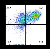
ARTICLE                                                                                NATURE COMMUNICATIONS | https://doi.org/10.1038/s41467-020-19094-0

migration assay for carbomer hydrogels. C.M., T.C., L.H., and S.S. performed data          Reprints and permission information is available at http://www.nature.com/reprints
analysis. C.M. and T.C. generated the figures. C.M., T.C., B.F., and C.J.V. wrote the
manuscript.                                                                                Publisher’s note Springer Nature remains neutral with regard to jurisdictional claims in
                                                                                           published maps and institutional affiliations.
Funding
Open Access funding enabled and organized by Projekt DEAL.
                                                                                                             Open Access This article is licensed under a Creative Commons
                                                                                                             Attribution 4.0 International License, which permits use, sharing,
Competing interests                                                                        adaptation, distribution and reproduction in any medium or format, as long as you give
The authors declare no competing interests.                                                appropriate credit to the original author(s) and the source, provide a link to the Creative
                                                                                           Commons license, and indicate if changes were made. The images or other third party
Additional information                                                                     material in this article are included in the article’s Creative Commons license, unless
Supplementary information is available for this paper at https://doi.org/10.1038/s41467-   indicated otherwise in a credit line to the material. If material is not included in the
020-19094-0.                                                                               article’s Creative Commons license and your intended use is not permitted by statutory
                                                                                           regulation or exceeds the permitted use, you will need to obtain permission directly from
Correspondence and requests for materials should be addressed to B.F.                      the copyright holder. To view a copy of this license, visit http://creativecommons.org/
                                                                                           licenses/by/4.0/.
Peer review information Nature Communications thanks Evren Alici and the other,
anonymous reviewers for their contribution to the peer review of this work. Peer review
reports are available.                                                                     © The Author(s) 2020

8                     NATURE COMMUNICATIONS | (2020)11:5224 | https://doi.org/10.1038/s41467-020-19094-0 | www.nature.com/naturecommunications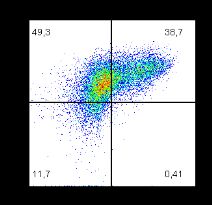
Supplementary Information

Cryopreservation impairs 3-D migration and cytotoxicity of natural killer cells

                                  Mark et al.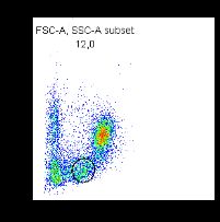
Supplementary Figure 1: Cytotoxicity (mean ± se) of 5 independent isolations (4 subjects, 3 different
expansions) for fresh (blue) and cryopreserved (red) NK cells with different effector to target ratio
(20:1,10:1,5:1,2.5:1) at different times (0.5h, 1h, 2h, 3h, 4h). At shorter incubation times (0.5 and 1 h)
and low NK-to-target ratio, the cytotoxicity between fresh and cryopreserved cells equalizes. Error bars
denote 1 SEM. Source data are provided as a Source Data file.
Supplementary Figure 2: Motile fraction, migration cell speed and migration persistence of fresh (blue)
and cryopreserved (red) NK cells from n = 10 independent experiments from 5 subjects (1-5) and
different expansion (a,b,c) in 1.2 mg/ml collagen. Each symbol represents mean ± se from cells
measured in 5 fields of view, with approximately 80 cells (motile plus non-motile) in each field of view.
Migration speed and persistence is computed from on average of n = 125 motile fresh cells and n = 12
motile cryopreserved cells for each subject and expansion (each data point). In total, n = 1248 motile
fresh and n = 122 motile cryopreserved NK cells are measured. Paired (fresh versus cryopreserved)
data from each subject and expansion are connected by lines. a: Motile fraction. b: Cell speed. c:
Directional persistence. Source data are provided as a Source Data file.

* indicates a measurement where not a single NK cell in 5 fields of view was migratory. Therefore,
mean cell speed and mean directional persistence could not be determined.
Supplementary Figure 3: Motile fraction, migration cell speed and migration persistence of fresh (blue)
and cryopreserved (red) NK cells from n = 3 independent isolations (3 subjects) in 1.2 mg/ml collagen
for different incubation times in RPMI 1640 medium (2h, 20h, 44h). After expansion or thawing, cells are
mixed in collagen solution after 2h (= 6h measurement) in medium, after 20h in medium, and after 44h
in medium. After 1 hour of gel polymerization we incubate the samples for three more hours so that
measurements start 6h, 24h and 48h after thawing or expansion. Each symbol represents mean ± se
from 10 fields of view for each subject, with approximately 80 cells in each field of view. Paired (fresh
versus cryopreserved) data from each subject are connected by lines. Data for migration speed and
persistence are based on in total n = 1508 motile fresh and n = 628 motile cryopreserved NK cells. a:
Motile fraction b: Cell speed c: Directional persistence. Source data are provided as a Source Data file.
Supplementary Figure 4: Motile fraction, migration cell speed and migration persistence of NK92 cells
(ATCC CRL-2407) from n=3 independent experiments in 1.2 mg/ml collagen. NK92 cells are grown in
Alpha MEM without nucleosides with 15% FCS, 15 HS and 500 IU/ml IL-2. Cryopreservation and
thawing is performed as for ex-vivo expanded NK cells (see Methods). Each symbol represents mean
± se from 10 fields of view for each independent experiment, with approximately 80 cells in each field of
view. Paired (fresh versus cryopreserved) data from each independent experiment are connected by
lines. Data for migration speed and persistence are based on in total n = 650 motile fresh (blue) and n
= 125 motile cryopreserved (red) NK92 cells. a: Motile fraction b: Cell speed c: Directional persistence.
Source data are provided as a Source Data file.
Supplementary Figure 5: Motile fraction, migration speed and migration persistence of fresh (blue) and
cryopreserved (red) NK cells from n=3 independent isolations (3 subjects) in non-adhesive copolymer
hydrogels of acrylic acid and alkyl-methacrylate (9mg/ml carbomer) and in 1.2 mg/ml collagen. Each
symbol represents mean ± se from 10 fields of view for each subject, with approximately 75 cells in each
field of view. Paired (fresh versus cryopreserved) data from each subject are connected by lines. Data
for migration speed and persistence are based on in total n = 657 motile fresh and n = 125 motile
cryopreserved NK cells. a: Motile fraction b: Cell speed c: Directional persistence. Source data are
provided as a Source Data file.
Supplementary Figure 6: a: Killing efficiency for individual fields-of-view as a function of the number
of motile fresh (blue) and motile cryopreserved (red) NK cells as determined by the 3-D cytotoxicity
assay (same donors and expansions as in Fig. 4 in the main text). b: Killing rate for individual fields-of-
view as a function of the number of K562 target cells. We find no systematic dependence of the killing
rate on either the number of NK cells or the number of target cells, demonstrating that the killing rate
estimation is not biased by different concentrations of NK cells and target cells. Source data are provided
as a Source Data file.
day 0

                                                                                      day 14 fresh
                       Life dead

                                                                                      day 14

                                             SSC-A
FSC-H

                                                                   CD56
                                                                                      cryopreserved/
                                                                                      thawed

         FSC-A                     SSC-A              FSC-A                CD16

Supplementary Figure 7: Cells are first gated for singlets (FSC-H versus FSC-A) and life cells (Life
dead versus SSC-A). Next, lymphocytes are gated based on size and granularity by forward and side
scatter (SSC-A versus FCS-A). Finally, the lymphocyte gate is analyzed for expression of NK cell-
specific markers (e.g. CD3, CD56, CD16, CD107a).
Supplementary Figure 8: Recovery of cell speed after time in collagen for fresh (blue) and cryopreserved (red) NK cells from 3 different subjects. Measurement of cell migration started immediately following the completion of the collagen polymerization process 60 min after embedding the cells. For computing the mean cell speed, only the motile fraction of the cells is analyzed. Cells can migrate into and out of the microscope's field of view so that the number of cells from which the mean cell speed is computed fluctuates over time. On average, the mean value is computed from 115 motile fresh cells (blue circles) and 105 motile cryopreserved cells (red circles). Error bars denote 1 SEM. Source data are provided as a Source Data file.
Supplementary Figure 9: Size distribution of K562 cells (blue) and non-motile (round) NK cells (orange)
(K562 n = 162, NK n = 117). The overlap in the size distributions is less than 2%. Source data are
provided as a Source Data file.
Supplementary Figure 10: Composite confocal and bright-field maximum intensity projections of a cell
population expanded from PBMCs in a 3D collagen gel. The brightfield maximum intensity projection at
the beginning of the experiment is shown in greyscale. The red overlay shows the confocal maximum
intensity projection of a CD56-APC staining that was recorded 10 minutes after recording the brightfield
image stack. The green overlay shows the confocal maximum intensity projection of a CD3-Alexa488
staining that was also recorded 10 minutes after recording the brightfield image stack. Thus, cells that
appear red on a dark background are motile NK cells, while cells that appear red on a bright background
have not moved during the last 10 minutes and represent non-motile NK cells. Cells that appear yellow
(double positive for CD56 and CD3) are NKT cells, and cells that appear green are T cells. Based on
three field-of-views, we find that 82% of all motile cells are NK cells. The experiment was set up similar
to the 3D cell motility assay described in the Methods section and was carried out once.
You can also read