Drosophila as a Model to Study Cellular Communication Between the Hematopoietic Niche and Blood Progenitors Under Homeostatic Conditions and in ...
←
→
Page content transcription
If your browser does not render page correctly, please read the page content below
REVIEW
                                                                                                                                   published: 16 August 2021
                                                                                                                            doi: 10.3389/fimmu.2021.719349

                                            Drosophila as a Model to Study
                                            Cellular Communication Between
                                            the Hematopoietic Niche and Blood
                                            Progenitors Under Homeostatic
                                            Conditions and in Response to an
                                            Immune Stress
                                            Ismaël Morin-Poulard †, Yushun Tian †, Nathalie Vanzo and Michèle Crozatier *

                               Edited by:   MCD/UMR5077, Centre de Biologie Intégrative (CBI), Toulouse, France
               Stéphane J. C. Mancini,
       UMR1236 Microenvironnement,
Différenciation cellulaire, Immunologie    In adult mammals, blood cells are formed from hematopoietic stem progenitor cells, which
           et Cancer (INSERM), France       are controlled by a complex cellular microenvironment called “niche”. Drosophila
                         Reviewed by:       melanogaster is a powerful model organism to decipher the mechanisms controlling
                            Jiwon Shim,
    Hanyang University, South Korea
                                            hematopoiesis, due both to its limited number of blood cell lineages and to the
                        Lolitika Mandal,    conservation of genes and signaling pathways throughout bilaterian evolution. Insect
 Indian Institute of Science Education
                                            blood cells or hemocytes are similar to the mammalian myeloid lineage that ensures innate
           and Research Mohali, India
                                            immunity functions. Like in vertebrates, two waves of hematopoiesis occur in Drosophila.
                  *Correspondence:
                     Michèle Crozatier      The first wave takes place during embryogenesis. The second wave occurs at larval
 michele.crozatier-borde@univ-tlse3.fr      stages, where two distinct hematopoietic sites are identified: subcuticular hematopoietic
     †
      These authors have contributed        pockets and a specialized hematopoietic organ called the lymph gland. In both sites,
                 equally to this work
                                            hematopoiesis is regulated by distinct niches. In hematopoietic pockets, sensory neurons
                   Specialty section:       of the peripheral nervous system provide a microenvironment that promotes embryonic
         This article was submitted to      hemocyte expansion and differentiation. In the lymph gland blood cells are produced from
                Cytokines and Soluble
               Mediators in Immunity,
                                            hematopoietic progenitors. A small cluster of cells called Posterior Signaling Centre (PSC)
               a section of the journal     and the vascular system, along which the lymph gland develops, act collectively as a
              Frontiers in Immunology       niche, under homeostatic conditions, to control the balance between maintenance and
           Received: 02 June 2021           differentiation of lymph gland progenitors. In response to an immune stress such as wasp
           Accepted: 26 July 2021
         Published: 16 August 2021          parasitism, lymph gland hematopoiesis is drastically modified and shifts towards
                            Citation:       emergency hematopoiesis, leading to increased progenitor proliferation and their
              Morin-Poulard I, Tian Y,      differentiation into lamellocyte, a specific blood cell type which will neutralize the
     Vanzo N and Crozatier M (2021)
             Drosophila as a Model to
                                            parasite. The PSC is essential to control this emergency response. In this review, we
        Study Cellular Communication        summarize Drosophila cellular and molecular mechanisms involved in the communication
          Between the Hematopoietic         between the niche and hematopoietic progenitors, both under homeostatic and stress
  Niche and Blood Progenitors Under
       Homeostatic Conditions and in        conditions. Finally, we discuss similarities between mechanisms by which niches regulate
     Response to an Immune Stress.          hematopoietic stem/progenitor cells in Drosophila and mammals.
          Front. Immunol. 12:719349.
    doi: 10.3389/fimmu.2021.719349          Keywords: Drosophila, lymph gland, niche, hematopoiesis, immune stress

Frontiers in Immunology | www.frontiersin.org                                     1                                August 2021 | Volume 12 | Article 719349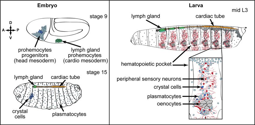
Morin-Poulard et al.                                                                                                                          Drosophila Hematopoietic Niche

INTRODUCTION                                                                               itself is heterogeneous, raising the possibility that distinct and
                                                                                           specific niche cell types control subsets of HSPCs (6, 17, 18).
Hematopoiesis is the process that leads to the constant formation                              For 15 years, Drosophila melanogaster has proven to be a
of blood cells throughout metazoan life. In vertebrates,                                   suitable model organism to investigate the mechanisms
hematopoietic stem and progenitor cells (HSPCs) give rise to                               controlling hematopoiesis, based both on limited blood cell
all blood cell types. In adults, HSPCs are found in the bone                               lineages and on functional parallels with the vertebrate system.
marrow, and its microenvironment, termed ‘niche’, ensures                                  In flies, blood/immune cells are called hemocytes and are related
hematopoietic homeostasis by controlling the proliferation and                             to vertebrate myeloid cells
differentiation of HSPCs, both under normal conditions and in                                  Drosophila hematopoiesis occurs in two waves during
response to a stress such as infection or systemic inflammation                             development (19, 20). The first wave takes place during
(1–4). The ‘niche’ concept was proposed in 1978 by R. Schofield                             embryogenesis (21). A cluster of cells derived from the head
(5) and refers to the cellular context that maintains and regulates                        mesoderm gives rise to hematopoietic progenitors, which
HSPC self-renewal and differentiation. The bone marrow                                     differentiate into plasmatocytes and crystal cells. Plasmatocytes,
hematopoietic niche is now described as a complex                                          which are involved in phagocytosis of cellular debris and
multicellular network that supports HSPCs, either via direct                               pathogens, are equivalent to mammalian macrophages (21–24).
adhesive interactions or via the secretion of many different                               Crystal cells contain crystalline inclusions of prophenoloxidases,
factors acting in a paracrine manner to control their                                      which are required for the synthesis of melanin (25, 26) and are
localization, maintenance, proliferation and differentiation. At                           involved in clotting and wound healing (27–29). These hemocytes
least two anatomically distinct HSPC niches exist in the bone                              of embryonic origin persist in larval and adult stages (Figures 1A, B
marrow. Imaging studies indicate that HSPCs localize around                                and (30–34). The second wave of hematopoiesis takes place in larval
arterioles in the endosteal area, which is in close proximity to the                       stages at two distinct hematopoietic sites: the hematopoietic pockets
bone surface and is called the endosteal niche (6), and around                             and the hematopoietic organ called the lymph gland. Hematopoietic
sinusoids located in the inner bone marrow and called the                                  pockets are aggregate of embryo-derived hemocytes segmentally
vascular niche (7–12). Recent advances in single cell                                      repeated in epidermal-muscular clusters underneath the larval
technologies allowed the identification of various populations                              cuticle (Figure 1C). In addition, de novo blood cell specification
of niche cells with distinct transcriptional profiles, revealing the                        occurs in the lymph gland (see below) from hematopoietic
huge complexity of the cell population within the bone marrow                              progenitors and give rise to plasmatocytes, crystal cells and a
hematopoietic niches (8, 13–16). Furthermore, the HSPC pool                                third blood cell type called lamellocyte. Lamellocytes are not

               A                                                                   C

               B

 FIGURE 1 | Embryonic and larval hematopoiesis. (A, B) Embryonic hemocytes (blood cells) originate from the head mesoderm in the embryo and differentiate into
 plasmatocytes (macrophages, light blue) and a small number of crystal cells (dark blue). Lymph gland progenitors (green) are specified from the thoracic cardiogenic
 mesoderm in the embryo (A) Anterior (A)/Posterior (P) and Dorsal (D)/Ventral (V) axes are indicated. (B) At the end of embryogenesis, crystal cells remain clustered in
 the anterior part, whereas plasmatocytes are dispersed throughout the embryo. The lymph gland is composed of one pair of lobes and is localized at the anterior
 part of the dorsal vessel/cardiac tube. (C) In third instar larvae, plasmatocytes (light blue) and crystal cells (dark blue) of embryonic origin are found in circulation and
 colonizing local microenvironments, in particular the hematopoietic pockets, where they expand. Close up of a hematopoietic pocket where neurons are in red,
 oenocytes in grey, and plasmatocytes and crystal cells in light and dark blue, respectively. Activin-b produced by PNS neurons promotes plasmatocyte proliferation
 and adhesion. The lymph gland (green) is composed of several pairs of lobes aligned along the cardiac tube.

Frontiers in Immunology | www.frontiersin.org                                          2                                          August 2021 | Volume 12 | Article 719349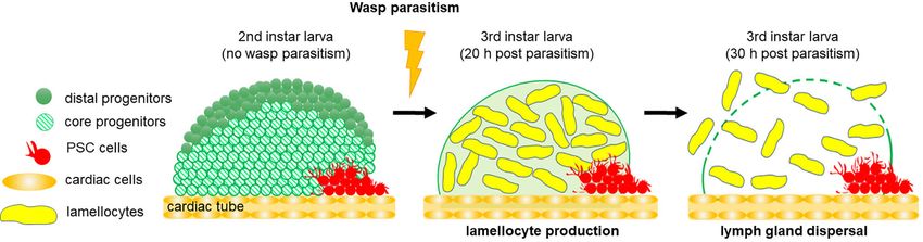
Morin-Poulard et al.                                                                                                Drosophila Hematopoietic Niche

found in larvae under normal conditions, but they massively                 inflammation (49, 50). Resident macrophage proliferation is
differentiate in response to an immune stress such as wasp                  strongly dependent on the tissue microenvironment, and
parasitism. Lamellocytes are required for the encapsulation of              whether vertebrate neuronal sensing, as described in Drosophila,
foreign bodies too large to be engulfed by phagocytosis (29, 35).           regulates locally macrophage behavior remains to be addressed.
The lymph gland is localized dorsally, in close association with the            Finally, several studies report on the plasticity of embryonic-
Drosophila dorsal vessel, which is the vascular system. At                  derived hemocytes. Within hematopoietic pockets, plasmatocytes
metamorphosis, the lymph gland disrupts and all cells are                   can trans differentiate into crystal cells (51, 52). Furthermore,
released into the circulation (29, 36). Both embryo and lymph               embryonic-derived hemocytes can also give rise to lamellocytes
gland-derived blood cells are present in the adult fly and accumulate        following parasitism (32, 53–56). A puzzling question was
in the respiratory epithelia and fat body (33, 34, 37). Their numbers       whether signals from the neuronal niche might also regulate
continuously decrease with aging (29, 38), and whether adult flies           blood cell plasticity in hematopoietic pockets. A recent study
are able to produce new blood cells is currently under debate. Ghosh        established that in hematopoietic pockets localized at the caudal
et al. identified active hematopoietic hubs, localized in the abdomen,       end of the larva, the trans differentiation of macrophages into
and supporting hematopoiesis in adults (34). However, this                  crystal cells is promoted by the neuronal activity of a specific
conclusion is strongly questioned by a recent analysis which                subset of oxygen sensing neurons (52). This study establishes
studies hemocytes localized in the head and thorax regions and              that environmental conditions, such as oxygen levels, control
where no indication of de novo blood cell production was observed,          in vivo blood cell trans differentiation. Whether neuronal
even after bacterial infections (37). Since recent single cell RNAseq       control of blood cell trans differentiation in response to
analyses identify different hemocyte populations (39–42), it is             environmental conditions is conserved during evolution,
possible that hemocytes characterized in these two distinct                 deserves further investigation.
locations might have a different potential. This point deserves
further investigation.
    While no data indicate that embryonic hematopoiesis is niche-
dependent, several studies established that larval hematopoiesis is
                                                                            THE PSC ACTS AS A NICHE TO CONTROL
under the control of distinct niches. In this review, we will give an       LYMPH GLAND HEMATOPOIESIS
overview of the various Drosophila hematopoietic niches
                                                                            In third instar larvae, the mature lymph gland is composed of
identified so far and of the molecular cascades that regulate the
                                                                            paired lobes: one primary pair and several secondary pairs. The
communication between niche cells and progenitors, both under
                                                                            anterior lobes, which are the largest in size contain progenitors,
homeostatic and immune stress conditions.
                                                                            differentiating hemocytes and mature blood cells, while posterior
                                                                            lobes are composed of a heterogeneous population of progenitors,
                                                                            which do not undergo terminal differentiation (23, 36, 57, 58).
                                                                            Each anterior lobe is divided into several zones (Figure 2A). A
NEURONS AS A MICROENVIRONMENT                                               central zone, called the medullary zone (MZ), contains tightly
CONTROLLING EMBRYONIC-DERIVED                                               packed blood cell progenitors (prohemocytes) characterized by the
HEMOCYTES IN HEMATOPOIETIC                                                  expression of the Janus kinase/signal transducer and activator of
POCKETS                                                                     transcription (JAK/STAT) receptor domeless (dome) (23, 59).
                                                                            Recently, the most internally localized subpopulation of MZ
At larval stages, most embryonic-derived hemocytes are                      progenitors was further characterized by expression of specific
differentiated macrophages. They are either circulating in the              markers such as the Thioester-containing protein-4 (Tep4) and
hemolymph or residing in clusters, which are segmentally                    Col (60). This subpopulation is defined as “core progenitors”. The
repeated along the larval body wall and called hematopoietic                neighboring progenitors lacking tep4 and col expression are called
pockets (29, 43, 44) (Figure 1C). There is a continuous and                 “distal progenitors” (61). Recent advances in single cell
dynamic exchange between circulating and resident/pocket                    technologies established the transcriptional profiles of lymph
macrophages (44–47). Large hepatocyte-like cells called                     gland cells under homeostatic conditions and at various
oenocytes, and sensory neurons from the peripheral neuronal                 developmental time points (41). The molecular signatures,
system (PNS), are in close contact with resident macrophages in             provided by single cell transcriptomic analysis, define an
the hematopoietic pockets (Figure 1C). A subset of sensory                  additional prohemocyte sub-cluster called PH1 (prohemocyte 1).
neurons that produces Activin-b, a ligand of the TGF-b family,              At the periphery of anterior lobes, the cortical zone (CZ) is
regulates their proliferation and adhesion to hematopoietic                 composed of differentiated blood cells that can be identified
pockets (48). It should be emphasized that the neuronal niche               through the expression of specific markers for plasmatocytes
in hematopoietic pockets and the niche in the lymph gland (see              and crystal cells. Between the MZ and the CZ, cells undergo the
below) have distinct functions. While the neuronal niche is                 transition from progenitors to specified blood cells and
regulating differentiated macrophages, the niche in the lymph               correspond to intermediate progenitors. They simultaneously
gland is controlling both differentiated hemocytes and                      express markers for prohemocytes and for early differentiating
hematopoietic progenitors. In vertebrate, tissue-resident                   cells [Figure 2A and (59)]. At the posterior end of the primary
macrophages regulate tissue homeostasis and contribute to                   lobe is the PSC, identified by its expression of the Notch ligand

Frontiers in Immunology | www.frontiersin.org                           3                                 August 2021 | Volume 12 | Article 719349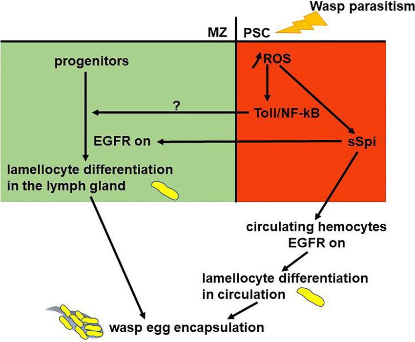
Morin-Poulard et al. Drosophila Hematopoietic Niche
A B
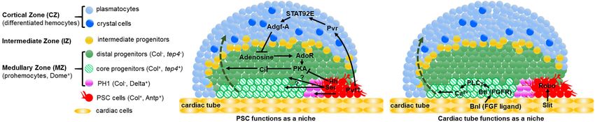
FIGURE 2 | Two niches control lymph gland homeostasis. (A, B) Schematic representation of third instar larva lymph gland anterior lobes. The medullary zone (MZ)
contains three types of progenitors: distal progenitors and core progenitors are in green and hatched green, respectively, and the PH1 is in pink. Intermediate
progenitors are in yellow, plasmatocytes and crystal cells in the cortical zone (CZ) are in light and dark blue, respectively. The PSC and the cardiac tube/vascular
system are in red and orange, respectively. (A) Differentiated hemocytes result from progenitors’ differentiation (green dashed arrow) In a wildtype (WT) lymph gland,
under homeostatic conditions, the PSC regulates the maintenance of a subset of MZ progenitors. Hedgehog (Hh) is required for maintaining distal progenitors. PSC
signals required for controlling PH1 remain to be identified, as well as the progenitor subset controlled by Ser expressed in the PSC. Pvf1 secreted by the PSC,
controls progenitor maintenance via differentiated hemocytes. (B) The cardiac tube corresponds to a second niche present in the lymph gland. The FGF ligand
Branchless (Bnl) activates its receptor Breathless (Btl) in progenitors. Btl-FGF activation regulates intracellular Ca2+ levels via PLCg, and controls the maintenance of
core progenitors and in turn the whole progenitor pool. The ligand Slit produced by cardiac cells activates its Robo receptors in the PSC. Robo signaling controls
PSC cell clustering and proliferation.
Serrate (Ser) (23, 62), the homeobox protein Antennapedia (Antp) [Figure 2A and (60, 78)]. More recent studies shed some light on
(63) and high levels of Collier/Knot (Col/Kn), an orthologue of these discrepancies. Baldeosingh et al. examined the effects on
mammalian Early B cell factor (EBF) (57, 64). In late third instar MZ progenitors of specific PSC cell ablation induced by rpr
larvae the mature PSC is composed of 30-40 cells [Figure 2A and expression. For this, they analyzed the expression of
(57, 63, 64)] and it plays a role similar to a niche to control lymph Odd-skipped (Odd), a transcription factor expressed in all MZ
gland homeostasis. progenitors. In the absence of PSC cells, a prohemocyte
Most studies on the PSC’s role as a niche were performed in subpopulation with Odd-positive/Col-negative cells
third instar larvae. The number of PSC cells is tightly controlled differentiated into mature hemocytes, whereas the Odd-positive/
and several intrinsic and extrinsic factors regulate their Col-positive cells remained undifferentiated (79). The study
proliferation. Since several recent reviews report on genes and further reported that Hh from the PSC is required to maintain
molecular mechanisms that control PSC cell numbers, we shall Odd-positive but not Col-positive prohemocytes, establishing that
not develop this specific issue but provide a table summarizing the MZ cell population is composed of Hh-independent (core
the information [see Table 1 and reviews by (19, 20, 77)]. PSC progenitors) and Hh-dependent (distal progenitors) progenitors
cells produce cytoplasmic processes called filopodia that extend (Figure 2A). Altogether these data confirm that the PSC only
over 2 to 3 cell diameters. An interesting possibility is that regulates a subset of MZ progenitors and that this is achieved
filopodia could be engaged in direct cellular contacts between through Hh signaling.
PSC cells and MZ progenitors (63, 64). Blanco et al. investigated the role of Ser in the PSC. Ser
The PSC requirement to control the balance between knockdown in PSC cells leads to increased plasmatocyte and
hemocyte differentiation and progenitor maintenance crystal cell numbers, which is in agreement with previously
(homeostasis) in third instar larvae was first reported by published data (64). Furthermore, they report that Notch
Mandal et al. and Krzemien et al. In this context, Hedgehog knockdown specifically in core progenitors leads to a reduction
(Hh) secreted by PSC cells is a key regulator of lymph gland of their numbers. These data indicate that Ser in PSC cells restricts
homeostasis (63) and Col is required for PSC specification hemocyte differentiation [Figure 2A and (61)]. Ser requires cell-
during embryonic development (64). It has been proposed that cell contact to activate the Notch pathway, raising the possibility
by controlling hematopoietic progenitor maintenance within the that PSC filopodia could mediate Notch signaling, although the
lymph gland, the PSC plays a role similar to a niche. However, subset of progenitors controlled by Ser remains to be identified.
several studies questioned the genuine interactions that take A recent study further established a role of the PSC in L1
place between MZ and PSC cells, since ablation of PSC cells larvae (80). At this stage the lymph gland is composed of PSC
driven by the expression of the proapoptotic gene reaper (rpr) cells and hematopoietic progenitors and no differentiation
does not affect MZ progenitor maintenance but rather reduces occurs. The PSC counts 2-4 cells that express Col and Antp.
crystal cell differentiation (78). Another study reported that Through the expression of different markers, it has been shown
reduction of PSC cell numbers or alteration of PSC signaling that two types of progenitors are present. One subset of
increases hemocyte differentiation without affecting the pool of progenitors, expressing Notch, is aligned along the cardiac
“core progenitors”. Altogether, these two studies establish that tube, and this cell state is transient, since Notch positive cells
core progenitors are maintained independently from the PSC are only found during the first 20 hours of larval development.
Frontiers in Immunology | www.frontiersin.org 4 August 2021 | Volume 12 | Article 719349Morin-Poulard et al. Drosophila Hematopoietic Niche
TABLE 1 | Genes and pathways involved in controlling the number of PSC cells and their cohesion.
Gene Cell type Genetic conditions Function References
Collier/knot PSC LOF (col RNAi) Reduces PSC cell number (65)
Wnt/Wingless PSC LOF (UAS-Dfz2DN) GOF (UAS-wg) Promotes PSC cell proliferation (66)
BMP / Decapentaplegic PSC LOF (dpp RNAi; UAS-tkv DN ) Inhibits PSC cell proliferation (65)
Dally-like Not determined dlp mutant Reduces PSC cell number (65)
Dmyc PSC LOF (dmyc RNAi) Increases PSC cell number (65)
GOF (UAS-dmyc )
Insulin/TOR PSC LOF (InR RNAi) GOF (UAS-PI3K CAAX ) Increases PSC cell number (67, 68)
Bantam PSC LOF (UAS-sponge) GOF (UAS-bantam ) Increases PSC cell number (69)
Bag of Marbles PSC LOF (bam RNAi) Inhibits PSC cell proliferation (70)
Thor/4EBP PSC LOF (eIf4A RNAi) Increases PSC cell number (70)
Retinoblastoma-family protein PSC LOF (Rbf RNAi) Inhibits PSC cell proliferation (70)
GOF (UAS-Rbf )
ARF1-GTP PSC and hemocytes LOF (arf1 RNAi) Increases PSC cell number (71)
Jumu progenitors LOF (jumu RNAi) Inhibits PSC cell proliferation (72)
Promotes PSC cell clustering
Jumu PSC LOF (jumu RNAi) Increases PSC cell number (72)
GOF (UAS-jumu )
Slit/Robo PSC and cardiac cells LOF (robo and slit Inhibits PSC cell proliferation (73)
RNAi) Promotes PSC cell clustering
DE-cadherin PSC LOF (DE-cad RNAi) Reduces PSC cell number (73)
Promotes PSC cell clustering
Cdc42 PSC LOF (UAS-cdc42DN) GOF (UAS-cdc42CA Increases PSC cell number (73)
Promotes PSC cell clustering
Coracle PSC LOF (cora RNAi) Reduces PSC cell number (74)
Neurexin IV PSC LOF (nrxIV RNAi) Reduces PSC cell number (74)
Lar PSC LOF (Lar RNAi) Reduces PSC cell number (75)
GOF (UAS-Lar )
NUP98-HOXA9 PSC and hemocytes GOF (UAS-NA9) Promotes PSC cell proliferation (76)
E2F PSC LOF (E2F RNAi) Increases PSC cell number (70)
Lineage tracing experiments established that this cell population factor 1 (Pvf1), which binds and activates the Pvr tyrosine
gives rise to most lymph gland cells at later larval stages, leading kinase receptor. Pvf1 is produced by PSC cells and transported
the authors to propose that they correspond to genuine by vesicles into CZ cells that express Pvr. Pvr activation in the CZ
Hematopoietic Stem Cells (HSCs). The presence of these HSCs induces a Stat92E-dependent but JAK-independent signaling
in L1 larvae is also niche-dependent. They rely on Dpp/BMP cascade, leading to the overexpression of adenosine deaminase-
signaling issued from the PSC. related growth factor A (Adgf-A). Stat92E activation is
In summary, these studies reveal a temporal role for the dependent on the ARF1/Asrij complex that encodes a ras small
PSC during larval development to regulate lymph gland GTPase and an endocytic protein (71). Adgf-A downregulates
hematopoiesis and further establish that different signals, long adenosine levels in neighboring MZ cells, leading to a reduced
versus short distance, are produced by PSC cells throughout larval activity of PKA (cAMP-dependent protein kinase 1). PKA
development to regulate different progenitor subsets. Thus, the controls the degradation of active Cubitus interruptus (Ci), the
lymph gland is a valuable model to investigate the spatial and transcription factor mediating Hh signal transduction. This
temporal role of the niche. Additional analyses are required to backward signal to the MZ is called the “equilibrium signal”
identify other yet undetected PSC signals, define which (81, 82). Overall, signals from CZ and PSC cells regulate the
progenitor sub-clusters respond to which PSC signals, and balance of Ci activity within the MZ, thereby controlling
finally define how these various niche signals are integrated in progenitor maintenance (Figure 2A).
progenitor subtypes to control the balance between progenitor
maintenance and blood cell differentiation.
THE CARDIAC TUBE FUNCTIONS AS A
THE PSC INDIRECTLY CONTROLS HEMATOPOIETIC NICHE
HEMATOPOIETIC PROGENITOR Within the MZ, “core progenitors” that express col and tep4, are
MAINTENANCE VIA DIFFERENTIATED in close contact with the cardiac tube and are maintained
HEMOCYTES independently from PSC activity [Figure 2A and (60, 78, 79)].
This raises the possibility that cardiac cells contribute to the
In third instar larvae, the PSC indirectly controls hematopoietic regulation of lymph gland homeostasis. Two recent studies
progenitor maintenance via differentiated hemocytes. The PSC investigated the role of the cardiovascular system under
also secretes another ligand called PDGF and VEGF-related homeostatic conditions and established that cardiac cells act
Frontiers in Immunology | www.frontiersin.org 5 August 2021 | Volume 12 | Article 719349Morin-Poulard et al. Drosophila Hematopoietic Niche both i) indirectly via the PSC, and ii) directly on MZ progenitors EMERGENCY HEMATOPOIESIS: to control lymph gland hematopoiesis. An initial study showed KEY ROLE FOR THE PSC that the Drosophila cardiac tube is required to maintain the integrity and function of the PSC through Slit/Robo signaling. Drosophila blood cells are the effectors of the cellular arm of the The Slit ligand secreted by cardiac cells activates Robo signaling innate immune response (24). Wasp parasitism is commonly in the PSC. Slit/Robo activation controls both the number of PSC used to induce an emergency hematopoiesis, which culminates in cells and their cohesion, and in turn PSC function (Table 1). It the massive differentiation of lamellocytes, a cryptic blood cell controls PSC size by repressing BMP signaling, and maintains type (29, 85). Lamellocytes are specialized hemocytes, which PSC cell clustering by regulating the activity of the Cdc42 small mediate the encapsulation and killing of pathogens too large to GTPase and the accumulation of DE-Cadherin. This study was be phagocytosed. Resistance to wasp parasitism depends on the the first to highlight an inter-organ communication between the ability of the Drosophila larva to reroute basal hematopoiesis and cardiac tube and the lymph gland in order to control PSC produce lamellocytes, in a timely manner, to neutralize wasp eggs morphology and consequently its function [Figure 2B and before they hatch inside the fly larva. Following wasp egg-laying (73)]. In a second study, which investigated whether cardiac in a Drosophila second instar larva, the egg is identified as a cells can directly act on MZ progenitors via secreted signals, the foreign body and differentiation of lamellocytes from lymph authors performed a candidate RNAi screen in cardiac cells to gland MZ progenitors and circulating/sessile hemocytes is identify new potential signaling pathways involved in the triggered (29, 32, 53–57). In response to wasp parasitism, crosstalk between the vascular and the hematopoietic systems. lymph gland hematopoiesis is drastically modified and shifts to This study provided evidence that cardiac cells play a role similar emergency hematopoiesis, leading to increased progenitor to a niche through the activation of Fibroblast Growth Factor proliferation 4-6 hours post-parasitism (59, 86). 20 hours post (FGF) signaling. The FGF ligand Branchless (Bnl) secreted by parasitism, lamellocytes massively differentiate at the expense of cardiac cells was detected in MZ progenitors as cytoplasmic MZ progenitors, ultimately leading to the premature dispersal of punctate dots; it is internalized by MZ cells, most likely through lymph gland anterior lobes [Figure 3 and (86)]. The PSC is FGF-receptor-mediated endocytosis. Bnl binding to its receptor absolutely required for this emergency response, since Breathless (Btl) leads to Bnl/Btl-FGF pathway activation in MZ lamellocytes fail to differentiate when PSC cells are ablated by progenitors, where it controls calcium levels via the activation of targeted expression of rpr (60, 64, 78). It has been shown that in phospholipase C (PLCg). A previous study showed that Drosophila larvae, parasitization increases Reactive Oxygen reduction of cytosolic Ca2+ in lymph gland progenitors leads to Species (ROS) levels in PSC cells, leading to the secretion of the loss of progenitor markers and to increased blood cell Spitz (sSpi), one ligand of the Epidermal Growth Factor Receptor differentiation (83). Altogether, these data indicate that (EGFR) signaling pathway (87). Spi issued from the PSC through the activation of Fibroblast Growth Factor (FGF) activates the EGFR pathway, both in circulating embryo- signaling, the vascular system prevents hematopoietic derived hemocytes and in MZ progenitors, which triggers their progenitors from massive differentiation, ensuring the proper differentiation into lamellocytes. [Figure 4 and (86, 87)]. balance between blood cell populations within the lymph Furthermore, it has been established that the Toll/NF-kB gland. For the first time, this study provides evidence that the pathway is activated in PSC cells in response to wasp vascular system, which directly controls blood cell progenitors parasitism (86, 88). Activation of the pathway is triggered by independently from the PSC, acts as a niche [Figure 2B and high ROS levels in PSC cells, which leads to expression of Spätzle (84)]. In conclusion, two distinct niches, the PSC and the cardiac (Spz), the Toll/NF-kB pathway ligand, and subsequent activation tube, control lymph gland homeostasis. of the pathway in the PSC. This pathway controls, in a non-cell FIGURE 3 | Lymph gland response to wasp parasitism. Schematic representation of 2nd instar larval lymph gland, composed of PSC cells and progenitors. Twenty hours post parasitism, lamellocytes differentiate at the expense of progenitor maintenance. Thirty hours post parasitism, the lymph gland disrupts and cells are released into the hemolymph, where they encapsulate the wasp egg. Frontiers in Immunology | www.frontiersin.org 6 August 2021 | Volume 12 | Article 719349
Morin-Poulard et al.                                                                                                                       Drosophila Hematopoietic Niche

 FIGURE 4 | Gene regulatory network controlling larval emergency hematopoiesis. The PSC (red) plays an essential role in mounting the cellular immune response. In
 response to wasp parasitism, increased Reactive Oxygen Species (ROS) levels in the PSC cause lamellocyte differentiation from lymph gland progenitors (green) and
 circulating hemocytes. ROS in PSC cells activate Toll/NF-kB and Spitz secretion (sSpi). sSpi, the EGFR ligand, induces lamellocyte fate. Toll/NF-kB activation in the PSC
 regulates non cell-autonomously lamellocyte differentiation in the lymph gland. EGFR and Toll/NF-kB activation are required to regulate lymph gland stress hematopoiesis.

autonomous manner, lymph gland lamellocyte differentiation in                            differential response to parasitism between anterior and
the MZ, which leads to premature disruption of lymph gland                               posterior lobes. Finally, under wasp infection, cell coalescence
anterior lobes, and in fine successful wasp egg encapsulation by                          is observed in posterior lobes, and this response is prevented
lamellocytes. It seems that in response to wasp infection, the                           when the PSC is ablated, suggesting a role for the PSC in this
EGFR and Toll/NF-kB pathways act in parallel to trigger                                  response (58). In conclusion, a complex regulation of JAK/STAT
lamellocyte differentiation from MZ cells [Figure 4 and (86)].                           signaling is induced in response to wasp parasitism, and whether
How Toll/NF-kB activation in the PSC acts on MZ progenitors                              JAK/STAT activity in the different cell types could depend on the
remains to be investigated.                                                              two niches, namely PSC or/and cardiac tube, certainly deserves
   JAK/STAT signaling is one of the evolutionarily conserved                             further investigation.
signaling pathways involved in immunity (89) and specifically in                              Besides wasp parasitism, Drosophila can be infected by
Drosophila for lamellocyte differentiation upon parasitism (90,                          bacteria, fungi or viruses, which activate the humoral response
91). Under normal conditions, JAK/STAT is activated in lymph                             (24). Interestingly, a recent study established that bacterial
gland MZ hematopoietic progenitors; in response to wasp                                  infection also alters lymph gland hematopoiesis, since it
parasitism, the pathway is switched off in progenitors, thus                             reported increased plasmatocyte and crystal cell differentiation
triggering their differentiation into lamellocytes (90).                                 at the expense of MZ progenitors upon infection. However, in
Furthermore, wasp parasitism leads to JAK/STAT activation in                             contrast to wasp parasitism, no lamellocytes differentiated. The
larval somatic muscles, which in turn controls the number of                             study further showed that septate junctions form a permeability
circulating lamellocytes and the efficiency of wasp egg                                   barrier at the PSC that is disrupted following bacterial infection
encapsulation (92).                                                                      that trigger prohemocyte differentiation probably by enabling
   In depth analysis of lymph gland hematopoiesis focuses on                             PSC signals to extend into the MZ (74). The authors further
lymph gland anterior lobes. In response to wasp parasitism,                              established that activation of the Toll/NF-kB and Immune
hemocytes from posterior lobes do not differentiate into                                 Deficiency (Imd) pathways in PSC cells leads to the loss of the
lamellocytes (23, 36, 58). The JAK/STAT pathway, which is                                PSC permeability barrier. However, whether bacterial infection
activated in posterior lobes in response to parasitism, is required                      disrupts the niche permeability barrier via the activation of NF-
to prevent lamellocyte differentiation. Furthermore, while the                           kB pathways in the PSC is not known yet. Since the Toll/NF-kB
PSC is essential in anterior lobes for the response to wasp                              pathway is activated in PSC cells and is required for lamellocyte
parasitism, it plays no role in posterior lobes to prevent                               differentiation, it is possible that the permeability barrier
lamellocyte differentiation. Altogether, these data indicate a                           modification in PSC cells in response to wasp parasitism

Frontiers in Immunology | www.frontiersin.org                                        7                                         August 2021 | Volume 12 | Article 719349
Morin-Poulard et al.                                                                                              Drosophila Hematopoietic Niche

contributes to niche hematopoietic progenitor signaling. These            plasmatocytes (40, 42, 103). These results raised many questions
data open novel insights into the cellular communication                  about the heterogeneity of the Drosophila blood cell pool and their
between the PSC and MZ progenitors.                                       regulation by different niche cell types. Likewise, in mammalian
   In mammals, systemic bacterial infection activates the Toll/           bone marrow, single cell approaches revealed a considerable
NF-kB pathway in mouse bone marrow endothelial cells,                     heterogeneity among both niche and hematopoietic stem
provoking an “emergency granulopoiesis” (93, 94). This, again,            progenitor cells (10). Further analyses are now necessary to
underlines the evolutionary conservation of molecular                     decipher which niche cells control which progenitor subset,
mechanisms controlling stress-induced hematopoiesis between               to identify the signals involved in this crosstalk, and finally to
Drosophila and mammals. As a conclusion, our comprehension                determine how information provided by the diverse niche cells is
of the mechanisms regulating emergency hematopoiesis in                   integrated to control hematopoiesis under homeostatic conditions
Drosophila should improve our fundamental understanding of                and after infection.
how inflammatory signaling regulates hematopoiesis in health                  Mechanisms regulating emergency hematopoiesis are poorly
and disease conditions.                                                   understood. Oxidative stress regulates hematopoiesis via ROS
                                                                          both in Mammals and in Drosophila (87, 104, 105). In
                                                                          mammalian bone marrow, bacterial infection induces an
                                                                          “emergency granulopoiesis” that leads to de novo production of
CONCLUSIONS AND PERSPECTIVES                                              neutrophils. In this context, the TLR (Toll-like Receptor)/NF-kB
Drosophila is a powerful in vivo model system to study the                pathway is activated via TLR4 in mouse bone marrow
dialogue between a hematopoietic niche and progenitor cells,              endothelial cells, a component of the vascular niche (93, 94).
since several signaling pathways and transcription factors                In Drosophila, the cellular immune response to parasitism is a
involved in the Drosophila microenvironment play comparable               typical emergency hematopoiesis. ROS levels increase in the PSC,
roles in mammals. Under homeostatic conditions, the                       thus activating both Toll/NF-kB and EGFR signaling pathways,
transcription factor Col/EBF, expressed in PSC cells, is required         which act in parallel to mount a stress hematopoiesis (86).
for PSC specification (57, 64) and controls PSC cell numbers and           Whether the EGFR pathway plays a role in mammalian
function through BMP/Dpp pathway activation (65). In mouse                hematopoiesis has not yet been established (106). Altogether,
osteoblasts, EBF2 is an essential component of the endosteal niche,       those studies are in favor of evolutionary parallels between
where it controls osteoblast numbers and regulates HSPC                   Drosophila and mouse in the control of stress-induced
maintenance (95, 96). The Notch pathway is also involved both             hematopoiesis. The recent identification of the cardiac tube as
organism. The Notch ligand Serrate is expressed in the Drosophila         a niche controlling lymph gland homeostasis under homeostatic
PSC, where it prevents progenitor differentiation (61, 62).               conditions obviously raises the question about its potential role
Similarly, in mammalian osteoblasts, Notch1 and 3 and the                 during emergency hematopoiesis.
ligands Jagged1 and Delta1 are all expressed and regulate                    Malignant hematopoiesis and inflammation in mammals is
hematopoiesis, although the precise regulatory mechanisms                 often associated with an abnormal microenvironment (2, 3, 12,
remain unclear (8, 97, 98). Furthermore, in Drosophila, Slit              107, 108). Thus, deciphering the mechanisms at play in the
secreted by cardiac cells activates Robo receptors expressed by           HSPC/niche dialogue is of most importance and Drosophila
PSC cells. Silt/Robo activation controls PSC cell numbers and their       stands as an invaluable model to do so.
function (73), while, in mouse bone marrow Slit2/Robo4 controls
HSPC localization in the perivascular niche (99–101). In
Drosophila, the FGF pathway is a key player in the
communication between cardiac tube and hematopoietic                      AUTHOR CONTRIBUTIONS
progenitors. In mammals, this pathway remodels bone and the               IM-P, YT, NV, and MC wrote the manuscript. IM-P and YT
bone marrow microenvironment to support bone integrity, HSPC              made the figures and NV the table. IM-P, YT, NV, and MC
maintenance and expansion, and plays a crucial role for proper            designed the review. All authors contributed to the article and
hematopoiesis during stress recovery (102). Finally, the high             approved the submitted version.
similarity between Drosophila and mammalian bone marrow
hematopoiesis is further emphasized by our recent identification
of the cardiac tube as a second niche for lymph gland
hematopoiesis, reminiscent of the two niches, endosteal and               ACKNOWLEDGMENTS
perivascular, controlling HSPC self-renewal and differentiation
in mammals.                                                               We thank J. L. Frendo, V. Gobert, M. Haenlin, G. Lebreton, M.
    Recent data based on single cell analysis revealed an                 Meister and C. Monod for critical reading of the manuscript.
unsuspected heterogeneity among lymph gland hematopoietic                 Research in the authors’ laboratory is supported by the CNRS,
progenitors (41). Single-cell RNA sequencing performed on                 University Toulouse III, ARC (Association pour la Recherche sur
circulating Drosophila larval hemocytes highlighted a similarly           le Cancer), La Ligue contre le Cancer 31, La Socié té Française
unexpected heterogeneity among these cells, which were so far             d’Hé matologie (SFH), China Scholarship Council (CSC) and
believed to consist of merely two cell types, crystal cells and           FRM (Fondation pour la Recherche Mé dicale).

Frontiers in Immunology | www.frontiersin.org                         8                                 August 2021 | Volume 12 | Article 719349
Morin-Poulard et al. Drosophila Hematopoietic Niche
REFERENCES Immune Stress. FEBS Lett (2016) 590:4034–405. doi: 10.1002/1873-
3468.12327
1. Haltalli MLR, Watcham S, Wilson NK, Eilers K, Lipien A, Ang H, et al. 21. Tepass U, Fessler LI, Aziz A, Hartenstein V. Embryonic Origin of
Manipulating Niche Composition Limits Damage to Haematopoietic Stem Hemocytes and Their Relationship to Cell Death in Drosophila.
Cells During Plasmodium Infection. Nat Cell Biol (2020) 22:1399–410. doi: Development (1994) 120:1829–37. doi: 10.1242/dev.120.7.1829
10.1038/s41556-020-00601-w 22. Evans CJ, Hartenstein V, Banerjee U. Thicker Than Blood: Conserved
2. Batsivari A, Haltalli MLR, Passaro D, Pospori C, Lo Celso C, Bonnet D. Mechanisms in Drosophila and Vertebrate Hematopoiesis. Dev Cell (2003)
Dynamic Responses of the Haematopoietic Stem Cell Niche to Diverse 5:673–90. doi: 10.1016/S1534-5807(03)00335-6
Stresses. Nat Cell Biol (2020) 22:7–17. doi: 10.1038/s41556-019-0444-9 23. Jung SH, Evans CJ, Uemura C, Banerjee U. The Drosophila Lymph Gland as
3. Ho YH, Del Toro R, Rivera-Torres J, Rak J, Korn C, Garcia-Garcia A, et al. a Developmental Model of Hematopoiesis. Development (2005) 132:2521–
Remodeling of Bone Marrow Hematopoietic Stem Cell Niches Promotes 33. doi: 10.1242/dev.01837
Myeloid Cell Expansion During Premature or Physiological Aging. Cell 24. Lemaitre B, Hoffmann J. The Host Defense of Drosophila Melanogaster. Annu
Stem Cell (2019) 25:407–18.e406. doi: 10.1016/j.stem.2019.06.007 Rev Immunol (2007) 25:697–743. doi: 10.1146/annurev.immunol.
4. Upadhaya S, Krichevsky O, Akhmetzyanova I, Sawai CM, Fooksman DR, 25.022106.141615
Reizis B. Intravital Imaging Reveals Motility of Adult Hematopoietic Stem 25. Dudzic JP, Kondo S, Ueda R, Bergman CM, Lemaitre B. Drosophila Innate
Cells in the Bone Marrow Niche. Cell Stem Cell (2020) 27:336–45.e334. doi: Immunity: Regional and Functional Specialization of Prophenoloxidases.
10.1016/j.stem.2020.06.003 BMC Biol (2015) 13:81. doi: 10.1186/s12915-015-0193-6
5. Schofield R. The Relationship Between the Spleen Colony-Forming Cell and 26. Binggeli O, Neyen C, Poidevin M, Lemaitre B. Prophenoloxidase Activation
the Haemopoietic Stem Cell. Blood Cells (1978) 4:7–25. is Required for Survival to Microbial Infections in Drosophila. PloS Pathog
6. Kunisaki Y, Bruns I, Scheiermann C, Ahmed J, Pinho S, Zhang D, et al. (2014) 10:e1004067. doi: 10.1371/journal.ppat.1004067
Arteriolar Niches Maintain Haematopoietic Stem Cell Quiescence. Nature 27. Rizki TM, Rizki RM, Carton Y. Leptopilina Heterotoma and L. Boulardi:
(2013) 502:637–43. doi: 10.1038/nature12612 Strategies to Avoid Cellular Defense Responses of Drosophila Melanogaster.
7. Pinho S, Frenette PS. Haematopoietic Stem Cell Activity and Interactions Exp Parasitol (1990) 70:466–75. doi: 10.1016/0014-4894(90)90131-U
With the Niche. Nat Rev Mol Cell Biol (2019) 20:303–20. doi: 10.1038/ 28. Ramet M, Manfruelli P, Pearson A, Mathey-Prevot B, Ezekowitz RA.
s41580-019-0103-9 Functional Genomic Analysis of Phagocytosis and Identification of a
8. Tikhonova AN, Dolgalev I, Hu H, Sivaraj KK, Hoxha E, Cuesta-Dominguez Drosophila Receptor for E. Coli. Nature (2002) 416:644–8. doi: 10.1038/
A, et al. The Bone Marrow Microenvironment at Single-Cell Resolution. nature735
Nature (2019) 569:222–8. doi: 10.1038/s41586-019-1104-8 29. Lanot R, Zachary D, Holder F, Meister M. Postembryonic Hematopoiesis in
9. Acar M, Kocherlakota KS, Murphy MM, Peyer JG, Oguro H, Inra CN, et al. Drosophila. Dev Biol (2001) 230:243–57. doi: 10.1006/dbio.2000.0123
Deep Imaging of Bone Marrow Shows non-Dividing Stem Cells are Mainly 30. Lebestky T, Chang T, Hartenstein V, Banerjee U. Specification of Drosophila
Perisinusoidal. Nature (2015) 526:126–30. doi: 10.1038/nature15250 Hematopoietic Lineage by Conserved Transcription Factors. Science (2000)
10. Crane GM, Jeffery E, Morrison SJ. Adult Haematopoietic Stem Cell Niches. 288:146–9. doi: 10.1126/science.288.5463.146
Nat Rev Immunol (2017) 17:573–90. doi: 10.1038/nri.2017.53 31. Kurucz E, Markus R, Zsamboki J, Folkl-Medzihradszky K, Darula Z, Vilmos
11. Ramasamy SK, Kusumbe AP, Itkin T, Gur-Cohen S, Lapidot T, Adams RH. P, et al. Nimrod, a Putative Phagocytosis Receptor With EGF Repeats in
Regulation of Hematopoiesis and Osteogenesis by Blood Vessel-Derived Drosophila Plasmatocytes. Curr Biol (2007) 17:649–54. doi: 10.1016/
Signals. Annu Rev Cell Dev Biol (2016) 32:649–75. doi: 10.1146/annurev- j.cub.2007.02.041
cellbio-111315-124936 32. Honti V, Csordá s G, Má rkus R, Kurucz E, Jankovics F, Andó I. Cell Lineage
12. Baryawno N, Przybylski D, Kowalczyk MS, Kfoury Y, Severe N, Gustafsson Tracing Reveals the Plasticity of the Hemocyte Lineages and of the
K, et al. A Cellular Taxonomy of the Bone Marrow Stroma in Homeostasis Hematopoietic Compartments in Drosophila Melanogaster. Mol Immunol
and Leukemia. Cell (2019) 177:1915–32.e1916. doi: 10.1016/j.cell.2019. (2010) 47:1997–2004. doi: 10.1016/j.molimm.2010.04.017
04.040 33. Holz A, Bossinger B, Strasser T, Janning W, Klapper R. The Two Origins of
13. Baccin C, Al-Sabah J, Velten L, Helbling PM, Grunschlager F, Hernandez- Hemocytes in Drosophila. Development (2003) 130:4955–62. doi: 10.1242/
Malmierca P, et al. Combined Single-Cell and Spatial Transcriptomics dev.00702
Reveal the Molecular, Cellular and Spatial Bone Marrow Niche 34. Ghosh S, Singh A, Mandal S, Mandal L. Active Hematopoietic Hubs in
Organization. Nat Cell Biol (2020) 22:38–48. doi: 10.1038/s41556-019- Drosophila Adults Generate Hemocytes and Contribute to Immune
0439-6 Response. Dev Cell (2015) 33:478–88. doi: 10.1016/j.devcel.2015.03.014
14. Al-Sabah J, Baccin C, Haas S. Single-Cell and Spatial Transcriptomics 35. Rizki TM, Rizki RM. Lamellocyte Differentiation in Drosophila Larvae
Approaches of the Bone Marrow Microenvironment. Curr Opin Oncol Parasitized by Leptopilina. Dev Comp Immunol (1992) 16:103–10. doi:
(2020) 32:146–53. doi: 10.1097/CCO.0000000000000602 10.1016/0145-305X(92)90011-Z
15. Severe N, Karabacak NM, Gustafsson K, Baryawno N, Courties G, Kfoury Y, 36. Grigorian M, Mandal L, Hartenstein V. Hematopoiesis at the Onset of
et al. Stress-Induced Changes in Bone Marrow Stromal Cell Populations Metamorphosis: Terminal Differentiation and Dissociation of the
Revealed Through Single-Cell Protein Expression Mapping. Cell Stem Cell Drosophila Lymph Gland. Dev Genes Evol (2011) 221:121–31. doi:
(2019) 25:570–83.e577. doi: 10.1016/j.stem.2019.06.003 10.1007/s00427-011-0364-6
16. Ranzoni AM, Tangherloni A, Berest I, Riva SG, Myers B, Strzelecka PM, 37. Sanchez Bosch P, Makhijani K, Herboso L, Gold KS, Baginsky R, Woodcock
et al. Integrative Single-Cell RNA-Seq and ATAC-Seq Analysis of Human KJ, et al. Adult Drosophila Lack Hematopoiesis But Rely on a Blood Cell
Developmental Hematopoiesis. Cell Stem Cell (2021) 28:472–87.e477. doi: Reservoir at the Respiratory Epithelia to Relay Infection Signals to
10.1016/j.stem.2020.11.015 Surrounding Tissues. Dev Cell (2019) 51:787–803.e785. doi: 10.1016/
17. Pinho S, Marchand T, Yang E, Wei Q, Nerlov C, Frenette PS. Lineage-Biased j.devcel.2019.10.017
Hematopoietic Stem Cells Are Regulated by Distinct Niches. Dev Cell (2018) 38. Mackenzie DK, Bussiere LF, Tinsley MC. Senescence of the Cellular Immune
44:634–41.e634. doi: 10.1016/j.devcel.2018.01.016 Response in Drosophila Melanogaster. Exp Gerontol (2011) 46:853–9. doi:
18. Itkin T, Gur-Cohen S, Spencer JA, Schajnovitz A, Ramasamy SK, Kusumbe 10.1016/j.exger.2011.07.004
AP, et al. Distinct Bone Marrow Blood Vessels Differentially Regulate 39. Cattenoz PB, Giangrande A. Tailoring the Immune Response to the
Haematopoiesis. Nature (2016) 532:323–8. doi: 10.1038/nature17624 Availability of Nutrients. FEBS J (2020) 287:3396–8. doi: 10.1111/febs.15304
19. Banerjee U, Girard JR, Goins LM, Spratford CM. Drosophila as a Genetic 40. Tattikota SG, Cho B, Liu Y, Hu Y, Barrera V, Steinbaugh MJ, et al. A Single-
Model for Hematopoiesis. Genetics (2019) 211:367–417. doi: 10.1534/ Cell Survey of Drosophila Blood. Elife (2020) 9. doi: 10.7554/eLife.54818
genetics.118.300223 41. Cho B, Yoon SH, Lee D, Koranteng F, Tattikota SG, Cha N, et al. Single-Cell
20. Letourneau M, Lapraz F, Sharma A, Vanzo N, Waltzer L, Crozatier M. Transcriptome Maps of Myeloid Blood Cell Lineages in Drosophila. Nat
Drosophila Hematopoiesis Under Normal Conditions and in Response to Commun (2020) 11:4483. doi: 10.1038/s41467-020-18135-y
Frontiers in Immunology | www.frontiersin.org 9 August 2021 | Volume 12 | Article 719349Morin-Poulard et al. Drosophila Hematopoietic Niche
42. Fu Y, Huang X, Zhang P, van de Leemput J, Han Z. Single-Cell RNA 63. Mandal L, Martinez-Agosto JA, Evans CJ, Hartenstein V, Banerjee U. A
Sequencing Identifies Novel Cell Types in Drosophila Blood. J Genet Hedgehog- and Antennapedia-Dependent Niche Maintains Drosophila
Genomics (2020) 47:175–86. doi: 10.1016/j.jgg.2020.02.004 Haematopoietic Precursors. Nature (2007) 446:320–4. doi: 10.1038/nature05585
43. Honti V, Kurucz E, Csordas G, Laurinyecz B, Markus R, Ando I. In Vivo 64. Krzemien J, Dubois L, Makki R, Meister M, Vincent A, Crozatier M. Control
Detection of Lamellocytes in Drosophila Melanogaster. Immunol Lett (2009) of Blood Cell Homeostasis in Drosophila Larvae by the Posterior Signalling
126:83–4. doi: 10.1016/j.imlet.2009.08.004 Centre. Nature (2007) 446:325–8. doi: 10.1038/nature05650
44. Makhijani K, Alexander B, Tanaka T, Rulifson E, Bruckner K. The 65. Pennetier D, Oyallon J, Morin-Poulard I, Dejean S, Vincent A, Crozatier M.
Peripheral Nervous System Supports Blood Cell Homing and Survival in Size Control of the Drosophila Hematopoietic Niche by Bone
the Drosophila Larva. Development (2011) 138:5379–91. doi: 10.1242/ Morphogenetic Protein Signaling Reveals Parallels With Mammals. Proc
dev.067322 Natl Acad Sci USA (2012) 109:3389–94. doi: 10.1073/pnas.1109407109
45. Makhijani K, Bruckner K. Of Blood Cells and the Nervous System: 66. Sinenko SA, Mandal L, Martinez-Agosto JA, Banerjee U. Dual Role of
Hematopoiesis in the Drosophila Larva. Fly (Austin) (2012) 6:254–60. doi: Wingless Signaling in Stem-Like Hematopoietic Precursor Maintenance in
10.4161/fly.22267 Drosophila. Dev Cell (2009) 16:756–63. doi: 10.1016/J.DEVCEL.2009.03.003
46. Gold KS, Bruckner K. Macrophages and Cellular Immunity in Drosophila 67. Benmimoun B, Polesello C, Waltzer L, Haenlin M. Dual Role for Insulin/
Melanogaster. Semin Immunol (2015) 27:357–68. doi: 10.1016/j.smim. TOR Signaling in the Control of Hematopoietic Progenitor Maintenance in
2016.03.010 Drosophila. Development (2012) 139:1713–7. doi: 10.1242/DEV.080259
47. Gold KS, Bruckner K. Drosophila as a Model for the Two Myeloid Blood Cell 68. Tokusumi Y, Tokusumi T, Shoue DA, Schulz RA. Gene Regulatory
Systems in Vertebrates. Exp Hematol (2014) 42:717–27. doi: 10.1016/ Networks Controlling Hematopoietic Progenitor Niche Cell Production
j.exphem.2014.06.002 and Differentiation in the Drosophila Lymph Gland. PLoS One (2012) 7
48. Makhijani K, Alexander B, Rao D, Petraki S, Herboso L, Kukar K, et al. (7). doi: 10.1371/JOURNAL.PONE.0041604
Regulation of Drosophila Hematopoietic Sites by Activin-Beta From Active 69. Lam V, Tokusumi T, Tokusumi Y, Schulz RA. Bantam miRNA is Important
Sensory Neurons. Nat Commun (2017) 8:15990. doi: 10.1038/ncomms15990 for Drosophila Blood Cell Homeostasis and a Regulator of Proliferation in
49. Nobs SP, Kopf M. Tissue-Resident Macrophages: Guardians of Organ the Hematopoietic Progenitor Niche. Biochem Biophys Res Commun (2014)
Homeostasis. Trends Immunol (2021) 42:495–507. doi: 10.1016/ 453:467–72. doi: 10.1016/J.BBRC.2014.09.109
j.it.2021.04.007 70. Tokusumi T, Tokusumi Y, Hopkins DW, Schulz RA. Bag of Marbles
50. Sieweke MH, Allen JE. Beyond Stem Cells: Self-Renewal of Differentiated Controls the Size and Organization of the Drosophila Hematopoietic
Macrophages. Science (2013) 342:1242974. doi: 10.1126/science.1242974 Niche Through Interactions with the Insulin-Like Growth Factor Pathway
51. Leitao AB, Sucena E. Drosophila Sessile Hemocyte Clusters are True and Retinoblastoma-Family protein. Development (2015) 142:2261–67. doi:
Hematopoietic Tissues That Regulate Larval Blood Cell Differentiation. 10.1242/DEV.121798
Elife (2015) 4. doi: 10.7554/eLife.06166 71. Khadilkar RJ, Rodrigues D, Mote RD, Sinha AR, Kulkarni V, Magadi SS,
52. Corcoran S, Mase A, Hashmi Y, Ouyang D, Augsburger J, Jacobs T, et al. et al. ARF1-GTP Regulates Asrij to Provide Endocytic Control of Drosophila
Regulation of Blood Cell Transdifferentiation by Oxygen Sensing Neurons in Blood Cell Homeostasis. Proc Natl Acad Sci USA (2014) 111:4898–903. doi:
Drosophila. Biorxiv (2020). doi: 10.1101/2020.04.22.056622 10.1073/pnas.1303559111
53. Anderl I, Vesala L, Ihalainen TO, Vanha-Aho LM, Ando I, Ramet M, et al. 72. Hao Y, Jin LH. Dual Role for Jumu in the Control of Hematopoietic
Transdifferentiation and Proliferation in Two Distinct Hemocyte Lineages in Progenitors in the Drosophila Lymph Gland. Elife (2017) 6. doi: 10.7554/
Drosophila Melanogaster Larvae After Wasp Infection. PloS Pathog (2016) ELIFE.25094
12:e1005746. doi: 10.1371/journal.ppat.1005746 73. Morin-Poulard I, Sharma A, Louradour I, Vanzo N, Vincent A. And
54. Avet-Rochex A, Boyer K, Polesello C, Gobert V, Osman D, Roch F, et al. An Crozatier, M. Vascular Control of the Drosophila Haematopoietic
In Vivo RNA Interference Screen Identifies Gene Networks Controlling Microenvironment by Slit/Robo Signalling. Nat Commun (2016) 7:11634.
Drosophila Melanogaster Blood Cell Homeostasis. BMC Dev Biol (2010) doi: 10.1038/ncomms11634
10:65. doi: 10.1186/1471-213X-10-65 74. Khadilkar RJ, Vogl W, Goodwin K, Tanentzapf G. Modulation of Occluding
55. Markus R, Laurinyecz B, Kurucz E, Honti V, Bajusz I, Sipos B, et al. Sessile Junctions Alters the Hematopoietic Niche to Trigger Immune Activation.
Hemocytes as a Hematopoietic Compartment in Drosophila Melanogaster. Elife (2017) 6. doi: 10.7554/eLife.28081
Proc Natl Acad Sci USA (2009) 106:4805–9. doi: 10.1073/pnas.0801766106 75. Kaur H, Sharma SK, Mandal S, Mandal L. Lar Maintains the Homeostasis of
56. Stofanko M, Kwon SY, Badenhorst P. Lineage Tracing of Lamellocytes the Hematopoietic Organ in Drosophila by Regulating Insulin Signaling in
Demonstrates Drosophila Macrophage Plasticity. PloS One (2010) 5:e14051. the Niche. Development (2019) 146(24). doi: 10.1242/DEV.178202
doi: 10.1371/journal.pone.0014051 76. Baril C, Gavory G, Bidla G, Knævelsrud H, Sauvageau G, Therrien M.
57. Crozatier M, Ubeda JM, Vincent A, Meister M. Cellular Immune Response Human NUP98-HOXA9 Promotes Hyperplastic Growth of Hematopoietic
to Parasitization in Drosophila Requires the EBF Orthologue Collier. PloS Tissues in Drosophila. Dev Biol (2017) 421:16–26. doi: 10.1016/J.YDBIO.
Biol (2004) 2:E196. doi: 10.1371/journal.pbio.0020196 2016.11.003
58. Rodrigues D, Renaud Y, VijayRaghavan K, Waltzer L, Inamdar MS. 77. Yu S, Luo F, Jin LH. The Drosophila Lymph Gland is an Ideal Model for
Differential Activation of JAK-STAT Signaling Reveals Functional Studying Hematopoiesis. Dev Comp Immunol (2018) 83:60–9. doi: 10.1016/
Compartmentalization in Drosophila Blood Progenitors. Elife (2021) 10. j.dci.2017.11.017
doi: 10.7554/eLife.61409 78. Benmimoun B, Polesello C, Haenlin M, Waltzer L. The EBF Transcription
59. Krzemien J, Oyallon J, Crozatier M, Vincent A. Hematopoietic Progenitors Factor Collier Directly Promotes Drosophila Blood Cell Progenitor
and Hemocyte Lineages in the Drosophila Lymph Gland. Dev Biol (2010) Maintenance Independently of the Niche. Proc Natl Acad Sci USA (2015)
346:310–9. doi: 10.1016/j.ydbio.2010.08.003 112:9052–7. doi: 10.1073/pnas.1423967112
60. Oyallon J, Vanzo N, Krzemien J, Morin-Poulard I, Vincent A, Crozatier M. 79. Baldeosingh R, Gao H, Wu X, Fossett N. Hedgehog Signaling From the
Two Independent Functions of Collier/Early B Cell Factor in the Control of Posterior Signaling Center Maintains U-Shaped Expression and a
Drosophila Blood Cell Homeostasis. PloS One (2016) 11:e0148978. doi: Prohemocyte Population in Drosophila. Dev Biol (2018) 441(1):132–45.
10.1371/journal.pone.0148978 doi: 10.1016/j.ydbio.2018.06.020
61. Blanco-Obregon D, Katz MJ, Durrieu L, Gandara L, Wappner P. Context- 80. Dey NS, Ramesh P, Chugh M, Mandal S, Mandal L. Dpp Dependent
Specific Functions of Notch in Drosophila Blood Cell Progenitors. Dev Biol Hematopoietic Stem Cells Give Rise to Hh Dependent Blood Progenitors
(2020) 462:101–15. doi: 10.1016/j.ydbio.2020.03.018 in Larval Lymph Gland of Drosophila. Elife (2016) 5. doi: 10.7554/
62. Lebestky T, Jung SH, Banerjee U. A Serrate-Expressing Signaling Center eLife.18295
Controls Drosophila Hematopoiesis. Genes Dev (2003) 17:348–53. doi: 81. Mondal BC, Mukherjee T, Mandal L, Evans CJ, Sinenko SA, Martinez-
10.1101/gad.1052803 Agosto JA, et al. Interaction Between Differentiating Cell- and Niche-
Frontiers in Immunology | www.frontiersin.org 10 August 2021 | Volume 12 | Article 719349You can also read