Serum derived exosomes from house dust mite sensitized guinea pigs contribute to inflammation
←
→
Page content transcription
If your browser does not render page correctly, please read the page content below
Molecular Medicine REPORTS 24: 747, 2021

                   Serum‑derived exosomes from house dust
             mite‑sensitized guinea pigs contribute to inflammation
                in BEAS‑2B cells via the TLR4‑NF‑κB pathway
                           CHAO LIU1,2, XIAO‑LIN HUANG3, JIAN‑PING LIANG1, XU ZHONG2,
                                    ZI‑FENG WEI2, LI‑XUE DAI2 and JUN WANG2

1
    Department of Respiratory Disease, Zhongshan People's Hospital, Zhongshan, Guangdong 528403; 2The Second Department
    of Respiratory Disease, Jiangxi Provincial People's Hospital Affiliated to Nanchang University, Nanchang, Jiangxi 330006;
        3
         Dental Implant and Restoration Centre, Zhongshan People's Hospital, Zhongshan, Guangdong 528403 P.R. China

                                          Received April 15, 2021; Accepted July 21, 2021

                                                    DOI: 10.3892/mmr.2021.12387

Abstract. Airway epithelial cells, which are the first physical        Introduction
defense barrier against allergens, play a pivotal role in
immunity, airway inflammation and airway remodeling. The               Inflammatory airway diseases, including asthma, chronic
damage and dysfunction of these cells trigger the development          obstructive pulmonary disease (COPD), and other pulmonary
of airway inflammatory diseases. Exosomes, which exist in              inflammatory diseases, are characterized by limited airflow
various bodily fluids, mediate cell‑cell communication and             and airway hyperresponsiveness, which can aggravate the
participate in the immune response process. The present                process of lung disorders further (1,2). As these lung diseases
study aimed to investigate whether serum exosomes play a               are associated with abnormal immune responses of the airway
pro‑inflammatory role in bronchial epithelial cells (BEAS‑2B           to inhaled allergens or toxic substances, an improved under‑
cells) and, if so, explore the underlying molecular mechanisms.        standing of their physiopathology is required, particularly
A guinea pig model of House dust mite (HDM)‑induced                    that of the first‑line epithelial tissues (3). As innate immune
asthma was established by sensitizing the rodents with HDM             sensors and modulators, airway epithelial cells play a
and PBS, and serum‑derived exosomes were harvested. It was             pivotal role in mediating local innate and adaptive immune
found that serum‑derived exosomes from HDM‑sensitized                  responses in airway microenvironments (4). Damaged
guinea pigs displayed higher levels of exosomal markers                airway epithelium‑instigated abnormal airway barrier func‑
than those from controls. Additionally, western blot analysis          tion responses trigger inflammatory airway diseases (5,6).
and reverse transcription‑quantitative PCR indicated that              When injured, airway epithelial cells release cytokines,
serum‑derived exosomes from HDM‑sensitized guinea pigs                 including IL‑25, IL‑33 and thymic stromal lymphopoietin, that
carried heat shock protein 70 and triggered an inflamma‑               mediate the inflammatory response and airway remodeling
tory response in BEAS‑2B cells via the toll‑like receptor 4            of asthma (7‑9). Therefore, airway epithelial cell‑produced
(TLR4)‑NF‑κ B pathway. However, TAK‑242, an inhibitor                  functional molecules mediate intercellular interactions and
of the expression of TLR4, blocked the activation of the               communications (10).
TLR4‑NF‑κ B pathway. These findings provided a novel                       Intercellular informational communication plays a critical
mechanism for exosome‑mediated inflammatory responses                  role in modulating physiopathological functions in organ‑
and a new perspective for the intervention of inflammatory             isms (11). Exosomes have recently drawn widespread attention
airway disorders.                                                      for facilitating cell‑to‑cell communication and participating
                                                                       in various pathophysiological processes, including immune
                                                                       responses (12), antigen presentation (13), inflammatory
                                                                       responses (14), cell migration and cell proliferation (15). Various
                                                                       cell types, such as alveolar macrophages, stem cells, airway
                                                                       epithelial cells and eosinophils, secrete 30 to 120 nm‑sized
Correspondence to: Dr Jun Wang, The Second Department of
                                                                       exosomes (3,16). Exosomes are also widely present in body
Respiratory Disease, Jiangxi Provincial People's Hospital Affiliated
to Nanchang University, 152 Aiguo Road, Donghu, Nanchang,              fluids, such as plasma, serum, urine, breast milk and bron‑
Jiangxi 330006, P.R. China                                             choalveolar lavage fluids (BALF) (17,18). These nano‑vesicles
E‑mail: wangjun5087@163.com                                            are formed by the inward budding of late endosomes that fuse
                                                                       with the cytoplasmic membrane and release intracellular vesi‑
Key words: exosomes, airway inflammation, epithelial cells, house      cles into the extracellular space (19). Receptor cells can uptake
dust mite, Toll like receptor 4, NF‑κ B, heat shock protein 70         exosomes in various ways to alter their phenotypic appearances
                                                                       and functions (20). Therefore, exosomes, comprising proteins,
                                                                       lipids and nucleic acids can release their contents to participate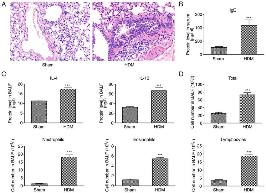
2        LIU et al: SERUM-DERIVED EXOSOMES INDUCE PRO-INFLAMMATORY ACTIVITY IN THE BRONCHIAL EPITHELIUM

in the intercellular transfer of information (19). In particular,   were from MilliporeSigma, interleukin (IL)‑4 and IL‑13 ELISA
exosomes can alter the biological functions of recipient cells      kits were from Nanjing Jiancheng Bioengineering Institute.
via the transfer of mitochondria (21).                              The Immunoglobulin E (IgE; cat. no. SFE40020) ELISA kit
    Recent studies have shown close associations between            was purchased from Shanghai Shifeng, Inc. (https://shfeng‑edu.
exosomes and inflammatory airway diseases (22‑24).                  biomart.cn), and IL‑6 (cat. no. EK 106/2‑96) and nerve growth
Exosomes are crucial in informational communication                 factor (NGF; cat. no. EK 1141‑96) ELISA kits were from
between asthma microenvironments and various cells (25).            Hangzhou Multi Sciences (Lianke) Biotech Co., Ltd. TAK‑242
Eosinophils secrete exosomes in healthy subjects and patients       (cat. no. HY‑11109) and BAY 11‑7082 (cat. no. HY‑13453) were
with asthma, the latter of which is particularly enhanced (16),     purchased from MedChemExpress, recombinant human heat
but stable patients with asthma and healthy subjects express        shock protein 70 (rHSP70; cat. no. 11660‑H07H) was from
exosomal microRNAs (miRNAs/miRs) differently (26).                  Sino Biological Technology Co., Ltd, and heat shock protein 70
Additionally, miR‑34a, miR‑92b and miR‑210 are enriched in          (HSP70)‑IN‑1 (cat. no. M9273) was from AbMole Bioscience,
exosomes, altering airway microenvironments upon asthma             Inc. Primary antibodies against CD63 (cat. no. ab68418),
development (27). One study found exosome secretion in mice         IKKα /β (cat. no. ab178870), phosphorylated (p)‑IKKα /β
BALF to be significantly enhanced upon contact with oval‑           (cat. no. ab194528) and HSP70 (cat. no. ab31010) were procured
bumin compared with controls (28). Paredes et al (29) noted         from Abcam, anti‑TLR4 (cat. no. BA1717) was from Boster
that exosomes from asthmatics induced leukotriene C4 and            Biological Technology, and anti‑p65 (cat. no. GB11997),
interleukin‑8 (IL‑8) secretions in airway epithelial cells (29).    anti‑p‑p65 (cat. no. GB11142‑1), anti‑β‑actin (cat. no. GB11001)
These extracellular vesicles can also mediate the pathogenesis      and a HRP‑conjugated goat anti‑rabbit IgG secondary anti‑
of COPD (30). Plasma exosome concentrations are higher in           body (cat. no. G1213) were purchased from Wuhan Servicebio
patients with acute exacerbations of COPD and stable COPD           Technology Co., Ltd.
than in healthy individuals (31). Reportedly, exosomal miR‑21
from bronchial epithelial cells in patients with COPD promotes      Cell culture. BEAS‑2B cells were cultivated in DMEM
myofibroblast differentiation through hypoxia‑inducible factor      supplemented with 20% serum replacement (a cell culture
1α (32). Clearly, exosomes from various sources mediate the         supplement that replaces fetal bovine serum to maintain cell
pathogenesis of inflammatory airway disorders.                      growth and reproduction in vitro), streptomycin (100 mg/ml)
    While most studies have focused on the promotion of             and penicillin (100 U/ml) at 37˚C in a 5% CO2 atmosphere.
the development of various cells or BALF exosome‑induced            The cells were passaged every 2 days in a 1:3 ratio. In cellular
airway inflammatory disorders, few have paid attention to           co‑culture with serum‑derived exosomes, serum replace‑
serum‑derived exosomes facilitating the pathogenesis of             ment was used to prevent interference from fetal bovine
inflammatory airway diseases. Thus, the present study inves‑        serum‑derived exosomes. BEAS‑2B cells were co‑cultured
tigated whether serum‑derived exosomes from House dust              with serum‑derived exosomes isolated from the HDM
mite (HDM)‑sensitized guinea pigs could alter the phenotypic        group and PBS group at 37˚C for 24 or 48 h separately.
appearances of bronchial epithelial cells and, if so, explore the   BEAS‑2B cells were also pretreated with or without
underlying molecular mechanisms.                                    5 µg/ml proteinase K (Beijing Solarbio Science & Technology
                                                                    Co., Ltd.), 5 µM BAY 11‑7082, 5 µM HSP70‑IN‑1 and different
Materials and methods                                               concentrations of TAK‑242 (100 or 300 nM) at 37˚C for 1 h,
                                                                    and then co‑cultured with serum‑derived exosomes from the
Animals. Female guinea pigs (6‑8 weeks old, n=20) were              HDM group at 37˚C for 24 h. BEAS‑2B cells were treated with
obtained from Hunan Changsha Tianqin Biotechnology                  or without rHSP70 (1 or 10 µg/ml) at 37˚C for 4 h.
(http://cstqsw.com) and acclimated for a week before the
experiments. The animals were kept in a pathogen‑free               Animal model. The guinea pigs (200‑250 g, female) were
environment and fed ad libitum. Our research protocol was           housed in a room maintained at moderate temperature
approved by the Animal Care Committee of Jiangxi Provincial         (22±2˚C) and humidity (40‑70%) with a 12 h light/dark cycle,
People's Hospital Affiliated with Nanchang University               and free access to food and water for the duration of the present
(approval no. 2021‑052; Nanchang, China).                           study. They were adaptively fed for 7 days and randomly
                                                                    divided into two groups: Sham group (PBS treatment group,
Cells, reagents and antibodies. The human bronchial epithe‑         n=10) and HDM group (n=10). HDM extracts (100,000 U/ml)
lial cell line BEAS‑2B was obtained from The Cell Bank              were diluted in a 0.1 mol/l PBS solution at concentrations of
of Type Culture Collection of The Chinese Academy of                2,000 U/ml, 4,000 U/ml and 8,000 U/ml. A HDM‑induced
Sciences. HDM extract was purchased from ALK‑Abelló A/S,            asthma model was created as shown in Fig. 1. The guinea
Dulbecco's modified Eagle's medium (DMEM) and ExoQuick              pigs in each group were intraperitoneally injected with pento‑
Exosome Isolation Reagent were acquired from Thermo Fisher          barbital (35 mg/kg). Subsequently, blood samples (5 ml per
Scientific, Inc. Serum replacement was from Stemboscience,          rodent) were withdrawn from the heart via cardiac puncture.
Inc., the PKH67 Green Fluorescent Cell Linker kit and DAPI          The guinea pigs were euthanized by the immediate removal of
were from Sigma‑Aldrich (Merck KGaA), while parafor‑                the heart after exsanguination; death was confirmed when the
maldehyde, Giemsa's stain and Phosphotungstic acid hydrate          animals developed cardiac arrest, respiratory arrest, corneal
were purchased from Beijing Solarbio Science & Technology           reflex arrest and rigor mortis. All procedures were conducted
Co., Ltd. The BCA Protein Quantitative kit was from CoWin           strictly in accordance with the National Institutes of Health
Biosciences, polyvinylidene difluoride (PVDF) membranes             Guide for the Care and Use of Laboratory Animals (33).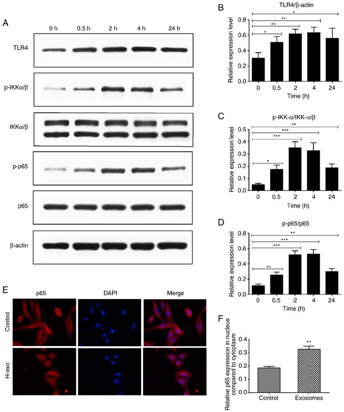
Molecular Medicine REPORTS 24: 747, 2021                                                            3

Lung histology. Harvested lung tissues from the guinea pigs
were fixed in 10% formalin solution for 24 h at 37˚C and
embedded in paraffin. After deparaffinization, 5 µm sections of
these tissues were stained with hematoxylin for 5 min at 37˚C
and eosin for 5 min at 37˚C (H&E) to observe morphology,
including pulmonary edema, airway inflammation and airway
epithelial injury under a light microscope (ECLIPSE CI;
Nikon Corporation).

Total cell counts. Precipitated cell suspension was conducted     Figure 1. Summary of the study protocol. The HDM group guinea pigs were
with 1 ml PBS. A few droplets from the suspension were taken      injected subcutaneously with 1,000 µl of 2,000 U/ml HDM on day 1, intra‑
to the cell‑count boards to determine the total cell count in     peritoneally injected with 1,000 µl of 2,000 U/ml HDM on days 3, 5 and 7, and
BALF per ml. The remaining precipitated cells, including          intraperitoneally with 500 µl of 4,000 U/ml HDM on days 9, 11 and 13. Guinea
                                                                  pigs in the experimental group were then sensitized and challenged in atom‑
eosinophils, neutrophils and lymphocytes, were fixed in 4%        ized boxes crafted for the present study with 8,000 U/ml HDM extract from
paraformaldehyde solution for 30 min at 37˚C and stained          days 15 to 21, each time for 30 min. Sham group rodents were sensitized
with Wright‑Giemsa for 20 min at 37˚C (at least 200 cells per     and challenged with PBS, instead of HDM. Subsequently, the animals were
sample) to deduce the percentage of cells under a light micro‑    subjected to tracheotomy and washed with 5 ml ice‑cold PBS three times
                                                                  before BALF was collected. Serum from the heart was collected using
scope [ECLIPSE CI; Nikon Corporation (magnification, x40)].       disposable needles, and lung tissues were harvested. HDM, house dust mite;
                                                                  BALF, bronchoalveolar lavage fluid.
ELISA. BALF IL‑4 and IL‑13 concentrations and serum IgE
levels from treated‑guinea pigs and IL‑6 and NGF contents in
cell supernatants were quantified using ELISA kits. In brief,
the guinea pigs were subjected to tracheotomy and washed with     at room temperature. The sample was then dried for 2 min
5 ml ice‑cold PBS three times before BALF was collected. The      under incandescent light, and the results were observed and
obtained BALF supernatants was centrifuged at 4˚C (250 x g,       images captured using a transmission electron microscope
10 min). Different concentrations of serum‑derived exosomes       (JEM‑1200EX; JEOL, Ltd.) at an acceleration voltage of 80 kV.
(0, 50, 100, 200 µg/ml) from the HDM group were added to          The TEM images were cropped and scaled by Photoshop CS6
BEAS‑2B cells simultaneously and incubated at 37˚C for            (Adobe Systems Incorporated).
24 and 48 h. After incubation, cell supernatants were collected
by centrifugation at 4˚C (1,000 x g, 15 min).                     Western blotting. After washing three times with precooled
                                                                  PBS and a protease inhibitor cocktail on ice for 30 min, cells
Exosome isolation and quantitation. Serum exosomes were           were harvested in RIPA lysis buffer with 1 mM PMSF. The
isolated using the ExoQuick Exosomes Isolation Reagent,           concentration of protein was measured using a BCA protein
according to the manufacturer's recommended protocol.             assay kit. Total proteins (30 µg per lane) were loaded, sepa‑
Briefly, serum from HDM‑sensitized and PBS‑sensitized             rated with 8‑10% SDS‑polyacrylamide gels, and transferred
guinea pigs was differentially centrifuged at 4˚C (2,000 x g,     onto PVDF membranes. The PVDF membranes were blocked
30 min) to remove cells and debris. The supernatants              with 5% non‑fat milk at room temperature for 2 h and then
were filtered through 0.22‑µm filters to eliminate particles      incubated with a 1:1,000 dilution of the specific primary
>220 nm, and a reagent mixture was added to the well until        antibodies at 4˚C overnight. Followed by washing with TBS
the solution was homogenous. The mixed suspension was then        with 1% Tween‑20 three times (10 min each time), and incu‑
incubated at 4˚C for 30 min and centrifuged at 10,000 x g         bation with horseradish peroxidase‑conjugated secondary
for 10 min at room temperature. Exosomes contained in the         antibodies (1:3,000) at room temperature for 1 h. Then,
pellet at the bottom of the tube were re‑suspended in 200 µl      the blots was visualized with enhanced chemiluminescent
PBS. Serum‑produced exosomal proteins were quantitated            solution (Beijing Solarbio Science & Technology Co., Ltd.).
using the BCA Protein Assay kit, and estimated by reference       Primary antibodies against the following were used: CD63,
to a standard curve generated from proteins [bovine serum         HSP70, TLR4, IKKα /β, p‑IKKα /β, p65, p‑p65 and β ‑actin.
albumin (BSA)] of known concentration.                            Band densities were analyzed using ImageJ software 6.0
                                                                  (National Institutes of Health); the β‑actin protein was used
Experimental groups. The samples were divided into three          as an internal reference.
groups: Control group (untreated cells), S‑exo treatment group
(exosomes from the sham group) and H‑exo treatment group          Exosome labeling. Exosomes were labeled with PKH67
(exosomes from the house dust mite group).                        (a novel fluorescent dye that labels living cells by binding
                                                                  to lipid molecules in membrane structures) for general
Transmission electron microscopy (TEM). The ultrastruc‑           cell membrane labelling according to the manufacturer's
ture of exosomes was observed using TEM, referring to the         instructions, with minor modifications. In brief, 100 µl
methods described in a previous study (34). A 20 µl drop of       serum‑derived exosomes from the HDM group were mixed
the exosomal suspension was placed on parafilm and loaded         with 1 ml Diluent C, and for control, 1 ml Diluent C was
to a carbon‑coated grid for 2 min. A 2% phosphotungstic           mixed with PBS. The Diluent C added to the experiment
acid solution prepared with triple distilled water was used       and control was prepared by mixing 1 µl PKH67 dye with
to stain the carbon‑coated grid‑loaded suspension for 30 sec      750 µl Diluent C. Next, 1 ml of 1% BSA (Beijing Solarbio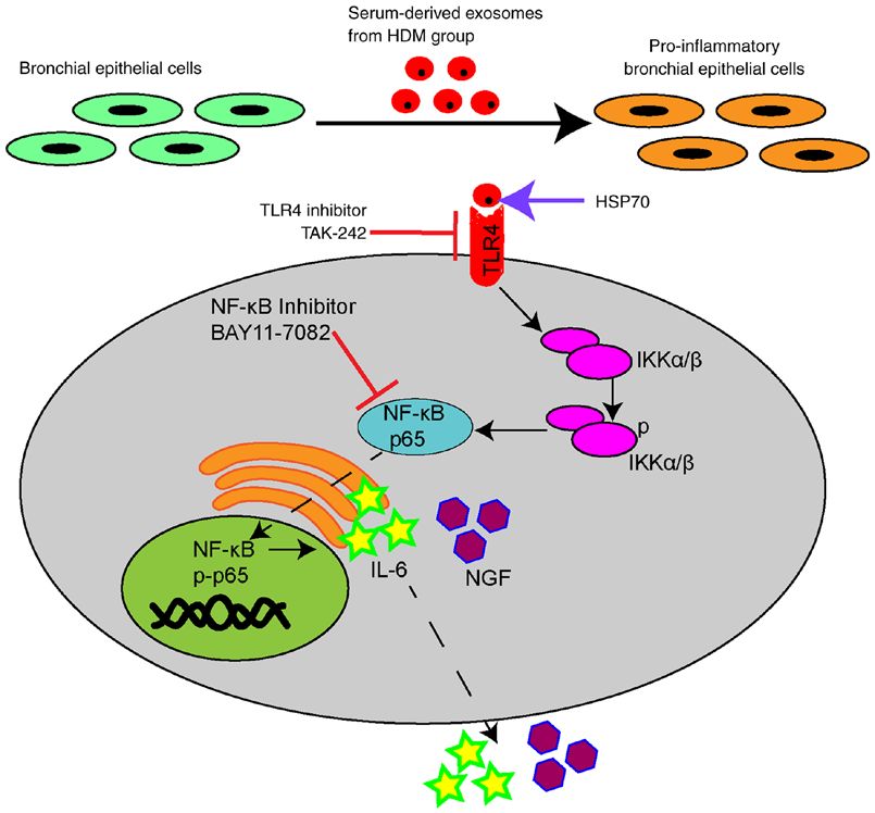
4         LIU et al: SERUM-DERIVED EXOSOMES INDUCE PRO-INFLAMMATORY ACTIVITY IN THE BRONCHIAL EPITHELIUM

Table I. Sequences of primers used in reverse transcription-        Statistical analysis. GraphPad Prism 6.0 (GraphPad Software,
quantitative PCR.                                                   Inc.) was used for all statistical analyses. Data are expressed
                                                                    as the mean ± SD. An unpaired Student's t‑test was used for
Primer                         Sequences (5'→3')                    comparisons between two groups, and one‑way ANOVA
                                                                    followed by the Bonferroni post hoc test were employed for
IL‑6            F:   GTAGTGAGGAACAAGCCAGAGC                         multiple comparisons. P
Molecular Medicine REPORTS 24: 747, 2021 5 Figure 2. Allergy asthma model in guinea pigs was established successfully via HDM sensitization and challenge. (A) A lung tissue section depicting the level of airway inflammation (hematoxylin and eosin staining). Magnification, x200. (B) ELISA‑determined Serum IgE levels. (C) ELISA‑determined BALF IL‑4 and IL‑13 contents. (D) Total cell count and inflammatory cell count in BALF. ***P
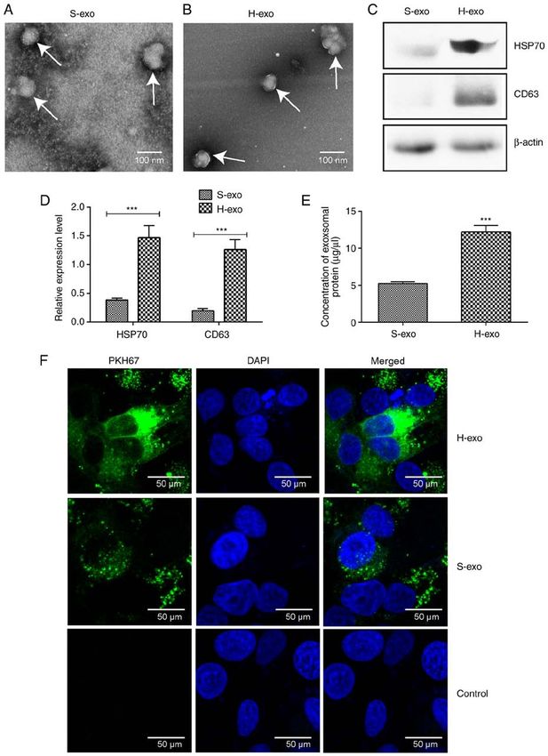
6         LIU et al: SERUM-DERIVED EXOSOMES INDUCE PRO-INFLAMMATORY ACTIVITY IN THE BRONCHIAL EPITHELIUM

Figure 3. Characterization of serum‑derived exosomes. (A and B) Morphology of S‑exo and H‑exo under transmission electron microscopy. Scale bar, 100 nm.
(C and D) Immunoblot‑determined CD63 and HSP70 expression levels. β‑actin was used as a loading control. (E) BCA Protein Assay kit‑determined S‑exo
and H‑exo concentrations. (F) S‑exo and H‑exo uptake by BEAS‑2B cells. Fluorescence microscopy images of BEAS‑2B cells incubated with PKH67‑labeled
serum exosomes (green) or non‑incubated exosomes. BEAS‑2B cell nuclei were stained with DAPI stain (blue). Scale bar, 50 µm. ***P
Molecular Medicine REPORTS 24: 747, 2021                                                                7

Figure 4. Effects of serum‑derived exosomes on immune‑related cytokine secretion in BEAS‑2B cells. (A) RT‑qPCR‑determined mRNA levels of cytokines in
BEAS‑2B cells after addition of H‑exo or S‑exo. ***P
8          LIU et al: SERUM-DERIVED EXOSOMES INDUCE PRO-INFLAMMATORY ACTIVITY IN THE BRONCHIAL EPITHELIUM

Figure 5. TLR4‑NF‑κB pathway activation in BEAS‑2B cells. (A‑D) TLR4‑NF‑κB pathway protein expression changes in BEAS‑2B cells. *P
Molecular Medicine REPORTS 24: 747, 2021                                                               9

Figure 6. Inhibition of mRNA and protein expression levels in the TLR4‑NF‑κB pathway following TAK‑242 addition to BEAS‑2B cells. (A and B) IL‑6 and
NGF mRNA expression changes in BEAS‑2B cells. (C‑F) Western blotting‑determined activation of TLR4‑NF‑κB in BEAS‑2B cells. BEAS‑2B cells were
pretreated with various concentrations of TAK‑242 and incubated with H‑exo. *P
10         LIU et al: SERUM-DERIVED EXOSOMES INDUCE PRO-INFLAMMATORY ACTIVITY IN THE BRONCHIAL EPITHELIUM

Figure 7. Exosomal surface HSP70 stimulation of IL‑6 and NGF expression levels via the TLR4 pathway. (A and B) Relative mRNA expression levels of IL‑6
and NGF following the addition of proteinase K. (C) Cytokine mRNA levels following the addition of various concentrations of rHSP70 to BEAS‑2B cells.
(D) IL‑6 and NGF mRNA expression changes in the BEAS‑2B cells incubated with 1 µg/ml rHSP70 after treatment with 100 nM TAK‑242 for 24 h. *P
Molecular Medicine REPORTS 24: 747, 2021                                                           11

Figure 8. Schematic representation of exosome‑mediated inflammatory response in BEAS‑2B cells. Serum‑derived exosomes from HDM group induces the
transformation of bronchial epithelial cells into pro‑inflammatory bronchial epithelia by activating the TLR4‑NF‑κB pathway via HSP70. HDM, house dust
mite; IL‑6, interleukin‑6; NGF, nerve growth factor; TLR4, toll‑like receptor 4; HSP70, heat shock protein 70; p‑, phosphorylated.

research on the involvement of serum‑derived exosomes in the                 alter BEAS‑2B cell phenotypic appearances by regulating the
pathogenesis of asthma is far from complete. Previous analysis               TLR4‑NF‑κ B signaling pathway (Fig. 7A‑D). A previous study
of the expression of miR‑125b in the serum exosomes of patients              has shown that treatment with proteinase K‑digested HSP70
with different severities of asthma compared with healthy                    in bone marrow‑derived dendritic cells results in a reduc‑
subjects showed an altered miR‑125b content, and thus may                    tion in HSP70‑dependent cytokines (57). A previous study
have potential as a diagnostic marker for asthma (55). Therefore,            reported that the increased secretion of inflammatory factors
further research must be conducted to determine whether                      could be markedly suppressed following the pretreatment of
serum‑derived exosomes from asthmatic patients of different                  mesenchymal stem cells with proteinase K compared with a
severity play a pro‑inflammatory role in bronchial epithelial                A549 exosome‑treated group (58). As expected, the results
cells or other receptor cells, and this is our next research focus.          of the current study indicated that the increases in cytokine
    Exosomal surface molecules can mediate intracellular                     concentrations were partially repressed by proteinase K and
signaling pathways through direct contact with receptors on                  the HSP70 blocker compared with the exosome‑treated group
target cells. Anand et al (37) demonstrated that exosomal                    without the enzyme. Therefore, it should be considered that
surface HSP70 levels from macrophages infected with myco‑                    other exosomal components, including nucleic acids and other
bacteria are expressed higher than in controls, and HSP70 in                 proteins, possibly participate in this inflammatory response
exosome‑treated macrophages activates NF‑κ B signaling to                    and this should be further explored.
stimulate the release of TNF‑α in uninfected macrophages.                        In conclusion, it was demonstrated that serum‑derived
Circulating HSP70 levels from patients with asthma are                       exosomes interacted with BEAS‑2B cells and could alter their
relevant to the severity of disorders and the symptom of                     phenotypic appearance. Additionally, the HSP70‑modulated
asthma and, therefore, may contribute to the pathogenesis of                 inflammatory effect on the surface of serum‑derived exosomes
the disease (56). The present study showed that serum‑derived                from the HDM group upregulated IL‑6 and NGF expres‑
exosomal HSP70 in the HDM group was higher than in the                       sion levels by activating the TLR4‑NF‑κ B pathway (Fig. 8).
sham group, suggesting that exosomal surface HSP70 may be                    Overall, exosomal presence in HDM‑sensitized guinea pigs
involved in the pathogenesis process. HSP70 on the surface of                could be influential in the underlying mechanism of inflam‑
serum‑derived exosomes from the HDM group could, therefore,                  matory airway diseases; however, blocking exosome‑mediated
12 LIU et al: SERUM-DERIVED EXOSOMES INDUCE PRO-INFLAMMATORY ACTIVITY IN THE BRONCHIAL EPITHELIUM
communication between cells would attenuate the inflamma‑ 6. Gon Y and Hashimoto S: Role of airway epithelial barrier dysfunc‑
tion in pathogenesis of asthma. Allergol Int 67: 12‑17, 2018.
tion, potentially partially relieving symptoms of inflammatory 7. Mitchell PD and O'Byrne PM: Epithelial‑derived cytokines in
airway diseases in guinea pigs. asthma. Chest 151: 1338‑1344, 2017.
8. Mitchell PD and O'Byrne PM: Biologics and the lung: TSLP
and other epithelial cell‑derived cytokines in asthma. Pharmacol
Acknowledgements Ther 169: 104‑112, 2017.
9. Sun Z, Ji N, Ma Q, Zhu R, Chen Z, Wang Z, Qian Y, Wu C, Hu F,
Not applicable. Huang M and Zhang M: Epithelial‑mesenchymal transition in
asthma airway remodeling is regulated by the IL‑33/CD146 axis.
Front Immunol 11: 1598, 2020.
Funding 10. Kato A and Schleimer RP: Beyond inflammation: Airway epithe‑
lial cells are at the interface of innate and adaptive immunity.
Curr Opin Immunol 19: 711‑720, 2007.
The present study was supported by the Natural Science 11. Videira RF and da Costa Martins PA: Non‑coding RNAs in
Foundation of China (grant nos. 81460004 and 82160006); cardiac intercellular communication. Front Physiol 11: 738, 2020.
the Jiangxi Provincial Cultivation Program for Academic 12. Barros FM, Carneiro F, Machado JC and Melo SA: Exosomes and
immune response in cancer: Friends or foes? Front Immunol 9:
and Technical Leaders of Major Subjects (grant no. 730, 2018.
20172BCB22025); and the Jiangxi Provincial Natural Science 13. Smith VL, Cheng Y, Bryant BR and Schorey JS: Exosomes func‑
Foundation General Project (grant no. 20202BAB206003). tion in antigen presentation during an in vivo mycobacterium
tuberculosis infection. Sci Rep 7: 43578, 2017.
14. Chan BD, Wong WY, Lee MM, Cho WC, Yee BK, Kwan YW and
Availability of data and materials Tai WC: Exosomes in inflammation and inflammatory disease.
Proteomics 19: e1800149, 2019.
15. Zhao B, Li X, Shi X, Shi X, Zhang W, Wu G, Wang X, Su L
The datasets used and/or analyzed during the current study are and Hu D: Exosomal microRNAs derived from human amni‑
available from the corresponding author on reasonable request. otic epithelial cells accelerate wound healing by promoting the
proliferation and migration of fibroblasts. Stem Cells Int 2018:
5420463, 2018.
Authors' contributions 16. Mazzeo C, Cañas JA, Zafra MP, Rojas Marco A, Fernández-
Nieto M, Sanz V, Mittelbrunn M, Izquierdo M, Baixaulli F,
JW and CL conceived and designed the present study. CL, Sastre J and Del Pozo V: Exosome secretion by eosinophils: A
possible role in asthma pathogenesis. J Allergy Clin Immun 135:
XLH, XZ, ZFW and LXD performed the experiments. CL, 1603‑1613, 2015.
JPL and XLH analyzed the experimental data. CL wrote 17. Zhao L, Yu J, Wang J, Li H, Che J and Cao B: Isolation and
the manuscript. CL and JW confirm the authenticity of all identification of miRNAs in exosomes derived from serum of
colon cancer patients. J Cancer 8: 1145‑1152, 2017.
the raw data. All authors have read and approved the final 18. Alipoor SD, Mortaz E, Garssen J, Movassaghi M, Mirsaeidi M
manuscript. and Adcock IM: Exosomes and exosomal miRNA in respiratory
diseases. Mediat Inflamm 2016: 1‑11, 2016.
19. Mathivanan S, Ji H and Simpson RJ: Exosomes: Extracellular
Ethics approval and consent to participate organelles impor tant in intercellular communication.
J Proteomics 73: 1907‑1920, 2010.
The present study was approved by the Animal Care and 20. Turturici G, Tinnirello R, Sconzo G and Geraci F: Extracellular
membrane vesicles as a mechanism of Cell‑to ‑ Cell
Committee of Jiangxi Provincial People's Hospital Affiliated communication: Advantages and disadvantages. Am J Physiol
to Nanchang University (approval no. 2021‑052; Nanchang, Cell Physiol 306: C621‑C633, 2014.
China). 21. Hough KP and Deshane JS: Exosomes in allergic airway diseases.
Curr Allergy Asthma Rep 19: 26, 2019.
22. Kadota T, Fujita Y, Yoshioka Y, Araya J, Kuwano K and Ochiya T:
Patient consent for publication Extracellular vesicles in chronic obstructive pulmonary disease.
Int J Mol Sci 17: 1801, 2016.
23. Mortaz E, Alipoor SD, Varahram M, Jamaati H, Garssen J,
Not applicable. Mumby SE and Adcock IM: Exosomes in severe asthma: Update
in their roles and potential in therapy. Biomed Res Int 2018:
Competing interests 2862187, 2018.
24. Cañas JA, Sastre B, Rodrigo‑Muñoz JM and Del PV: Exosomes:
A new approach to asthma pathology. Clin Chim Acta 495:
The authors declare that they have no competing interests. 139‑147, 2019.
25. Huang F, Jia H, Zou Y, Yao Y and Deng Z: Exosomes: An impor‑
tant messenger in the asthma inflammatory microenvironment.
References J Int Med Res 48: 1220702772, 2020.
26. Levanen B, Bhakta NR, Torregrosa PP, Barbeau R, Hiltbrunner S,
1. Zanini A, Cherubino F, Zampogna E, Croce S, Pignatti P and Pollack JL, Skold CM, Svartengren M, Grunewald J,
Spanevello A: Bronchial hyperresponsiveness, airway inflam‑ Gabrielsson S, et al: Altered microRNA profiles in bronchoal‑
mation, and reversibility in patients with chronic obstructive veolar lavage fluid exosomes in asthmatic patients. J Allergy Clin
pulmonary disease. Int J Chron Obstruct Pulmon Dis 10: Immunol 131: 894‑903, 2013.
1155‑1161, 2015. 27. Bartel S, La Grutta S, Cilluffo G, Perconti G, Bongiovanni A,
2. Alashkar AB, Miethe S, Pogge VSE, Potaczek DP and Garn H: Giallongo A, Behrends J, Kruppa J, Hermann S, Chiang D, et al:
Epigenetic regulation of airway epithelium immune functions in Human airway epithelial extracellular vesicle miRNA signature
asthma. Front Immunol 11: 1747, 2020. is altered upon asthma development. Allergy 75: 346‑356, 2020.
3. Fujita Y, Kosaka N, Araya J, Kuwano K and Ochiya T: Extracellular 28. Kulshreshtha A, Ahmad T, Agrawal A and Ghosh B:
vesicles in lung microenvironment and pathogenesis. Trends Mol Proinflammatory role of epithelial cell‑derived exosomes in
Med 21: 533‑542, 2015. allergic airway inflammation. J Allergy Clin Immun 131:
4. Weitnauer M, Mijošek V and Dalpke AH: Control of local immu‑ 1194‑1203, 2013.
nity by airway epithelial cells. Mucosal Immunol 9: 287‑298, 29. Paredes PT, Esser J, Admyre C, Nord M, Rahman QK, Lukic A,
2016. Radmark O, Gronneberg R, Grunewald J, Eklund A, et al:
5. Holgate ST: Epithelium dysfunction in asthma. J Allergy Clin Bronchoalveolar lavage fluid exosomes contribute to cytokine and
Immunol 120: 1233‑1246, 2007. leukotriene production in allergic asthma. Allergy 67: 911‑919, 2012.Molecular Medicine REPORTS 24: 747, 2021 13
30. Hough KP, Chanda D, Duncan SR, Thannickal VJ and 46. Bretz NP, Ridinger J, Rupp AK, Rimbach K, Keller S, Rupp C,
Deshane JS: Exosomes in immunoregulation of chronic lung Marmé F, Umansky L, Umansky V, Eigenbrod T, et al: Body
diseases. Allergy 72: 534‑544, 2017. fluid exosomes promote secretion of inflammatory cytokines in
31. Tan D, Armitage J, Teo TH, Ong NE, Shin H and Moodley YP: monocytic cells via Toll‑like receptor signaling. J Biol Chem 288:
Elevated levels of circulating exosome in COPD patients are 36691‑36702, 2013.
associated with systemic inflammation. Respir Med 132: 47. Rincon M and Irvin CG: Role of IL‑6 in asthma and other inflam‑
261‑264, 2017. matory pulmonary diseases. Int J Biol Sci 8: 1281‑1290, 2012.
32. Xu H, Ling M, Xue J, Dai X, Sun Q, Chen C, Liu Y, Zhou L, 48. Abram M, Wegmann M, Fokuhl V, Sonar S, Luger EO, Kerzel S,
Liu J, Luo F, et al: Exosomal microRNA‑21 derived from bron‑ Radbruch A, Renz H and Zemlin M: Nerve growth factor and
chial epithelial cells is involved in aberrant epithelium‑fibroblast neurotrophin‑3 mediate survival of pulmonary plasma cells
cross‑talk in COPD induced by cigarette smoking. Theranostics 8: during the allergic airway inflammation. J Immunol 182:
5419‑5433, 2018. 4705‑4712, 2009.
33. Care NRCU and Animals AUOL: Guide for the Care and Use of 49. Othman N, Jamal R and Abu N: Cancer‑derived exosomes as
Laboratory Animals. National Academies Press US, Washington, effectors of key inflammation‑related players. Front Immunol 10:
DC, 2011. 2103, 2019.
34. Tang YT, Huang YY, Zheng L, Qin SH, Xu XP, An TX, Xu Y, 50. Cañas JA, Sastre B, Rodrigo‑Muñoz JM, Fernández‑Nieto M,
Wu YS, Hu XM, Ping BH and Wang Q: Comparison of isola‑ Barranco P, Quirce S, Sastre J and Del PV: Eosinophil‑derived
tion methods of exosomes and exosomal RNA from cell culture exosomes contribute to asthma remodelling by activating struc‑
medium and serum. Int J Mol Med 40: 834‑844, 2017. tural lung cells. Clin Exp Allergy 48: 1173‑1185, 2018.
35. Livak KJ and Schmittgen TD: Analysis of relative gene expres‑ 51. Gao W, Liu H, Yuan J, Wu C, Huang D, Ma Y, Zhu J, Ma L,
sion data using real‑time quantitative PCR and the 2(‑Delta Delta Guo J, Shi H, et al: Exosomes derived from mature dendritic
C(T)) method. Methods 25: 402‑408, 2001. cells increase endothelial inflammation and atherosclerosis via
36. Zhang L and Yu D: Exosomes in cancer development, metas‑ membrane TNF‑α mediated NF‑κ B pathway. J Cell Mol Med 20:
tasis, and immunity. Biochim Biophys Acta Rev Cancer 1871: 2318‑2327, 2016.
455‑468, 2019. 52. Ye W, Tang X, Yang Z, Liu C, Zhang X, Jin J and Lyu J:
37. Anand PK, Anand E, Bleck CK, Anes E and Griffiths G: Plasma‑derived exosomes contribute to inflammation via the
Exosomal Hsp70 induces a pro‑inflammatory response to foreign TLR9‑NF‑κ B pathway in chronic heart failure patients. Mol
particles including mycobacteria. PLoS One 5: e10136, 2010. Immunol 87: 114‑121, 2017.
38. Dickson RP, Erb‑Downward JR and Huffnagle GB: Homeostasis 53. Zandi E, Rothwarf DM, Delhase M, Hayakawa M and Karin M:
and its disruption in the lung microbiome. Am J Physiol Lung The IkappaB kinase complex (IKK) contains two kinase subunits,
Cell Mol Physiol 309: L1047‑L1055, 2015. IKKalpha and IKKbeta, necessary for IkappaB phosphorylation
39. Mendez R, Banerjee S, Bhattacharya SK and Banerjee S: Lung and NF‑kappaB activation. Cell 91: 243‑252, 1997.
Inflammation and disease: A perspective on microbial homeo‑ 54. Schuliga M: NF‑kappaB signaling in chronic inflammatory
stasis and metabolism. Iubmb Life 71: 152‑165, 2019. airway disease. Biomolecules 5: 1266‑1283, 2015.
40. Paplinska‑Goryca M, Misiukiewicz‑Stepien P, Nejman‑Gryz P, 55. Zhao M, Juanjuan L, Weijia F, Jing X, Qiuhua H, Hua Z, Fuhe L
Proboszcz M, Mlacki M, Gorska K and Krenke R: Epithelial- and Hao P: Expression levels of microRNA‑125b in serum
macrophage‑dendritic cell interactions impact alarmins exosomes of patients with asthma of different severity and its
expression in asthma and COPD. Clin Immunol 215: 108421, diagnostic significance. Curr Drug Metab 20: 781‑784, 2019.
2020. 56. Hou C, Zhao H, Li W, Liang Z, Zhang D, Liu L, Tong W, Cai SX
41. Cornwell WD, Kim V, Song C and Rogers TJ: Pathogenesis of and Zou F: Increased heat shock protein 70 levels in induced
inflammation and repair in advanced COPD. Semin Respir Crit sputum and plasma correlate with severity of asthma patients.
Care Med 31: 257‑266, 2010. Cell Stress Chaperones 16: 663‑671, 2011.
42. Admyre C, Bohle B, Johansson SM, Focke‑Tejkl M, Valenta R, 57. Spiering R, van der Zee R, Wagenaar J, van Eden W and Broere F:
Scheynius A and Gabrielsson S: B cell‑derived exosomes can Mycobacterial and mouse HSP70 have immuno‑modulatory
present allergen peptides and activate allergen‑specific T cells effects on dendritic cells. Cell Stress Chaperones 18: 439‑446,
to proliferate and produce TH2‑like cytokines. J Allergy Clin 2013.
Immunol 120: 1418‑1424, 2007. 58. Li X, Wang S, Zhu R, Li H, Han Q and Zhao RC: Lung tumor
43. Valadi H, Ekström K, Bossios A, Sjöstrand M, Lee JJ and exosomes induce a pro‑inflammatory phenotype in mesenchymal
Lötvall JO: Exosome‑mediated transfer of mRNAs and stem cells via NFκ B‑TLR signaling pathway. J Hematol Oncol 9:
microRNAs is a novel mechanism of genetic exchange between 42, 2016.
cells. Nat Cell Biol 9: 654‑659, 2007.
44. Vargas A, Roux‑Dalvai F, Droit A and Lavoie JP: Neutrophil- This work is licensed under a Creative Commons
derived exosomes: A new mechanism contributing to airway Attribution-NonCommercial-NoDerivatives 4.0
smooth muscle remodeling. Am J Respir Cell Mol Biol 55: International (CC BY-NC-ND 4.0) License.
450‑461, 2016.
45. Sahoo S and Losordo DW: Exosomes and cardiac repair after
myocardial infarction. Circ Res 114: 333‑344, 2014.You can also read


