Identification of Novel Isoforms of the BH3 Domain Protein Bim Which Directly Activate Bax To Trigger Apoptosis
←
→
Page content transcription
If your browser does not render page correctly, please read the page content below

MOLECULAR AND CELLULAR BIOLOGY, June 2002, p. 3577–3589 Vol. 22, No. 11
0270-7306/02/$04.00⫹0 DOI: 10.1128/MCB.22.11.3577–3589.2002
Copyright © 2002, American Society for Microbiology. All Rights Reserved.
Identification of Novel Isoforms of the BH3 Domain Protein Bim
Which Directly Activate Bax To Trigger Apoptosis
Michela Marani,1 Tencho Tenev,1† David Hancock,2 Julian Downward,2 and Nicholas R. Lemoine1*
Molecular Oncology Unit, Imperial College School of Medicine, Hammersmith Hospital, London W12 0NN,1 and
Signal Transduction Laboratory, Cancer Research UK, London WC2A 3PX,2 United Kingdom
Received 6 August 2001/Returned for modification 7 September 2001/Accepted 4 March 2002
Bim (Bcl-2-interacting mediator of cell death) is a member of the BH3 domain-only subgroup of Bcl-2 family
Downloaded from http://mcb.asm.org/ on February 26, 2021 by guest
members, for which three splice variants have been described. Bim is expressed in many healthy cell types,
where it is maintained in an inactive conformation through binding to the microtubule-associated dynein
motor complex. Upon certain apoptotic stimuli, Bim is released from microtubules and mediates caspase-
dependent apoptosis through a mechanism that is still unclear. Here, we have identified and characterized
novel splice variants of human Bim mRNA. In particular, we show that a newly discovered, small protein
isoform, BimAD, is also able to induce apoptosis strongly in several human cell lines. BimAD and the
previously characterized isoform BimS are shown to be capable of heterodimerizing in vivo with both death
antagonists (Bcl-2 and Bcl-XL) and death agonists (Bax). Mutants of BimAD that bind to Bax but not to Bcl-2
still promote apoptosis, indicating that Bim can regulate apoptosis through direct activation of the Bax-
mediated cell death pathway without interaction with antiapoptotic Bcl-2 family members. Furthermore, we
have shown that the interaction of the BimS and BimAD isoforms with Bax leads to a conformational change
in this protein analogous to that triggered by the BH3-only protein Bid.
Members of the Bcl-2 family of proteins have come to be other family members, allowing the activated protein to exert
regarded as central players in the “decision” step triggering its proapoptotic activity (reviewed in reference 17).
apoptosis. Some proteins within this family, such as Bcl-2, Although it has been suggested that BH3-only proteins pro-
Bcl-XL, and Mcl-1, inhibit apoptosis, while others, including mote apoptosis by binding and inhibiting the activity of anti-
Bax, Bak, Bad, and Bim, promote cell death. Members of this apoptotic members of the family, recent data also point to their
family contain at least one of four conserved motifs known as potential role as direct enhancers of the proapoptotic function
the Bcl-2 homology (BH) domains, termed BH1, BH2, BH3, of Bax and Bak. In support of this view, Bid has been shown to
and BH4 (reviewed in references 1 and 2). These domains are interact with either survival factors or proapoptotic Bax and
important in determining the pro- and antiapoptotic properties Bak, in which it produces a conformational change that pro-
of the various family members and in mediating interactions motes their death activity (9, 37).
between them. Structural studies and mutational analyses have Among the different Bcl-2 family members, we were inter-
highlighted the key role of the BH3 domain in the death ested in characterizing in more detail the function of the BH3-
agonist activity of proapoptotic members. Furthermore, the only death agonist, Bim. Bim was originally identified as a
increasing number of cell death agonists with no similarity to Bcl-2-interacting protein by screening a bacteriophage
Bcl-2 beyond their BH3 homology has confirmed the key role cDNA expression library constructed from a mouse thymic
of this domain in triggering apoptosis. The proapoptotic pro- lymphoma (21). Three isoforms were characterized, BimEL,
teins fall into two groups, those having only the BH3 domain in BimL, and BimS. These differed from each other in cytotoxic-
common, such as Bad, Bid, and Bim, and those having the ity, with BimS being the most potent. This is partly explained
BH1, BH2, and BH3 domains in common, such as Bax and by the sequestration of BimEL and BimL in the cytoskeleton-
Bak. Several of these proteins appear to exist in an inactive
associated motor complex bound to dynein light chain LC8,
conformation in viable cells, and depending on the specific
from which they are released upon various apoptotic stimuli
death signals and the particular cell type, one or more death
(26). The expression of forkhead transcription factors may also
agonists can undergo a posttranslational modification resulting
contribute to Bim expression and activity (10). BimS protein
in an active conformation. This then determines the intracel-
has been only recently detected in 293 human embryonic kid-
lular localization and defines the pattern of interactions with
ney cells (25), while BimEL and BimL have been found in a
variety of tissues and cell types (22). Inhibition of Bim expres-
sion has been shown to have specific effects on hematopoietic
* Corresponding author. Mailing address: Molecular Oncology
homeostasis, and Bim proteins appear to play an important
Unit, Imperial College School of Medicine, Hammersmith Hospital, role in the prevention of autoimmunity (6).
DuCane Road, London W12 0NN, United Kingdom. Phone: In this report, we describe the cloning and functional char-
44(0)2083833975. Fax: 44(0)2083833258. E-mail: nick.lemoine@cancer acterization of novel Bim isoforms which are the products of
.org.uk.
† Present address: The Breakthrough Toby Robins Breast Cancer
alternative splicing. Apoptosis assays reveal clear differences
Research Centre, Institute of Cancer Research, London SW3 6JB, in the activities of the new isoforms, and only one, named
United Kingdom. BimAD, has a proapoptotic activity comparable to that of
3577
3578 MARANI ET AL. MOL. CELL. BIOL.
BimS. Analysis of the human Bim gene indicates that BimAD centage of apoptotic cells was quantified by microscopically counting the number
results from the splicing out of exons 3 and 4 during mRNA of apoptotic (round) blue cells versus the total number of blue cells counted (500
to 800 cells) from five randomly chosen fields.
processing. The BimAD protein retains the BH3 domain but The apoptosis assays were repeated with incubation of the cells with the
lacks the predicted carboxyl-terminal hydrophobic region broad-spectrum caspase inhibitor zVAD-fmk (Calbiochem; 50 M).
found in the previously characterized Bim isoforms due to an Antibodies. The antibodies used in the study of Bim protein isoforms and
early stop codon in a newly discovered exon. In transient trans- members of the Bcl-2 family were as follows: a rabbit polyclonal antibody against
amino acids 22 to 40 of human Bim protein (anti-Bim; N terminal; Calbiochem),
fection assays, BimAD induces apoptosis that is suppressible
a rat monoclonal antibody against human Bim [Bim (Ab-1); Calbiochem], a
by coexpression of Bcl-2 or the pan-caspase inhibitor zVAD- rabbit polyclonal antibody against amino acids 11 to 30 of human Bax (Bax N20:
fmk. Binding studies show that BimS and BimAD are able to sc-493; Santa Cruz Biotechnology), a mouse monoclonal antibody against human
bind not only to the antiapoptotic proteins Bcl-2 and Bcl-XL Bax (YTH-6A7; Trevigen), a mouse monoclonal antibody against human Bcl-2
but also to proapoptotic Bax. Engineering mutations in Bim [Bcl-2 (100): sc-509; Santa Cruz Biotechnology], a goat polyclonal antibody
against actin [actin (C-11): sc-1615; Santa Cruz Biotechnology], a rabbit poly-
that distinguish between Bcl-2 and Bax binding indicate that clonal antibody against amino acids 18 to 233 of human Bcl-XS/L (B22630;
Bim can induce apoptosis through direct interaction with Bax Transduction Laboratories), and a goat polyclonal antibody against glutathione
Downloaded from http://mcb.asm.org/ on February 26, 2021 by guest
as well as through interaction with Bcl-2 and Bcl-XL. Further- S-transferase (GST) (Amersham Pharmacia Biotech).
more, we show that the expression of BimS and BimAD results TAP tag purification and immunoblot analysis. Each Bim variant was sub-
in a conformational change in Bax that has been proposed as cloned as a fusion product with the C-terminal tandem affinity purification (TAP)
tag sequence (24, 28) in pcDNA3.1, and derived clones were verified by auto-
a critical step in Bax activation (9, 20). The data presented here mated sequencing (as described above). 293 cells in a logarithmic phase of
provide new insights for understanding the cell death pathway growth were seeded on 100-mm-diameter tissue culture dishes at a density of 2
triggered by Bim. ⫻ 105 cells/ml in fresh medium the day before transfection. The cells were
transiently cotransfected with 10 g of each of the expression plasmids by using
a CalPhos mammalian transfection kit (Clontech) according to the manufactur-
MATERIALS AND METHODS er’s recommended procedure. Adherent cells were lysed 48 h after transfection
Cloning of Bim cDNAs. An oligonucleotide primer (5⬘-TGATATCAATGCA in 1 ml of 1% Triton buffer (50 mM HEPES [pH 7.5], 150 mM NaCl, 10%
TTCTCCACACCAGGCGGAC-3⬘) complementary to the human BimEL se- glycerol, 1% Triton X-100) supplemented with the protease inhibitor cocktail
quence was designed and used for reverse transcription-PCR, employing re- Complete (Boehringer Mannheim). After centrifugation at 13,000 ⫻ g for 15 min
agents from a 5⬘/3⬘RACE cDNA amplification kit (Boehringer Mannheim) and at 4°C, the supernatant was recovered and added to 100 l of immunoglobulin G
RNA from 293 human embryonal kidney cells or OVCAR3 ovarian cancer cells. Sepharose bead suspension (Amersham) previously washed once in 1 ml of
The cDNA products were amplified by PCR with the reverse oligonucleotide IPP150 (10 mM Tris-Cl [pH 8.0], 150 mM NaCl, 0.1% NP-40). The samples were
primer (see above) and a forward oligonucleotide (5⬘-AGAATTCATGGCAA rotated for 2 h at 4°C to allow the binding of the fusion protein. The beads were
AGCAACCTTCTGATGTAAG-3⬘) complementary to the human BimEL se- washed three times with 1 ml of IPP150 and once with 1 ml of Tev cleavage buffer
quence, and the amplification products were subcloned into the pcDNA3.1/V5- (10 mM Tris-Cl [pH 8.0], 150 mM NaCl, 0.1% NP-40, 0.5 mM EDTA, 1 mM
His-TOPO vector by using the TOPO TA cloning kit (Invitrogen). The dithiothreitol) and then resuspended in 200 l of Tev cleavage buffer containing
sequences of derived clones were verified by automated sequencing with a Se- 20 U of Tev protease (Gibco Life Technologies). The samples were rotated at
quiTherm EXCEL II DNA sequencing kit-LC kit (Epicentre Technologies). The room temperature for 2 h to cleave the tagged protein. One hundred microliters
sequences were determined with a Li Cor DNA sequencer 4200 series. of calmodulin binding buffer (Cbb) (10 mM -mercaptoethanol, 10 mM Tris-Cl
Site-directed mutagenesis of Bim. Site-directed mutagenesis of Bim was per- [pH 8.0], 150 mM NaCl, 1 mM magnesium acetate, 1 mM imidazole, 2 mM
formed with a QuickChange site-directed mutagenesis kit (Stratagene) according CaCl2, 0.1% NP-40) was added to the suspension and centrifuged at 13,000 ⫻ g
to the manufacturer’s recommended procedure. Bim-BH3 mutants were gener- for 1 min. The supernatant was collected and added to a calmodulin bead
ated as follows: the G156E substitution mutation was generated with the muta- suspension (Amersham) previously washed in 1 ml of Cbb and resuspended in
genic forward primer 5⬘-GAGTTGCGGCGTATCGAAGACGAGTTTAACG 900 l of Cbb. Ten microliters of 100 mM CaCl2 solution was added, and the
C-3⬘ and a complementary reverse primer; the G156A point mutation was samples were rotated for 1 h at 4°C to allow binding to the beads. After being
generated with the forward primer 5⬘-GAGTTGCGGCGTATCGCAGACGAG washed three times in Cbb, 200 l of calmodulin elution buffer (10 mM -mer-
TTTAACGC-3⬘ and its complementary reverse primer; N160A was generated captoethanol, 10 mM Tris-Cl [pH 8.0], 150 mM NaCl, 1 mM magnesium acetate,
with the forward primer 5⬘-CGGAGACGAGTTTGCCGCTTACTATGC-3⬘ 1 mM imidazole, 2 mM EGTA, 0.1% NP-40) was added and the samples were
and its complementary reverse primer. The pcDNA3.1/BimSTaptag and incubated at room temperature for 5 min followed by centrifugation (13,000 ⫻
pcDNA3.1/BimADTaptag plasmids (see below) were used as DNA templates. g, 1 min). The supernatant was subjected to sodium dodecyl sulfate-polyacryl-
Mutations were confirmed by DNA sequencing (as described above). amide gel electrophoresis (SDS-PAGE), and the resolved proteins were trans-
Cell culture. Human embryonal kidney 293 cells and HeLa cells were cultured ferred onto Immobilon-P membranes (Millipore). After the blocking of nonspe-
in Dulbecco’s modified Eagle’s medium supplemented with 10% fetal calf serum, cific binding for 1 h at room temperature with 1.5% bovine serum albumin in
50 g of streptomycin/ml, and 50 IU of penicillin/ml. Mouse NIH 3T3 cells were TPBS (0.1% Tween 20 in PBS), the membrane was incubated for 1 h at room
grown in Dulbecco’s modified Eagle’s medium supplemented with 5% donor calf temperature with the appropriate primary antibody (1:2,000 dilution). To ensure
serum, 50 g of streptomycin/ml, and 50 IU of penicillin/ml. The cultures were equal loading and transfer, the membranes were also probed for actin. The
incubated at 37°C in a humidified atmosphere of 10% CO2. immunoreactive proteins were visualized with the appropriate (antirabbit or
The Burkitt lymphoma-derived Daudi cell line, the human HL60 leukemia cell antimouse) horseradish peroxidase-linked secondary antibody, and the antigen-
line, the Jurkat T-cell line, and HeLa-S3 cervical carcinoma cells were cultured antibody complex was visualized by using an ECL system (Amersham).
in RPMI medium supplemented with 7.5% fetal calf serum, 50 g of strepto- Parallel experiments were performed by lysing 293 cells in 1% CHAPS buffer
mycin/ml, and 50 IU of penicillin/ml. The cultures were incubated at 37°C in a (150 mM NaCl, 10 mM HEPES [pH 7.4], 1% CHAPS {3-[(3-cholamidopropyl)-
humidified atmosphere of 5% CO2. dimethylammonio]-1-propanesulfonate}) supplemented with the protease inhib-
Apoptosis assays. NIH 3T3 cells in a logarithmic phase of growth were seeded itor cocktail Complete. NP-40 was replaced with the same concentration of
at a density of 2.6 ⫻ 105 per 35-mm-diameter dish the day before transfection. CHAPS in all of the buffers used in the purification. Western blot analysis was
The cells were transiently cotransfected with 1.3 g of each of the expression performed as described above.
plasmids or a vector (negative control) and 0.7 g of pcDNA3.1/V5-His-TOPO/ Purification of recombinant GST-BimS, GST-BimAD, and His-tagged
lacZ (Invitrogen) containing the gene for -galactosidase (-Gal) by using Su- Bax⌬TM. BimS and BimAD were cloned in pGEX-5X-1 (Amersham), and GST
perfect (Qiagen). At 14 h after transfection, the cells were washed once with fusion proteins were expressed in competent BL21/pLysS Escherichia coli. An
phosphate-buffered saline (PBS) and then fixed in 3.7% formaldehyde for 10 min overnight Luria broth culture was diluted 1/10 and grown to an optical density at
on ice. The cells were washed twice again with PBS, stained in X-gal (5-bromo- 600 nm of 0.6. Expression of GST-Bim protein was induced with 2 mM IPTG
4-chloro-3-indolyl--D-galactopyranoside) buffer (0.1% X-gal [Bioline], 4 mM (isopropyl--D-thiogalactopyranoside) for 2 h, and cells were then harvested by
potassium ferricyanide, 4 mM potassium ferrocyanide, 2 mM MgCl in PBS) to centrifugation at 4,000 ⫻ g at 4°C. The pellet from a 1-liter culture was resus-
indicate the -Gal expressing cells, and incubated at 37°C overnight. The per- pended in 50 ml of lysis buffer (50 mM HEPES [pH 7.5], 150 mM NaCl, 10%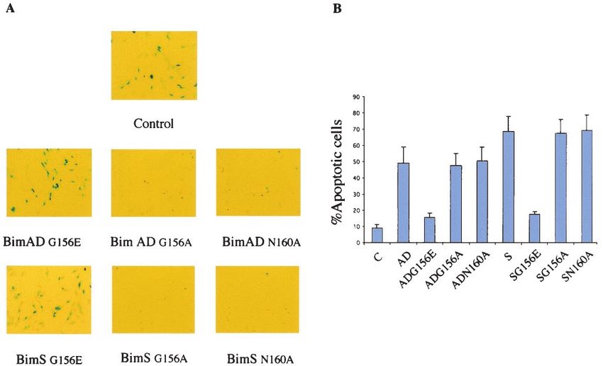
VOL. 22, 2002 Bim ISOFORMS ACTIVATE Bax TO TRIGGER APOPTOSIS 3579
glycerol, 1% Triton X-100) supplemented with Complete protease inhibitor
cocktail. The lysate was centrifuged at 13,000 ⫻ g for 30 min, and the supernatant
was recovered. A 2-ml aliquot of Glutathione-Uniflow Resin (Clontech) was
washed in washing buffer (10 mM Tris-Cl [pH 8.0], 150 mM NaCl, 0.1% NP-40)
and then incubated with the supernatant for 1 h at 4°C. The beads were washed
three times with washing buffer, and the Bim-GST product was eluted with 1.5 ml
of reduced glutathione (10 mM). The eluted proteins were dialyzed against PBS.
His-tagged Bax-␣ lacking 21 amino acids at the COOH terminus (His-
Bax⌬TM) cloned in the pTrcHis vector (Invitrogen) was kindly donated by
Ingram Iaccarino. This construct was expressed in E. coli BL21(DE3)/pLysS
cells. An overnight Luria broth culture was diluted 1/10 and grown to an optical
density at 600 nm of 0.6. Expression of His-tagged Bax protein was induced with
2 mM IPTG for 2 h, and cells were then harvested by centrifugation at 4°C. The
pellet from a 100-ml culture was resuspended in 5 ml of lysis buffer (50 mM
Tris-Cl [pH 8.0], 150 mM NaCl, 1 mM EDTA, 10% Glycerol, 1% Triton X-100)
supplemented with Complete protease inhibitor cocktail. The lysate was soni-
Downloaded from http://mcb.asm.org/ on February 26, 2021 by guest
cated for 10 s and centrifuged at 13,000 ⫻ g for 30 min. The supernatant was
incubated with 0.5 ml of washed His beads (Qiagen) for 1 h at 4°C. The beads
were washed three times with washing buffer (10 mM Tris-Cl [pH 8.0], 150 mM
NaCl, 20 mM imidazole, 0.1% Triton), and the His-tagged Bax product was
eluted with 0.5 ml of elution buffer (10 mM Tris-Cl [pH 8.0], 150 mM NaCl, 250
mM imidazole, 0.1% Triton). The eluted protein was dialyzed against PBS.
In vitro binding assay. Fifty microliters of GST-Sepharose beads (Amersham)
was washed in PBS and resuspended in 0.5 ml of PBS. Recombinant Bim-GST
proteins or control GST (final concentration of 100 nM each) were added to
different bead preparations, and the samples were rotated for 1 h at 4°C. The
beads were recovered by centrifugation at 4,000 ⫻ g for 1 min and washed three
times in 1 ml of IPP150 washing buffer and one time in 1% Triton buffer.
Recombinant His-Bax (final concentration of 10 nM) was added to the beads
resuspended in 1 ml of 1% Triton buffer, and the samples were rotated for 1 h
at 4°C. The beads were washed five times in IPP150, each time followed by
rotation for 5 min at 4°C. Material bound to the beads was eluted by the addition FIG. 1. Isolation of human Bim isoforms. Agarose gel electro-
of 100 l of reduced glutathione (10 mM), and the eluted proteins were analyzed phoresis of Bim cDNAs after reverse transcription-PCR on 293 or
by SDS-PAGE and Western blotting for Bax immunoreactivity as described OVCAR3 cell lines is shown, and the three known Bim isoforms
above. The intensity of each band was quantitated with a Typhoon 8600 optical together with six other related cDNAs (ⴱ) are identified. Leftmost
scanner and ImageQuant 5.1 image analysis software (Molecular Dynamics). lane, 100-bp DNA marker (New England Biolabs).
Data were normalized to the appropriate GST intensity.
Immunoprecipitation. Fifty microliters of protein A Sepharose beads (Amer-
sham) was washed, resuspended in 1 ml of Triton buffer, and then incubated with
for 1 h in the dark. After being washed three times with PBS, the coverslips were
rotation for 1 h at 4°C with 3 l of polyclonal anti-Bim antibody (500 g/ml).
mounted with ProLong Antifade mounting medium (Molecular Probes) on mi-
Similarly, 50 l of protein G Sepharose beads (Amersham) was incubated with
croscope slides and analyzed with a fluorescence microscope (Zeiss UK). The
8 l of monoclonal anti-Bim antibody (100 g/ml). The beads were recovered by
percentage of Bax-positive cells was quantified by microscopically counting the
centrifugation and washed twice with 1 ml of Triton buffer. 293 cells seeded at 2
number of cells showing a red punctate distribution versus the total number of
⫻ 105 cells/ml on two 100-mm-diameter dishes were lysed in 1 ml of 1% Triton
green (green fluorescent protein [GFP]-expressing) cells. Approximately 500
buffer with freshly added protease inhibitors. The lysates were centrifuged at
cells were counted from 20 randomly chosen fields for each experiment.
13,000 ⫻ g for 5 min to remove nuclei and cellular debris, and the supernatant
As a positive control for the induction of Bax immunoreactivity, the cells were
was added to the bead suspension and then mixed by rotation for 2 h at 4°C.
treated with staurosporine (Sigma) at 1.5 M for 5 h.
Immunoprecipitates were collected by a brief spin and washed three times with
1 ml of lysis buffer before fractionation by SDS-PAGE, which was followed by
Western blot analysis with the polyclonal anti-Bim antibody. For immunopre-
RESULTS
cipitation of Bax, a 50-l aliquot of protein G Sepharose beads (Amersham) was
incubated with 7.5 l of polyclonal anti-Bax antibody (200 g/ml) followed by Identification of Bim splice variants and isolation of
incubation with 293 cell lysates as described above. For immunoprecipitation of
BimAD. Bim-specific cDNA was amplified from both 293 hu-
Bim from HL60 and Daudi cells, approximately 5 ml of cell pellet was lysed in 10
ml of 1% Triton buffer with freshly added protease inhibitors. The lysates were man embryonic kidney cells and OVCAR3 ovarian cancer cells
incubated with 5 l of polyclonal anti-Bim antibody (500 g/ml) or 50 l of following reverse transcription-PCR with specific Bim primers
monoclonal anti-Bim antibody (100 g/ml) and mixed by rotation for 2 h at 4°C. (see Materials and Methods). Several transcripts were ampli-
Protein G Sepharose beads (50-l aliquot; Amersham) were added to pull down fied together with the previously described BimEL, BimL, and
immune complexes and then mixed by rotation for 1 h at 4°C. Immunoprecipi-
tates were collected and washed as described above before fractionation by
BimS (Fig. 1). Negative control reactions with only a single
SDS-PAGE, which was followed by Western blot analysis with the polyclonal primer, with RNA without reverse transcription, or in the
anti-Bax antibody. presence of RNase did not generate any product (data not
Immunostaining. The day before transfection, HeLa cells were plated at a shown).
density of 1.2 ⫻ 105 cells/ml on 24-mm glass coverslips in 35-mm-diameter
Sequence analysis of the six unknown variants revealed the
culture dishes. The cells were transiently cotransfected with 0.3 g of each of the
expression plasmids or a vector (negative control) and 0.1 g of pEGFP-Actin presence of a novel sequence, 130 bp long, in three of them,
(Clontech) by using Effectene (Qiagen). At 12 h after transfection, the cells were BimAD, BimACD, and BimABCD (Fig. 2). The position of
washed twice with PBS and fixed for 15 min in 1% paraformaldehyde at room the insertion corresponded exactly with the junction of the
temperature. After being washed with PBS, the cells were incubated in PBS previously described exons 5 and 6 of the Bim gene (7) and was
containing the permeabilizing agent digitonin (500 g/ml; Fluka) and the mouse
anti-Bax 6A7 monoclonal antibody (1:100 dilution) overnight at 4°C. The cov-
therefore designated 5a. Although the variants contained exon
erslips were washed three times with PBS and incubated with cy3-conjugated 6 in the cDNA, a stop codon at the beginning of the novel
antimouse antibody (Amersham) diluted 1:200 in 1% bovine serum albumin-PBS insert resulted in truncated proteins, all lacking the carboxyl-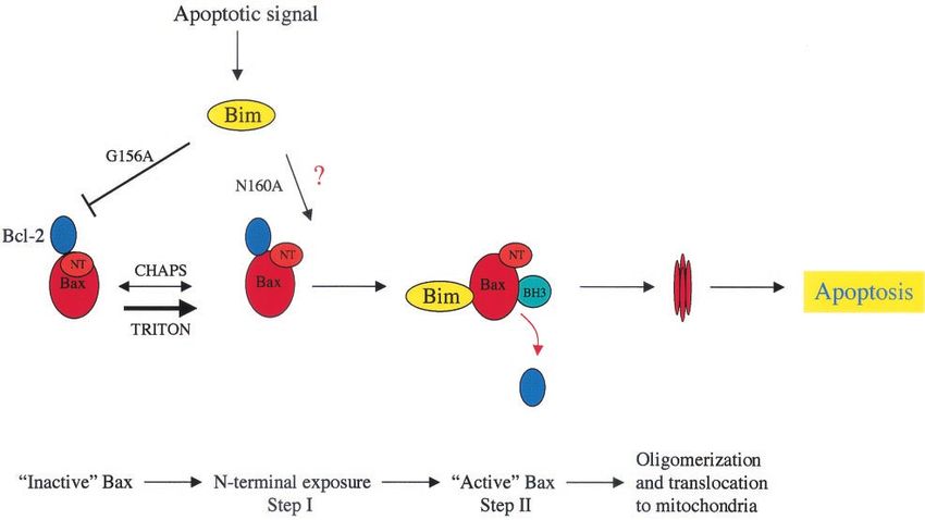
3580 MARANI ET AL. MOL. CELL. BIOL.
Downloaded from http://mcb.asm.org/ on February 26, 2021 by guest
FIG. 2. Bim proteins and predicted amino acid sequences of human Bim variants (the BH3 domain is underlined). The names of the new
variants were assigned by reference to the domain junctions. The novel exon (5a), identified in the human Bim gene, is present in BimAD,
BimACD, and BimABCD (short product LEK). Termination codon positions are indicated (ⴱ) according to the reading frame predicted by the
junction between exons 5 and 5a. The short product (GIFE) in BimAC, BimABC, and BimA comes from a translational frameshift due to the
junction between exons 4 and 6 or 2 and 6.
terminal hydrophobic domain identified in exon 6; only a short ity (Fig. 3A and data not shown). The percentage of apoptotic
C-terminal amino acid stretch (LEK) was present as a result of -Gal-positive (blue) cells cotransfected with BimAD was in-
translation of the novel insert. Similarly, the other three vari- termediate between those for BimS (the most potent) and
ants, named BimAC, BimABC, and BimA, also lacked the BimL. No morphological changes were observed in parallel
exon 6-encoded C terminus due to a stop codon resulting from cultures transfected with the control pcDNA3 vector (Fig. 3A).
translational frameshift. Immunoblot analysis of lysates from transfected cells that were
Comparison of the cDNA sequences of each variant with cultured in the presence of the caspase inhibitor zVAD-fmk to
genomic data indicates that they are produced by alternative inhibit apoptosis induced by the transgenes demonstrated that
splicing events. The coding sequence of BimAD, the principal the differential effects of the isoforms did not arise from dif-
subject of this study, is derived from the second and fifth exons. ferent levels of protein expression (Fig. 3C).
BimAD therefore contains the BH3 domain found in BimEL, Overexpression of Bcl-2 effectively blocked the apoptotic
BimL, and BimS. effect of BimAD as well as those of BimEL, BimL, and BimS
Induction of apoptosis by Bim proteins. The function of the but not that of caspase 8, which is known to mediate apoptosis
Bim variants was explored by transient transfection in NIH 3T3 independent of the Bcl-2 checkpoint. zVAD-fmk, a wide-spec-
mouse fibroblasts, where apoptotic cells are readily detectable trum caspase inhibitor, was able to inhibit the proapoptotic
by a change in morphology. The cells were cotransfected with activity of the Bim isoforms as well as that of caspase 8, as
expression plasmids encoding each Bim isoform along with shown in Fig. 4. These data suggest that the novel isoform
pCMV--Gal as a marker for transfected cells. At 14 h post- BimAD also mediates apoptosis through a caspase-mediated
transfection, the cells were fixed, stained for -Gal expression, pathway, as has been shown for the previously characterized
and examined for the morphological features of cell death. As Bim variants (21), and that this can be blocked by the overex-
shown in Fig. 3A, BimEL, BimL, and BimS all induced apo- pression of antiapoptotic Bcl-2 family proteins.
ptosis, as evidenced by the appearance of shrunken and The effect of BimAD in promoting apoptosis is not re-
rounded blue cells. The new variants BimAC, BimABC, and stricted to NIH 3T3 cells, since overexpression of BimAD was
BimACD lacked apoptotic activity; BimABCD had only a able to induce apoptosis in both the HeLa and 293 cell lines
weak effect, whereas BimAD had a higher proapoptotic activ- (data not shown).VOL. 22, 2002 Bim ISOFORMS ACTIVATE Bax TO TRIGGER APOPTOSIS 3581
Downloaded from http://mcb.asm.org/ on February 26, 2021 by guest
FIG. 3. Effect of Bim variants after transient transfection into NIH 3T3 cells. (A) NIH 3T3 cells stained for -Gal expression 14 h after
transfection with 1.3 g of the various Bim constructs together with 0.7 g of the -Gal expression vector. (B) Percentages of round blue cells
relative to the total population of blue cells. Bars indicate the standard deviation of the results of three independent experiments. (C) Expression
of the Bim variants in NIH 3T3 cells.
BimS and BimAD heterodimerize with both death antago-
nists (Bcl-2 and Bcl-XL) and death agonists (Bax). It has been
suggested that BH3-only members of the Bcl-2 family exert
their proapoptotic activity by heterodimerizing with antiapop-
totic Bcl-2 family members, antagonizing their protective func-
tion. We therefore explored whether Bim isoforms are capable
of associating with other Bcl-2 family proteins by using a TAP
method (24, 28). This is a two-step strategy to purify efficiently
and rapidly complexes between TAP-tagged proteins and as-
sociated components present in the cell at their endogenous
levels. Initially, we studied the isoforms BimEL and BimS by
subcloning them fused to the C-terminal TAP tag sequence in
pcDNA3.1, and the derived clones were transiently transfected
into 293 cells. The fusion proteins and associated endogenous
proteins were recovered from extracts by two affinity steps and
subjected to SDS-PAGE-immunoblot assays with antibodies
FIG. 4. Bim-induced cell death is blocked by Bcl-2 and zVAD-fmk.
specific for several Bcl-2 family members. As shown in Fig. 5, Bcl-2 cotransfection blocks Bim-induced apoptosis but not caspase
both isoforms were able to associate with Bcl-2. BimS was also 8-mediated killing. The caspase inhibitor zVAD-fmk inhibits apoptosis
shown to interact with the proapoptotic protein Bax. In view of induced by Bim as well as by caspase 8, further validating the data
these data, the study was extended to include the other Bim obtained. The data shown are the percentages of round blue cells
relative to the total population of blue cells. The apoptotic activities of
isoforms. All of the variants containing the BH3 domain were the different variants in the presence or absence of Bcl-2 and zVAD-
able to associate with Bcl-2, thus correlating with the data fmk are compared. Bars indicate the standard deviation of the results
demonstrating that Bcl-2 can inhibit Bim-induced apoptosis of three independent experiments.3582 MARANI ET AL. MOL. CELL. BIOL.
It has been shown that in the presence of nonionic deter-
gents, such as NP-40 or Triton-X, Bax can undergo a confor-
mational change, leading to the exposure of the amino-termi-
nal epitope. Hence, we performed the protein complex
purification in transient transfection analysis by permeabilizing
293 cells in a buffer containing 1% CHAPS, a zwitterionic
detergent that has been shown to maintain Bax in its native
conformation (13, 14). Under these conditions, no specific
signal was detected, although BimS and BimAD could still
bind to Bcl-2 (data not shown).
To explore whether the interaction between BimS or
BimAD with activated Bax occurs directly, we generated re-
combinant BimS-GST and BimAD-GST proteins as well as a
Downloaded from http://mcb.asm.org/ on February 26, 2021 by guest
His-tagged Bax⌬TM protein to test for binding in an in vitro
assay. Recombinant GST was used as a negative control. Con-
sistent with the coimmunoprecipitation data, both BimAD-
GST and BimS-GST were shown to bind to His-Bax⌬TM in
FIG. 5. Association of BimEL and BimS with various Bcl-2 family
the activated conformation (Fig. 7). The intensity of the im-
members. Immunoblot analysis with anti-Bcl-2 antibody (A) or anti- munoreactive bands was quantitated and normalized to the
Bax antibody (B) of 293 cells transiently transfected with BimEL- appropriate GST intensity (Fig. 7). The amount of His-
TAPtag, BimS-TAPtag, or TAPtag empty vector (⫺C) followed by Bax⌬TM pulled down by BimAD-GST was 12.8-fold greater
TAP tag purification (left panels) is shown. Equal loading of the two
Bim isoforms was confirmed with the polyclonal anti-Bim antibody than that pulled down by BimS-GST.
(right panels). Expression of endogenous BimAD and association of endog-
enous Bim proteins with Bax. To demonstrate that endoge-
(Fig. 6B). Importantly, the novel isoform BimAD was also nous BimAD mRNA is translated into protein in 293 cells,
capable of binding to Bax. Furthermore, no binding was ob- immunoprecipitation assays were performed with anti-Bim an-
served with the empty TAP tag parental vector, confirming the tibodies. Immunoprecipitates from either untransfected cells
specificity of these interactions (Fig. 6B). or cells transfected with BimEL, BimS, or BimAD were ob-
FIG. 6. Proteins associating with Bim variants. 293 cells were transiently transfected with each of the Bim-TAPtag-expressing plasmids followed
by TAP tag purification and immunoblot analysis. (A) Expression of each variant detected by Western blot analysis with an anti-Bim polyclonal
antibody. (B) BimEL, BimL, BimS, BimAD, BimACD, and BimABCD, all containing the BH3 domain, heterodimerize with Bcl-2. Both BimS and
BimAD associate with Bax. Negative control (⫺C) cells were transfected with TAP tag parental vector and purified by the same TAP tag
procedure.VOL. 22, 2002 Bim ISOFORMS ACTIVATE Bax TO TRIGGER APOPTOSIS 3583
not to Bcl-2 (Fig. 9). These mutants or control wild-type Bim
plasmids were transiently transfected into NIH 3T3 cells to-
gether with pCMV--Gal as a reporter gene, and the -Gal
assay was performed as previously described. Neither of the
Bim G156E mutants (of BimS and BimAD) was able to pro-
mote apoptosis in this system, while both the Bim G156A and
Bim N160A mutants still retained death agonist activity (Fig.
10). In view of these data, we postulate a model in which Bim
can induce apoptosis by two distinct mechanisms. It can bind
directly to Bax, leading to its activation and targeting to mito-
chondria, and it can also bind to Bcl-2, neutralizing its ability to
sequester Bax in an inactive state.
Bim induces Bax conformational change. Since one of the
Downloaded from http://mcb.asm.org/ on February 26, 2021 by guest
mechanisms proposed for Bax activation is the conversion to
FIG. 7. Interactions between BimAD-GST, BimS-GST, or control an active conformation (9), we explored whether the binding of
GST (C-GST) with His-tagged Bax⌬TM in vitro. Immunoblotting was Bim to Bax could correlate with Bax conformational change.
performed with the polyclonal anti-Bax antibody or anti-GST antibody. For this purpose, HeLa cells were transiently transfected with
BimS or BimAD together with a GFP-expressing vector as a
marker for transfected cells. At 12 h after transfection, Bax
tained by using either a polyclonal or a monoclonal anti-Bim immunostaining was assessed by microscopy by employing a
antibody, and the products were subjected to SDS-PAGE- monoclonal antibody that specifically recognizes the epitope
immunoblot assay with the polyclonal anti-Bim antibody. A exposed by the conformational change in the Bax protein
protein migrating at the same level as BimAD was immuno- (6A7). The assay was performed at 12 h, since this time cor-
precipitated from the untransfected 293 cells (Fig. 8A). Con- responds with the onset of Bim expression (data not shown)
trol experiments with preimmune serum and a specific peptide and Bax conformational change has been shown to be an early
to block the polyclonal anti-Bim antibody showed that this event in the apoptotic cascade (11, 19). As shown in Fig. 11A,
represented specific Bim immunoreactivity. Furthermore, Bim BimS or BimAD expression leads to the appearance of acti-
was not detected in 293 cell lysates previously immunodepleted vated Bax immunoreactivity, showing a punctate distribution
with the polyclonal antibody (Fig. 8A). In addition, immuno- within the cytoplasm. This result was similar to that observed in
blot analysis of total lysates from parental 293 cells or 293 cells HeLa cells treated with staurosporine, which were previously
transiently transfected with pcDNA-BimEL or pcDNA-BimAD reported to induce Bax activation in this system (Fig. 11A) (9).
revealed the presence of a band comigrating with BimAD (Fig. Control cells transfected with the empty vector together with
8B). GFP-expressing plasmid did not display positive immunoreac-
Since the loss of Bim in Bim⫺/⫺ mice was shown to affect tivity for Bax (Fig. 11A). Similarly, BimS mutants were as-
markedly the hematopoietic compartment (6), we also ana- sessed for Bax immunoreactivity. Mutations in BimS that block
lyzed the expression of the Bim variants in a panel of hema- its binding either to Bax (BimS-G156A) or to Bcl-2 (BimS-
topoietic cell lines. While the longer isoforms EL, L, and S N160A) did not affect its ability to induce Bax conformational
were expressed in all of the lines tested, the predicted Bi- change. However, the mutant BimS-G156E, which inhibits
m(AD) protein was found in HL60 and Daudi (Fig. 8C). As binding to both Bax and Bcl-2, was unable to activate Bax (Fig.
these two cell lines express BimAD (and BimS) in readily 11A).
detectable amounts, lysates from either HL60 or Daudi were The effect of Bim on Bax conformation was further con-
subjected to immunoprecipitation studies to assess whether firmed by immunoprecipitation assays with another Bax anti-
Bim could coimmunoprecipitate with Bax when they were ex- body (N20) that recognizes only the activated conformation
pressed at their endogenous levels. Bim could be immunopre- (Fig. 11C). 293 cells were transiently transfected with BimS,
cipitated with Bax in Daudi but not in HL60 cells under the BimAD, or the BimS mutants and lysed in buffer containing
experimental conditions used (Fig. 8D). Control immunopre- 1% CHAPS. Consistent with the microscopic data, Bax was
cipitation experiments with 293 cells transfected with exoge- detected in the immunoprecipitates from BimS- or BimAD-
nous BimAD or BimS showed coimmunoprecipitation of Bax transfected cells but not in control untransfected cells (Fig.
under the same conditions (Fig. 8D). 11C). Both the BimS-G156A and BimS-N160A mutants were
Interaction with Bax requires the BH3 domain of both BimS also able to induce Bax conformational change, while the mu-
and BimAD. Since Bid, another proapoptotic BH3-only mem- tant BimS-G156E was impaired in this activity (Fig. 11C). Bax
ber of the Bcl-2 family, has been shown to interact with Bax could also be immunoprecipitated in cells lysed in the presence
through its BH3 domain in a manner distinct from that of its of Triton X-100, a nonionic detergent previously shown to
interaction with Bcl-2 (36), we made mutants of Bim that expose the N-terminal epitope of Bax (14).
might be able to bind differentially to Bax and Bcl-2. BH3
mutants of BimS-TAPtag and BimAD-TAPtag were tested for DISCUSSION
their binding to Bcl-2 and Bax in vivo by using the TAP tag
method. We derived three mutants with different specificities: We have identified six new splice variants of Bim in human
Bim G156E does not bind to either Bax or Bcl-2; Bim G156A cells and investigated in detail the function of one variant,
binds to Bcl-2 but not to Bax; and Bim N160A binds to Bax but BimAD. BimAD encodes a protein of 80 amino acids andDownloaded from http://mcb.asm.org/ on February 26, 2021 by guest
FIG. 8. Expression of BimAD. (A) Immunoprecipitation (I.P.) of lysates from 293 cells after transient transfection of BimEL, BimS, or BimAD
or of lysates from untransfected cells. Both polyclonal (poly) and monoclonal (mono) anti-Bim antibodies were employed in the study. Western
blot analysis was performed with the polyclonal anti-Bim antibody. Control experiments involved immunoprecipitation with preimmune serum
(pre-imm.), blocking of the anti-Bim polyclonal antibody with a specific peptide (⫹pept.), or immunodepletion of 293 cell lysates with the
polyclonal antibody (imm.depl.). (B) Western blot of whole-cell lysate from 293 cells or 293 cells transiently transfected with BimEL or BimAD
as controls. The expression of Bim was detected by immunoblotting with an anti-Bim polyclonal antibody. (C) Expression of Bim isoforms in
Jurkat, HelaS3, HL60, and Daudi cells. (D) Immunoprecipitation of endogenous Bim from Daudi and HL60 cells and of exogenous Bim in
transfected 293 cells with either a polyclonal antiserum (PoAb) or a monoclonal antibody (MoAb). The presence of Bax in the immunoprecipitated
complexes was detected with the polyclonal Bax N20 antiserum. IgG, immunoglobulin G.
3584VOL. 22, 2002 Bim ISOFORMS ACTIVATE Bax TO TRIGGER APOPTOSIS 3585
splice variants of Bim were described (35), but our study is the
first to identify BimAD, the novel proapoptotic form.
To assess the nature of the unknown sequence, a Blast
search was performed against the latest draft of the human
genome. The sequence was located in chromosome 2 within an
intron of the human Bim gene, as deduced from sequence
accession numbers AC013332 and AC046192. The 130-bp se-
quence was found in the intronic region flanked by intron-exon
junction sequences corresponding to the GT-AG rule (27). In
addition, we identified a good candidate for the branch point
FIG. 9. The BH-3 domain of Bim is required for its association with (CCTGAC) lying 24 nucleotides upstream of the 3⬘ splice
both Bcl-2 and Bax. Western blot analysis was performed with anti-
junction, which has been found to be required for lariat for-
Bcl-2 antibody (upper panels) or anti-Bax antibody (lower panels).
mation during splicing (27). Finally, a pyrimidine stretch is
Downloaded from http://mcb.asm.org/ on February 26, 2021 by guest
Lanes 1 and 5, control from BimAD-TAPtag and BimS-TAPtag, re-
spectively, after TAP tag purification; lanes 2 and 6, G156E mutants located between the branch site and the AG at the 3⬘ splice
are impaired in their binding to both Bcl-2 and Bax; lanes 3 and 7, site, although this deviates from the respective consensus se-
G156A mutants retain binding to Bcl-2 but not to Bax; lanes 4 and 8,
N160A mutants retain binding to Bax but not to Bcl-2.
quence (Pyr)12CAG, with several purines interrupting the
polypyrimidine tract. Weak 3⬘ splice sites with higher purine
content have been shown to be common in alternatively spliced
results from an alternative splicing event in which exons 3 and exons (30).
4 of the Bim gene are lost while a unique 130-bp sequence is The formation of BimAD is analogous to that of N-Bak,
inserted after exon 5. This generates a change in the reading which has been shown to result from the insertion of a 20-bp
frame with a premature termination codon and elimination of sequence that causes a translational frameshift. This in turn
the C-terminal hydrophobic tail. Therefore, except for the C- leads to a truncated protein due to a premature stop codon
terminal deletion, the amino acid sequence predicted for the (31).
variant matches that of the previously described BimS. While It has become clear that alternative splicing plays an impor-
this manuscript was in revision, a number of nonapoptotic tant role in the expression of many genes in the programmed
FIG. 10. Effect of Bim mutants after transient transfection into NIH 3T3 cells. (A) A -Gal assay after transient transfection of NIH 3T3 cells
shows differential effects of single or double mutations. Control cells were transfected with empty vector along with -Gal expression vector.
(B) Percentages of round blue cells relative to the total population of blue cells. Bars indicate the standard deviation of the results of three
independent experiments. C, control.3586 MARANI ET AL. MOL. CELL. BIOL.
Downloaded from http://mcb.asm.org/ on February 26, 2021 by guest
FIG. 11. BimS and BimAD promote Bax conformational change. (A) HeLa cells growing on glass coverslips were transiently transfected with
BimAD, BimS, or BimS mutants. Twelve hours after transfection, the cells were immunostained with a monoclonal antibody that specifically
recognizes the conformationally active Bax (6A7). Bax-associated fluorescence was visualized with a cy3-conjugated secondary antibody (red). As
a positive control, the cells were treated with staurosporine (1 M) for 6 h. As a negative control, the cells were transiently transfected with the
parental pcDNA3 plasmid. (B) Percentages of positive (red) cells relative to the total population of GFP-positive (green) cells. In the case of
staurosporine, positive cells were counted relative to the total population. Bars indicate the standard deviation of the results of three independent
experiments. (C) 293 cells were transiently transfected with BimAD, BimS, BimS-G156A, BimS-N160A, BimS-G156E, or control empty vector.
Cells were lysed in 1% CHAPS (C) and subjected to immunoprecipitation with the conformation-specific Bax antibody (N20). Conformationally
active Bax was detected by immunoblotting with polyclonal anti-Bax antibody. As a positive control, 293 cells were lysed in 1% Triton (T) and
treated as described above. STP, staurosporine.
cell death pathway (15). Within the Bcl-2 family, splicing vari- totic activity of the longer forms. However, BimAD is less
ants have also been reported for Bcl-2, Bcl-X, Bcl-W, Bax, efficient in inducing apoptosis than BimS when overexpressed
Mcl-1, Bok, Bak, and Bcl-G. As we have shown for Bim, in in cells. This might be explained by the fact that BimAD lacks
some cases variants of the same gene may exhibit different the C-terminal hydrophobic tail, which has been shown to have
biological functions. The long form of Bcl-X (L) is a strongly proapoptotic potential for other Bcl-2 family members (2, 16).
antiapoptotic protein, while Bcl-X (S) and Bcl-X- are pro- For the same reason, overexpression of BimEL in NIH 3T3
apoptotic (5, 29). Further, Mcl-1s is a splicing variant of the results in a more potent killing effect than that of BimAD.
antiapoptotic Bcl-2 family member Mcl-1 but exhibits pro- Surprisingly, BimL was less efficient in inducing apoptosis in
apoptotic activity (3, 4). It has recently been shown that the our system, raising the possibility that some region in exon 3
BH-3 domain-only splice variant of proapoptotic Bak, N-Bak, might also be important for the death agonist activity of these
is antiapoptotic in neurons (31). Similarly, the five Bim iso- molecules. To summarize the data on the apoptotic potency of
forms we isolated that do not show death-promoting potential the different splice products, it appears that the BH3 domain
could have intrinsic antiapoptotic activity or, alternatively, (exon 5) is required for killing and the dynein binding domain
could operate as transdominant inhibitors of the proapoptotic (exon 4) reduces the strength of apoptosis induction, presum-
forms. ably by causing sequestration of Bim to the cytoskeleton, while
As with BimS, BimAD contains the BH3 region but lacks the the hydrophobic C-terminal domain (exon 6) increases the
dynein-binding domain responsible for the reduced proapop- apoptotic strength.VOL. 22, 2002 Bim ISOFORMS ACTIVATE Bax TO TRIGGER APOPTOSIS 3587
Downloaded from http://mcb.asm.org/ on February 26, 2021 by guest
FIG. 12. Model for the apoptotic pathway triggered by Bim. See the text for details.
Most BH3-only proteins are thought to induce apoptosis by tectable through immunofluorescence and immunoprecipita-
interacting with antiapoptotic Bcl-2 family members, neutral- tion studies (9, 23). Hence, the scenario for Bid appears to be
izing their ability to antagonize the function of the proapop- very similar to that which we have found for Bim.
totic Bax and Bak. Our binding experiments with the TAP tag Recent reports on Bax and Bak activation highlight a pos-
method showed that BimS and BimAD are capable of het- sible model to explain our results (12, 32). These data imply
erodimerizing in vivo with Bcl-2 and Bcl-XL as well as with Bax that the change at the N terminus of Bax and Bak is not the
in the presence of nonionic detergents. Furthermore, immu- only critical event in the activation of these molecules but that
noprecipitation studies showed that Bim can associate with a second and distinct conformational change at the C terminus
Bax even when expressed at the endogenous level. These in- is necessary for complete Bax activation and dissociation of the
teractions require an intact BH3 domain, as point mutations inhibitory Bcl-2–Bcl-XL interaction. In this scenario, Triton
within this domain disrupt the binding activity of BimS and X-100 could only trigger the exposure of an epitope at the
BimAD to the other proteins. The observation that Bim cannot amino terminus, leaving the carboxy terminus unaltered (32)
bind Bax stably in the presence of 1% CHAPS, while it can still and Bcl-XL still able to bind (12). Only in the presence of a
bind Bcl-2 and induce the exposure of the N terminus of Bax, damage signal could both of the epitopes be exposed (12, 32),
prompted us to review the available literature in more detail. and in this situation, Bcl-XL binding is lost (12). Importantly,
To date, only three proapoptotic proteins have been shown to Bcl-XL overexpression did not alter the kinetics of the Bak
associate directly with Bax: the BH3-only proteins Bid and N-terminal conformational change after etoposide treatment,
MAP-1 (34, 36) and the SH-3 domain-containing protein Bif-1 although it was found to delay the second change (12). Simi-
(8). Notably, Bif-1 was reported to bind to Bax only in its native larly, expression of the viral Bcl-2 homolog E1B19K protein
conformation, and the addition of nonionic detergents was did not prevent the first N-terminal change in Bax after treat-
able to disrupt this interaction. On the other hand, the study of ment with tumor necrosis factor plus cycloheximide, although
the interaction of MAP-1 with Bax was performed only in the it prevented the conformational change at the C terminus and
presence of 0.2% NP-40, rendering it difficult to discern the the formation of Bax cross-linked products (32).
relevance of this interaction under native conditions. Surpris- These data agree with those of previous studies on the so-
ingly, previous reports of Bid interaction with Bax or Bak also lution structure of monomeric Bax (33), which suggest that the
used nonionic detergents in the lysis of cells expressing ectopic N terminus is highly mobile and remains flexible and solvent-
Bid and in the subsequent purification steps (9, 23, 36, 37). exposed in the presence of detergents and that this event does
Only in two cases was this interaction studied in the presence not necessarily induce exposure of the BH3 domain, which
of CHAPS, and there was no indication that either full-length remains occluded by the C terminus. Indeed, the exposure of
or cleaved Bid could bind to Bax under these conditions (19, the N terminus of Bax is rapid, reversible, and not sufficient
32). As in the case of Bim, Bid has also been extensively alone to commit the cell to apoptosis (18).
reported to induce a change in Bax conformation that is de- In this scenario, Bim (and possibly Bid) could represent the3588 MARANI ET AL. MOL. CELL. BIOL.
necessary signal for Bax activation, and such a role has already able to interact directly with Bax and Bak when appropriately
been proposed in Taxol-induced damage in SH-SY5Y cells stimulated.
(18). In our model (Fig. 12), the Bax N terminus exposure
ACKNOWLEDGMENTS
induced by Bim (or Bid) is a reversible event kept under
control by the inhibitory interaction of Bcl-2 (indeed, Bim This work was supported by Cancer Research UK and the Special
associated with Bcl-2 even in the presence of CHAPS). A Trustees of the Hammersmith and Acton Hospitals.
We thank Bim Laguda and Zoë Leech for expert technical assis-
stable interaction between Bim and Bax could become detect- tance and Rita Lopes, Miguel Martins, Ingram Iaccarino, and Sophie
able only if the N terminus remained exposed for a sufficient Khanna for helpful discussions. We are grateful to Helen Hurst for
period of time, as we have seen in the presence of Triton as critical reading of the manuscript.
well as in the in vitro binding experiment where His-Bax⌬TM REFERENCES
was constitutively active. Indeed, the amount of Bax that could 1. Adams, J. M., and S. Cory. 2001. Life-or-death decisions by the Bcl-2 protein
be pulled down by the Bax N20 conformation-specific antibody family. Trends Biochem. Sci. 26:61–66.
was severalfold higher in the presence of Triton than in the 2. Antonsson, B., and J. C. Martinou. 2000. The Bcl-2 protein family. Exp. Cell
Res. 256:50–57.
Downloaded from http://mcb.asm.org/ on February 26, 2021 by guest
presence of CHAPS, even in control cells treated with stauro- 3. Bae, J., C. P. Leo, S. Y. Hsu, and A. J. Hsueh. 2000. MCL-1S, a splicing
sporine (Fig. 11C). The formation of the active Bax (step II), variant of the antiapoptotic BCL-2 family member MCL-1, encodes a pro-
apoptotic protein possessing only the BH3 domain. J. Biol. Chem. 275:
with the BH3 domain exposed and Bim bound, could be a 25255–25261.
rapid event leading to Bax oligomerization and translocation to 4. Bingle, C. D., R. W. Craig, B. M. Swales, V. Singleton, P. Zhou, and M. K.
mitochondria. Indeed, the interactions between the C-terminal Whyte. 2000. Exon skipping in Mcl-1 results in a bcl-2 homology domain 3
only gene product that promotes cell death. J. Biol. Chem. 275:22136–22146.
helix and the BH3 binding pocket are mainly hydrophobic, and 5. Boise, L. H., M. Gonzalez-Garcia, C. E. Postema, L. Ding, T. Lindsten, L. A.
rotation at this level would lead to a very energetically unfa- Turka, X. Mao, G. Nunez, and C. B. Thompson. 1993. bcl-x, a bcl-2-related
vorable conformation (33). The formation of oligomers could gene that functions as a dominant regulator of apoptotic cell death. Cell
74:597–608.
be the energy-driven process, and Bim is unlikely to bind to 6. Bouillet, P., D. Metcalf, D. C. Huang, D. M. Tarlinton, T. W. Kay, F.
Bax when oligomers are found. In another report, Bid did not Kontgen, J. M. Adams, and A. Strasser. 1999. Proapoptotic Bcl-2 relative
Bim required for certain apoptotic responses, leukocyte homeostasis, and to
cofractionate with the 500-kDa Bax complex in cells treated preclude autoimmunity. Science 286:1735–1738.
with tumor necrosis factor plus cycloheximide (32), and a hit- 7. Bouillet, P., L. C. Zhang, D. C. Huang, G. C. Webb, C. D. Bottema, P. Shore,
and-run model for truncated Bid-dependent Bax activation H. J. Eyre, G. R. Sutherland, and J. M. Adams. 2001. Gene structure
alternative splicing, and chromosomal localization of pro-apoptotic Bcl-2
was proposed in that study. Further studies are needed to relative Bim. Mamm. Genome 12:163–168.
elucidate such a model for Bim. 8. Cuddeback, S. M., H. Yamaguchi, K. Komatsu, T. Miyashita, M. Yamada, C.
From the behavior of the BimS and BimAD BH3 domain Wu, S. Singh, and H. G. Wang. 2001. Molecular cloning and characterization
of Bif-1. A novel Src homology 3 domain-containing protein that associates
point mutants, it appears that the ability of these Bim isoforms with Bax. J. Biol. Chem. 276:20559–20565.
to cause apoptosis can occur via two pathways (Fig. 12). Bim 9. Desagher, S., A. Osen-Sand, A. Nichols, R. Eskes, S. Montessuit, S. Lauper,
K. Maundrell, B. Antonsson, and J. C. Martinou. 1999. Bid-induced con-
and the mutant G156A can bind to Bcl-2 and shift the equi- formational change of Bax is responsible for mitochondrial cytochrome c
librium towards the first step (indirect pathway). In addition, release during apoptosis. J. Cell Biol. 144:891–901.
Bim and the mutant N160A may bind directly to Bax and 10. Dijkers, P. F., R. H. Medemadagger, J. W. Lammers, L. Koenderman, and
P. J. Coffer. 2000. Expression of the pro-apoptotic Bcl-2 family member Bim
induce an activated conformation. There are thus direct and is regulated by the forkhead transcription factor FKHR-L1. Curr. Biol.
indirect pathways to Bax activation. While most BH3-only pro- 10:1201–1204.
teins have been suspected to use the indirect pathway, Bid 11. Gilmore, A. P., A. D. Metcalfe, L. H. Romer, and C. H. Streuli. 2000.
Integrin-mediated survival signals regulate the apoptotic function of Bax
activates Bax directly and Bim appears to act in both ways. Bim through its conformation and subcellular localization. J. Cell Biol. 149:431–
is only the second BH3-only protein to be shown to be able to 446.
12. Griffiths, G. J., B. M. Corfe, P. Savory, S. Leech, M. Degli Esposti, J. S.
activate Bax directly. In the case of the indirect mechanism Hickman, and C. Dive. 2001. Cellular damage signals promote sequential
through binding to Bcl-2 or other antiapoptotic family mem- changes at the N-terminus and BH-1 domain of the pro-apoptotic protein
bers, the activation of Bax still results with high efficiency. Bak. Oncogene 20:7668–7675.
13. Hsu, Y. T., and R. J. Youle. 1998. Bax in murine thymus is a soluble mono-
While the exact mechanism for this is unknown, it is likely that meric protein that displays differential detergent-induced conformations.
the effective removal of Bcl-2 and Bcl-XL leads to the accu- J. Biol. Chem. 273:10777–10783.
mulation of BH3 proteins (including Bim) in a free form that 14. Hsu, Y. T., and R. J. Youle. 1997. Nonionic detergents induce dimerization
among members of the Bcl-2 family. J. Biol. Chem. 272:13829–13834.
are able to target Bax directly. Alternatively, the formation of 15. Jiang, Z. H., and J. Y. Wu. 1999. Alternative splicing and programmed cell
Bim–Bcl-2 heterodimers may lead to increased levels of free death. Proc. Soc. Exp. Biol. Med. 220:64–72.
16. Kataoka, T., N. Holler, O. Micheau, F. Martinon, A. Tinel, K. Hofmann, and
Bax that could be targeted for activation by proteins unrelated J. Tschopp. 2001. Bcl-rambo, a novel Bcl-2 homologue that induces apopto-
to the Bcl-2 family or that could become spontaneously acti- sis via its unique C-terminal extension. J. Biol. Chem. 276:19548–19554.
vated. 17. Kelekar, A., and C. B. Thompson. 1998. Bcl-2-family proteins: the role of the
BH3 domain in apoptosis. Trends Cell Biol. 8:324–330.
The possibility has been raised that other molecules in ad- 18. Makin, G. W., B. M. Corfe, G. J. Griffiths, A. Thistlethwaite, J. A. Hickman,
dition to Bid could be responsible for Bax activation (38, 39). and C. Dive. 2001. Damage-induced Bax N-terminal change, translocation to
We show here that BimS and BimAD are likely candidates, but mitochondria and formation of Bax dimers/complexes occur regardless of
cell fate. EMBO J. 20:6306–6315.
others may exist as well. At present it is not clear whether the 19. Murphy, K. M., U. N. Streips, and R. B. Lock. 2000. Bcl-2 inhibits a Fas-
more highly expressed isoforms of Bim, BimL and BimEL, also induced conformational change in the Bax N terminus and Bax mitochon-
drial translocation. J. Biol. Chem. 275:17225–17228.
act directly on Bax. While it was not possible to see complexes 20. Nechushtan, A., C. L. Smith, I. Lamensdorf, S. H. Yoon, and R. J. Youle.
of BimL and BimEL with Bax in the experiments performed 2001. Bax and Bak coalesce into novel mitochondria-associated clusters
here, it is possible that they may form under conditions where during apoptosis. J. Cell Biol. 153:1265–1276.
21. O’Connor, L., A. Strasser, L. A. O’Reilly, G. Hausmann, J. M. Adams, S.
BimL and BimEL are released from microtubules following Cory, and D. C. Huang. 1998. Bim: a novel member of the Bcl-2 family that
apoptotic stimuli. Similarly, other BH3-only proteins might be promotes apoptosis. EMBO J. 17:384–395.You can also read