A new non classical fold of varroa odorant binding proteins reveals a wide open internal cavity
←
→
Page content transcription
If your browser does not render page correctly, please read the page content below
www.nature.com/scientificreports
OPEN A new non‑classical fold of varroa
odorant‑binding proteins reveals
a wide open internal cavity
Beatrice Amigues1,6, Jiao Zhu2,4,6, Anais Gaubert1, Simona Arena3, Giovanni Renzone3,
Philippe Leone1, Isabella Maria Fischer2, Harald Paulsen4, Wolfgang Knoll2,5, Andrea Scaloni3,
Alain Roussel1, Christian Cambillau1* & Paolo Pelosi2*
Odorant-binding proteins (OBPs), as they occur in insects, form a distinct class of proteins that
apparently has no closely related representatives in other animals. However, ticks, mites, spiders
and millipedes contain genes encoding proteins with sequence similarity to insect OBPs. In this work,
we have explored the structure and function of such non-insect OBPs in the mite Varroa destructor,
a major pest of honey bee. Varroa OBPs present six cysteines paired into three disulphide bridges,
but with positions in the sequence and connections different from those of their insect counterparts.
VdesOBP1 structure was determined in two closely related crystal forms and appears to be a
monomer. Its structure assembles five α-helices linked by three disulphide bridges, one of them
exhibiting a different connection as compared to their insect counterparts. Comparison with classical
OBPs reveals that the second of the six α-helices is lacking in VdesOBP1. Ligand-binding experiments
revealed molecules able to bind only specific OBPs with a moderate affinity, suggesting that either
optimal ligands have still to be identified, or post-translational modifications present in the native
proteins may be essential for modulating binding activity, or else these OBPs might represent a failed
attempt in evolution and are not used by the mites.
Odorant-binding proteins (OBPs) are carriers of pheromones and odorants in vertebrates and i nsects1–4. Insect
OBPs, which represent a class of proteins structurally different5, 6 from those of vertebrates7, 8, have been detected
so far only in Hexapoda4, 9. They are polypeptides of 120–140 amino acids, folded in a compact structure generally
made of six α-helical domains. A pattern of six conserved cysteines paired in three interlocked disulphide bridges
is a key feature of these proteins10–12. A single hydrophobic pocket binds small organic volatile compounds, such
as pheromones and general odorants, with dissociation constants of the micromolar order13. Besides OBPs, other
soluble carriers such as chemosensory proteins (CSPs), Nieman-Pick C2 (NPC2) proteins and lipocalins, may
be involved in chemical communication of a rthropods4, 9, 14–16. In particular, NPC2 proteins are expressed with
several members in all arthropods and represented until a couple of years ago the only known class of putative
semiochemical carriers in Chelicerata and Crustacea.
However, recent work unveiled the presence of proteins with some sequence similarity to insect OBPs in
spiders, ticks and mites, as well as in Myriapoda, which have been tentatively named “OBP-like”17–21. Such poly-
peptides exhibit values of amino acid identity with insect OBPs around 10–15%. However, such low similarity
can be significant when compared with the low values of conserved amino acids between the OBPs of different
insect Orders. Given their phylogenetic relationship with their insect counterparts, we shall simply refer to these
proteins as OBPs. Members of this family have not been found so far in Crustacea nor in other species of living
organisms.
In order to understand the role of OBPs in Chelicerata, we have focused our attention on the mite Varroa
destructor, the well-known honey bee parasite. The genome of this species contains 5 genes encoding members
1
Architecture et Fonction des Macromolécules Biologiques (AFMB, UMR 6098), Centre National de la Recherche
Scientifique (CNRS), Aix-Marseille Université (AMU), Campus de Luminy, Case 932, 13288 Marseille Cedex 09,
France. 2Biosensor Technologies, Austrian Institute of Technology GmbH, Konrad‑Lorenz Straße, 24, 3430 Tulln,
Austria. 3Proteomics and Mass Spectrometry Laboratory, ISPAAM, National Research Council, 80147 Naples,
Italy. 4Faculty of Biology, Institute of Molecular Physiology, Johannes Gutenberg-Universität Mainz, 55099 Mainz,
Germany. 5Department of Physics and Chemistry of Materials, Faculty of Medicine/Dental Medicine, Danube
Private University, Krems, Austria. 6These authors contributed equally: Beatrice Amigues and Jiao Zhu. *email:
christian.cambillau@univ-amu.fr; paolo.pelosi@ait.ac.at
Scientific Reports | (2021) 11:13172 | https://doi.org/10.1038/s41598-021-92604-2 1
Vol.:(0123456789)
www.nature.com/scientificreports/
Figure 1. (a) Alignment of the five OBPs of V. destructor. The six conserved cysteines are highlighted.
VdesOBP1 and VdesOBP2 are very similar (72% identity), as VdesOBP3 and VdesOBP4 (60%), but proteins
of the two groups share only about 15% of their residues between them as well as with VdesOBP5. The six
conserved cysteines are highlighted and in red font. Signal peptides are underlined. (b) Phylogenetic tree of
tick and mite OBPs. Vdes: Varroa destructor; Turt: Tetranychus urticae; Hqin: Haemaphysalis qinghaiensis;
Hfla: Haemaphysalis flava; Hlon: Haemaphysalis longicornis; Isca: Ixodes scapularis; Gocc: Galendromus
occidentalis; Tmer: Tropilaelaps mercedesae; Rann: Rhipicephalus annulatus; Rsan: Rhipicephalus sanguineus;
Ldel: Leptotrombidium delicense; Dtin: Dinothrombium tinctorium; Dpte: Dermatophagoides pteronyssinus; Ssca:
Sarcoptes scabiei; Emay: Euroglyphus maynei. All sequences used to build the tree are reported in Supplementary
Table S1 online. Names of ticks are in red, those of mites are in green. Highlighted clades contain VdesOBP1 and
VdesOBP2 (pink), VdesOBP3 and VdesOBP4 (light blue), VdesOBP5 (light green).
of the OBP family18, 19, 22. The sequences of the five encoded proteins are aligned in Fig. 1a. At the mature protein
level, VdesOBP1 and VdesOBP2 are very similar to each other with 72% identity. Also VdesOBP3 and VdesOBP4
are closely related sharing 60% of their residues, but the identity levels drop to 15% or less between the two
groups and with VdesOBP5. Orthologues of these sequences are found also in other mites. Some representative
examples are reported in the phylogenetic tree of Fig. 1b, where the three clusters (OBP1-2, OBP3-4 and OBP5)
are clearly separated. A number of sequences from other ticks and mites fall into different groups. In particular,
all four OBPs of the spider mite Tetranychus urticae belong to a cluster well distinct from the three highlighted
for varroa. Besides, the four OBPs of T. urticae are much more similar to each other with identities around 40%.
This suggests that different mites may have adopted different sub-classes of OBPs for their chemoreception
systems, although we cannot exclude that such differences might be partly due to incomplete annotation. In any
case, OBPs are present in each species of Chelicerata with a small number of genes, poorly conserved between
species and highly divergent within the same species. Unfortunately, due to only few genomes available and their
poor annotation, the picture provided by this tree is to be considered as partial and preliminary. The amino acid
sequences used to build the phylogenetic tree are reported in Supplementary Table S1 online.
Transcriptomic studies reported VdesOBP2 (XP_022645714) as the member with the highest expression in
the forelegs18, while the gene encoding VdesOBP3 (XP_022653293) was found to be upregulated in the forelegs,
where chemosensory appendages are located, with respect to the second pair of legs19. At the proteomic level,
four OBPs were detected: VdesOBP1 (XP_022672530), VdesOBP2, VdesOBP3 and VdesOBP4 (XP_022653281),
the last two being more highly expressed in the forelegs than in hindlegs22.
In this work, we have expressed and characterised the OBPs of this mite. We have also solved the crystal
structure of VdesOBP1, which has a fold different from that of insect OBPs.
Results
Protein expression and purification. First we confirmed the five OBP sequences reported in the NCBI
database by cloning the five genes from RNA extracted from whole individuals. Sequences were identical to the
published ones at the amino acid level, while few single base substitutions were likely due to differences between
the population of mites used in our study with respect to those used for the sequences stored in the NCBI data-
base.
The mature proteins, containing an additional methionine present at the N-terminus, were expressed in bacte-
ria, following standard protocols, as reported in the Methods section. We have successfully obtained VdesOBP1,
VdesOBP2, VdesOBP4 and VdesOBP5 in medium to good yields, but we were unable to express VdesOBP3 in
amounts adequate for purification and consequently for its characterization. The proteins were purified by two
or three chromatographic steps on anion-exchange columns, such as DE-52 and HiPrep-Q. Figure 2 reports a
summary of expression and purification for the four OBPs.
Cysteine pairing assessment. VdesOBP1-5 contains six cysteines occurring at conserved positions
(Fig. 1), although in a pattern different from the one typical of insect OBPs. This fact suggested possible dif-
ferences in the cysteine pairing with respect to insects10–12. To assess this issue, VdesOBP1, VdesOBP2 and
Scientific Reports | (2021) 11:13172 | https://doi.org/10.1038/s41598-021-92604-2 2
Vol:.(1234567890)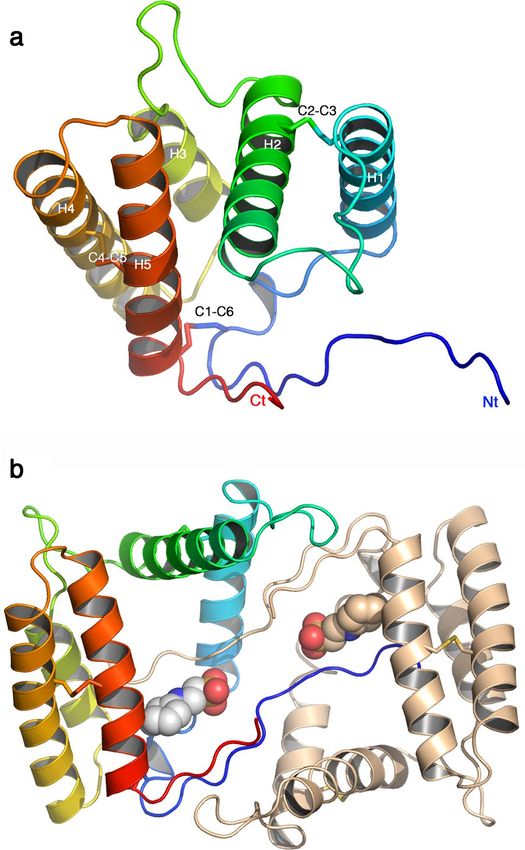
www.nature.com/scientificreports/
Figure 2. Expression and purification of four OBPs of V. destructor. While VdesOBP1, VdesOBP2 and
VdesOBP5 were easily produced in bacteria, VdesOBP3 and VdesOBP4, which are structurally related,
proved difficult to express. In fact, VdesOBP4 was obtained only in very low yield, while we could not observe
expression of VdesOBP3 in any of the different constructs adopted. M: Molecular weight markers; bef: crude
sample before culture induction; aft: crude sample after induction with IPTG; SN: supernatant after lysis and
centrifugation; P: pellet after lysis and centrifugation; OBP: sample of purified protein.
VdesOBP5 were subjected to extensive alkylation with iodoacetamide under non-reducing denaturing condi-
tions; after purification, they were then digested with trypsin followed by a second treatment with chymot-
rypsin. Resulting digests were analyzed by nanoLC-ESI-Q-Orbitrap-MS/MS, and S–S-crosslinked peptides were
assigned using dedicated bioinformatics procedures (Supplementary Table S2 online). Examples of mass spectra
of disulphide-bridged peptides from VdesOBP1 and VdesOBP5 digests are reported in Fig. 3 and Supplementary
Figure S1 online, respectively.
These results and the absence of carboxyamidomethylated peptides in the protein digests demonstrated that
all cysteines present in VdesOBPs are involved in disulphide bridges. Surprisingly, the ascertained, conserved
disulphide pattern of VdesOBPs was unlike that of insect OBPs. In fact, a C1–C6, C2–C3 and C4–C5 arrange-
ment was identified in VdesOBPs, which was completely different form the C1–C3, C2–C5, C4–C6 typical of
insect OBPs10–12. This phenomenon brings two major consequences in the structure of VdesOBPs: (1) the pairing
of adjacent cysteines should make V. destructor proteins more flexible and less compact than insect OBPs; (2)
linking C1–C6 brings together the protein N- and C-termini, while these two ends point generally in opposite
directions in insect OBPs.
Protein characterization in solution. The protein oligomeric state of VdesOBP1 was characterized in
solution at different pH values, namely 4.5, 7.0 and 9.7, using HPLC with multi-angle laser light scattering /
UV/ refractometry (SEC–MALLS-RI) coupled detectors (Wyatt, Santa-Barbara, USA). The results point to a
monomeric state, with measured mass values of 15,708, 16,143 and 15,306 Da (16.496 kDa theoretical mass),
respectively (Supplementary Fig. S2 online).
Overall structure of VdesOBP1. The three-dimensional structure of VdesOBP1 was solved by sulphur
single-wavelength anomalous dispersion (SSAD) with a wavelength λ = 1.512 Å on beamline proxima 2A at
SOLEIL synchrotron (Saint-Aubin, France). Data were collected at 1.968 Å resolution on a P3221 crystal form
and a second dataset was collected at 1.81 Å resolution with wavelength λ = 0.98011 Å (Table 1). Another crystal
form was collected, with space group P21 at 1.2 Å resolution, and its structure was determined by molecular
replacement using the P 3221 crystal form. VdesOBP1 crystal forms P 3221 and P 21 were crystallized at pH val-
ues of 7.5 and 9.5, respectively. They contain one and two molecules in the asymmetric unit, respectively. After
refinement, the final R/Rfree values were 18.6/22.6% and 18.0/20.1% for the two crystal forms, respectively
(Table 1).
The cloned protein is 147 residues long and possesses 6 cysteine residues (Fig. 4). The chain was built in
the electron density map of the P21 crystal form with the exception of residues 1–6 at the N-terminus, while
the P3221 crystal form starts at residue 11 and lacks loop 50–53. We checked the integrity of the protein in the
crystal and our analysis showed that the protein was intact, confirming that the N-terminal segment is present
but not ordered in the crystal. The r.m.s.d. value between the structures of the two VdesOBP1 crystal forms is
1.55 Å (Fig. 5a).
Being the structure best defined, our description will apply to the P21 form. VdesOBP1 is mostly helical
(Fig. 4). In VdesOBP1, five α-helices are observed in contrast with all classical insect OBP or PBPs (pheromone-
binding proteins), such as BmorPBP5, Lush23 or LmadPBP24, which assemble six α-helices. The N- and C-terminal
stretches are elongated forming a pseudo β-sheet of two strands (Fig. 4). Three disulphide bridges (21/141;C1–C6,
41/68;C2–C3, and 111/132;C4–C5, Fig. 4a) strengthen the 3D fold of VdesOBP1.
It has been reported that OBPs may form functional dimers25–27. However, it cannot be concluded firmly for
or against dimerization of OBPs in their biological context. In the crystal, dimers have been observed in the case
of AgamOBP1, but this observation may not apply to solutions, due to an insufficient contact area26. In the case
of VdesOBP1, we observed a monomer using multi-angle static light scattering (MALS)/dynamic light scattering
and RI coupled on-line with a size-exclusion chromatography (SEC) column (MALS/UV/RI/SEC) at three pH
Scientific Reports | (2021) 11:13172 | https://doi.org/10.1038/s41598-021-92604-2 3
Vol.:(0123456789)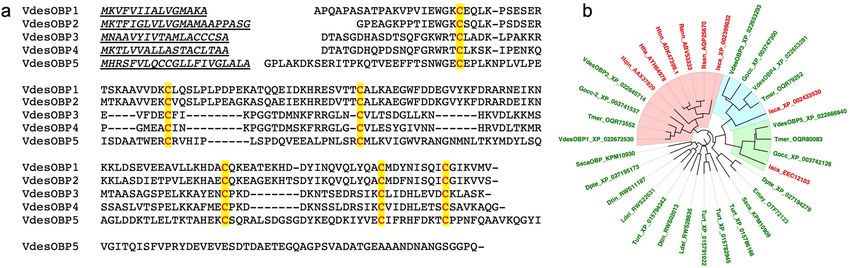
www.nature.com/scientificreports/
Figure 3. Fragmentation spectra of disulfide-bridged peptides identified in the tryptic-chymotryptic digest
of V. destructor VdesOBP1 as revealed by nanoLC-ESI-Q-Orbitrap-MS/MS analysis. The fragments are
highlighted in different colours depending on peptides present in S–S-linked species, and on the corresponding
b and y ion series. Complete data on disulphide-bridged peptides are reported in Supplementary Table S2
online.
values (Supplementary Fig. S2 online). In contrast, the P21 form contains two molecules in the asymmetric unit
forming a crystallographic dimer (Fig. 4b). A crystallographic dimer can be reconstituted in the P 3221 form,
using a symmetrical molecule in the crystal packing (Fig. 5b). However, the second monomer of one form does
not display the same orientation relative to that of the second form when the first monomers are superimposed
(Fig. 5b,c). Finally, the PISA server reported an ambiguous response for the P 21 form (score = 0.49) but discarded
the possibility of a biological dimer in form P3221 (score = 0). All in all, it seems highly probable that the biologi-
cally relevant form is a monomer.
The binding crevice. The binding crevice, as detected by C avityPlus28, is large and wide open in the mono-
mer, with dimensions of the floor of ~ 18 × 18 Å (Fig. 6a). This large surface implies that numerous amino acid
side-chains form the walls of the binding site: hydrophobic side-chains (Pro14, Ile16, Trp18, Leu24, Pro 26,
Val37, Val38, Leu70, Trp75, Tyr82, Ala87, Ile91, Tyr129, Met133, Ile137), polar non-charged residues (Thr66,
Gln130) and charged residues (Glu30, Arg31, Lys34, Glu73, Glu90, Asp134). Hence, although most side chains
are from hydrophobic/aromatic residues (16/24), two basic and four acidic residues complete the wall. The active
site is empty, except in form P21 where a buffer molecule, 2-(N-cyclohexylamino)-ethane sulfonic acid (NCES),
is docked against a wall of the binding site (Fig. 6a). Examination of the binding site in the crystallographic
dimer revealed that the N-terminus of each monomer protrudes deeply in the binding site of the other mono-
mer, drastically reducing its size. This effect explains the serendipitous binding of NCES as this ligand interacts
with the cell wall of one monomer and the C-terminal extension of the other one (Fig. 6b). The sulfonic acid
moiety of NCES forms an ionic bond with Arg31A side chain (atom NH2) and with the main chain N–H of
Lys12B.
Comparison of VdesOBP1 with other OBPs/PBPs structures. A search on the PDB with the DALI
server reported hits with good statistics, taking into account the Z-score and the r.m.s.d.29. The best hit is that
of entry 5dic (Z = 7; r.m.s.d. 2.9 Å on 96/115 residues), the fatty acid binding protein OBP56a from the oral disk
of the blowfly Phormia regina (PDB ID: 5dic, by Ishida, Y., Leal, W.S., Wilson, D.K., unpublished) with a non-
Scientific Reports | (2021) 11:13172 | https://doi.org/10.1038/s41598-021-92604-2 4
Vol:.(1234567890)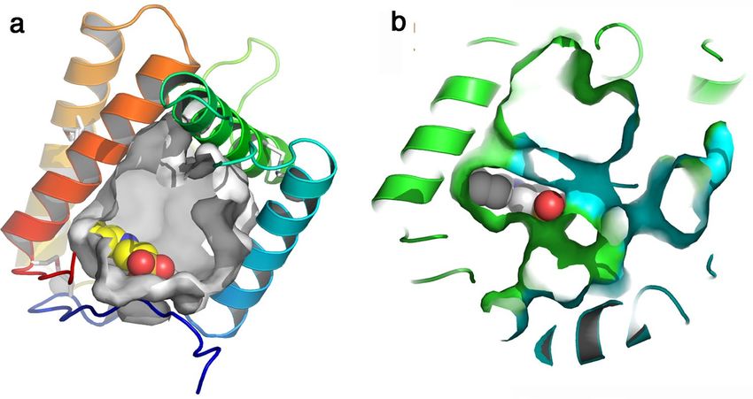
www.nature.com/scientificreports/
Data collection
Beamline SOLEIL Proxima2 SOLEIL Proxima1
PDB ID 7NYJ 7NZA
Space group P3221 P21
Cell parameters, a, b, c (Å), γ/β (°) 53.8, 53.8, 91.7 γ = 120° 31.6, 113.0, 41.8 β = 98.95°
Wavelength (Å) 0.98011 0.97856
Resolution (Å) 46.6–1.81 (1.95–1.81) 40.09–1.20 (1.27–1.20)
Rmergea (%) 7.95 (110) 6.5 (88.5)
Mean((I)/sd(I))a 13.26 (1.87) 13.15 (1.19)
cc(1/2) 99.9 (76.3) 99.8 (78.8)
No. of observationsa 284,352 (57,758) 634,776 (86,086)
No. of unique reflectionsa 13,957 (2889) 87,891 (13,358)
Completenessa (%) 100.0 (100.0) 97.2 (91.4)
Multiplicitya 20.4 (20.0) 7.0 (6.4)
Refinement
Resolution (Å) 46.59–1.81 (1.857–1.810) 38.8–1.20 (1.23–1.20)
No. of reflectionsa 13,827 (1009) 83,470 (5555)
atoms:protein/ligands/solvent 2289/36/393 2325/36/340
No. of test set reflections 728 (53) 4402 (308)
Rwork/Rfreea (%) 18.6 (28.6)/22.6 (31.0) 18.0 (33.0)/20.1(34.1)
rmsd bonds (Å)/angles (°) 0.015/1.83 0.018/2.20
B-factors (Å2) 40.5 19.0
Ramachandran: favored/allowed (%) 98.5/1.5 96.0/4.0
Table 1. Data collection and refinement statistics. a Refers to the highest-resolution bin.
classical OBP fold. This FBP/OBP has been shown to bind the C16 palmitic acid (but not the acetate), as well as
the C18 stearic, oleic and linoleic acids, but not decanoic acid30. The structure is reported in complex with two
molecules of palmitic acid in its wide cavity. Superposition of VdesOBP1 and PregOBP56a shows that VdesOBP1
helices 1, 2, 3, 4 and 5 superimpose well with PregOBP56a helices 1, 3, 4, 5 and 6. Helix 2 of PregOBP56a closes
its binding cavity, while a similar effect is not observed in VdesOBP1. This PregOBP56a helix 2 is replaced in
VdesOBP1 by a loop between helices 1 and 2, projected out of the protein core (Fig. 7a). However, the Pre-
gOBP56 disulphide bridges obey the C1–C3, C2–C5 and C4–C6 connectivity also present in classical OBPs, in
contrast with VdesOBP1 (C1–C6, C2–C3, C4–C5). Another good fit (1ow4; Z = 5.4; r.m.s.d 3.3 Å on 8–90/120
residues) is observed with the PBP of the cockroach Leucopheae maderae (LmadPBP) (1ORG). In this case
again, LmadPBP exhibits an extra helix instead of a loop between helices 1 and 2 in VdedOBP1 (Fig. 7b). Two
disulphide bonds are conserved between the two proteins, the first bridging helices 1 and 2, the second bridging
helices 4 and 5 of VdesOBP1 (Figs. 4a, 7b), although as result of different (C1–C6, C2–C3, C4–C5 in VdesOBP1
and C1–C3, C2–C5, C4–C6 in LmadPBP) cysteine linkages. The third disulphide bond that bridges helices 3 and
6 in LmadPBP is not observed in VdesOBP1. Instead, the third disulphide bridge is observed between helix 5
(corresponding to helix 6 in LmadPBP) and the extended N-terminus in VdesOBP1 (Fig. 7b).
Ligand‑binding assays. The purified VdesOBP1 did not show any affinity for either of the most common
probes used in ligand-binding assays with OBPs, namely N-phenyl-1-naphthylamine (1-NPN) and 1-amino-
anthracene (1-AMA). Extraction with dichloromethane to remove possible endogenous ligands did not prove
effective. We also subjected the protein to a denaturing treatment with 8 M urea and 1 mM DTT, followed by
slow renaturation by extensive dialysis, but we still could not observe any binding with either probes. We also
tried 8-phenylamino-naphthalenesulfonic acid (ANS) as well as larger fluorescent reporters31, such as N-naph-
thyl-1-naphthylamine (1-NNN) and N-phenyl-1-aminoanthracene (PAA) with no effect.
It is still possible that the fluorescent probe binds the protein, but the event is not accompanied by a change in
the emission spectrum. In fact, when adding 1-NPN to the protein, we observed a dose-dependent quenching of
the intrinsic tryptophan fluorescence, suggestive of some affinity of this probe for VdesOBP1 (data not shown). In
any case, competitive binding using a fluorescent reporter could not be used with VdesOBP1 or with VdesOBP2.
Therefore, we attempted to find ligands for these two proteins monitoring the quenching of intrinsic tryptophan
fluorescence. On the other hand, this method is limited to chemicals able to accept the energy transferred by
the tryptophan (therefore structures with a region of conjugated π-electrons), and is not suitable to evaluate
binding affinities. Instead, with VdesOBP4 and VdesOBP5 we obtained clear binding curves using 1-NPN as the
fluorescent reporter (Fig. 8a). Therefore, we performed competitive binding experiments only with these two
OBPs. Some of the displacement curves obtained with VdesOBP4 and VdesOBP5, using the ligands shown in
Fig. 8b, are reported in Fig. 8c,d respectively. We can notice that only coniferyl aldehyde is able to decrease the
1-NPN fluorescence to 50% at the maximum concentration used, qualifying for a modest ligand. None of the
other chemicals used in the experiments (listed in Supplementary Table S3 online) with the four proteins proved
Scientific Reports | (2021) 11:13172 | https://doi.org/10.1038/s41598-021-92604-2 5
Vol.:(0123456789)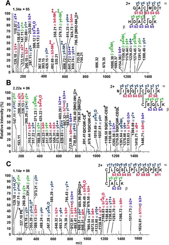
www.nature.com/scientificreports/
Figure 4. Crystal structure of VdesOBP1 form P21. (a) Ribbon view of a monomer displaying helices
1–5 and disulphide bridges. The polypeptide chain is rainbow coloured, from blue (N-terminus) to red
(C-terminus). The disulphide bonds are identified by the secondary structures they belong to. (b) Ribbon
view of the crystallographic dimer. One monomer is rainbow coloured, the other one is coloured brown. The
serendipitously bound buffer molecule 2-(N-cyclohexylamino)-ethane sulfonic acid (NCES) is displayed as
atomic spheres (C: white, N: blue and O: red). Figure made with PyMOL (version 1.5.0.2. http://www.pymol.
org).
to be a better ligand. Representative curves relative to quenching of the intrinsic fluorescence of tryptophan in
VdesOBP1 and VdesOBP2 are reported in Supplementary Fig. S3 online.
Summarising these data, we can first observe that some of the tested chemicals proved to be ligands for Vdes-
OBP4 and VdesOBP5 in 1-NPN displacement assays, while they determined significant quenching of intrinsic
tryptophan fluorescence in VdesOBP1 and VdesOBP2. The same few compounds (coniferyl aldehyde, safranal
and vanillin) were the best ligands with all four proteins, although with poor dissociation constants between
10 and 20 μM. These ligands are associated with plants, in particular with leaf damage, and do not bear any
relationship to the varroa habits. In particular, none of the honey bee pheromones, such as 9-ketodecanoic acid,
homovanillyl alcohol or fatty acid esters were able to bind any of the tested proteins.
Discussion
This work reports the original structural and functional characterization of four out of the five OBPs encoded in
the genome of the mite Varroa destructor, a major pest for honey bee. Mass spectrometric analysis demonstrated
that the six cysteines VdesOBPs, whose sequence position do not match the pattern in insect OBPs, are paired
into three disulphide bridges (C1–C6, C2–C3 and C4–C5) different from that (C1–C3, C2–C5, C4–C6) of their
insect counterparts. The overall folding, determined for VdesOBP1, is also novel and different from the conserved
structure of insect OBPs, although some similar aspects can be recognised.
VdesOBP1 structure was determined in two crystal forms closely related. Its structure assembles 5 α-helices
linked by three disulphide bridges. Comparison with classical OBPs reveals that the second of the six α-helices
found in them is lacking in VdesOBP1. Although dimer formation of VdesOBP1 is observed in the crystal,
Scientific Reports | (2021) 11:13172 | https://doi.org/10.1038/s41598-021-92604-2 6
Vol:.(1234567890)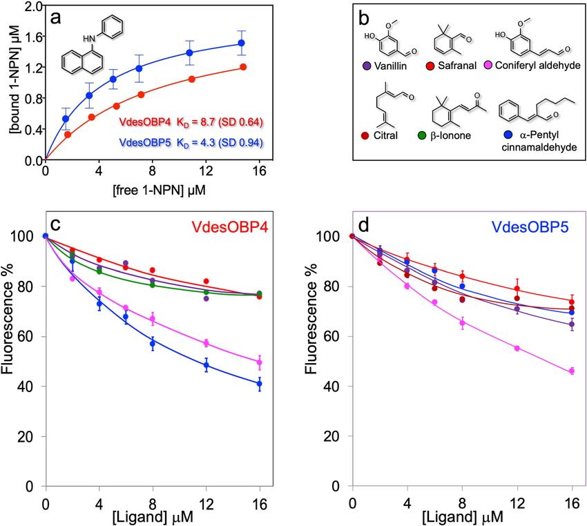
www.nature.com/scientificreports/
Figure 5. Comparison of crystal structures of VdesOBP1 forms P21 and P3221. (a) Ribbon view of the
superposition of monomers belonging to forms P21 (rainbow coloured) and P 3221 (grey). (b) Ribbon view of
the crystallographic dimers of forms P21 and P3221. In form P21 one monomer is rainbow coloured, the other
one is coloured brown. The crystallographic dimer of P3221 form is coloured light grey. One monomer of each
form has been superimposed (left). The second monomers do not superimpose. B/same view rotated 90° around
a vertical axis. Note the rotation of ~ 80° of the second monomer of form P3221 relative to the second monomer
of form P 21. Figure made with PyMOL (version 1.5.0.2. http://www.pymol.org).
it most probably does not represent the functional form in vivo. This dimerization is probably driven by the
presence of a wide and fully open cavity, able to accommodate the extended N- and C-terminal stretches of the
opposite monomer. The large cavity opening can be ascribed to the absence of helix 2 in OBPs classical fold,
which restrains and closes the internal binding site. In VdesOBP1, this helix is absent and replaced by a loop
projected outside the protein core. Two disulphide bridges are conserved as compared to classical OBPs, although
associated with different sequential cysteine linkages. These observations suggest that VdesOBP1 possesses an
original fold remotely related to classical OBPs. Comparison with PregOBP56a suggests that two to three fatty
acids could bind inside the VdesOBP1 cavity. Such an arrangement of three ligands was observed in Mamestra
brassicae CSP (CSPMbraA6)30.
The unique structure of VdesOBP1 and the lack of strong ligands pose questions that, at this stage, can only
be addressed by formulating hypotheses and models to be verified by further experimental work. A first idea,
based on the limited binding activity of these proteins and on the small number of genes (4–5) expressed in V.
destructor, as well as in other mites and ticks, might suggest that these genes represent by-products of evolution,
which are not used any more. In fact, at least two more families of carrier proteins could be active in Chelicerata,
namely NPC2 proteins and lipocalins, apart from a third family (CCPs: candidate carrier proteins) that so far
seems to be specific of spiders21. NPCs and lipocalins are represented in varroa by 6 and 9 members, respectively,
while in other Chelicerata often 10–20 members of NPC2s have been identified, with exceptions of nearly 50
genes, as in the spider mite Tetranychus urticae. Although making sense for the reason reported above, the idea
that OBPs in Chelicerata might represent a dead end in evolution and a failed attempt to make efficient carrier
proteins contrasts with proteomic findings showing significant expression of such proteins22. In fact, four (Vdes-
OBP1-4) of the five VdesOBPs were identified in different parts of V. destructor, with one of them (VdesOBP4)
particularly abundant in sensory organs, namely the first pair of legs and the mouthparts.
Scientific Reports | (2021) 11:13172 | https://doi.org/10.1038/s41598-021-92604-2 7
Vol.:(0123456789)www.nature.com/scientificreports/
Figure 6. The internal cavity of VdesOBP1 form P 21. (a) Slabbed ribbon view of a monomer rainbow coloured.
The cavity is represented by a grey surface and is wide open. The serendipitously bound buffer molecule
2-(N-cyclohexylamino)-ethane sulfonic acid (NCES) is displayed as atomic spheres (C: white, N: blue and
O: red), and is stacked against the cavity wall. (b) Slabbed ribbon view of a crystallographic dimer rainbow
coloured. The cavity is represented by a green surface originating from monomer 1 and a blue surface from
monomer 2. The serendipitously bound buffer molecule NCES is displayed as atomic spheres (C: white, N: blue
and O: red), and is stacked against the cavity walls from both monomers. Figure made with PyMOL (version
1.5.0.2. http://www.pymol.org).
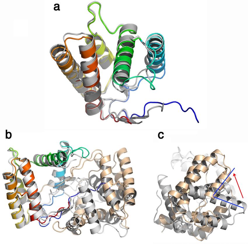
Figure 7. Comparison of crystal structures of VdesOBP1 with the closest related OBPs from Phormia regina
(PregOBP56) and Leucophera madereae (LmadPBP). (a) Ribbon view of the superposition of monomers from
VdesOBP1 (rainbow coloured) and PregOBP56 (light grey) with its two fatty acid ligands (grey sticks). Note the
absence of the classical OBP fold helix 2 (red squared) in VdesOBP1. (b) Ribbon view of the superposition of
monomers from VdesOBP1 (rainbow coloured) and LmadPBP. Note the absence of the classical OBP fold helix
2 (red squared) in VdesOBP1 as compared to LmadPBP (red squared) and the shift of one disulphide bridge
(red arrow). The 3D positions of the two other disulphide bridges are conserved (purple arrow), in the final
fold, although as result of different C1-C6, C2-C3, C4-C5 (VdesOBP1) and C1-C3, C2-C5, C4-C6 (LmadPBP)
connectivities. Figure made with PyMOL (version 1.5.0.2. http://www.pymol.org).
Scientific Reports | (2021) 11:13172 | https://doi.org/10.1038/s41598-021-92604-2 8
Vol:.(1234567890)www.nature.com/scientificreports/
Figure 8. Ligand-binding assays. (a) Binding of the fluorescent probe N-phenylnaphthylamine (1-NPN) to
VdesOBP4 and VdesOBP5. With the other two proteins expressed (VdesOBP1 and VdesOBP2), we could not
observe the blue shift usually associated with binding of the fluorescent probe. (b) Structures of the ligand
used in the competitive binding experiments. (c,d) Competitive binding assays. Only few chemicals were able
to displace 1-NPN from the complex with VdesOBP4 and VdesOBP5. The same chemicals were also the best
ligands for the other VdesOBPs (Supplementary Fig. S3 online).
If then the expression of OBPs at the protein level indicates that they are functional, then, along with a
second hypothesis, their poor affinity might be related to the absence of post-translational modifications. It
has been recently shown that the pig OBP1 can bind fatty acids or steroids depending on the phosphorylation
and glycosylation status of the protein32. Our varroa OBPs, being expressed in bacteria, are devoid of any post-
translational modification. On the other hand, we do not have any information on possible phosphorylation and/
or glycosylation of these proteins in their native forms. This is certainly an interesting aspect, although not easy
to investigate, not only for the tiny size of sensory organs, but also because phosphorylation, in particular, may
be present only in certain time windows related to the physiology and the behaviour of the mite. Indeed, protein
phosphorylation is highly related to signal transduction events regulating essential physiological process, and it
has been reported to drastically modify the binding properties of O BPs32. Thus, we searched for putative phos-
phorylation sites in the five OBP sequences, and we found a very large number of serine and threonine residues
that could be potentially phosphorylated (Supplementary Fig. S4 online). In addition, we found few tyrosine
residues as potential phosphorylation sites (2 in VdesOBP1, 1 in VdesOBP2 and VdesOBP3, 4 in VdesOBP5).
In the three-dimensional structure of VdesOBP1, no serine or threonine residues are found inside the bind-
ing pocket. However, phosphorylation of serine and threonine residues outside the binding pocket (as well as
glycosylation of the protein) can affect the folding of the protein and influence its binding affinities. Further
speculation would be inappropriate based on the available experimental data. Certainly, a structural study of the
native proteins, regarding their post-translational modifications would be essential before proposing alternative
modes of action.
Materials and methods
Biological material. V. destructor mites were collected from the cells of an infected honeybee hive and used
immediately for RNA extraction.
Reagents. Chemical compounds for buffers and solutions, as well as ligands for binding assays were of
analytical grade from Merck, Austria. Ligands were dissolved in spectroscopic grade (Uvasol) methanol. Oligo-
Scientific Reports | (2021) 11:13172 | https://doi.org/10.1038/s41598-021-92604-2 9
Vol.:(0123456789)www.nature.com/scientificreports/
nucleotides and synthetic genes were custom synthesised at Eurofins Genomics, Germany. Enzymes and kits for
DNA extraction and purification were from New England Biolabs, USA.
RNA extraction and gene cloning. Total RNA was extracted using Tri-reagent (Sigma-Aldrich) accord-
ing to the protocol provided. cDNA was prepared from 2 μL of total RNA using the kit qScript cDNA SuperMix
(Quanta Bio) and the manufacturer’s procedure. For sequence verification, PCR was performed on cDNA using
specific primers at the 5’ end and at the 3’ end. The primers, which were also used for cloning into the expres-
sion vector, also contained the restriction sites NdeI and EcoRI at the 5’ and the 3’ ends, respectively. After
digestion, the PCR products were inserted into pET30 vector previously linearized with the same enzymes. This
procedure was adopted for all the genes of V. destructor, except for VdesOBP1 used for structural studies, which
was produced from a synthetic gene (Eurofins Genomics), whose nucleotide sequence was optimized for E. coli
expression.
Gene cloning. Synthetic genes encoding VdesOBP1 and VdesOBP2 were custom synthesised at Euro-
fins Genomics, Germany, and optimised for E. coli expression. After digestion with restriction enzymes Nde
I and Eco RI, the genes were subcloned into the pET-30 expression vector (Novagen, Darmstadt, Germany).
The sequences encoding VdesOBP4 and VdesOBP5, instead, were amplified directly from cDNA, digested and
cloned into the pET-30. The constructs encoded only the mature protein sequences preceded by an additional
methionine, which is not considered here for amino acid numbering. Expression was performed in E. coli BL-21
(DE3) competent cells. The sequences of the primers used are reported in Supplementary Table S4 online.
Protein expression and purification. Protein synthesis was induced by adding IPTG (to a final concen-
tration of 0.4 mM) to the E. coli culture, grown for two additional hours at 37 °C, then harvested by centrifuga-
tion, sonicated in 50 mM Tris/HCl pH 7.4 and centrifuged at high speed. The VdesOBPs were obtained in good
yield in the supernatant, except for VdesOBP4, which was produced in poor yield and was present in the pellet,
from which it was solubilized with 8 M urea and 1 mM DTT. Refolding was obtained by simple dialysis for
three days (with changes of buffer every day) against 50 mM Tris–HCl, pH 7.425. All VdesOBPs were purified
by 2–3 chromatographic steps on anion-exchange columns DE-52 and HiPrep-Q 16/10, 20 mL (Bio-Rad). The
purification process was monitored at each step by polyacrylamide gel electrophoresis in denaturing conditions
(SDS-PAGE).
Protein alkylation and digestion, and mass spectrometry analysis. Samples of V. destructor
OBP1, OBP2 and OBP5 (20–50 μg) dissolved in 0.1 M tetraethylammonium bicarbonate (TEAB), pH 6.5, con-
taining 4 M guanidinium chloride, were treated with iodoacetamide (0.5 M final concentration) for 30 min, in
the dark; then, proteins were insolubilized by addition of 6 vol of cold acetone, leaving the samples at -20 °C,
overnight. After centrifugation at 12,000 rpm at 4° C, for 20 min, supernatants were removed, and recovered
pellets were dried with a SpeedVac device (ThermoFisher Scientific, USA). Recovered proteins were dissolved in
0.05 M TEAB, pH 6.5 (2 µg/μL final concentration), treated with trypsin (1:10 w/w enzyme/substrate) for 16 h,
at 37 °C, then with chymotrypsin (1:8 w/w enzyme/substrate) for 16 h, at 37 °C. Protein digests were desalted
with ZipTip C18 (Millipore, USA) and directly analyzed with a UltiMate 3000 HPLC RSLC nano-chromatogra-
pher (ThermoFisher Scientific) linked on-line through a nano-Spray ion source (Thermo Fisher Scientific) to a
Q-ExactivePlus mass spectrometer (Thermo Fisher Scientific)33. Peptides were separated on an Acclaim PepMap
RSLC C18 column (150 mm × 75 μm ID; 2 μm particle size; 100 Å pore size) (Thermo Fisher Scientific), at a flow
rate of 300 nL/min, using a gradient of solvent B (19.92/80/0.08 v/v/v water/acetonitrile/formic acid) in solvent
A (99.9/0.1 v/v water/formic acid). Solvent B started at 3%, increased linearly to 40% in 45 min, then raised to
80% in 5 min, where it remained for additional 4 min, before rapidly returning to 3%. Mass spectrometer worked
in positive polarity using a data-dependent mode, performing a full MS1 scan in the range m/z 345–1350, at a
nominal resolution of 70,000, followed by MS/MS scans of the 10 most abundant ions in high energy collisional
dissociation mode34. MS/MS spectra were acquired in a dynamic m/z range, with a nominal resolution of 17,500,
a normalized collision energy of 28%, an automatic gain control target of 50,000, a maximum ion injection time
of 110 ms, an isolation window of 1.2 m/z and a dynamic exclusion setting of 20 s.
Bioinformatics for peptide identification. Firstly, raw mass data files were analyzed with Proteome
Discoverer v. 2.4 package (Thermo Fisher Scientific), running by Mascot v. 2.6.1 (Matrix Science, UK) and
Byonic v. 2.6 (Protein Metrics, USA) software. Database searching was performed against a database containing
the sequences of V. destructor OBP1, OBP2 and OBP5, trypsin, chymotrypsin and common protein contami-
nants. Parameters for database searching were variable oxidation at Met, deamidation at Asn/Gln, pyroglutamate
formation at Gln and carbamidomethylation at Cys. Mass tolerance setting included ± 10 ppm for precursor
ions and ± 0.05 Da for fragmentation ions35. Proteolytic enzyme and maximum number of missed cleavages
were set to trypsin, chymotrypsin and 5, respectively. Proteome Discoverer peptide candidates were considered
confidently identified only when the following criteria were satisfied: (1) protein and peptide false discovery rate
(FDR) confidence: high; (2) peptide Mascot score: > 30; (3) peptide spectrum matches (PSMs): unambiguous;
(4) peptide rank (rank of the peptide match): 1; (5) Delta CN (normalized score difference between the selected
PSM and the highest-scoring PSM for that spectrum): 0. Byonic peptide candidates were considered confi-
dently identified only when the following criteria were satisfied: (1) PEP 2D and PEP 1D: < 10 × 10−5; (2) FDR: 0;
(3) q-value 2D and q-value 1D: < 10 × 10−5. Finally, disulphide bridge assignment was obtained using dedicated
BioPharma Finder v. 4.0 (Thermo Fisher Scientific) and pLink v. 2.3.9 s oftware36. Both programs were used
for database searching, enabling the specific function of disulphide-linked peptides attribution, and applying
Scientific Reports | (2021) 11:13172 | https://doi.org/10.1038/s41598-021-92604-2 10
Vol:.(1234567890)www.nature.com/scientificreports/
the additional settings reported above for Proteome Discoverer and Byonic examinations. Disulphide-bridged
peptide identification was considered reliable when BioPharma Finder and pLink results showed a “confidence
score” > 95 and/or an “E-value” < 1.0–10, respectively. Manual interpretation and verification of the candidate
spectra were always performed.
Crystallization, data collection and structure determination of the P3221 form. The protein
was concentrated to 20 mg/ml. Screening experiments were performed using several commercial kits. The sit-
ting-drop method in Swissci plates was used. The reservoirs of the Swcissi Plates were filled using a Genesis II
(TECAN) pipetting robot. The nanodrops were dispensed by a MOSQUITO Crystal (TTP Labtech). Respec-
tively 300 nL, 200 nL and 100 nL of protein were dispensed onto the central of three-sitting-drop shelves. 100
nL of the reservoir solution were added to the protein drops. The plates were sealed with a transparent film and
stored in a R1000 crystallization hotel (Formulatrix). Small crystals were obtained in 0.2 M cadmium chloride,
2.2 M ammonium sulfate. Those crystals were used for micro-seeding. The sitting-drop method in Greiner
plates was used and new crystals were obtained by doing a re-screen in the commercial screen PEGII with the
initial condition in which the first crystals were obtained. The droplet with the thicker crystals contained 2/3 of
the initial condition (0.2 M cadmium chloride, 2.2 M ammonium sulfate) + 1/3 of PEG II screen (0.2 M calcium
chloride, 0.1 M HEPES pH 7.5; 30% w/v PEG 4000).
The crystal was cryo-cooled at 100 °K within this crystallization liquor, with the addition of 10% glycerol as
cryoprotectant. Structure determination used SAD-phasing method at remote wavelengths (λ = 1.51200 Å) with
sulphur atom anomalous dispersion on native crystals. A strong theoretical Bijvoet ratio of 1.12% was expected
from the 10 sulphur atoms of the protein that contribute to the anomalous signal at wavelength of 1.512 Å. The
sulphur-SAD data set was collected on the PROXIMA-2 beamline (SOLEIL) equipped with a high performance
micro-diffractometer with a mini-kappa (MD2), an X-ray fluorescence detector (KETEK), and a fast, low-noise,
photon-counting, area detector EIGER X 9 M (238 fps in 9 M mode, 750 fps in 4 M mode). A redundant data
set was collected at 1.97 Å resolution. including 3600 images of 0.1° rotation collected with four K orientations
of 0°, 30°, 45° and 60°. The data set was processed with X DS37. The data sets were indexed and integrated with
Pointless and scaled with CCP4 S CALA38. VdesOBP1 crystallized in space group P 3221 with cell dimensions
a = 53.84 Å, b = 53.84 Å, and c = 91.74 Å and and γ = 120 with one molecule in the asymmetric unit, corresponding
to a solvent content of 50.87%. Six sulfur atom sites were found by using PHENIX Autosol39. The results from
PHENIX include a PDB containing the anomalous scatterers, a MTZ file containing the experimentally deter-
mined phases, and the optimized phases from density modification. Structure initial construction was performed
with PHENIX Autosol39 and was not further refined. A second data set was collected at 1.20 Å resolution at a
wavelength of 0.97856 Å. The ab-initio determined structure was used as a starting model for model refinement.
Refinement was performed with R EFMAC540 and AutoBUSTER41 alternated with rebuilding using COOT42.
Crystallization, data collection and structure determination of the P21 form. The protein at
20 mg/mL was subjected to screening experiments that were performed using several commercial kits. Vdes-
OBP1 was set in 2/3 of the initial crystallization conditions (0.1 M CHES pH 9.5, , 30% w/v PEG3000) and 1/3
of ammonium sulfate screen (0.2 M sodium nitrate, 2.2 M ammonium sulfate). Crystals of VdesOBP1 were
briefly soaked in crystallization solution supplemented with 10% (v/v) polyethylenglycol. Diffraction data were
collected at 1.2 Å resolution on beamline Proxima-1 at SOLEIL, Paris, France. The data sets were integrated with
XDS37 and were scaled with CCP4 SCALA38. VdesOBP1 crystallized in space group P 21 with cell dimensions
a = 31.6 Å, b = 112.96 Å, c = 41.82 Å and β = 98.95, with two molecules in the asymmetric unit, correspond-
ing to a solvent content of 44.48%. The structure was solved by molecular replacement with MOLREP43 using
the structure of VdesOBP1 form P3221 as starting model. Refinement was performed with REFMAC540 and
AutoBUSTER41, and the structures were corrected with COOT42. Figures 4, 5, 6 and 7 were made with PyMOL
(version 1.5.0.2. http://www.pymol.org).
Multi‑angle light scattering. 100 μL of purified VdesOBP1 were analyzed by SEC-MALS using a HPLC
system (Thermo Scientific) equipped with a Superdex 75 column coupled with a multi-angle light scattering
device (Dawn 8, Wyatt Technology) and an Optilab TrEX differential refractive index detector (Optilab TrEX,
Wyatt Technology). At first, the column was first equilibrated with a buffer containing 0.02% w/v NaN3 for sev-
eral hours, until the baseline of the Optilab TrEX signal was stable. Then, light scattering and concentration data
were analysed in ASTRA to determine molecular weight and size distribution. Inter-detector band broadening
and alignment were applied using the algorithms in ASTRA 7 (Wyatt Technology, USA) using BSA protein
standard according to manufacturer’s recommended methods. Three different buffers were used to understand
the behaviour of the protein at different pH values, namely 50 mM CHES pH 10, 50 mM Hepes pH 7 and 50 mM
sodium acetate pH 5. The temperature-controlled autosampler was kept at 10 °C.
Ligand‑binding assays. Direct binding of fluorescent probes was monitored by adding aliquots of 1 mM
methanol solution of the probe to a 2 μM solution of the protein in 50 mM Tris–HCl, pH 7.4 to final concentra-
tions of 2 to 16 μM. The excitation wavelength was 337 nm and intensities were measured on the maximum of
the peak, usually between around 410 and 420 nm. For VdesOBP1 and VdesOBP2, binding of ligands was evalu-
ated by monitoring the intrinsic tryptophan quenching of the protein. Excitation wavelength was 295 nm and
intensities were recorded around 335–340 nm. For VdesOBP4 and VdesOBP5, competitive binding experiments
were performed by titrating a solution of protein and 1-NPN both at 2 μM in Tris–HCl buffer, pH 7.4 with 1 mM
methanolic solutions of ligands to final concentrations of 2–16 μM. All measurements were performed with a FL
Scientific Reports | (2021) 11:13172 | https://doi.org/10.1038/s41598-021-92604-2 11
Vol.:(0123456789)www.nature.com/scientificreports/
6500 spectrofluorometer (PerkinElmer); slits were set at 5 nm for both excitation and emission, and 1 cm path
quartz cuvettes were used.
Dissociation constants for 1-NPN were evaluated using Prism software. Affinities of other ligands were
calculated from the corresponding [IC]50 values (the concentration of each ligand halving the initial value of
fluorescence), from the equation:
Kd = [IC]50 /1 + [1 − NPN]/KNPN
where [1-NPN] is the concentration of free 1-NPN and K
NPN is the dissociation constant of the complex
OBP/1-NPN.
Data availability
The three-dimensional structure of VdesOBP1 reported in this paper has been deposited in the database with
PDB ID 7NYJ and 7NZA.
Received: 29 March 2021; Accepted: 11 June 2021
References
1. Pelosi, P., Baldaccini, N. E. & Pisanelli, A. M. Identification of a specific olfactory receptor for 2-isobutyl-3-methoxypyrazine.
Biochem. J. 201, 245–248 (1982).
2. Vogt, R. G. & Riddiford, L. M. Pheromone binding and inactivation by moth antennae. Nature 293, 161–163 (1981).
3. Leal, W. S. Odorant reception in insects: roles of receptors, binding proteins, and degrading enzymes. Annu. Rev. Entomol. 58,
373–391 (2013).
4. Pelosi, P., Iovinella, I., Zhu, J., Wang, G. & Dani, F. R. Beyond chemoreception: diverse tasks of soluble olfactory proteins in insects:
Soluble olfactory proteins in insects. Biol. Rev. 93, 184–200 (2018).
5. Sandler, B. H., Nikonova, L., Leal, W. S. & Clardy, J. Sexual attraction in the silkworm moth: structure of the pheromone-binding-
protein–bombykol complex. Chem. Biol. 7, 143–151 (2000).
6. Tegoni, M., Campanacci, V. & Cambillau, C. Structural aspects of sexual attraction and chemical communication in insects. Trends
Biochem. Sci. 29, 257–264 (2004).
7. Bianchet, M. A. et al. The three-dimensional structure of bovine odorant binding protein and its mechanism of odor recognition.
Nat. Struct. Biol. 3, 934 (1996).
8. Tegoni, M., Ramoni, R., Bignetti, E., Spinelli, S. & Cambillau, C. Domain swapping creates a third putative combining site in bovine
odorant binding protein dimer. Nat. Struct. Biol. 3, 863–867 (1996).
9. Pelosi, P., Iovinella, I., Felicioli, A. & Dani, F. R. Soluble proteins of chemical communication: an overview across arthropods.
Front. Physiol. 5, 320 (2014).
10. Scaloni, A. Analysis of post-translational modifications in soluble proteins involved in chemical communication from mammals
and insects. Methods Enzymol. 642, 103–124 (2020).
11. Scaloni, A., Monti, M., Angeli, S. & Pelosi, P. Structural analysis and disulfide-bridge pairing of two odorant-binding proteins from
Bombyx mori. Biochem. Biophys. Res. Commun. 266, 386–391 (1999).
12. Leal, W. S., Nikonova, L. & Peng, G. Disulfide structure of the pheromone binding protein from the silkworm moth Bombyx mori.
FEBS Lett. 464, 85–90 (1999).
13. Pelosi, P., Zhu, J. & Knoll, W. From gas sensors to biomimetic artificial noses. Chemosensors 6, 32 (2018).
14. Angeli, S. et al. Purification, structural characterization, cloning and immunocytochemical localization of chemoreception proteins
from Schistocerca gregaria. Eur. J. Biochem. 262, 745–754 (1999).
15. Zhu, J. et al. Niemann-pick C2 proteins: a new function for an old family. Front. Physiol. 9, 52 (2018).
16. Ishida, Y. et al. Niemann-Pick type C2 protein mediating chemical communication in the worker ant. PNAS 111, 3847–3852 (2014).
17. Renthal, R. et al. The chemosensory appendage proteome of Amblyomma americanum (Acari: Ixodidae) reveals putative odorant-
binding and other chemoreception-related proteins. Insect Sci. 24, 730–742 (2017).
18. Eliash, N. et al. Chemosensing of honeybee parasite, Varroa destructor : Transcriptomic analysis. Sci. Rep. 7, 1–11 (2017).
19. Eliash, N. et al. Varroa chemosensory proteins: some are conserved across Arthropoda but others are arachnid specific. Insect Mol.
Biol. 28, 321–341 (2019).
20. Vizueta, J. et al. Evolution of chemosensory gene families in arthropods: insight from the first inclusive comparative transcriptome
analysis across spider appendages. Genome Biol. Evol. 9, 178–196 (2017).
21. Vizueta, J., Rozas, J. & Sánchez-Gracia, A. Comparative genomics reveals thousands of novel chemosensory genes and massive
changes in chemoreceptor repertories across chelicerates. Genome Biol. Evol. 10, 1221–1236 (2018).
22. Iovinella, I. et al. Proteomic analysis of chemosensory organs in the honey bee parasite Varroa destructor: A comprehensive
examination of the potential carriers for semiochemicals. J. Proteomics 181, 131–141 (2018).
23. Kruse, S. W., Zhao, R., Smith, D. P. & Jones, D. N. M. Structure of a specific alcohol-binding site defined by the odorant binding
protein LUSH from Drosophila melanogaster. Nat. Struct. Mol. Biol. 10, 694–700 (2003).
24. Lartigue, A. et al. The crystal structure of a cockroach pheromone-binding protein suggests a new ligand binding and release
mechanism. J. Biol. Chem. 278, 30213–30218 (2003).
25. Qiao, H. et al. Cooperative interactions between odorant-binding proteins of Anopheles gambiae. Cell. Mol. Life Sci. 68, 1799–1813
(2011).
26. Biessmann, H. et al. The Anopheles gambiae odorant binding protein 1 (AgamOBP1) mediates indole recognition in the antennae
of female mosquitoes. PLoS ONE 5, e9471 (2010).
27. Lartigue, A. et al. Sulfur single-wavelength anomalous diffraction crystal structure of a pheromone-binding protein from the
honeybee Apis mellifera L. J. Biol. Chem. 279, 4459–4464 (2004).
28. Xu, Y. et al. CavityPlus: a web server for protein cavity detection with pharmacophore modelling, allosteric site identification and
covalent ligand binding ability prediction. Nucl. Acids Res. 46, W374–W379 (2018).
29. DALI and the persistence of protein shape - Holm - 2020 - Protein Science - Wiley Online Library. https://doi.org/10.1002/pro.
3749.
30. Campanacci, V. et al. Moth chemosensory protein exhibits drastic conformational changes and cooperativity on ligand binding.
PNAS 100, 5069–5074 (2003).
31. Mastrogiacomo, R., Iovinella, I. & Napolitano, E. New fluorescent probes for ligand-binding assays of odorant-binding proteins.
Biochem. Biophys. Res. Commun. 446, 137–142 (2014).
32. Nagnan-Le Meillour, P. et al. Binding Specificity of Native Odorant-Binding Protein Isoforms Is Driven by Phosphorylation and
O-N-Acetylglucosaminylation in the Pig Sus scrofa. Front. Endocrinol. 9, (2019).
Scientific Reports | (2021) 11:13172 | https://doi.org/10.1038/s41598-021-92604-2 12
Vol:.(1234567890)www.nature.com/scientificreports/
33. Zhu, J. et al. Reverse chemical ecology: Olfactory proteins from the giant panda and their interactions with putative pheromones
and bamboo volatiles. Proc. Natl. Acad. Sci. USA 114, E9802–E9810 (2017).
34. Arena, S., Renzone, G. & Scaloni, A. A multi-approach peptidomic analysis of hen egg white reveals novel putative bioactive
molecules. J. Proteomics 215, 103646 (2020).
35. Arena, S. & Scaloni, A. An extensive description of the peptidomic repertoire of the hen egg yolk plasma. J. Agric. Food Chem. 66,
3239–3255 (2018).
36. Chen, Z.-L. et al. A high-speed search engine pLink 2 with systematic evaluation for proteome-scale identification of cross-linked
peptides. Nat. Commun. 10, 3404 (2019).
37. Kabsch, W. XDS. Acta Crystallogr. D Biol. Crystallogr. 66, 125–132 (2010).
38. Winn, M. D. et al. Overview of the CCP4 suite and current developments. Acta Crystallogr. D Biol. Crystallogr. 67, 235–242 (2011).
39. Adams, P. D. et al. PHENIX: a comprehensive Python-based system for macromolecular structure solution. Acta Crystallogr. D
Biol. Crystallogr. 66, 213–221 (2010).
40. Murshudov, G. N. et al. REFMAC5 for the refinement of macromolecular crystal structures. Acta Crystallogr. D Biol. Crystallogr.
67, 355–367 (2011).
41. Blanc, E. et al. Refinement of severely incomplete structures with maximum likelihood in BUSTER-TNT. Acta Crystallogr. D Biol.
Crystallogr. 60, 2210–2221 (2004).
42. Emsley, P., Lohkamp, B., Scott, W. G. & Cowtan, K. Features and development of Coot. Acta Crystallogr. D Biol. Crystallogr. 66,
486–501 (2010).
43. Vagin, A. & Teplyakov, A. Molecular replacement with MOLREP. Acta Crystallogr. D Biol. Crystallogr. 66, 22–25 (2010).
Author contributions
J.Z., P.P. and C.C. designed research; J.Z., B.A., S.A, P.L., G.R. and A.G. performed research; P.P., C.C., A.R., J.Z.,
B.A., S.A, G.R., A.S., H.P. and W.K. analysed data and discussed results; P.P., A.S., A.R. and W.K. contributed all
reagents, analytical tools and laboratory equipment; C.C., P.P. and A.S. wrote the paper. All authors read, revised
and approved the final manuscript.
Competing interests
The authors declare no competing interests.
Additional information
Supplementary Information The online version contains supplementary material available at https://doi.org/
10.1038/s41598-021-92604-2.
Correspondence and requests for materials should be addressed to C.C. or P.P.
Reprints and permissions information is available at www.nature.com/reprints.
Publisher’s note Springer Nature remains neutral with regard to jurisdictional claims in published maps and
institutional affiliations.
Open Access This article is licensed under a Creative Commons Attribution 4.0 International
License, which permits use, sharing, adaptation, distribution and reproduction in any medium or
format, as long as you give appropriate credit to the original author(s) and the source, provide a link to the
Creative Commons licence, and indicate if changes were made. The images or other third party material in this
article are included in the article’s Creative Commons licence, unless indicated otherwise in a credit line to the
material. If material is not included in the article’s Creative Commons licence and your intended use is not
permitted by statutory regulation or exceeds the permitted use, you will need to obtain permission directly from
the copyright holder. To view a copy of this licence, visit http://creativecommons.org/licenses/by/4.0/.
© The Author(s) 2021
Scientific Reports | (2021) 11:13172 | https://doi.org/10.1038/s41598-021-92604-2 13
Vol.:(0123456789)You can also read