Synergistic broad spectrum antibacterial activity of Hypoxis hemerocallidea derived silver nanoparticles and streptomycin against respiratory ...
←
→
Page content transcription
If your browser does not render page correctly, please read the page content below
www.nature.com/scientificreports
OPEN Synergistic broad‑spectrum
antibacterial activity of Hypoxis
hemerocallidea‑derived silver
nanoparticles and streptomycin
against respiratory pathobionts
Oluwole S. Aremu1,3*, T. Qwebani‑Ogunleye1, Lebogang Katata‑Seru2*, Zimbili Mkhize2 &
John F. Trant3*
Respiratory tract infections arise due to the introduction of microbes into the airway, disrupting
the normal, healthy, complex interdependent microbiome. The selective disruption of this
community can be either beneficial or dangerous. Nanoparticles are a potential tool for modifying
this population. Coated silver nanoparticles (AgNPs) were synthesized using ethanolic extracts of
Hypoxis hemerocallidea (EEHH), a Southern African plant used extensively in traditional medicine
and the source of many bioactive secondary metabolites. The room temperature reaction between
silver nitrate and EEHH forms largely spherical AgNPs with an average diameter of 6–20 nm.
These nanoparticles show similar levels of antibacterial activity as the broad-spectrum antibiotic
streptomycin against Bacillus cereus, Streptococcus pneumonia, Escherichia coli, Pseudomonas
aeuroginosa, and Moraxella catarrhalis. However, the AgNPs synergistically increase the antibacterial
activity of streptomycin when they are applied in combination (30–52%). AgNPs are reiterated to be
promising dual-function antibiotics, synergistically enhancing activity while also acting as delivery
agents for small molecules.
Acute respiratory infections and lung disease remain a deadly public health issue with an estimated 3.5 million
deaths in 2019 prior to the emergence of SARS-CoV-21. The death rates peak during both infancy and late adult-
hood. Over 2 million pediatric cases have been reported, more than for any other public health d isease2,3. Lung
disease and acute middle ear infections also harm global health. Many pediatric and adult patients experience
inflammation from middle ear infection with reoccurrence, which is common in developing c ountries4.
Pathobionts, a mixed population of bacteria universally present in the human upper respiratory tract, include
Streptococcus pneumoniae (pneumococcus), Hemophilus influenza, Moraxella catarrhalis, and Staphylococcus
aureus5,6. Changes in this respiratory microbial community can open niches allowing for colonization by new
pathogens that can lead to respiratory disease, especially in individuals with compromised or naïve immune
systems7. The use of metallic nanoparticles in personal protective equipment and/or consumer goods could
prove useful in limiting the availability of these pathogens in the environment, protecting these populations
from infection.
Nanoparticle synthesis from inorganic salts requires the addition of reducing and capping β-agents to provide
the organic passivating shell around the metal core. Although many reagents can be used, employing complex
matrices from plant extracts offers several advantages including likely biocompatibility, ready access to the start-
ing material from non petroleum sources, and, often, low cost by repurposing otherwise waste material8. Some of
the phytochemicals can survive the synthetic process endowing the nanoparticles with additional functionality
beyond that provided by the metal i tself9,10. Fortunately, many secondary metabolites from plants, like carbohy-
drates and flavonoids, have been shown to be capable of reducing of Ag+ to AgNPs11. Furthermore, the stability
1
Institute of Traditional Knowledge and Traditional Medicine, Vaal University of Technology Science and
Technology Park, 5 Moshoeshoe Road, Sebokeng 1911, South Africa. 2Department of Chemistry, North-West
University, Mafikeng, South Africa. 3Department of Chemistry and Biochemistry, University of Windsor, 401
Sunset Avenue, Windsor, ON N9B 3P4, Canada. *email: oluwolea@vut.ac.za; lebo.Seru@nwu.ac.za; j.trant@
uwindsor.ca
Scientific Reports | (2021) 11:15222 | https://doi.org/10.1038/s41598-021-93978-z 1
Vol.:(0123456789)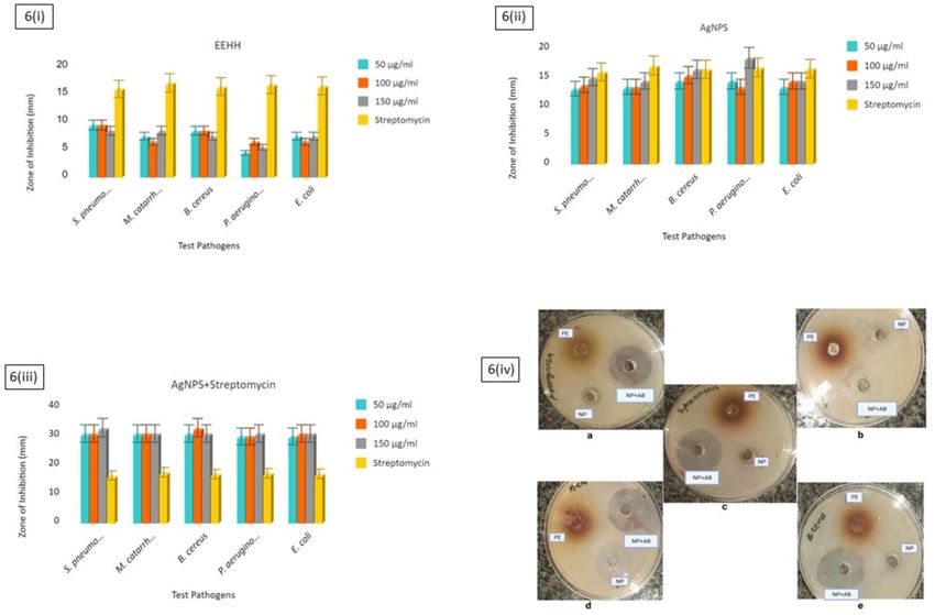
www.nature.com/scientificreports/
OH
O
HO
HO
OH
O
HO HO
OH
Hypoxoside
O O
OH
HO OH
HO
OH
Rooperol
OH
O
OH
O
O
HO
HO O H
OH
OH
O OH
Harpagoside
H
H
H H
HO
b
Figure 1. Structures of major known secondary metabolites in Hypoxis hemerocallidea.
of the resulting AgNPs and the kinetics of their growth and consequent resulting size, can be tuned through
the changing the identity of the capping agents. The capping agents determine these parameters through their
electrostatic and non-covalent bonding interactions with the nascent AgNP s urface12,13.
Hypoxis hemerocallidea (HH), the “African potato,” is used in traditional medicine across Southern Africa,
especially for endocrine gland dysplasia14, and as a purgative and remedy for delirium, bad dreams, impotency,
and apprehension by the Z ulu15,16. HH is a vascular plant with bright yellow flowers inspiring its common name:
"yellow stars." HH contains some rather unusual phytochemicals like the ene-yne-containing hypoxoside and
rooperol, the iridoid glycoside harpagoside, and the unusual steroid β-sitosterol (Fig. 1), which collectively
have demonstrated pharmacological potential for antibacterial, anti-inflammatory, antioxidant, and anticancer
activity17–19. It is often consumed by HIV/AIDS patients to boost their immunity and improve their general
wellbeing20; however, as its constituents have been shown to inhibit Cytochorme p450 metabolism, it could pos-
sibly interfere with many retroviral d rugs21, although the evidence remains inconclusive to d ate22. Best practice
recommends that its use be discussed with clinicians when starting antiretroviral t herapy23. This species could
be used in the synthesis of AgNPs to manufacture a synergistic product with therapeutic potential. To the best
of our knowledge, AgNPs have never been prepared using HH, and the synergistic impact of co-administering
HH ethanolic extract (EEHH) and its AgNPs with antibiotics to treat pathogens has never been studied. This is
the goal of this work.
Materials and methods
Extraction of phytochemicals from Hypoxis heamerocallidea for the synthesis of AgNPs. Fresh
HH corms, cultivated, were purchased from local commercial farms in Sebokeng in Gauteng, South Africa.
Cultivation of these plants was conducted using normal commercial farming practices in line with South Afri-
can regulations. Authentication of their identity was conducted by botanist Dr. Bukola Aremu of the School of
Scientific Reports | (2021) 11:15222 | https://doi.org/10.1038/s41598-021-93978-z 2
Vol:.(1234567890)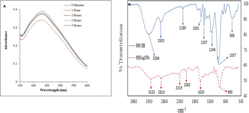
www.nature.com/scientificreports/
Biological Sciences, North-West University, Mafikeng, South Africa, and the University of Windsor, Windsor,
Canada. The corms were then extensively washed under running water, dried at room temperature (24 °C), and
pulverized into a powder with a commercial blender. Pulverized corms (500 g) were soaked in absolute ethanol
(1.5 L) with mechanical shaking for 72 h. The extract was filtered to remove the powder, then concentrated under
reduced pressure using a rotary evaporator at 70.1 °C to obtain a dark brown powder. After drying under high
vacuum for an additional 16 h to constant mass, 23.6 g of a dark brown powder was obtained18. This fraction, the
ethanolic extract of Hypoxis heamerocallidea is referred to as EEHH.
Synthesis of AgNPs‑HH. A stock solution of EEHH was prepared by suspending 2 g of the crude powder
in 50 mL of 70% ethanol (v/v). Next, it was sonicated for 10 min to solubilize the solution to a translucent deep
yellow solution with no observable turbidity. To a 40 mL aliquot of this solution was then added 400 µL of 0.1 M
AgNO3 in one portion with vigorous stirring. The reaction mixture immediately changed to a dark brown colour,
indicative of the formation of the AgNPs. The process was carried out at ambient room temperature open to the
atmosphere, and progress was monitored by measuring the UV–Vis spectrum of the reactant mixture at hourly
intervals over 4 h according to our previously reported p rocedure24. With no further changes being observed
between 3 and 4 h, the particles were purified by dialysis (against water, cut-off of 3.5 kDa), then dried by lyophi-
lization and stored until resuspended when needed.
Phytochemical screening. A qualitative phytochemical analysis of the EEHH (independent of the pres-
ence of the silver) was carried out using standard procedures25–27. The crude extract was screened for the pres-
ence of saponins, tannins, phenols, coumarins, flavonoids, terpenoids, glycosides, alkaloids, and proteins. To test
for tannins, we performed a ferric chloride test by placing 5 mL of the extracts from nine plants inside test tubes.
A few drops of 0.1% ferric chloride were added. The presence of brownish green or blue–black color indicated
the presence of tannins in the sample. For sterols and triterpenoids, we performed the Salkowski test. 5 mL of the
extracts from nine plants were placed in test tubes, and 2 mL of chloroform and 3 mL of concentrated sulfuric
acid were added consecutively. We shook them well and allowed them to stand for a few minutes. Red color in
the lower layer indicates the presence of sterols, and yellow color in the lower layer indicates the presence of trit-
erpenoids. To test for flavonoids, we performed an alkaline reagent test. A few drops of 1% liquor ammonia were
put in a test tube containing the sample. The emergence of a yellow color confirmed the presence of flavonoids.
To test for glycosides, we performed a bromine water test. We added 5 mL of bromine water to the test extract
solutions, and a yellow precipitate indicated the presence of glycosides. To test for saponins, we did a foam test.
To 10 mL of the sample, 3 mL of distilled water was added and shaken well to obtain a froth. A few drops of
olive oil were added to the froth, and the formation of an emulsion indicates the presence of saponins. To test
for cardiac glycosides, we did the Kellar–Kiliani test. To 5 mL of a sample, 2 mL of glacial acetic acid containing
a drop of ferric chloride was added. This was followed by the addition of 1 mL of concentrated sulfuric acid. A
brown ring obtained indicates a positive result for the test. To test for alkaloids, 0.1 mg of the extract was added
to 6 mL of dilute hydrochloric acid (1 M) and boiled, cooled, and filtered. The filtrate was divided into three parts
and subjected to the following tests. To the first aliquot, 2 drops of Dragendorff ’s reagent were added. The forma-
tion of red precipitate indicated the presence of alkaloids. To the second aliquot, 2 drops of Meyer’s reagent were
added. A creamy white precipitate revealed the presence of alkaloids. To the third aliquot, 2 drops of Wagner’s
reagent were added. The formation of a reddish-brown precipitate indicated the presence of alkaloids. To test for
phenol, 0.5 mL of extract was added to 5 mL of Foalin Ciocletu reagent and 4 mL of aqueous sodium carbonate.
The generation of a blue colour indicated the presence of phenol.
Characterization of the nanoparticles. The absorption spectra of the AgNPs were measured between
300 and 700 nm using a PerkinElmer (Germany) 365 UV–Vis spectrometer at 24 °C. The morphology of the
AgNPs was examined on a JEOL 3010 high-resolution transmission electron microscope equipped with energy-
dispersive X-ray (EDX) functionality (Bruker, Germany). The FTIR spectra of the crude extract and the AgNPs
were obtained on a Bruker Alpha-P FTIR spectrophotometer (Germany) from 500 to 4000 cm−1. The structural
Bruker equipment with monochromatic Cu kα radiation (λ = 1:5406 Å) at 40 kV. Scanning was conducted in
characterization of the AgNPs was carried out using an X-ray diffractometer. XRD analysis was conducted using
the region of 20–100 2θ angles. Dynamic light scattering (Malvern Zetasizer Nano-ZS) was used to analyze
the zeta potential of the synthesized AgNPs. For the DLS measurements, powder AgNPs were resuspended
in distilled water and sonicated for 15–20 min to properly disperse the particles in water. Zeta potential, and
hydrodynamic diameter values were obtained from the triplicate analysis of the nanoparticles in the aqueous
media24. Gas Chromatography–Mass Spectrometry (GC–MS) analysis was performed using a Bruker GC–TOF–
MS Gas Chromatography coupled with a 5973 Mass selective detector. The capillary column (Rxi-5SilMS) with
an internal diameter of 30 × 25 mm and 0.2 μm film thickness (Restek, Bellefonte, PA, USA) was used. Ultrahigh
purity helium (Afrox, South Africa) was used as the carrier gas at a constant flow rate of 1.0 mL/min and a linear
velocity of 37 cm/s. An aliquot of 1 μL of sample diluted in the respective solvents was injected into the column
with an inlet temperature of 250 °C in a splitless mode at time of 30 s. An initial oven temperature of 40 °C was
set and programmed to increase up to 300 °C at the rate of 10 °C per min with a holding time of 3 min at each
increment. The electron ionization mode of 70 eV (EI+) and electron multiplier/detector voltage at 1750 V was
used to operate the mass spectrometry. The other operating parameters were as follows: ion source temperature
of 230 °C, MS transfer line temperature 280 °C, MS solvent delay time of 5 min and mass acquisition range of
40–550 DA and data acquisition rate of 10 spectra/S. The compounds were identified by direct comparison of the
mass spectrum of the analyte at a particular retention time to that of reference standards found in the National
Scientific Reports | (2021) 11:15222 | https://doi.org/10.1038/s41598-021-93978-z 3
Vol.:(0123456789)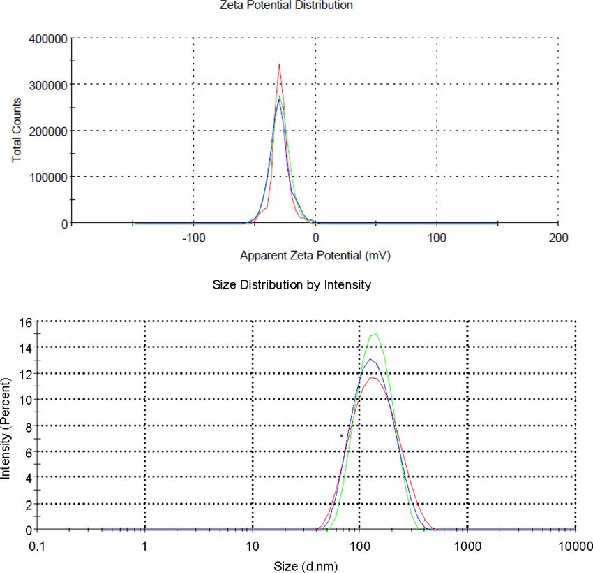
www.nature.com/scientificreports/
Metabolite Ethanolic extract
Glycosides +
Flavonoids +
Alkaloids +
Terpenes +
Steroids +
Tannins +
Saponins +
Phenol +
Cardiac glycosides −
Table 1. Phytochemical analysis of the crude extract of Hypoxis hemerocallidea.
Institute of Standards and Technology (NIST) library. The area percentage of each component was calculated by
comparing its average peak area to the total areas obtained28.
Antibacterial susceptibility. The antibacterial activity of the EEHH, AgNPs, and a combination treatment
of AgNPs and streptomycin (50 µL:50 µL), were investigated against five pathogenic bacterial strains: Gram-
positive S. pneumoniae (ATCC 27336) and Bacillus cereus (ATCC 10876); and Gram-negative M. catarrhalis
(ATCC 25240), Escherichia coli (ATCC 25922), and Pseudomonas aeruginosa (ATCC 27853)29. Using the disc
diffusion technique, pure cultures of these microorganisms were sub-cultured on nutrient agar and incubated
at 37 °C for 24 h.
Fresh overnight cultures were inoculated on Mueller Hinton agar (MHA) plates using sterile swabs and
allowed to stand for 20 min. Wells of 6-mm diameter were made on MHA plates with the bacterial lawn. Each
well was filled with 50 μL of different concentrations (50, 100, and 150 μg/mL) EEHH in distilled water and HH
AgNPs in dimethyl sulfoxide (DMSO) prepared from 10 mg/mL stock. DMSO (5%) was used as the negative
control, and streptomycin (10 μg/mL) served as the reference standard. The plates were incubated at 37 °C for
24 h, and the diameters of the inhibition zones around the wells were measured. Experiments were carried out
in triplicate to reduce e rror30. The minimum inhibition concentration (MIC) and minimum bactericidal concen-
tration (MBC) of green synthesized AgNPs were determined using the modified method described in the CLSI
guideline (2012)31. The MIC test was performed in a 96-well round bottom microtiter plate using standard broth
microdilution methods while the MBC test was performed on the MHA plates. The bacterial inoculums were
adjusted to the concentration of 1 06 CFU/mL. For the MIC test, 100 μL of the synthesized AgNPs stock solution
(500 μg/mL) was added and diluted twofold with the bacterial inoculums in 100 μL of MHB started from column
12 to column 3. Column 12 of the microtiter plate contained the highest concentration of AgNPs, while column
3 contained the lowest concentration. Column 1 served as negative control (only medium) and the column 2
served as positive control (medium and bacterial inoculums). Each well of the microtiter plate was added with
30 μL of the resazurin solution and incubated at 37 °C for 24 h. Any colour changes were observed. Blue/purple
colour indicated no bacterial growth while pink/colourless indicated bacterial growth. The MIC value was taken
at the lowest concentration of antibacterial agents that inhibits the growth of bacteria (colour remained in blue).
Streptomycin (Sigma, St. Louis) treatments were all conducted at 10 μg/mL. This was prepared as needed from
a stock solution by diluting tenfold with the assay media. The stock solution was prepared by transferring 10 mg
of streptomycin into a 1 mL volumetric flask, and making up the volume with a 1:1 ethanol: water solution, pro-
viding a 10 mg/mL solution. This was then diluted 100-fold by transferring 10 μL of this solution to a new 1 mL
volumetric flask, and making up the volume with the 50% ethanolic solution. This provided the 100 μg/mL stock.
The MBC was defined as the lowest concentration of the antibacterial agents that completely kill the bacteria.
MBC test was performed by plating the suspension from each well of microtiter plates that exhibited no colour
change into MHA plate. The plates were incubated at 37 °C for 24 h. The lowest concentration with no visible
growths on the MHA plate was taken as MBC value. The log of reduction RFvalue were enumerated accordingly.
Results and discussion
Phytochemical screening. The results from the qualitative analysis of the EEHH are provided as Table 1.
The EEHH ethanolic extract was positive for alkaloids, flavonoids, steroids, phenols, terpenes, glycosides, carbo-
hydrates, saponins, and tannins as expected, but the tests were negative for cardiac g lycosides32.
GC–MS profiling. The volatile phytochemicals present in the ethanolic crude extract were subjected to GC–
MS analysis (Table 2). The extract is dominated with high molecular mass unsaturated fatty acid palmitoleic
acid, unusual sugar d-allose, and the unusual chlorinated long-chain hydrocarbon 2-chloroethyl linoleate. Con-
siderable amounts of siloxanes were observed, these are likely environmental contaminants from the growing
conditions, but are not naturally produced by the plant.
UV–Vis analysis. The UV–vis absorption spectra of the AgNPs as a function of time during synthesis are
provided as Fig. 2A. EEHH-AgNPs produced surface plasmon resonance (SPR) peaks at 430–434 nm, which is
Scientific Reports | (2021) 11:15222 | https://doi.org/10.1038/s41598-021-93978-z 4
Vol:.(1234567890)www.nature.com/scientificreports/
No Name Retention time (m) % Area
1 Palmitoleic acid 19.31 12.84
2 d-Allose 12.56 10.58
3 2-Chloroethyl linoleate 19.250 10.16
4 Pentadecanoic acid 15.59 4.83
5 1,2,3,5-Cyclohexanetetrol 14.01 3.61
6 O-geranyl-β-d-Mannofuranoside 21.68 2.68
7 5-Hydroxymethylfurfural 8.79 2.50
8 (E,E,E)-9-Octadecenoic acid, 1,2,3-propanetriyl ester 23.84 2.47
9 Stearic acid 19.49 2.18
10 5,6,6-trimethyl-Undeca-3,4-diene-2,10-dione 13.40 2.11
11 3-Deoxy-d-mannoic lactone 13.83 1.91
12 Benzaldehyde, 3,4-dihydroxy- 13.54 1.61
13 Octadecamethyl-cyclononasiloxane 23.09 1.44
14 2,5-Monomethylene-l-rhamnitol 10.03 1.41
15 Hexadecamethylheptasiloxane 24.05 1.41
16 eicosamethyl-Cyclodecasiloxane 22.07 1.26
17 N-Nitrosoazacyclononane 6.63 1.25
18 Hexadecamethylheptasiloxane 20.99 1.19
19 Tetracosamethyl-cyclododecasiloxane 24.93 1.19
Table 2. Major constituents of the chemical composition of the ethanolic crude extract of HH corms using
GC–MS. Other trace components < 1.00%.
Figure 2. Nanoparticle formation: (A) UV spectrum of the AgNPs formed, (B) surface charge: FTIR spectra of
the extract and AgNPs.
consistent with no agglomeration. The generated AgNPs were stable over the reaction period, and the UV–vis
peak stabilized at 434 nm without further movement beyond 4 h indicating the presence of a steady state. How-
ever, because the response time increased, the SPR peak position became bathochromically shifted, indicating a
gradual increase in nanoparticle size32–34.
FTIR analysis. The FTIR spectrum is consistent with the incorporation of the secondary metabolites and
their degradation products into the AgNPs as the same functional groups are present in both the extract and the
nanoparticles. As noted by others, many classes of phytochemicals can reduce silver salts to metallic silver, while
others can act as capping agents. This will, of course, affect the chemical structure of these organic compounds,
and these transitions are noted by the changes in the relative intensity of the vibrational bands.
The pronounced peaks of the extract were at 3284, 2920, 1591, 1507, 1246, 1037, and 586 cm−1, whereas
those of the AgNPs were at 3225, 2910, 1620, and 990 cm−1. The broad vibration at 3284/3225 cm−1 is typical of
hydroxyl groups on carbohydrates, flavonoids, and saponins. The peaks at 2920/2910 cm−1 arise from aliphatic
Scientific Reports | (2021) 11:15222 | https://doi.org/10.1038/s41598-021-93978-z 5
Vol.:(0123456789)www.nature.com/scientificreports/
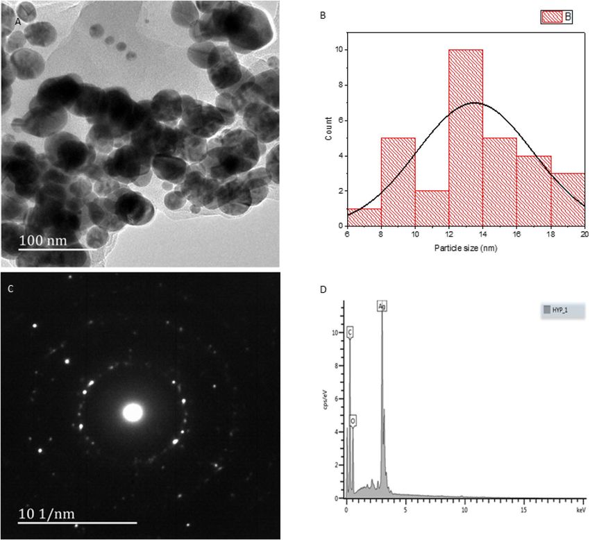
Figure 3. Morphology: (A) TEM, (B) Particle size distribution (C) TEM-SAED, (D) EDX.
C–H stretches in alkyl groups, 1591 and 1507 cm−1 from aromatic ortho disubstituted C–H stretches, 1246 cm-1
is typical of phenol O–H stretches, while 1037 cm−1 suggests the presence of aliphatic ethers and alcohol C–O
bonds35,36. These alcohols and phenols are probably involved in the reduction of ionic silver to zero-valent AgNPs
and decrease upon reaction (Fig. 2B)37.
The reduction of the relative intensity of the aliphatic C–H stretching region during AgNPs synthesis sug-
gests conversion of C–H bonds in the phytocompounds to multiple bonds or o xidation38. The FTIR analysis is
consistent with the formation of AgNPs through reduction of Ag(I) to Ag (0) with concomitant oxidation of
the HH phytochemicals.
Phytochemical screening of crude EEHH confirmed the presence of flavonoids and carbohydrates. Flavonoids
release free reactive hydrogen during their tautomeric transformations (keto-enol rearrangement), which can
assist in the reduction of A gNPs39. Furthermore, alcohols facilitate the reduction of ionic silver to zero-valent
AgNPs as they oxidize to c arbonyls40.
TEM, SAED, and EDX analysis. TEM analysis supports a largely spherical morphology for the AgNPs
(Fig. 3A). Through sizing 30 randomly selected particles observed by TEM we observed the mode was between
12 and 14 nm, with the mean value being 13.3 nm (Fig. 3B). As noted above, these are core–shell structures;
this is suggested in the TEM images, but the distinct rings found in the SAED patterns (Fig. 3C) of the AgNPs
confirm the polycrystalline property of the as-synthesized A gNPs41. Elemental composition analysis by EDX
supports the contention that the nanoparticles comprise a metallic core with an organic shell: strong silver (Ag)
signals and weaker signals from C and O atoms are consistent with this hypothesis. There is no significant con-
tamination from the nitrogen in the nitrate or from other opportunistic metals. The position of the signal at
13cps/eV suggest that the silver core is crystalline rather than amorphous (Fig. 3D)41.
X‑ray diffraction. The XRD pattern of the AgNPs shows sharp diffraction peaks corresponding to the (111),
(200), (220), and (311) crystal planes (Fig. 4), which are associated with the face-centered cubic lattice of silver.
The XRD profile indicates that our materials crystallize in a monoclinic phase, and this spectrum is in line with
Scientific Reports | (2021) 11:15222 | https://doi.org/10.1038/s41598-021-93978-z 6
Vol:.(1234567890)www.nature.com/scientificreports/
100000
80000
60000
Intensity
40000
21-1
20000
111 220
200 311
0
20 40 60 80
2θ (degree)
Figure 4. XRD pattern of green synthesized AgNPs using HH.
those reported for other organic-silver nanoparticles prepared from other botanical extracts42–44. This confirms
the formation of silver nanocrystals as the metallic core of the nanoparticles24.
Dynamic light scattering and Zeta potential analysis of the nanoparticles. The zeta potential
value of HH mediated AgNPs in aqueous suspension was established as − 29.2 mV (Fig. 5). This suggests that the
surface of the nanoparticles is negatively charged and that the particles are uniformly dispersed in the aqueous
medium. The high negative value is evident of the extreme stability of the nanoparticles because of electrostatic
repulsive forces between the particles. Zeta potential value of about − 29 mV ensures a high energy barrier for
the stabilization of the nanosuspension. DLS suggests a hydrodynamic diameter of 119 nm with a polydispersity
index of 0.188. This is an order of magnitude larger than that observed by TEM. Although there are many small
particles generated, the TEM images suggest the presence of larger structures. These might move as an assembly
in solution together to provide this larger observed size in solution.
Antibacterial susceptibility. The antibacterial activity of the crude extracts, AgNPs, and the AgNPs co-
administered with streptomycin, were investigated against both Gram-positive and Gram-negative bacteria.
This was quantified using a standard Kirby–Bauer disc diffusion assay with DMSO as the negative control and
pure streptomycin as the positive control (Fig. 6).
The extract alone only shows mild activity. The components of EEHH have been a traditional medicine, and
like most secondary plant metabolites likely have some role in plant defense against infection. However, the
AgNPs show good activity against all bacteria regardless of Gram-status and are similar in potency to streptomy-
cin. However, when streptomycin and the AgNPs are used together, the effect is synergistic: the area of disinfec-
tion is considerably greater than a simple addition of their individual activities would indicate. DMSO showed
no activity with no area of inhibition (data not shown) confirming the viability of the tested bacteria strains.
Silver salts have long been known to be potent antimicrobials, and the development of silver nanoparticles, with
their far higher surface area of activated silver metal, has greatly accelerated their i nvestigation45. However, the
overuse of silver can decrease its efficacy against microorganisms as they develop resistance46.
The MIC result (Table 3) shows the bacteriostatic effect of the AgNP at 0.156 µg/mL for Streptococcus pneumo-
nia, Moraxella catarrhalis and Pseudomonas aeruginosa and 0.312 µg/mL upon exposure to Escherichia coli and
Bacillus cereus. The bactericidal effect of the AgNP ranges from 0.312 to 5 µg/mL for Streptococcus pneumonia,
Moraxella catarrhalis and Pseudomonas aeruginosa and 0.625–5 µg/mL with Escherichia coli and Bacillus cereus.
The log of reduction R Fvalue > 4 in all the bacteria challenge irrespective of their Gram status. The nanoparticle
thus possessed high efficacy with a percentage greater than 90%.
Scientific Reports | (2021) 11:15222 | https://doi.org/10.1038/s41598-021-93978-z 7
Vol.:(0123456789)www.nature.com/scientificreports/
Figure 5. Zeta potential and size distribution of the AgNPs. Analyses were conducted in triplicate for three
freshly prepared samples, and each run is plotted in the different colours.
This synergistic effect is expected and has been seen with other silver n anoparticles47,48. The mechanism of
action of the AgNPs is through adsorption to the bacterial cell membranes, followed by passive penetration into
the bacteria. The surface then becomes transiently exposed, and they can cause damage by interacting strongly
with essential phosphorous and sulfur-containing compounds such as DNA and proteins, resulting in bacterial
cell death49. They could also assist in preventing the bacteria from initiating gene expression changes due to
the presence of the streptomycin, or streptomycin might better enter the cell by adsorbing to the surface of the
nanoparticle and being carried into the cell itself along with the toxic silver p article50.
Conclusions
HH corm extract is readily obtained in large amounts from even small amounts of plant material (5% mass
recovery from crude material) and can be used to initiate the solution-phase synthesis of low dispersity AgNPs
under ambient conditions.
These biosynthesized AgNPs showed no agglomeration and had sizes typically ranging from 6 to 20 nm with a
roughly spherical or ovoid shape. These HH-AgNPs show broad spectrum antibacterial activity against common
respiratory pathobionts and synergistically enhance the antibacterial activity of streptomycin. We propose that
these could be useful agents for transporting largely insoluble antibiotics in the body as potential biologically-
active drug delivery vehicles, but much more analysis needs to be conducted.
Scientific Reports | (2021) 11:15222 | https://doi.org/10.1038/s41598-021-93978-z 8
Vol:.(1234567890)www.nature.com/scientificreports/
Figure 6. The antibacterial activity of (i) EEHH, (ii) AgNPs, (iii) AgNPs and Streptomycin (10 μg/mL for all
experiments), (iv) zone of inhibition on agar plate.
Bacteria MIC (µg/mL) MBC (µg/mL) RFvalue
Streptococcus pneumonia 0.156 0.312 5.36E+00 (100%)
Bacillus cereus 0.312 0.625 5.06E+00 (95%)
Moraxella catarrhalis 0.156 0.312 4.89E+00 (91%)
Escherichia coli 0.312 0.625 5.36E+00 (100%)
Pseudomonas aeruginosa 0.156 0.312 5.19E+00 (97%)
Table 3. Bacterial activity of the AgNP (µg/mL) with the mean log of reduction (RF).
Received: 11 August 2020; Accepted: 28 June 2021
References
1. Mazucanti, C. H. & Egan, J. M. SARS-CoV-2 disease severity and diabetes: Why the connection and what is to be done?. Immunity
Ageing 17, 1–11 (2020).
2. Bell, D. et al. Predicting the impact of COVID-19 and the potential impact of the public health response on disease burden in
Uganda. Am. J. Trop. Med. Hyg. 10, 1191 (2020).
3. Denis, M., Vandeweerd, V. & Van der Vliet, D. Covid-19 Living paper: Overview of information available to support the develop-
ment of medical countermeasures and interventions against COVID-19. J. Transdiscip. Insight. Accessible at: http://rega.kuleuven.
be/if/corona_covid-19. Accessed 20 April 2021.
4. Alam, M., Beig, S., Sultan, A. & Chandra, K. Bacteriological and antibiotic sensitivity profile of cholesteatomatous otitis media in
pediatric and adult population: Prevailing scenario in Northern India. Indian J. Otolaryngol. Head Neck Surg. https://doi.org/10.
1007/s12070-020-01956-0 (2020).
5. Watson, K. et al. Upper respiratory tract bacterial carriage in aboriginal and non-aboriginal children in a semi-arid area of Western
Australia. Pediatr. Infect. Dis. J. 25, 782–790 (2006).
6. Tanaka, S. C. et al. Behavioral Economics of Preferences, Choices, and Happiness 593–616 (Springer, 2016).
7. Murphy, T. F., Bakaletz, L. O. & Smeesters, P. R. Microbial interactions in the respiratory tract. Pediatr. Infect. Dis. J. 28, S121–S126
(2009).
8. Chung, I.-M., Park, I., Seung-Hyun, K., Thiruvengadam, M. & Rajakumar, G. Plant-mediated synthesis of silver nanoparticles:
Their characteristic properties and therapeutic applications. Nanoscale Res. Lett. 11, 40 (2016).
Scientific Reports | (2021) 11:15222 | https://doi.org/10.1038/s41598-021-93978-z 9
Vol.:(0123456789)www.nature.com/scientificreports/
9. Prabu, H. J. & Johnson, I. Plant-mediated biosynthesis and characterization of silver nanoparticles by leaf extracts of Tragia invo-
lucrata, Cymbopogon citronella, Solanum verbascifolium and Tylophora ovata. Karbala Int. J. Mod. Sci. 1, 237–246 (2015).
10. Anjum, S., Abbasi, B. H. & Shinwari, Z. K. Plant-mediated green synthesis of silver nanoparticles for biomedical applications:
Challenges and opportunities. Pak. J. Bot. 48, 1731–1760 (2016).
11. El-Moslamy, S. H., Elkady, M. F., Rezk, A. H. & Abdel-Fattah, Y. R. Applying Taguchi design and large-scale strategy for myco-
synthesis of nano-silver from endophytic Trichoderma harzianum SYA. F4 and its application against phytopathogens. Sci. Rep. 7,
45297 (2017).
12. Chiguvare, H. et al. Synthesis of silver nanoparticles using buchu plant extracts and their analgesic properties. Molecules 21, 774
(2016).
13. Chaudhuri, S. K. & Malodia, L. Phytosynthesis and characterization of silver nanoparticles synthesized from flower extract of
Roheda (Tecomella undulata G. Don). Defence Life Sci. J. 2, 65–73 (2017).
14. Marandola, P., Jallous, H., Bombardelli, F. & Morazzoni, P. Main phytoderivatives in the management of benign prostatic hyper-
plasia. Fitoterapia 68, 195–204 (1997).
15. Owira, P. M. & Ojewole, J. A. ‘African potato’ (Hypoxis hemerocallidea corm): a plant-medicine for modern and 21st century
diseases of mankind?–a review. Phytother. Res. Int. J. Devoted Pharmacol. Toxicol. Eval. Nat. Prod. Deriv. 23, 147–152 (2009).
16. Ojewole, J. A., Awe, E. O. & Nyinawumuntu, A. Antidiarrhoeal activity of Hypoxis hemerocallidea Fisch. & CA Mey. (Hypoxidaceae)
Corm (‘African potato’) aqueous extract in rodents. Phytother. Res. 23, 965–971 (2009).
17. Mugomeri, E., Chatanga, P., Raditladi, T., Makara, M. & Tarirai, C. Ethnobotanical study and conservation status of local medicinal
plants: towards a repository and monograph of herbal medicines in Lesotho. Afr. J. Tradit. Complement. Altern. Med. 13, 143–156
(2016).
18. Swayeb, A. A. The Possible Effect of Hypoxis Hemerocalledia (African Potato) on Blood Glucose Levels: An In Vitro Study. Masters
thesis, University of the Western Cape 1-109 (2015).
19. Ojewole, J. A., Awe, E. O. & Nyinawumuntu, A. Antidiarrhoeal activity of Hypoxis hemerocallidea Fisch. & CA Mey. (Hypoxidaceae)
Corm (‘African potato’) aqueous extract in rodents. Phytother. Res. Int. J. Devoted Pharmacol. Toxicol. Eval. Nat. Prod. Deriv. 23,
965–971 (2009).
20. Fasinu, P. S., Gutmann, H., Schiller, H., Bouic, P. J. & Rosenkranz, B. The potential of Hypoxis hemerocallidea for herb–drug
interaction. Pharm. Biol. 51, 1499–1507 (2013).
21. Mills, E. et al. Impact of African herbal medicines on antiretroviral metabolism. AIDS 19, 95–97 (2005).
22. Fasinu, P. S., Gurley, B. J. & Walker, L. A. Clinically relevant pharmacokinetic herb–drug interactions in antiretroviral therapy.
Curr. Drug Metab. 17, 52–64 (2016).
23. Peltzer, K. et al. Antiretrovirals and the use of traditional, complementary and alternative medicine by HIV patients in Kwazulu-
Natal South Africa: a longitudinal study. Afr. J. Tradit. Complement. Altern. Med. 8, 337–345 (2011).
24. Kgatshe, M., Aremu, O. S., Katata-Seru, L. & Gopane, R. Characterization and antibacterial activity of biosynthesized silver nano-
particles using the ethanolic extract of pelargonium sidoides DC. J. Nanomater. 2019, 1–10 (2019).
25. Edziri, H. et al. Phytochemical screening, butyrylcholinesterase inhibitory activity and anti-inflammatory effect of some Tunisian
medicinal plants. S. Afr. J. Bot. 114, 84–88 (2018).
26. Ochwang’I, D., Kimwele, C., Oduma, J., Gathumbi, P. & Kiama, S. Phytochemical screening of medicinal plants of the Kakamega
Country, Kenya commonly used against cancer. Med. Aromat. Plants (Los Angel) 5, 2167–412 (2016).
27. Subathraa, K. & Poonguzhali, T. Phytochemical screening of medicinal plant Lantana camera Linn. Res. Rev. J. Bot. 3, 1–4 (2018).
28. Mwinga, J. L. et al. In vitro antimicrobial effects of Hypoxis hemerocallidea against six pathogens with dermatological relevance
and its phytochemical characterization and cytotoxicity evaluation. J. Ethnopharmacol. 242, 112048 (2019).
29. Streeter, K. & Katouli, M. Pseudomonas aeruginosa: A review of their pathogenesis and prevalence in clinical settings and the
environment. Infect. Epidemiol. Microbiol. 2, 25–32 (2016).
30. de Morais, S. R. et al. Essential oil composition, antimicrobial and pharmacological activities of Lippia sidoides Cham. (Verbenaceae)
from Sao Goncalo do Abaete, Minas Gerais, Brazil. Pharmacogn. Mag. 12, 262 (2016).
31. Loo, Y. Y. et al. In vitro antimicrobial activity of green synthesized silver nanoparticles against selected gram-negative foodborne
pathogens. Front. Microbiol. 9, 1555 (2018).
32. Elbagory, A. M., Meyer, M., Cupido, C. N. & Hussein, A. A. Inhibition of bacteria associated with wound infection by biocompat-
ible green synthesized gold nanoparticles from South African plant extracts. Nanomaterials 7, 417 (2017).
33. Jalal, M. et al. Biosynthesis of silver nanoparticles from oropharyngeal Candida glabrata isolates and their antimicrobial activity
against clinical strains of bacteria and fungi. Nanomaterials 8, 586 (2018).
34. Salari, Z., Danafar, F., Dabaghi, S. & Ataei, S. A. Sustainable synthesis of silver nanoparticles using macroalgae Spirogyra varians
and analysis of their antibacterial activity. J. Saudi Chem. Soc. 20, 459–464 (2016).
35. Ghosh, S. et al. Gnidia glauca flower extract mediated synthesis of gold nanoparticles and evaluation of its chemocatalytic potential.
J. Nanobiotechnol. 10, 17 (2012).
36. Jancy, M. E. & Inbathamizh, L. Green synthesis and characterization of nano silver using leaf extract of Morinda pubescens. Asian
J. Pharm. Clin. Res. 5, 159–162 (2012).
37. Thilagam, M., Tamilselvi, A., Chandrasekeran, B. & Rose, C. Phytosynthesis of silver nanoparticles using medicinal and dye yield-
ing plant of Bixa orellana L. leaf extract. J. Pharm. Sci. Innov. 2, 9–13 (2013).
38. Kumar, V., Yadav, S. C. & Yadav, S. K. Syzygium cumini leaf and seed extract mediated biosynthesis of silver nanoparticles and their
characterization. J. Chem. Technol. Biotechnol. 85, 1301–1309 (2010).
39. Rauwel, P., Küünal, S., Ferdov, S. & Rauwel, E. A review on the green synthesis of silver nanoparticles and their morphologies
studied via TEM. Adv. Mater. Sci. Eng. 2015, 1–9 (2015).
40. Tanvi, et al. Effect of the crystallinity of silver nanoparticles on surface plasmon resonance induced enhancement of effective
absorption cross-section of dyes. J. Appl. Phys. 117, 083111 (2015).
41. Kumar, B., Smita, K., Cumbal, L. & Debut, A. Green synthesis of silver nanoparticles using Andean blackberry fruit extract. Saudi
J. Biol. Sci. 24, 45–50 (2017).
42. Vanaja, M. & Annadurai, G. Coleus aromaticus leaf extract mediated synthesis of silver nanoparticles and its bactericidal activity.
Appl. Nanosci. 3, 217–223. https://doi.org/10.1007/s13204-012-0121-9 (2013).
43. Anandalakshmi, K., Venugobal, J. & Ramasamy, V. Characterization of silver nanoparticles by green synthesis method using
Pedalium murex leaf extract and their antibacterial activity. Appl. Nanosci. 6, 399–408. https://doi.org/10.1007/s13204-015-0449-z
(2016).
44. Kota, S., Dumpala, P., Anantha, R. K., Verma, M. K. & Kandepu, S. Evaluation of therapeutic potential of the silver/silver chloride
nanoparticles synthesized with the aqueous leaf extract of Rumex acetosa. Sci. Rep. 7, 11566. https://doi.org/10.1038/s41598-017-
11853-2 (2017).
45. Kim, J. S. et al. Antimicrobial effects of silver nanoparticles. Nanomed. Nanotechnol. Biol. Med. 3, 95–101 (2007).
46. Yugandhar, P., Haribabu, R. & Savithramma, N. Synthesis, characterization and antimicrobial properties of green-synthesised
silver nanoparticles from stem bark extract of Syzygium alternifolium (Wt.) Walp. 3 Biotech 5, 1031–1039 (2015).
47. Singh, T., Jyoti, K., Patnaik, A., Singh, A. & Chauhan, S. Spectroscopic, microscopic characterization of Cannabis sativa leaf extract
mediated silver nanoparticles and their synergistic effect with antibiotics against human pathogen. Alex. Eng. J. 57, 3043–3051
(2018).
Scientific Reports | (2021) 11:15222 | https://doi.org/10.1038/s41598-021-93978-z 10
Vol:.(1234567890)www.nature.com/scientificreports/
48. Perveen, S., Safdar, N. & Yasmin, A. Antibacterial evaluation of silver nanoparticles synthesized from lychee peel: Individual versus
antibiotic conjugated effects. World J. Microbiol. Biotechnol. 34, 118 (2018).
49. Baker, C., Pradhan, A., Pakstis, L., Pochan, D. J. & Shah, S. I. Synthesis and antibacterial properties of silver nanoparticles. J. Nanosci.
Nanotechnol. 5, 244–249 (2005).
50. Doddam, S. N. et al. Mycobacterium tuberculosis DosR regulon gene Rv2004c contributes to streptomycin resistance and intracel-
lular survival. Int. J. Med. Microbiol. 309, 151353 (2019).
Acknowledgements
The authors thank the National Research Foundation of South Africa for grants (UID: 106379) that supported
this project, and the North-West University, University of Windsor, and the Vaal University of Technology for
their laboratory and personnel support for this research work.
Author contributions
O.S.A. conceived and designed the experiments; performed the experiments; analyzed the data; and wrote the
first draft of the manuscript. L.M.K., Q.T.O., Z.M., and J.F.T. provided academic input into study design, and the
editing of the final article. All authors edited and approved the final manuscript draft.
Funding
This research funded by the National Research Foundation of South Africa, grant number UID 106379 and the
Natural Sciences and Engineering Research Council of Canada (2018-06338 to JFT).
Competing interests
The authors declare no competing interests.
Additional information
Correspondence and requests for materials should be addressed to O.S.A., L.K.-S. or J.F.T.
Reprints and permissions information is available at www.nature.com/reprints.
Publisher’s note Springer Nature remains neutral with regard to jurisdictional claims in published maps and
institutional affiliations.
Open Access This article is licensed under a Creative Commons Attribution 4.0 International
License, which permits use, sharing, adaptation, distribution and reproduction in any medium or
format, as long as you give appropriate credit to the original author(s) and the source, provide a link to the
Creative Commons licence, and indicate if changes were made. The images or other third party material in this
article are included in the article’s Creative Commons licence, unless indicated otherwise in a credit line to the
material. If material is not included in the article’s Creative Commons licence and your intended use is not
permitted by statutory regulation or exceeds the permitted use, you will need to obtain permission directly from
the copyright holder. To view a copy of this licence, visit http://creativecommons.org/licenses/by/4.0/.
© The Author(s) 2021
Scientific Reports | (2021) 11:15222 | https://doi.org/10.1038/s41598-021-93978-z 11
Vol.:(0123456789)You can also read