Antitumor Anthraquinones from an Easter Island Sea Anemone: Animal or Bacterial Origin? - MDPI
←
→
Page content transcription
If your browser does not render page correctly, please read the page content below
marine drugs
Article
Antitumor Anthraquinones from an Easter Island Sea
Anemone: Animal or Bacterial Origin?
Ignacio Sottorff 1,2 , Sven Künzel 3 , Jutta Wiese 1 , Matthias Lipfert 4 , Nils Preußke 4 ,
Frank D. Sönnichsen 4 and Johannes F. Imhoff 1, *
 1   GEOMAR Helmholtz Centre for Ocean Research Kiel, Marine Microbiology, 24105 Kiel, Germany;
     isottorff@geomar.de (I.S.); jwiese@geomar.de (J.W.)
 2   Facultad de Ciencias Naturales y Oceanográficas, Universidad de Concepción, 4070386 Concepción, Chile
 3   Max Planck Institute for Evolutionary Biology, 24306 Plön, Germany; kuenzel@evolbio.mpg.de
 4   Otto Diels Institute for Organic Chemistry, University of Kiel, 24118 Kiel, Germany;
     mlipfert@oc.uni-kiel.de (M.L.); npreusske@oc.uni-kiel.de (N.P.); fsoennichsen@oc.uni-kiel.de (F.D.S.)
 *   Correspondence: jimhoff@geomar.de; Tel.: +49-431-600-4450

 Received: 31 January 2019; Accepted: 26 February 2019; Published: 5 March 2019

 Abstract: The presence of two known anthraquinones, Lupinacidin A and Galvaquinone B, which
 have antitumor activity, has been identified in the sea anemone (Gyractis sesere) from Easter Island.
 So far, these anthraquinones have been characterized from terrestrial and marine Actinobacteria only.
 In order to identify the anthraquinones producer, we isolated Actinobacteria associated with the sea
 anemone and obtained representatives of seven actinobacterial genera. Studies of cultures of these
 bacteria by HPLC, NMR, and HRLCMS analyses showed that the producer of Lupinacidin A and
 Galvaquinone B indeed was one of the isolated Actinobacteria. The producer strain, SN26_14.1, was
 identified as a representative of the genus Verrucosispora. Genome analysis supported the biosynthetic
 potential to the production of these compounds by this strain. This study adds Verrucosispora as
 a new genus to the anthraquinone producers, in addition to well-known species of Streptomyces
 and Micromonospora. By a cultivation-based approach, the responsibility of symbionts of a marine
 invertebrate for the production of complex natural products found within the animal’s extracts could
 be demonstrated. This finding re-opens the debate about the producers of secondary metabolites in
 sea animals. Finally, it provides valuable information about the chemistry of bacteria harbored in the
 geographically-isolated and almost unstudied, Easter Island.

 Keywords: Easter Island; Actinobacteria; anthraquinones; symbionts; sea anemone; marine
 invertebrates; spectroscopy; chromatography

1. Introduction
     Since the discovery of Easter Island, compelling explorations have characterized the flora [1]
and fauna [2–4] of this geographically isolated location. However, little has been done to understand
the chemistry harbored in this territory. The best-known finding is the discovery of rapamycin in
a soil dwelling actinobacterial representative, which serves as an immunosuppressive drug [5,6].
Beyond that, little progress has been made in exploiting the chemical diversity harbored by marine
invertebrates and microorganisms dwelling in this territory, despite the high degree of endemism
found [7].
     Marine invertebrates are immensely diverse, well distributed in the world oceans [8], and widely
known to contain medicinally relevant molecules [9–11]. While these metabolites play different roles in
nature, e.g., they act as chemical defense, chemical communication or reproductive signaling molecules,
they also find application as human medicines [12]. During the last decades, much effort has been

Mar. Drugs 2019, 17, 154; doi:10.3390/md17030154                          www.mdpi.com/journal/marinedrugs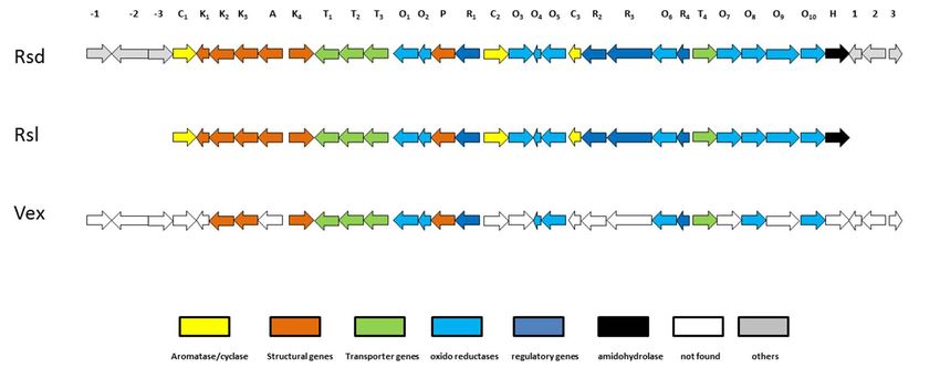
Mar. Drugs 2019, 17, 154                                                                                2 of 15

made to identify and characterize the chemicals contained in marine invertebrates. This has resulted in
the discovery of astonishing chemicals with novel biological activities and chemical scaffolds [12–14].
     Further, the immense progress in DNA sequencing technologies, the development of
bioinformatics, and an improvement in analytical techniques enables the identification of the source of
the chemicals. Thus, several molecules contained in marine invertebrates have now been shown to
actually be of microbial origin [15,16]. It is expected that with the increasing availability of metagenomic
information more identifications of the real producers of these metabolites will be made and will
establish the metabolite relevance for the interaction of host, symbiont, and the environment.
     Marine sponges represent a classic example of marine invertebrates that harbor microbes
producing secondary metabolites. They have been studied in detail to determine the origin of the
metabolites [17,18]. Another example is the producer of the approved anticancer drug Yondelis
(Ecteinascidin-743). This compound was first assigned to the tunicate Ecteinascidia turbinata, but later
identified as the product of a microbe, Candidatus Endoecteinascidia frumentensis [16].
     Anthraquinones have been characterized in different marine invertebrates, for example
crinoids [19] and sponges [20]. Anthraquinones have broad biological activity and are substances of
pharmaceutical relevance for revealing antitumor [21], antibacterial [22], antifungal [23] and epigenetic
modulator activities [24]. Two recently isolated anthraquinones, Lupinacidin A (1) and Galvaquinone
B (2), have been reported to be produced by Actinobacteria belonging to the genera Streptomyces and
Micromonospora. Lupinacidin A (1) was firstly reported as a specific inhibitor on murine colon 26-L5
carcinoma cells [25], and Galvaquinone B (2) showed moderate cytotoxicity against non-small-cell
lung cancer cells Calu-3 and H2887, in addition of epigenetic modulatory activity [24].
     Herein, we report the identification of two known anthraquinone molecules, Lupinacidin A (1)
and Galvaquinone B (2), contained in an Easter Island sea anemone, Gyractis sesere. Interestingly,
so far these molecules have been only characterized from microbial origin. Therefore, we undertook
the isolation and culture of the Actinobacteria associated with this marine invertebrate. The culture,
chemical and genomic evaluation of these bacteria showed that the real producer of the metabolites
was an Actinobacterium belonging to the genus Verrucosispora and not the sea anemone.

2. Results and Discussion

2.1. Sea Anemone Dereplication
      Samples of the sea anemone Gyractis sesere [26], also known as Actiniogeton rapanuiensis [27],
were collected in the intertidal zone of Easter Island, South Pacific Ocean, and extracted with
chloroform to give 8 mg of a yellowish residue. The HRLCMS analysis of this crude extract showed
the presence of two previously identified antitumor anthraquinones (Figure 1) [24,25], Lupinacidin A
(1) ([M + H]+ m/z 341.1378) and Galvaquinone B (2) ([M + H]+ m/z 369.3510), which were confirmed
by complete     NMR-spectroscopic
    Mar. Drugs 2019,                    characterization.
                     17, x FOR PEER REVIEW                                                  3 of 15

                 Figure 1. Identified molecules from the Easter Island sea anemone Gyractis sesere.

                   Figure 1. Identified molecules from the Easter Island sea anemone Gyractis sesere.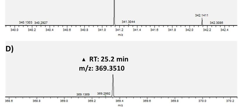
Mar. Drugs 2019, 17, 154                                                                                                    3 of 15

     Interestingly, the crude extract of the sea anemone (Figure 2) shows five resonances above 10 ppm;
a region which is characteristic for hydroxyl protons. The resonances at δ 14.18 and 12.96 ppm are
assigned to the groups at C-1 and C-6 of Lupinacidin A (1), as their vicinity and consequential hydrogen
bonding to the ketogroups slows down their exchange. Similarly, the resonances at δ 13.49, 12.50,
                 Figure 1. Identified
and 12.14 ppm originate     from themolecules from the Easter Island
                                      three hydroxylprotons          sea anemone Gyractis
                                                                in Galvaquinone   B (2). sesere.

         Figure
     Figure       2. Chemical
              2. Chemical      analysis
                             analysis ofof thecrude
                                         the    crudeextract
                                                      extractof
                                                              ofthe
                                                                  thesea
                                                                      sea anemone
                                                                          anemone Gyractis
                                                                                    Gyractis sesere.
                                                                                               sesere.A)
                                                                                                      (A)1H1HNMR
                                                                                                              NMR spectra
                                                                                                                     spectra
         of the
     of the crudecrude  extract
                     extract     of marine
                             of marine      anemone
                                        anemone        Gyractis
                                                    Gyractis     sesere
                                                             sesere     acquired
                                                                     acquired  in in CDCl
                                                                                   CDCl      , 600MHz.
                                                                                        3 , 3600   MHz.Highlighted
                                                                                                           Highlightedininthe
         the zoomed
     zoomed     area are area   are the frequencies
                          the frequencies               of characteristic
                                            of characteristic   resonancesresonances
                                                                             originatingoriginating
                                                                                            from hydroxyl fromexchangeable
                                                                                                                hydroxyl
     protons in vicinity to ketogroups. (B) UV chromatogram (254 nm) of the crude extract of the sea
     anemone Gyractis sesere highlighting the specific peaks for RT: 24.3 min, and N: RT: 25.2 min. (C)
     High resolution mass for  (m/z [M + H]+ 341.1378) and (D) high resolution mass for N (2) (m/z
     [M + H]+ 369.3510). *RT: Retention Time.

    Other main peaks found in the sea anemone crude extract were peaks at RT 14.2 min with a
HRMS [M + H]+ m/z 295.19009 and at RT 23 min with a HRMS [M + H]+ m/z 256.26312. Both exact
masses were evaluated using the MarinLit database, however their HRMS did not match any known
compound to-date.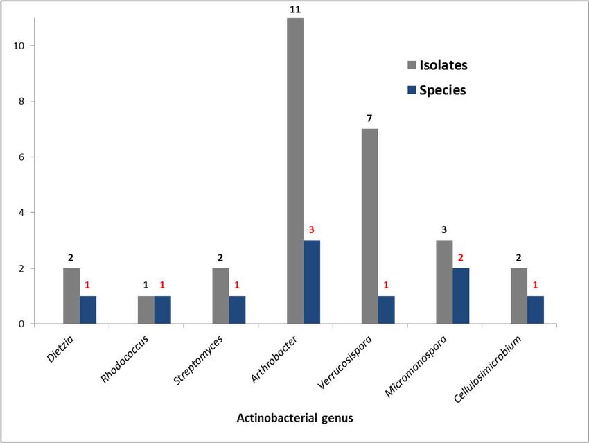
Mar. Drugs 2019, 17, x FOR PEER REVIEW 4 of 15
exchangeable protons in vicinity to ketogroups. B) UV chromatogram (254 nm) of the crude extract
of2019,
Mar. Drugs the sea
17, anemone
154 Gyractis sesere highlighting the specific peaks for RT: 24.3 min, and : RT: 25.2 4 of 15
min. C) High resolution mass for (m/z [M+H] 341.1378) and D) high resolution mass for (2) (m/z
+
[M+H]+ 369.3510). *RT: Retention Time.
2.2. Bacterial Metabolites and Harbored Bacteria
2.2. Bacterial metabolites and harbored bacteria
Lupinacidin A (1) and Galvaquinone B (2) have so far only been characterized in actinobacterial
Lupinacidin
representatives, A (1) and Galvaquinone
specifically from the genera B (2)Streptomyces
have so far only beenand
[24,28] characterized in actinobacterial
Micromonospora [25], raising
representatives, specifically from the genera Streptomyces [24,28] and Micromonospora
the question of the origin of these compounds in the sea anemone extract. Thus, we cultivated [25], raising thethe
question of the
Actinobacteria origin of
harbored bythese compounds
this sea anemoneintothedetermine
sea anemone extract.
if the Thus, we cultivated
anthraquinone producer wasthe a
Actinobacteria
bacterium harbored
or the sea by this
anemone. sea anemone
Isolation mediato anddetermine if the anthraquinone
the respective obtained strains producer was a in
are specified
bacterium or Table
Supplementary the sea
S1.anemone. Isolation
Ten strains media andthrough
were identified the respective obtained
analysis strains
of the 16S rRNA aregene
specified in
sequences
Supplementary Table 1. Ten strains were identified through analysis of the 16S rRNA gene sequences
as members of the genera Micromonospora, Streptomyces, Verrucosispora, Dietzia, Arthrobacter, Rhodococcus,
as members of the genera Micromonospora, Streptomyces, Verrucosispora, Dietzia, Arthrobacter,
and Cellulosimicrobium (Figure 3). Remarkably, Gyractis sesere harbors a high number of Actinobacteria
Rhodococcus, and Cellulosimicrobium (Figure 3). Remarkably, Gyractis sesere harbors a high number of
genera, in total seven; the most abundant genus being Micromonospora with three different species,
Actinobacteria genera, in total seven; the most abundant genus being Micromonospora with three
followed by Arthrobacter with two different species. Other actinobacterial genera were present with
different species, followed by Arthrobacter with two different species. Other actinobacterial genera
only one species each. Outstandingly, the only isolate belonging to the Verrucosispora genus was the
were present with only one species each. Outstandingly, the only isolate belonging to the
most abundant single
Verrucosispora genusActinobacterium
was the most abundantin thesingle
sea anemone.
Actinobacterium in the sea anemone.
Figure
Figure 3. Genera
3. Genera and
and numberofofActinobacteria
number Actinobacteriaspecies
species strains
strains isolated
isolatedfrom
fromthe
thesea
seaanemone
anemone Gyractis
Gyractis
sesere. sesere.
2.3.2.3. Bacterial
Bacterial growth
Growth
To To evaluate
evaluate thetheproduction
productionofofthe
the anthraquinones
anthraquinones by
by the
the isolated
isolatedbacteria,
bacteria,weweselected thethe
selected
actinobacterialrepresentatives
actinobacterial representativesforfor growth
growth experiments
experiments that
thatwere
weremost
mostclosely
closelyrelated to to
related known
known
producers
producers of the
of the genera
genera Streptomyces,
Streptomyces, Micromonospora
Micromonospora andVerrucosispora.
and Verrucosispora.InInaddition,
addition,wewealso
alsogrew
grewthe
the Dietzia
Dietzia representative
representative due lack
due to the to the lack of comprehensive
of comprehensive information
information about itsabout its secondary
secondary metabolite
metabolite production. On the other hand, Rhodococcus, Arthrobacter, and Cellulosimicrobium were
production. On the other hand, Rhodococcus, Arthrobacter, and Cellulosimicrobium were omitted here
omitted here because of their known poor production of secondary metabolites.
because of their known poor production of secondary metabolites.
The growth yield of the selected Actinobacteria was in the range of 20 to 100 mg crude extract.
The growth yield of the selected Actinobacteria was in the range of 20 to 100 mg crude extract.
The chromatograms of HPLC analyses of the crude extracts were compared in order to facilitate the
The chromatograms of HPLC analyses of the crude extracts were compared in order to facilitate the
metabolic comparison between grown bacteria, the sea anemone and the pure substances (Figure 4).
metabolic comparison between grown bacteria, the sea anemone and the pure substances (Figure 4).Mar. Drugs 2019, 17, 154 5 of 15
Mar. Drugs 2019, 17, x FOR PEER REVIEW 5 of 15
Figure
Figure 4. 4. HPLCchromatograms
HPLC chromatogramsof of the
the crude
crude extracts
extractsofofthe
thesea
seaanemone
anemoneGyractis
Gyractissesere, its respective
sesere, its respective
actinobacterialisolates,
actinobacterial isolates,and
andthe
the purified
purified anthraquinones,
anthraquinones,Lupinacidin AA
Lupinacidin (1)(1)
and Galvaquinone
and Galvaquinone B (2).
B (2).
Approximate
Approximate retentiontimes
retention timesofofLupinacidin
Lupinacidin AA (1)
(1) and
and Galvaquinone
GalvaquinoneBB(2)(2)are highlighted
are highlighted by by
boxes.
boxes.
ByBy comparisonofofchromatograms
comparison chromatograms and and retention
retentiontimes,
times,we
weobserved
observed that Lupinacidin
that Lupinacidin A (1)
A and
(1) and
GalvaquinoneBB(2)
Galvaquinone (2)were
wereonly
only present
present inin the
the crude
crude extract
extractofofthe
thesea
seaanemone
anemone Gyractis sesere
Gyractis andand
sesere
Verrucosispora
Verrucosispora sp. SN26_14.1,
sp. SN26_14.1, andinnot
and not in the
the other other actinobacterial
actinobacterial representatives.
representatives. Clearly,
Clearly, Verrucosispora
Verrucosispora must be the producer of the anthraquinones. Further, it is obvious that the metabolites
must be the producer of the anthraquinones. Further, it is obvious that the metabolites of Verrucosispora
of Verrucosispora sp. strain SN26_14.1 are dominant in the marine invertebrate. The chromatograms
sp. strain SN26_14.1 are dominant in the marine invertebrate. The chromatograms of Verrucosispora
of Verrucosispora and sea anemone extracts are nearly identical and differ only slightly in the region
and sea anemone extracts are nearly identical and differ only slightly in the region of retention time
of retention time 20–23 min. Notably the chromatogram of the sea anemone extract also does not
20–23 min. Notably the chromatogram of the sea anemone extract also does not show any peaks that
show any peaks that suggest the presence of metabolites of any other of the cultivated bacteria.
suggest the this
Together, presence of suggests
strongly metabolites of any other
Verrucosispora of thetocultivated
appeared be the most bacteria.
abundant Together,
microbe this
in thestrongly
sea
suggests Verrucosispora appeared
anemone biomass during the collection.to be the most abundant microbe in the sea anemone biomass during
the collection.
The subtle difference in the metabolite profiles between Verrucosispora and sea anemone extract
in The
the retention time region
subtle difference 20–23
in the min appears
metabolite to be
profiles to metabolites
between produced
Verrucosispora and by
seathe sea anemone
anemone extract in
theitself. Overall, the amount appears to be surprisingly small. This may however be caused by the
retention time region 20–23 min appears to be to metabolites produced by the sea anemone itself.
isolation
Overall, themethodology
amount appears (chloroform extraction),small.
to be surprisingly that prioritizes
This may lipophilic
however be substances
caused by and
theselects
isolation
against the isolation
methodology of polar
(chloroform compounds
extraction), such
that as peptides.
prioritizes lipophilic substances and selects against the
isolation of polar compounds such as peptides.
2.4. Actinobacterial producer
2.4. Actinobacterial Producer
To confirm and replicate the production of these metabolites, we undertook a scale up culture
of To
Verrucosispora
confirm and sp.replicate
SN26_14.1.theThus, 10 L of the
production Actinobacterium
of these metabolites,culture were grown,
we undertook andup
a scale extracted
culture of
through the use
Verrucosispora sp. of amberlite XAD-16
SN26_14.1. Thus, 10resin,
L of yielding 1 g of crude extract
the Actinobacterium culturewith a brownish
were grown, andcoloration.
extracted
This extract was subjected to stepwise flash chromatography using iso-octane and ethyl
through the use of amberlite XAD-16 resin, yielding 1 g of crude extract with a brownish coloration. acetate
gradients,
This extract which produced to
was subjected a total of tenflash
stepwise fractions. The fractions were
chromatography usingevaluated
iso-octanethrough HPLCacetate
and ethyl to
find the fractions containing Lupinacidin A (1) and Galvaquinone B (2). The chromatogram
gradients, which produced a total of ten fractions. The fractions were evaluated through HPLC to find
evaluation showed that only the orange colored fraction two, which was eluted with 90% iso-octane
the fractions containing Lupinacidin A (1) and Galvaquinone B (2). The chromatogram evaluation
and 10% ethyl acetate, contained 78 mg of metabolites enriched with Lupinacidin A (1) and
showed that only the orange colored fraction two, which was eluted with 90% iso-octane and 10%
Galvaquinone B (2). The purification of Lupinacidin A (1) and Galvaquinone B (2) was achieved
ethyl acetate, contained 78 mg of metabolites enriched with Lupinacidin A (1) and Galvaquinone B
through HPLC using normal and reverse phase chromatography.
(2). The purification of Lupinacidin A (1) and Galvaquinone B (2) was achieved through HPLC using
normal and reverse phase chromatography.Mar. Drugs 2019, 17, 154 6 of 15
Lupinacidin A (1) was isolated as a yellow powder, with a yield of 11 mg from a 10 L culture,
which suggested to be an intermediate yield compared with Streptomyces and Micromonospora
producers [24,25,28]. High resolution APCI-MS gave an [M + H]+ adduct of m/z 341.1378, which
results in a molecular formula of C20 H20 O5 . The calculation of the degree of unsaturation indicated
11 degrees. 1 H NMR showed the characteristic exchangeable protons of (1) at δ at 14.18 and 12.96 ppm,
in addition to the three neighboring aromatic proton signals δ 7.26, 7.62, and 7.79 ppm that showed
the expected coupling pattern for three neighboring aromatic protons in a para-ortho, ortho-meta
relationship (two duplets, and one duplet of duplets). The 13 C NMR experiment showed 20 carbons of
which two represented ketone signals (δ 190.2 and 186.9 ppm), 12 aromatic carbons, and six aliphatic
carbons (see Table 1). Two-dimensional NMR experiments, Homonuclear COrrelated SpectroscopY
(COSY), Heteronuclear Single Quantum Correlation (HSQC), and Heteronuclear Multiple Bond
Correlation (HMBC), helped to confirm the identity of the molecules. These data were in agreement
with the published information [24,25,28].
Table 1. Spectroscopic NMR data of Lupinacidin A (1) and Galvaquinone B (2).
Lupinacidin Galvaquinone
A B
δH Mult δH Mult
Position δC HMBC COSY δC HMBC COSY
(J in Hz) (J in Hz)
1 162.5 157.5
2 117.5 137.1
3 159.6 141.1
4 130.4 153.8
4a 127.9 116.2
5 190.2 190.7
5a 117.1 116.2
6 162.6 162.7
7.26, d 7.32, d
7 124.3 5a, 6, 9 H-8 124.8 5a, 6, 9 H-8
(8.2) (8.5, 1.3)
7.62, dd 7.72, dd
8 136 6, 9a H-7, H-9 137.1 6, 9, 9a H-7, H-9
(8.2, 7.5) (8.5, 7.6)
7.79, d 7.90, d
9 118.3 5a, 7, 10 H-8 119.7 5a, 7, 10, H-9
(7.5) (7.6, 1.3)
9a 133 133.4
10 186.9 186.5
10a 110.8 111.7
11 8.4 2.27, s 1, 2, 3 13.2 2.25, s 1, 2, 3
3.21, m 3, 4a, 13,
12 24.8 H-13 204.9
(6.6) 14
1.46, m 4a, 12, 14, H-12,
13 37.7 42.4 2.85, m 12, 14, 15 H-14
(6.6) 15 H-14
H-13,
1.80, m 12, 13, 15, 14, 15, 16,
14 28.4 H-15, 31.9 1.63, m H-15
(6.6) 16 17
H-16
H-13,
1.04, d 14, 15, 16,
15 22.5 13, 14, 16 H-14 27.6 1.63, m H-16,
(6.6) 17
H-17
1.04, d 0.93, d,
16 22.5 13, 14, 15 H-14 22.4 14, 15, 17 H-15
(6.6) (6.2)
0.93, d,
17 22.4 14, 15, 16 H-15
(6.2)
1-OH 14.18 1, 2, 10a, 13.49, s 1, 2, 10a
3-OH 5.62 2, 4a, 3
4-OH 12.50, s 3, 4, 10a
6-OH 12.96 5a, 6, 7 12.14, s 5a, 6, 8
** 1 H NMR (600 MHz) Solvent: CDCl3 (δ1 H, mult, J in Hz), *** 13 C NMR (125 MHz), Solvent: CDCl3.
Galvaquinone B (2) was isolated as a red powder, with a yield of 7 mg from a 10 L culture, which
is an intermediate yield compared with Streptomyces and Micromonospora producers [24,28]. HighMar. Drugs 2019, 17, 154 7 of 15
resolution APCI-MS gave an [M + H]+ adduct of m/z 369.3510 and a molecular formula of C21 H20 O6 .
The calculation of the degree of unsaturation indicated 12 degrees. Galvaquinone B (2) showed
characteristic exchangeable proton signals at δ 13.49, 12.50, and 12.14 ppm, respectively. Aromatic
signals were similar to those found in compound (1), showing three neighboring aromatic protons
in a para-ortho, ortho-meta relationship (two duplets, and one duplet of duplets), but with a higher
frequency (see Table 1). The 13 C NMR experiment showed 21 carbons of which three represented
ketone signals (δ 205, 190.2 and 186.9 ppm), 12 aromatic carbons, and six aliphatic carbons (see Table 1).
Mar. Drugs 2019, 17, x FOR PEER REVIEW 7 of 15
Two dimensional experiments (COSY, HSQC, HMBC) confirmed the identity of the molecules and
3-OH
were in agreement 5.62
with the published 2,data
4a, 3 [24,28].
4-OH 12.50, s 3, 4, 10a
6-OH 12.96 5a, 6, 7 12.14, s 5a, 6, 8
2.5. Phylogeny** of
1Hthe Producer
NMR (600 MHz) Solvent: CDCl3 (δ1H, mult, J in Hz), *** 13C NMR (125 MHz), Solvent: CDCl3.
To determine whether the present isolate was a new species and to evaluate its evolutionary
2.5. Phylogeny of the producer
relationship, we performed a phylogenetic evaluation of the strain based of the 16S rRNA gene
To determine whether the present isolate was a new species and to evaluate its evolutionary
sequence (Figure 5). The evaluation of the 16S gene showed a high similarity (99%) to the next related
relationship, we performed a phylogenetic evaluation of the strain based of the 16S rRNA gene
type strain, Verrucosispora maris DSM 45365T . However, analysis of the gyrase subunit B taxonomic
sequence (Figure 5). The evaluation of the 16S gene showed a high similarity (99%) to the next related
marker type
(gyrB) showed 94.4% maris
similarity to Verrucosispora marisof DSM 45365 T . The construction of
strain, Verrucosispora DSM 45365 T. However, analysis the gyrase subunit B taxonomic
marker (gyrB)
the phylogenetic treeshowed
with the94.4% similarity
closest to Verrucosispora
relatives in termsmaris
of theDSM
16S45365 T. The
rRNA construction
gene sequence of the
as well as
phylogeneticof
known producers tree
(1)with
andthe(2)closest relativesthat
confirmed in terms
the of the 16S rRNA
producer gene
strain sequence asbelongs
SN26_14.1 well as known
to the genus
producers
Verrucosispora, andofthat
(1) quite
and (2) confirmed
likely that thea producer
it represents new species strain SN26_14.1
within belongs
the genus. to the
This genus
result represents
Verrucosispora, and that quite likely it represents a new species within the genus. This result represents
the first report of anthraquinone production for the Verrucosispora genus. Interestingly, it appears
the first report of anthraquinone production for the Verrucosispora genus. Interestingly, it appears that
that anthraquinones
anthraquinonesare aremore widespread
more widespread metabolites
metabolites in the the Actinobacteria
inActinobacteria phylum,phylum, since compounds
since compounds (1)
and (2) have now been found in three actinobacterial genera, Micromonospora, Streptomyces, and
(1) and (2) have now been found in three actinobacterial genera, Micromonospora, Streptomyces, and
Verrucosispora
Verrucosispora producersproducers [24,25,28].
[24,25,28].
Figure 5. Phylogenetic tree based on 16S rRNA gene sequence of Verrucosispora sp. SN26_14.1. The
tree was calculated using a neighbor-joining statistical method and Jukes–Cantor model. • Red dots
Figure
highlight 5. Phylogenetic
Lupinacidin tree based
A (1) and on 16S rRNA
Galvaquinone gene sequence
B producers (2).of Verrucosispora sp. SN26_14.1. The
tree was calculated using a neighbor-joining statistical method and Jukes–Cantor model. • Red dots
highlight Lupinacidin A (1) and Galvaquinone B producers (2).Mar. Drugs 2019, 17, 154 8 of 15
Mar. Drugs 2019, 17, x FOR PEER REVIEW 8 of 15
2.6. Biosynthesis
2.6. Biosynthesis
Recently, the the
Recently, biosynthetic
biosyntheticmachinery
machinery forforthe
theproduction
productionof ofcompound
compound (1) (1) and
and(2)
(2)was
wasdescribed
described
as a as II polyketide synthase (PKS) that features a special Baeyer −
a type II polyketide synthase (PKS) that features a special Baeyer−Villiger type rearrangement, and
type Villiger type rearrangement, and
was was allocated
allocated to antoRsd
an gene
Rsd gene cluster
cluster in Streptomyces
in Streptomyces olivaceus
olivaceus SCSIO SCSIO
T05 T05
[28].[28].
TheTheRsdRsd biosynthetic
biosynthetic gene
gene
cluster cluster
(BGC) (BGC)great
showed showed great similarity
similarity to the RsltoBGCthe Rsl BGC reported
reported for the production
for the production of rishirilide
of rishirilide A and B
A and B in bottropensis
in Streptomyces Streptomyces(also
bottropensis
known (also known as Streptomyces.
as Streptomyces. sp. Gç C4/4)sp.[29]. Gç C4/4)
The BGC [29].Rsd
TheisBGC Rsd is
responsible
responsible for the production of six molecules (rishirilide B, rishirilide
for the production of six molecules (rishirilide B, rishirilide C, Lupinacidin A (1), Lupinacidin C, Lupinacidin A (1),D,
Lupinacidin
Galvaquinone A D,
andGalvaquinone
Galvaquinone A Band Galvaquinone
(2)), and among them B (2)),compound
and among(1)them and compound
(2) [28]. This (1)raised
and (2) the
[28]. This
question raised the
of whether question of whether
in Verrucosispora, in Verrucosispora,
compounds (1) and (2)compounds (1) andbiochemical
follow the same (2) follow the same
assembly
line biochemical
as describedassembly line as described
for Streptomyces. Thus, thefor Streptomyces.
genome Thus, the sp.
of Verrucosispora genome of Verrucosispora
SN26_14.1 was sequenced sp.
SN26_14.1 was sequenced using Illumina MiSeq. Although the obtained
using Illumina MiSeq. Although the obtained short reads were not complemented with a long read short reads were not
complemented with a long read sequencing technology as PacBio, we were still able to obtain a 6.9
sequencing technology as PacBio, we were still able to obtain a 6.9 Mb draft genome (NCBI Bioproject
Mb draft genome (NCBI Bioproject Access # PRJNA522941). This data was annotated with Prokka
Access # PRJNA522941). This data was annotated with Prokka and analyzed with the Antismash
and analyzed with the Antismash online platform [30] to identify the secondary metabolite
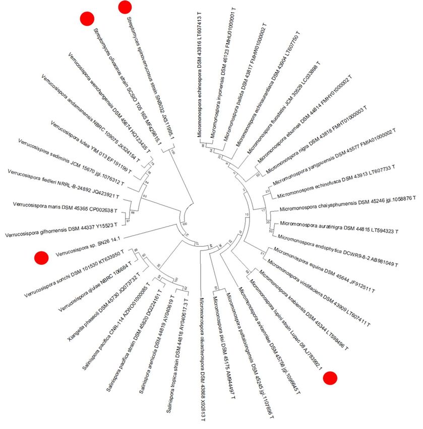
online platform [30] to identify the secondary metabolite biosynthesis gene clusters. As shown in
biosynthesis gene clusters. As shown in Figure 6, the draft genome of Verrucosispora sp. SN26_14.1
Figure 6, the draft genome of Verrucosispora sp. SN26_14.1 shared 60% of the genes of the Rsd gene
shared 60% of the genes of the Rsd gene cluster, as well as to an important percentage of the genes of
cluster,
the as
Rslwell
BGC.as The
to ansimilarity
important of percentage
the found genesof theranged
genes from
of the49% Rsl to
BGC.81%.The
The similarity of the found
genetic architecture
genesfound in Vex BGC was quite similar to that of the Rsd and Rsl BGC. Remarkably, we could nottodetect
ranged from 49% to 81%. The genetic architecture found in Vex BGC was quite similar that of
any and
the Rsd cyclase/aromatase and amidohydrolase
Rsl BGC. Remarkably, we could not sequences
detect anyin our draft genome. Likely,
cyclase/aromatase andthis relates to the
amidohydrolase
incompleteness of our sequence. Finally, it appears reasonable that Verrucosispora sp. SN26_14.1
sequences in our draft genome. Likely, this relates to the incompleteness of our sequence. Finally, it
follows
appears the samethat
reasonable biosynthetic machinery
Verrucosispora for the production
sp. SN26_14.1 follows the of Lupinacidin A (1) and
same biosynthetic Galvaquinone
machinery for the
B (2) as found
production for Streptomyces
of Lupinacidin species
A (1) and (Figure 6). B (2) as found for Streptomyces species (Figure 6).
Galvaquinone
Figure
Figure 6. Biosynthetic
6. Biosynthetic genegene cluster
cluster of of anthraquinonesproducers.
anthraquinones producers. Rsd:
Rsd: Streptomyces
Streptomyces olivaceus
olivaceusSCSIO
SCSIO
T05 T05
genegene cluster
cluster [28],
[28], Rsl:Streptomyces
Rsl: Streptomyces bottropensis
bottropensis (Streptomyces.
(Streptomyces.sp.sp.Gc C4/4) gene cluster
Gc C4/4) [29], Vex:
gene cluster [29],
Vex: Verrucosispora
Verrucosisporasp.sp.SN26_14.1.
SN26_14.1. C1:C1: aromatase,
aromatase,K1: K acyl carrier
1 : acyl protein,
carrier K2: ketosynthase
protein, K2 : ketosynthase (beta),(beta),
K3:
ketosynthase (alpha),
K3 : ketosynthase (alpha), A: acyl transferase,
A: acyl transferase, K K44:: 3-oxoacyl-ACP
3-oxoacyl-ACP synthase
synthaseIII,III, T
T11: : ABC-transporter
ABC-transporter
(substrate
(substrate binding),
binding), T2 :T2:ABC-transporter
ABC-transporter (ATP (ATP binding),
binding),T3T: 3ABC-transporter
: ABC-transporter trans-membrane,
trans-membrane, O1:
luciferase-like monooxygenase, O 2 : flavin reductase, P: phosphotransferase,
O1 : luciferase-like monooxygenase, O2 : flavin reductase, P: phosphotransferase, R1 : SARP family R 1 : SARP family
regulator,
regulator, C2: second
C2 : second ringring cyclase,
cyclase, O3O 3: 3-oxoacyl-ACPreductase,
: 3-oxoacyl-ACP reductase, OO44:: anthrone
anthrone monooxygenase,
monooxygenase,OO 5: :
5
NADH: flavin oxidoreductase, C 3: cyclase, R2: SARP regulatory protein, R3: LAL-family regulator, O6:
NADH: flavin oxidoreductase, C3 : cyclase, R2 : SARP regulatory protein, R3 : LAL-family regulator,
luciferase-like monooxygenase,
O6 : luciferase-like monooxygenase,RR 4: MarR family transcriptional regulator, T4: drug resistance
4 : MarR family transcriptional regulator, T4 : drug resistance
transporter, O7: putative NADPH quinone reductase, O8: putative NADPH: quinone oxidoreductase,
transporter, O7 : putative NADPH quinone reductase, O8 : putative NADPH: quinone oxidoreductase,
O9: FAD-dependent oxidoreductase, O10: C9-keto reductase, H: amidohydrolase, -3: unknown
O9 : FAD-dependent oxidoreductase, O10 : C9-keto reductase, H: amidohydrolase, -3: unknown
function, -2: major facilitator superfamily protein, -1: Transcriptional regulatory protein, 1: cupin, 2:
function, -2: +major facilitator superfamily protein, -1: Transcriptional regulatory protein, 1: cupin, 2:
citrate/H symporter, 3: transcriptional regulator.
citrate/H+ symporter, 3: transcriptional regulator.
2.7. Antibiotic
2.7. Antibiotic activity
Activity test
Test
We We performed a disc diffusion antibiotic test as a preliminary evaluation to determine if
performed a disc diffusion antibiotic test as a preliminary evaluation to determine if
Lupinacidin A (1) and Galvaquinone B (2) have an inhibition effect on bacteria. As a positive control,
Lupinacidin A (1) and Galvaquinone B (2) have an inhibition effect on bacteria. As a positive control,Mar. Drugs 2019, 17, 154 9 of 15
we used streptomycin at a concentration of 25 µg/disc. The results showed that Lupinacidin A (1)
and Galvaquinone B (2) did not produce any growth inhibition against the Gram-positive bacterium
Staphylococcus lentus DSM 20352T , and neither against the Gram-negative bacterium Escherichia coli
DSM 498T . In contrast, the positive control, streptomycin produced an inhibition halo of 22 mm for
Gram-negative and 18 mm for Gram-positive bacteria.
3. Materials and Methods
3.1. Sample Collection
The sea anemone Gyractis sesere (also known as Actiniogeton rapanuiensis) was sampled from
the coastal zone of Easter Island (27◦ 080 45.1”S, 109◦ 250 50.0”W) by the first author (Chilean citizen),
in March 2016. The sampling site was outside the Isla de Pascua national park, and the sample was
taken in agreement with regulations by the Chilean government. The sample was stored at 0 ◦ C one
hour after the sampling process.
3.2. Sea Anemone Dereplication
10 g of the sea anemone Gyractis sesere (wet weight) were thawed and homogenized with a mortar
and pestle. When a creamy consistency was obtained, the tissue was transferred to a 250 mL beaker
and 50 mL of chloroform was added. This extraction procedure was repeated three times. The obtained
chloroform extract was concentrated until dryness under reduced pressure in a rotatory evaporator.
The dried extract was resuspended in 1 L deionized water and transferred to a separation funnel,
where it was partitioned with chloroform (3 × 300 mL). This process produced 8 mg of crude extract
with a brownish coloration. Part of the crude extract (0.5 mg) was resuspended in methanol (HPLC
grade) and injected in a HPLC (Merck Hitachi LaChrom Elite, Darmstadt, Germany) and in a HRLCMS
Thermo Scientific™ Q Exactive™ Hybrid-Quadrupol-Orbitrap (Bremen, Germany), positive mode,
and a 30 minute gradient of H2 O and acetonitrile supplemented with 0.1% of formic acid. The gradient
developed as following: 0 min: 90% water, 10% acetonitrile, 25 min: 0% water, 100% acetonitrile,
28 min: 0% water, 100% acetonitrile, 30 min: 90% water, 10% acetonitrile. Mass spectroscopic data was
evaluated with Xcalibur® (Thermo Fisher Scientific, San Jose, CA, USA), and compared with online
databases (MarinLit, and Scifinder), and literature. The entire remaining sample was dissolved in
deuterated chloroform (Eurisotop™, Saint-Aubin, France) and analyzed by 1 H NMR using a Bruker
(Rheinstetten, Germany) Avance 600 MHz NMR spectrometer.
3.3. Bacterial Isolation
Approximately 1 cm3 (2 g) of the sea anemone Gyractis sesere (wet weight) were thawed and
homogenized with a sterile mortar and pestle. Subsequently, the homogenized tissue was mixed
with 9 mL of Ringer’s buffer 14 strength [31] to produce a final solution of 1:10. This solution was
incubated at 56 ◦ C for 10 min with the aim of reducing the viability of non-actinobacterial microbes.
After the incubation, 1 min of vortex was applied. The inoculation of the culture media was done by
adding 50 µL of the dilution into 15 cm diameter Petri dishes containing the media. The inoculum was
spread out on the plate with a triangular cell spreader made of glass. Finally, plates were incubated
at 25 ◦ C in darkness. Darkness was chosen as a filtering factor to eliminate potential microalgae
contamination. Four different media were prepared for the isolation of Actinobacteria from the sea
anemone Gyractis sesere. Medium SIMA1 (Salinispora isolation media A1) was selected from literature
and slightly modified as follows: 2.5 g starch, 1 g yeast extract, 0.5 g peptone, 1 L deionized water, and
25 g Tropic Marin™ salt (Wartenberg, Germany), 15 g/L agar [32]. The other media (BCM, BTM, and
BSEM) were generated for this study as follows: BCM, 3 g chitin, 0.5 g N-acetyl glucosamine, 0.2 g
K2 HPO4 , 0.25 g KNO3 , 0.25 g casein, 5 mL of mineral solution, 4 mL vitamin solution, 1 L deionized
water, 15 g/L Tropic Marin™ salt (Wartenberg, Germany), 12 g/L Gellan gum, pH = 7.35; BTM, 1 g
trehalose, 0.25 g histidine, 0,25 g proline, 0.2 g MgCl2* 6H2 O, 4 mL vitamin solution, 12 g/L GellanMar. Drugs 2019, 17, 154 10 of 15
gum, 1 L deionized water, 15 g Tropic Marin™ salt (Wartenberg, Germany), pH = 7.2; and BSEM,
0.1 g tyrosine, 0.1 g D-galactose, 4 mL vitamin solution, 5 mL mineral solution, 1 L Baltic Sea water,
16 g/L agar, pH = 7.4. Mineral salt solution contained 1 L distillated water, 50 mg FeSO4* 7H2 O, 50 mg
ZnCl2 , and 50 mg CuSO4 . Vitamin solution contained 1 L distillated water, 5 mg thiamine* HCl, 5 mg
riboflavin, 5 mg niacin, 5 mg pyrodoxine HCl, 5 mg inositol, 5 mg Ca-pantothenate, 5 mg p-amino
benzoic acid, and 2.5 mg biotin.
The media were autoclaved for 35 min at 121 ◦ C. Subsequently, the culture media were
supplemented with 50 mg/L of nalidixic acid (Sigma-Aldrich, St. Louis, MO, USA) and 100 mg/L of
cycloheximide (Carl Roth GmbH, Karlsruhe, Germany) [33], and poured into petri dishes. Once the
sample was inoculated onto the petri dish, they were incubated for six weeks. When bacterial colonies
were visually evident, we proceeded with the purification of the bacteria until obtaining an axenic
culture. The isolated bacteria were conserved using Cryobank™ (Mast Diagnostica GmbH, Reinfeld,
Germany) bacterial storage system.
3.4. Molecular Characterization and Phylogenetic Analysis
DNA was extracted from bacterial cells by use of a DNA isolation kit, DNeasy™ (Qiagen, Hilden,
Germany), following the manufacturer instructions. Subsequently, the 16S rRNA gene sequence was
amplified with PCR and the use of general bacterial primers in a concentration of 10 pmol/µL, i.e., 27f
and 1492r [34], 342f and 534r [35], 1387r [36] as well as 1525r [37]. PCR reagents were obtained from GE
Healthcare Illustra™ PuReTaq Ready-To-Go™ PCR Beads (GE Healthcare, Glattbrugg, Switzerland)
containing DNA polymerase, MgCl2 , and dNTPs. The PCR conditions were the same as reported by
Staufenberger et al. [35]. Once the PCR amplification process was terminated, a quality check of the
PCR products was performed by gel electrophoresis. The sequencing process was run at the Centre for
Molecular Biology at Kiel University (IKMB). The 16S rRNA gene sequences were manually curated
using Chromas pro software, version 1.7.6 (Technelysium Pty Ltd., Tewantin QLD, Australia), and
saved in FASTA format. Sequences were aligned with nucleotide BLAST [38] and EZbiocloud [39].
Phylogenetic analysis involved the alignment of the sequences with related reference strains in the web
platform SILVA-SINA [40]. MEGA was used to delete gap sites and to run bootstrapped phylogenetic
trees using a neighbor-joining model [41].
3.5. Bacterial Growth for Secondary Metabolites Production
For the evaluation of the secondary metabolites production, we grew the Easter Island isolated
strain Verrucosispora sp. SN26_14.1 in 10 × 2.5 L Thomson Ultra Yield® flasks (Thomson Instrument,
Oceanside, CA, USA), which contained 1 L each of a modified starch-glucose-glycerol (SGG) liquid
medium [31]. The composition of the production medium was: 5 g glucose, 5 g soluble starch, 5 g
glycerol, 1.25 g cornsteep powder, 2.5 g peptone, 1 g yeast extract, 1.5 CaCO3 , and 1 L deionized water.
The medium was also supplemented with 15 g/L Tropic Marin™ salt (Wartenberg, Germany). The pH
was adjusted to 7.7 using 1 M HCl and NaOH. The culture was kept in orbital agitation at 240 RPM,
28 ◦ C, for 14 days in darkness.
3.6. Chemical Extraction, Purification and Structure Elucidation
After the growth period, 20 g/L amberlite XAD-16 (Sigma-Aldrich, St. Louis, MO, USA) was
added to each culture medium flask and mixed for one hour using orbital agitation with 120 rpm.
Subsequently, the resin was separated through cheesecloth filtration [42], and the liquid was discarded.
Afterwards, amberlite plus cheesecloth was mounted on a glass funnel, washed with 3 L of deionized
water, and eluted with 1 L of acetone [42]. Acetone was then concentrated under reduced pressure
until an aqueous residue was obtained. One liter of deionized water was added to the acetone residue,
and it was brought to a separation funnel. The organic molecules were extracted using 3 × 1 L of ethyl
acetate. The organic phase was concentrated under reduced pressure until dryness.Mar. Drugs 2019, 17, 154 11 of 15
For the evaluation of the produced metabolites, we used HPLC-DAD (Merck Hitachi LaChrom
Elite, Darmstadt, Germany) and a 30 min gradient of H2 O-acetonitrile supplemented with 0.1% of
formic acid. The gradient was developed as following: 0 min: 90% water, 10% acetonitrile, 25 min:
0% water, 100% acetonitrile, 28 min: 0% water, 100% acetonitrile, 30 min: 90% water, 10% acetonitrile.
The gravity SB™ C-18 column was obtained from Macherey-Nagel (Düren, Germany).
The purification of chemicals involved three different steps: 1) Flash chromatography using
standard silica gel 60, pore size ~ 60 Å (Macherey-Nagel, Düren, Germany) as a stationary phase,
mounted in a glass Buchner funnel (D = 70 mm, H = 180 mm). The mobile phase solvents were
iso-octane and ethyl acetate. The chromatographic process was developed in a stepwise increase of
polarity (10% each), starting with 100% iso-octane, and 0% of ethyl acetate, and ending in 0% iso-octane
and 100% ethyl acetate, resulting in 10 different fractions. 2) The fraction that contained compound (1)
and (2) was selected and worked in HPLC (Merck Hitachi LaChrom Elite, Darmstadt, Germany) using a
normal phase NUCLEODUR® 100-5 column (4.6 × 250 mm) from Macherey-Nagel (Düren, Germany).
The method used for the purification was a combination of isocratic and gradient solvent mix, with a
flow rate of 1 mL/min, where A: iso-octane, B: ethyl acetate, and C: dichloromethane/methanol (50:50).
The method was developed as following: 0 min: 100% A and 0% B, 3 min: 100% A and 0% B, 5 min:
95% A and 5% B, 9 min: 95% A and 5% B, 11 min: 0% A and 100% B, 13 min: 0% A and 100% B, 14 min:
10% A, 50% B, and 40% C, 16 min: 10% A, 50% B, and 40% C, 18 min: 50% A and 50% B, 19 min: 100%
A and 0% B, 21 min: 100% A and 0% B. 3) The semi-purified compounds were purified through HPLC
(Merck Hitachi LaChrom Elite, Darmstadt, Germany using a reverse phase C-18 column, 10 × 250 mm
(YMC, Kyoto, Japan). The method used for the purification was a combination of isocratic and gradient
solvent mix, with a flow rate of 2.5 mL/min. The method was developed as following: 0 min: 90% A
and 10% B, 5 min: 20% A and 80% B, 9 min: 20% A and 80% B, 13 min: 0% A and 100% B, 19 min: 0%
A and 100% B, 23 min: 90% A and 10% B, 25 min: 90% A, 10% B (A. water, B: acetonitrile).
After these purification steps, Lupinacidin A (1) and Galvaquinone B (2) were obtained with
high purity to perform structural elucidation experiments. HRLCMS was performed with a Thermo
Scientific™ Q Exactive™ Hybrid-Quadrupol-Orbitrap (Thermo Scientific, Bremen, Germany), positive
mode, and a 30 min gradient of H2 O and acetonitrile supplemented with 0.1% of formic acid.
The gradient was developed as follows: 0 min: 90% water, 10% acetonitrile, 25 min: 0% water,
100% acetonitrile, 28 min: 0% water, 100% acetonitrile, 30 min: 90% water, 10% acetonitrile. Mass
spectroscopic data was evaluated with Xcalibur® (Thermo Fisher Scientific, San Jose, CA, USA), and
the compared with online databases (MarinLit, and Scifinder), and literature.
Additionally, 1 H and 13 C NMR and two-dimensional NMR experiments (HMBC, HSQC, COSY)
were acquired to characterize the main components of crude extract, and their chemical functionality.
For this, compound (1) and (2) were redissolved in CDCl3 (Eurisotop™, Saint-Aubin, France), and
transferred to NMR tubes (178 × 5.0 mm). Experiments were acquired on a Bruker (Rheinstetten,
Germany) Avance spectrometer operating at 600 MHz proton frequency equipped with a cryogenically
cooled triple resonance z-gradient probe head using stand pulse sequences from the Bruker experiment
library. Spectra were referenced against tetramethylsilane (Sigma-Aldrich, St. Louis, MO, USA) as
internal standard.
3.7. Genome Sequencing
The samples were prepared with the Nextera® XT DNA sample preparation kit from Illumina
(Illumina, San Diego, CA, USA) following the manufacturer’s protocol. Afterwards the samples
were pooled and sequenced on the Illumina MiSeq using the MiSeq® (Illumina, San Diego, CA, USA)
Reagent Kit v3 600 cycles sequencing chemistry. The library was clustered to a density of approximately
1200 K/mm2 .Mar. Drugs 2019, 17, 154 12 of 15
3.8. Genome Assembly
The quality control of reads was checked with FASTQC software [43] to evaluate the GC%,
number of k-mers, sequence length, and total reads. Trimmomatic v0.36 [44] was used to filter low
quality sequences and adapters. Filtered reads were assembled with SPAdes v3.11.0 [45] using default
k-mer lengths. The obtained contigs were evaluated with QUAST tool [46] to select the best quality
contig. Finally, Prokka [47] was used to annotate the draft genome.
3.9. Secondary Metabolites Gene Clusters Search
The online platform of Antismash [30] was used to detect the secondary metabolites gene clusters
present in the draft genome.
3.10. Antibiotic Activity Test
To test the antibiotic activity, we used the disc diffusion method [48] as a primary indicator. Thus,
compound (1) and (2) were tested to determine their activity on Staphylococcus lentus DSM 20352T ,
and Escherichia coli DSM 498T . These bacteria were cultured in GYM medium (4 g glucose, 4 g yeast
extract, 10 g malt extract, 2 g CaCO3 , 1 L deionized water, pH = 7.2, and 12 g agar). Lupinacidin A
(1), and Galvaquinone B (2) were transferred to a paper disc to reach a final concentration of 25 µg
and 50 µg each in triplicate. Additionally, we used an antibiotic susceptibility disc of streptomycin
(Oxoid® , Columbia, MD, USA) as a positive indicator of antibiotic activity. The plates were inoculated
with fresh culture of Staphylococcus lentus DSM 20352T , and Escherichia coli DSM 498T , and incubated at
37 ◦ C for 24 h. After the incubation period, the inhibition zone was measured and registered.
4. Conclusions
We established that the Easter Island sea anemone Gyractis sesere contained two anthraquinones,
Lupinacidin A (1) and Galvaquinone B (2), which were ultimately found to be produced by one of the
Actinobacteria associated with this marine invertebrate, Verrucosispora sp. SN26_14.1. The production
of the identified metabolites by the bacterial isolate apparently follows a recently characterized PKS
type II pathway with a Baeyer−Villiger type rearrangement assembly line. Our finding adds a new
actinobacterial genus to the producers of these anthraquinones, implying that these metabolites are
not exclusive to the genera Streptomyces and Micromonospora. It was demonstrated, that culture-based
approaches remain as effective tools for the isolation of polyketide producing Actinobacteria as sources
for secondary metabolites of potential use in drug discovery. Our study confirms that cnidarians, and
in specific sea anemones, can be a source of such pharmacologically relevant microorganisms. Finally,
these findings re-open the debate about the real producers of secondary metabolites in sea animals
and add another example of associated bacteria as producers of substances present in sea animals. In
addition, the study provides information on the chemistry harbored in biota of the geographically
isolated and almost unstudied, Easter Island.
Supplementary Materials: The following are available online at http://www.mdpi.com/1660-3397/17/3/154/s1.
Information on NMR spectra, HRLCMS data, and secondary metabolite gene cluster.
Author Contributions: I.S., J.F.I. and J.W. planned the experiments, I.S. performed the experiments, analyzed and
evaluated the data and wrote the first draft of the publication. J.F.I., F.D.S. and J.W. supervised the work and
revised the manuscript. S.K. sequenced the genome and supplied the genome data. M.L. and N.P. acquired LCMS
and NMR data. F.D.S. acquired and analyzed NMR data.
Funding: We thank the Deutscher Akademischer Austauschdienst (DAAD) for financial support under the
stipend # PKZ91564794. We acknowledge financial support by the Land Schleswig-Holstein within the funding
programme Open Access Publikationsfonds.
Acknowledgments: We thank Marion Höftmann and Gitta Kohlmeyer-Yilmaz for her valuable help with the NMR
data acquisition. We thank Ute Hentschel Humeida (GEOMAR Helmholtz Centre for Ocean Research Kiel) for her
support. We also thank Philip A. Thomas (ImagesByPT@PhilipT.com), for providing high quality photography of
the sea anemone Gyractis sesere. I.S. thanks Millaray Sierra for her support during the research. N.P. thanks the
Deutsche Bundesstiftung Umwelt (German Federal Environmental Foundation) for a predoctoral fellowship.Mar. Drugs 2019, 17, 154 13 of 15
Conflicts of Interest: The authors declare no conflict of interest.
References
1. Dransfield, J.; Flenley, J.R.; King, S.M.; Harkness, D.D.; Rapu, S. A recently extinct palm from Easter Island.
Nature 1984, 312, 750. [CrossRef]
2. Boyko, C.B. The endemic marine invertebrates of Easter Island: How many species and for how long?
In Easter Island: Scientific Exploration into the World’s Environmental Problems in Microcosm; Loret, J.,
Tanacredi, J.T., Eds.; Springer: Boston, MA, USA, 2003; pp. 155–175.
3. Skottsberg, C. The Natural History of Juan Fernández and Easter Island; Almqvist & Wiksells Boktryckeri:
Uppsala, Sweden, 1920.
4. Kohn, A.J.; Lloyd, M.C. Marine polychaete annelids of Easter Island. Int. Rev. Hydrobiol. 1973, 58, 691–712.
[CrossRef]
5. Sehgal, S.N. Rapamune® (RAPA, rapamycin, sirolimus): Mechanism of action immunosuppressive effect
results from blockade of signal transduction and inhibition of cell cycle progression. Clin. Biochem. 1998, 31,
335–340. [CrossRef]
6. Vezina, C.; Kudelski, A.; Sehgal, S. Rapamycin (AY-22, 989), a new antifungal antibiotic. J. Antibiot. 1975, 28,
721–726. [CrossRef] [PubMed]
7. Allen, G.R. Conservation hotspots of biodiversity and endemism for Indo-Pacific coral reef fishes. Aquat.
Conserv. 2008, 18, 541–556. [CrossRef]
8. Horton, T.; Kroh, A.; Ahyong, S.; Bailly, N.; Boyko, C.B.; Brandão, S.N.; Costello, M.J.; Gofas, S.; Hernandez, F.;
Holovachov, O.; et al. World Register of Marine Species (WoRMS); WoRMS Editorial Board: Ostend, Belgium, 2018.
9. Urda, C.; Fernández, R.; Pérez, M.; Rodríguez, J.; Jiménez, C.; Cuevas, C. Protoxenicins A and B, cytotoxic
long-chain acylated xenicanes from the soft coral Protodendron repens. J. Nat. Prod. 2017, 80, 713–719.
[CrossRef] [PubMed]
10. Nakamura, F.; Kudo, N.; Tomachi, Y.; Nakata, A.; Takemoto, M.; Ito, A.; Tabei, H.; Arai, D.; de Voogd, N.;
Yoshida, M.; et al. Halistanol sulfates I and J, new SIRT1–3 inhibitory steroid sulfates from a marine sponge
of the genus Halichondria. J. Antibiot. 2017, 71, 273. [CrossRef] [PubMed]
11. Aminin, D.; Menchinskaya, E.; Pisliagin, E.; Silchenko, A.; Avilov, S.; Kalinin, V. Anticancer activity of sea
cucumber triterpene glycosides. Mar. Drugs 2015, 13, 1202–1223. [CrossRef] [PubMed]
12. Hay, M.E. Marine chemical ecology: What’s known and what’s next? J. Exp. Mar. Biol. Ecol. 1996, 200,
103–134. [CrossRef]
13. Mehbub, M.F.; Lei, J.; Franco, C.; Zhang, W. Marine sponge derived natural products between 2001 and 2010:
Trends and opportunities for discovery of bioactives. Mar. Drugs 2014, 12, 4539–4577. [CrossRef] [PubMed]
14. Blunt, J.W.; Copp, B.R.; Keyzers, R.A.; Munro, M.H.G.; Prinsep, M.R. Marine natural products. Nat. Prod.
Rep. 2014, 31, 160–258. [CrossRef] [PubMed]
15. Piel, J.; Hui, D.; Wen, G.; Butzke, D.; Platzer, M.; Fusetani, N.; Matsunaga, S. Antitumor polyketide
biosynthesis by an uncultivated bacterial symbiont of the marine sponge Theonella swinhoei. Proc. Natl. Acad.
Sci. USA 2004, 101, 16222–16227. [CrossRef] [PubMed]
16. Rath, C.M.; Janto, B.; Earl, J.; Ahmed, A.; Hu, F.Z.; Hiller, L.; Dahlgren, M.; Kreft, R.; Yu, F.; Wolff, J.J.;
et al. Meta-omic characterization of the marine invertebrate microbial consortium that produces the
chemotherapeutic natural product ET-743. ACS Chem. Biol. 2011, 6, 1244–1256. [CrossRef] [PubMed]
17. Unson, M.D.; Holland, N.D.; Faulkner, D.J. A brominated secondary metabolite synthesized by the
cyanobacterial symbiont of a marine sponge and accumulation of the crystalline metabolite in the sponge
tissue. Mar. Biol. 1994, 119, 1–11. [CrossRef]
18. Schmidt, E.W.; Obraztsova, A.Y.; Davidson, S.K.; Faulkner, D.J.; Haygood, M.G. Identification of the
antifungal peptide-containing symbiont of the marine sponge Theonella swinhoei as a novel δ-Proteobacterium,
“Candidatus Entotheonella palauensis”. Mar. Biol. 2000, 136, 969–977. [CrossRef]
19. Feng, Y.; Khokhar, S.; Davis, R.A. Crinoids: Ancient organisms, modern chemistry. Nat. Prod. Rep. 2017, 34,
571–584. [CrossRef] [PubMed]
20. Khokhar, S.; Pierens, G.K.; Hooper, J.N.A.; Ekins, M.G.; Feng, Y.; Davis, R.A. Rhodocomatulin-type
anthraquinones from the australian marine invertebrates Clathria hirsuta and Comatula rotalaria. J. Nat.
Prod. 2016, 79, 946–953. [CrossRef] [PubMed]Mar. Drugs 2019, 17, 154 14 of 15
21. Tietze, L.F.; Gericke, K.M.; Schuberth, I. Synthesis of highly functionalized anthraquinones and evaluation of
their antitumor activity. Eur. J. Org. Chem. 2007, 2007, 4563–4577. [CrossRef]
22. Yang, K.-L.; Wei, M.-Y.; Shao, C.-L.; Fu, X.-M.; Guo, Z.-Y.; Xu, R.-F.; Zheng, C.-J.; She, Z.-G.; Lin, Y.-C.;
Wang, C.-Y. Antibacterial anthraquinone derivatives from a sea anemone-derived fungus Nigrospora sp.
J. Nat. Prod. 2012, 75, 935–941. [CrossRef] [PubMed]
23. Kim, Y.-M.; Lee, C.-H.; Kim, H.-G.; Lee, H.-S. Anthraquinones isolated from Cassia tora (Leguminosae) Seed
Show an Antifungal Property against Phytopathogenic Fungi. J. Agric. Food Chem. 2004, 52, 6096–6100.
[CrossRef] [PubMed]
24. Hu, Y.; Martinez, E.D.; MacMillan, J.B. Anthraquinones from a marine-derived Streptomyces spinoverrucosus.
J. Nat. Prod. 2012, 75, 1759–1764. [CrossRef] [PubMed]
25. Igarashi, Y.; Trujillo, M.E.; Martínez-Molina, E.; Yanase, S.; Miyanaga, S.; Obata, T.; Sakurai, H.; Saiki, I.;
Fujita, T.; Furumai, T. Antitumor anthraquinones from an endophytic actinomycete Micromonospora lupini sp.
nov. Bioorg. Med. Chem. Lett. 2007, 17, 3702–3705. [CrossRef] [PubMed]
26. Haddon, A.C.; Shackleton, A.M. Description of some new species of Actiniaria from Torres Straits. R. Dublin
Soc. 1893, 8.
27. Carlgren, O. Actiniaria und zoantharia von Juan Fernandez und der Osterinsel. In The Natural History of Juan
Fernandez and Easter Island; Skottsberg, C., Ed.; Almquist & Wiksells Boktryckeri: Uppsala, Sweden, 1922;
pp. 145–160.
28. Zhang, C.; Sun, C.; Huang, H.; Gui, C.; Wang, L.; Li, Q.; Ju, J. Biosynthetic Baeyer–Villiger chemistry enables
access to two anthracene scaffolds from a single gene cluster in deep-sea-derived Streptomyces olivaceus
SCSIO T05. J. Nat. Prod. 2018, 81, 1570–1577. [CrossRef] [PubMed]
29. Yan, X.; Probst, K.; Linnenbrink, A.; Arnold, M.; Paululat, T.; Zeeck, A.; Bechthold, A. Cloning and
heterologous expression of three type II PKS gene clusters from Streptomyces bottropensis. ChemBioChem 2012,
13, 224–230. [CrossRef] [PubMed]
30. Weber, T.; Blin, K.; Duddela, S.; Krug, D.; Kim, H.U.; Bruccoleri, R.; Lee, S.Y.; Fischbach, M.A.; Müller, R.;
Wohlleben, W.; et al. AntiSMASH 3.0—A comprehensive resource for the genome mining of biosynthetic
gene clusters. Nucleic Acids Res. 2015, 43, W237–W243. [CrossRef] [PubMed]
31. Goodfellow, M.; Fiedler, H.-P. A guide to successful bioprospecting: Informed by actinobacterial systematics.
J. Microb. 2010, 98, 119–142. [CrossRef] [PubMed]
32. Patin, N.V.; Duncan, K.R.; Dorrestein, P.C.; Jensen, P.R. Competitive strategies differentiate closely related
species of marine Actinobacteria. ISME J. 2015, 10, 478. [CrossRef] [PubMed]
33. Thaker, M.N.; Waglechner, N.; Wright, G.D. Antibiotic resistance–mediated isolation of scaffold-specific
natural product producers. Nat. Protoc. 2014, 9, 1469. [CrossRef] [PubMed]
34. Lane, D.J. 16S/23S rRNA sequencing. In Nucleic Acid Techniques in Bacterial Systematics; Stackebrandt, E.,
Goodfellow, M., Eds.; John Wiley and Sons: Chichester, UK, 1991; pp. 115–175.
35. Staufenberger, T.; Thiel, V.; Wiese, J.; Imhoff, J.F. Phylogenetic analysis of bacteria associated with Laminaria
saccharina. FEMS Microbiol. Ecol. 2008, 64, 65–77. [CrossRef] [PubMed]
36. Ellis, R.J.; Morgan, P.; Weightman, A.J.; Fry, J.C. Cultivation-dependent and independent approaches for
determining bacterial diversity in heavy-metal-contaminated soil. Appl. Environ. Microb. 2003, 69, 3223.
[CrossRef]
37. Frank, J.A.; Reich, C.I.; Sharma, S.; Weisbaum, J.S.; Wilson, B.A.; Olsen, G.J. Critical evaluation of two
primers commonly used for amplification of bacterial 16S rRNA genes. Appl. Environ. Microb. 2008, 74, 2461.
[CrossRef] [PubMed]
38. Altschul, S.F.; Gish, W.; Miller, W.; Myers, E.W.; Lipman, D.J. Basic local alignment search tool. J. Mol. Biol.
1990, 215, 403–410. [CrossRef]
39. Yoon, S.-H.; Ha, S.-M.; Kwon, S.; Lim, J.; Kim, Y.; Seo, H.; Chun, J. Introducing EzBioCloud: A taxonomically
united database of 16S rRNA gene sequences and whole-genome assemblies. Int. J. Syst. Evol. Microbiol.
2017, 67, 1613–1617. [CrossRef] [PubMed]
40. Quast, C.; Pruesse, E.; Yilmaz, P.; Gerken, J.; Schweer, T.; Yarza, P.; Peplies, J.; Glöckner, F.O. The SILVA
ribosomal RNA gene database project: Improved data processing and web-based tools. Nucleic Acids Res.
2013, 41, D590–D596. [CrossRef] [PubMed]You can also read