Annealing of ssDNA and compaction of dsDNA by the HIV-1 nucleocapsid and Gag proteins visualized using nanofluidic channels
←
→
Page content transcription
If your browser does not render page correctly, please read the page content below
Quarterly Reviews of                                  Annealing of ssDNA and compaction of dsDNA
           Biophysics
                                                                 by the HIV-1 nucleocapsid and Gag proteins
           cambridge.org/qrb                                     visualized using nanofluidic channels
                                                                 Kai Jiang1, Nicolas Humbert2, Sriram KK1, Thiebault Lequeu2, Yii-Lih Lin1,
           Report                                                Yves Mely2 and Fredrik Westerlund1
                                                                 1
           Cite this article: Jiang K, Humbert N, KK S,            Division of Chemical Biology, Department of Biology and Biological Engineering, Chalmers University of
           Lequeu T, Lin Y-L, Mely Y, Westerlund F (2019).       Technology, Gothenburg, SE 412 96, Sweden and 2Laboratoire de Bioimagerie et Pathologies, UMR 7021 CNRS,
           Annealing of ssDNA and compaction of dsDNA            Université de Strasbourg, Faculté de Pharmacie, Illkirch, F 67401, France
           by the HIV-1 nucleocapsid and Gag proteins
           visualized using nanofluidic channels.
           Quarterly Reviews of Biophysics 52, e2, 1–10.           Abstract
           https://doi.org/10.1017/S0033583518000124               The nucleocapsid protein NC is a crucial component in the human immunodeficiency virus
           Received: 5 September 2018
                                                                   type 1 life cycle. It functions both in its processed mature form and as part of the polyprotein
           Revised: 23 December 2018                               Gag that plays a key role in the formation of new viruses. NC can protect nucleic acids (NAs)
           Accepted: 24 December 2018                              from degradation by compacting them to a dense coil. Moreover, through its NA chaperone
                                                                   activity, NC can also promote the most stable conformation of NAs. Here, we explore the bal-
           Key words:
                                                                   ance between these activities for NC and Gag by confining DNA–protein complexes in nano-
           Gag; HIV-1; nanofluidics; nucleocapsid; single
           DNA molecules                                           channels. The chaperone activity is visualized as concatemerization and circularization of long
                                                                   DNA via annealing of short single-stranded DNA overhangs. The first ten amino acids of NC
           Author for correspondence:                              are important for the chaperone activity that is almost completely absent for Gag. Gag con-
           Fredrik Westerlund,                                     denses DNA more efficiently than mature NC, suggesting that additional residues of Gag are
           E-mail: fredrikw@chalmers.se
                                                                   involved. Importantly, this is the first single DNA molecule study of full-length Gag and we
                                                                   reveal important differences to the truncated Δ-p6 Gag that has been used before. In addition,
                                                                   the study also highlights how nanochannels can be used to study reactions on ends of long
                                                                   single DNA molecules, which is not trivial with competing single DNA molecule techniques.

                                                                 Introduction
                                                                 The synthesis of the viral DNA in human immunodeficiency virus type 1 (HIV-1) results from
                                                                 the reverse transcription (RTion) process, where two copies of single-stranded RNA (ssRNA)
                                                                 are transcribed into double-stranded DNA (dsDNA). The viral dsDNA is then trafficked to the
                                                                 nucleus of the target cell where it is integrated into the host cell chromatin, followed by tran-
                                                                 scription and translation processes by the cellular machinery. One of the transcription prod-
                                                                 ucts is an mRNA that codes for the Gag polyprotein, which is the main structural protein of
                                                                 HIV-1, and is on its own sufficient for assembly of new viral particles in cells (Campbell and
                                                                 Rein, 1999). The Gag polyprotein consists of four major domains; the N terminus matrix
                                                                 (MA), the capsid (CA), the nucleocapsid (NC) and the C-terminus p6, as well as two small
                                                                 spacer peptides, Sp1 and Sp2 (Fig. 1a). After the viral particle leaves the infected cell, the
                                                                 Gag polyprotein is sequentially cleaved by the virus-encoded protease to ultimately lead to
                                                                 the mature MA, CA, NC and p6 proteins. In the mature virion, MA associates with the inner
                                                                 viral membrane, CA assembles into the conical capsid and NC coats and condenses the viral
                                                                 RNA (Ganser-Pornillos et al., 2008; Briggs et al., 2009; Briggs and Kräusslich, 2011; Bell and
                                                                 Lever, 2013).
                                                                    The NC protein is a small structural protein that contains a basic N-terminal domain as
                                                                 well as two zinc finger motifs, separated by a short basic linker (Fig. 1b) (Darlix et al.,
                                                                 1995). This 55-amino-acid protein acts as a nucleic acid (NA) chaperone that favours the
                                                                 most thermodynamically stable conformation of NAs (Rein et al., 1998; Levin et al., 2005;
                                                                 Godet and Mély, 2010; Darlix et al., 2011). The NC chaperone activity relies on its ability
                                                                 to associate and dissociate rapidly from its NA targets, to destabilize their secondary structure
           © The Author(s) 2019. This is an Open Access          and to promote the annealing of complementary sequences. The chaperone activity of NC is
           article, distributed under the terms of the
                                                                 witnessed during the early phase of the virus life cycle, where it helps the two obligatory strand
           Creative Commons Attribution licence (http://
           creativecommons.org/licenses/by/4.0/), which          transfers in the RTion process, and also the integration of the viral DNA into the host genome.
           permits unrestricted re-use, distribution, and        In addition, during the late phase of the virus life cycle, NC, as a part of the Gag polyprotein,
           reproduction in any medium, provided the              plays a crucial role in the recognition and dimerization of the two copies of genomic RNA
           original work is properly cited.                      (Rein, 2010; Abd El-Wahab et al., 2014), Gag–Gag oligomerization and Gag trafficking to
                                                                 the plasma membrane (El Meshri et al., 2015; Freed, 2015). The ability of NC to aggregate
                                                                 NAs is due to its cationic character (Darlix et al., 1995; Mirambeau et al., 2006; Vo et al.,
                                                                 2006), notably in its N-terminal domain and removal of this domain reduces the aggregation
                                                                 capability greatly (Stoylov et al., 1997; Krishnamoorthy, 2003). On the other hand, properly

Downloaded from https://www.cambridge.org/core. IP address: 46.4.80.155, on 05 Nov 2021 at 16:20:05, subject to the Cambridge Core terms of use, available at https://www.cambridge.org/core/terms.
https://doi.org/10.1017/S0033583518000124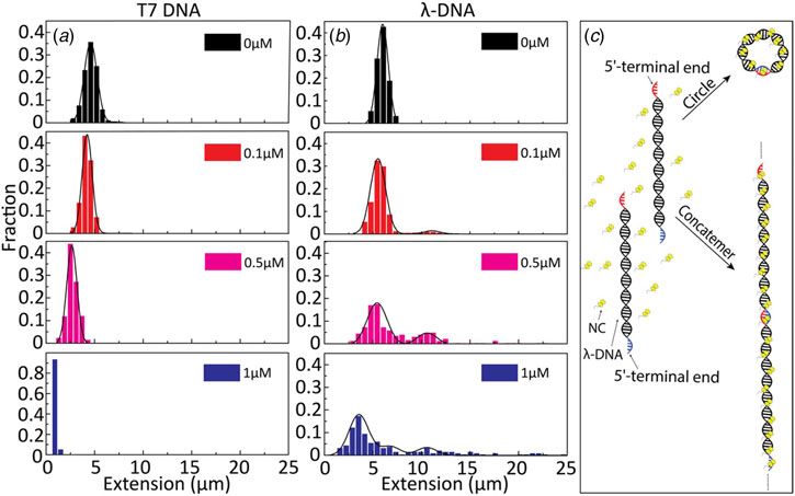
2                                                                                                                                                            Kai Jiang et al.

                                                                                                     of single DNA molecules in nanofluidic channels unique is that
                                                                                                     the single DNA molecules are suspended free in solution. This
                                                                                                     is in stark contrast to most single DNA molecule techniques,
                                                                                                     where at least one DNA end is attached to a bead or a surface,
                                                                                                     and opens up possibilities to study reactions occurring on DNA
                                                                                                     ends.
                                                                                                         Here, we studied the binding properties of two versions of Gag
                                                                                                     and several versions of NC, to long dsDNA (40–50 kb). Our
                                                                                                     results show that the ability to compact and condense dsDNA
          Fig. 1. (a) Schematic representation of the Gag polyprotein with major domains indi-
                                                                                                     is mainly related to the N-terminal domain of NC. In addition,
          cated. The p6 region highlighted in red is deleted in Δ-p6 Gag. (b) Amino acid             using dsDNA with short protruding ssDNA ends allowed us to
          sequence of NC(1–55) including its two-CCHC-zinc fingers. (c) Sequence of                  probe the chaperone activity of NC by studying concatemeriza-
          SSHS-SSHS NC(1–55) where the cysteine residues are replaced with serines.                  tion and circularization of DNA with complementary ssDNA
                                                                                                     overhangs. These studies demonstrated the key role of the
                                                                                                     N-terminal domain in the annealing of ssDNA. On the other
          folded zinc fingers are critical for NA destabilization (Bernacchi                         hand, compared with NC alone, the NC protein within the con-
          et al., 2002; Beltz et al., 2005; Godet et al., 2011; Wu et al.,                           text of Gag shows stronger ability of compaction and condensa-
          2013). Although both double-stranded (ds) and single-stranded                              tion of dsDNA, but weaker chaperone activity. Interestingly, the
          (ss) NAs can be bound by NC, the zinc fingers prefer ssNAs                                 chaperone activity is somewhat retained when the p6 domain is
          (Heath et al., 2003; Mirambeau et al., 2006; Darlix et al., 2011).                         deleted, and this is the first study where Gag and Δ-p6 are directly
              The binding of NC to single DNA molecules has been studied                             compared using single DNA molecule techniques. Importantly,
          thoroughly using optical tweezers, in particular by the Williams                           the use of nanofluidic channels allowed us to directly investigate
          group (Williams et al., 2001; Williams et al., 2002; Cruceanu,                             the competition between annealing and condensation on the sin-
          2006; Cruceanu et al., 2006; Wu et al., 2013, 2014). They used                             gle DNA molecule level. In addition to the specific studies on NC,
          single-molecule DNA stretching to probe the NA annealing,                                  the concatemerization demonstrates the usefulness of nanofluidic
          aggregation and destabilization activities of NC. Gag (in a version                        channels for probing intermolecular DNA–DNA interactions on
          where the p6 region is deleted, Δ-p6 Gag), wild-type NC, as well                           the single DNA molecule level, in particular involving DNA
          as different NC variants, designed to be defective in at least one of                      ends, and the example here is the first along those lines.
          these activities, by deletion or changes of key residues in the
          N-terminal domain, the zinc finger and the linker regions, have
          been investigated. Their results show that the NC protein within                           Materials and methods
          the context of Gag appears to have mostly a NA binding and pack-                           Protein expression and purification
          aging function, while the processed forms of NC appears to act
          mostly as a NA chaperone. In addition, both of the two zinc fingers                        The different NC peptides were prepared by solid-phase peptide
          are required for NA destabilization and the lack of, or changes in,                        synthesis on a 433A synthesizer (ABI, Foster City, CA, USA),
          the zinc fingers results in significantly weaker duplex destabiliza-                       HPLC purified and characterized by ion spray mass spectrometry,
          tion. On the other hand, by neutralizing the cationic residues, espe-                      as previously described (Shvadchak et al., 2009). To get the zinc-
          cially the N-terminal cationic residues, or by deleting the whole                          bound form of NC peptides, 2.2 molar equivalents of ZnSO4
          N-terminal domain, the ability of NC to interact with NAs is sig-                          was added to the peptide and pH was raised to 7.4. Peptide con-
          nificantly decreased (Vuilleumier et al., 1999; Beltz et al., 2005).                       centrations were determined using an extinction coefficient of
              We here use a complementary single DNA molecule method,                                5700 M−1 cm−1 at 280 nm.
          based on stretching single DNA molecules in nanofluidic chan-                                 Recombinant Gag and Δ-p6 Gag were prepared as follows:
          nels, to study the interaction between NC, in its free form and
          as part of Gag, and DNA. Nanofluidic channels have during the                              Bacterial strains and media
          last years emerged as a suitable tool for studying interactions be-                        All transformation steps were carried out using the Escherichia coli
          tween proteins and DNA (Persson and Tegenfeldt, 2010;                                      strains DH5a and BL21-CodonPlus, and the standard heat shock
          Frykholm et al., 2017). Single DNA molecules can be stretched                              protocol. For the production of plasmid DNA in DH5a strains
          in nanofluidic channels with an extension that scales linearly to                          and the production of recombinant protein in BL21-CodonPlus
          its contour length (Tegenfeldt et al., 2004). In combination with                          strains, bacteria were cultured in LB media (1% (w/v) peptone,
          fluorescence microscopy, conformational changes of DNA mole-                               0.5% (w/v) yeast extract and 0.5% NaCl). The media was supple-
          cules can be studied using nanofluidic channels with cross-                                mented with kanamycin (50 µg/ml; DH5a) or both kanamycin
          sectional diameters of tens to hundreds of nanometres (Levy                                and chloramphenicol (50 µg/ml; BL21-CodonPlus).
          and Craighead, 2010; Reisner et al., 2012; van der Maarel et al.,
          2014). Previous studies have demonstrated the use of nanofluidic                           Plasmid construction
          channels for studying DNA-binding proteins, including proteins                             The plasmid construction was adapted from McKinstry et al.
          that compact and condense DNA (Zhang et al., 2013a; Jiang                                  (2014), but using a cleavable TEV-His tag at the C-terminus
          et al., 2015; Frykholm et al., 2016; Malabirade et al., 2017) and                          end. The Pr55Gag-TEV-His and Pr55Gag-Δ-p6-TEV-His–pET28a
          proteins that form filaments on DNA (Zhang et al., 2013b;                                  expression plasmids were checked by DNA sequencing.
          Frykholm et al., 2014; Fornander et al., 2016). In addition,
          DNA can be condensed inside nanofluidic channels by neutral                                Large-scale protein production
          crowding agents (Zhang et al., 2009) or like-charged proteins                              Large-scale production of recombinant Pr55Gag-TEV-His and
          (Zhang et al., 2012). One important aspect that makes studies                              Pr55Gag-Δ-p6-TEV-His was performed by inoculating a single

Downloaded from https://www.cambridge.org/core. IP address: 46.4.80.155, on 05 Nov 2021 at 16:20:05, subject to the Cambridge Core terms of use, available at https://www.cambridge.org/core/terms.
https://doi.org/10.1017/S0033583518000124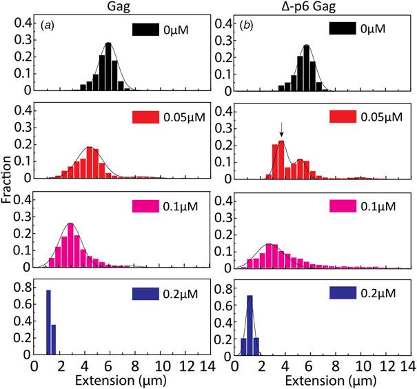
Quarterly Reviews of Biophysics                                                                                                                                                3

           colony into 25 ml LB media containing antibiotics and cultured at                          devices were fabricated using advanced nanofabrication described
           37 °C overnight with shaking at 200 rpm. The overnight culture                             elsewhere (Persson and Tegenfeldt, 2010). The channel system
           was used to inoculate 1 litre of LB media containing antibiotics                           consists of a pair of feeding channels (micro-size), spanned by a
           in a 2.5 litre glass flask. The culture was grown at 37 °C,                                set of parallel nanochannels. A schematic illustration of the nano-
           200 rpm, until an OD at 600 nm of approximately 0.5 was                                    fluidic chip is shown in Fig. 2a. The sample is loaded into the
           reached. Protein expression was induced with the addition of                               channel system from one of the four reservoirs that are connected
           0.2 mM IPTG, and the bacteria grown for a further 4 h at 37 °                              to the feeding channels and moved into the nanochannels by
           C. Bacteria were harvested by centrifugation (10 000 g; 4 °C;                              pressure-driven (N2) flow.
           15 min), and the pellet was stored at −80 °C. Bacterial pellets                                To avoid non-specific binding of protein to the negatively
           from the equivalent of 5 litres of culture were resuspended in                             charged channel walls, the channels were prior to the experiments
           40 ml of lysis buffer (50 mM TRIS-HCl pH = 8; 1 M NaCl;                                    coated with a lipid bilayer comprising 99% 1-palmitoyl-2-
           10 mM 2-mercaptoethanol; 25 mM imidazole; 1% Tween-20)                                     oleoyl-sn-glycero-3-phosphocholine (POPC, Avanti) and 1% N-
           supplemented with protease inhibitor cocktail and then sonicated.                          (fluorescein-5-thiocarbamoyl)-1,2-dihexadecanoyl-sn-Glycero-3-
           The suspension was supplemented with 500 units of Benzonase                                phosphoethanolamine, triethylammonium salt (fluorescein-
           Nuclease (Sigma-Aldrich, St. Louis, MO, USA; E1014) and incu-                              DHPE, Invitrogen). The coating procedure is described elsewhere
           bated for 30 min at 4 °C. DNA was sheared by repeated passage                              (Persson et al., 2012).
           through a 23-gauge needle. The lysate was centrifuged to remove                                The DNA and DNA–protein complexes were imaged using an
           insoluble material (27 000 g; 4 °C; 45 min). The clear supernatant                         epifluorescence microscope (Zeiss AxioObserver.Z1) equipped
           was filtered through a 0.45 µm syringe filter and loaded onto a                            with a Hamamatsu digital CMOS C11440-22CU camera, a 63×
           Nickel column (XK16) that had previously been equilibrated                                 oil immersion TIRF objective (NA = 1.46) and a 1.6× optovar
           with 50 mM TRIS-HCl pH = 8, 1 M NaCl, 10 mM 2-mercaptoe-                                   from Zeiss. Using the microscopy imaging software ZEN, 50 sub-
           thanol, 25 mM imidazole, 1% Tween-20 and 10% (v/v) glycerol.                               sequent images were recorded with an exposure time of 200 ms.
           The column was washed with equilibration buffer and bound pro-                             Data analysis was performed using a custom-written MATLAB-
           teins were eluted with a 0–1000 mM imidazole gradient in equil-                            based software. Microscopy image stacks were used as input to
           ibration buffer. Fractions containing Pr55Gag full-length or                               the program. Images were first binarized by thresholding with a
           Pr55Gag Δ-p6 were pooled and concentrated using a centrifugal                              global average plus onefold of standard deviation. Taking advan-
           Ultra-15, 30 000 molecular weight cut-off membrane (Millipore,                             tage of the high contrast of the YOYO-stained DNA fluorescence
           Burlington, MA, USA) and then desalted using a PD-10 column                                images, regions with higher brightness were directly considered as
           (GE Healthcare, Chicago, IL, USA). The protein was then incu-                              DNA objects without additional image filtering. Finally, the
           bated overnight at 4 °C with 1.2 kU of hexa-histidine-tagged                               lengths of the DNA molecules were extracted by identifying the
           TEV protease (Protean). To remove the cleaved His-tag, the                                 longest axis of the objects and the length was measured. In
           resulting mixture was passed over a HisTrap column at 1 ml/                                total, 50–100 DNA molecules were analysed for each sample con-
           min as above. The protein that did not bind was collected and                              centration. All the histograms are fit with Gaussian distributions.
           concentrated. The last step of purification consisted of a size
           exclusion chromatography using a Superdex 200 (high load 16/
                                                                                                      Results
           60) column previously equilibrated in 50 mM TRIS-HCl pH
           8.0, 1.0 M NaCl. Peak fractions from this column containing                                The goal of the study is to investigate how the binding of the NC
           the protein of interest were pooled and concentrated to 1–2 mg/                            protein, both in its isolated form and when inserted in its parent
           ml, snap frozen in liquid nitrogen and then stored at −80 °C.                              protein Gag, affects the physical properties of DNA. To do so, we
                                                                                                      mixed the proteins with pre-stained DNA (YOYO:bp ratio of
                                                                                                      1:50) at different ratios and observed individual complexes in
           Sample preparation
                                                                                                      nanochannels with a dimension of 100 nm × 150 nm. To scruti-
           DNA from phage T7 (T7-DNA, MABION, Konstantynów                                            nize the binding of the NC protein to ssDNA and dsDNA, bacter-
           Łódzki, Poland) or phage λ (λ-DNA, Roche, Basel, Switzerland)                              iophage T7 DNA (39 937 base pairs), which has blunt ends, and
           was pre-stained with YOYO-1 (Invitrogen, Waltham, MA, USA)                                 λ-DNA (48 502 base pairs), which has 12 bp-long ssDNA over-
           at a ratio of one dye molecule per 50 base pairs. This ratio min-                          hangs, were used as model DNAs in this study. In the following,
           imizes the effect of YOYO-1 on DNA conformation (Kundukad                                  the native nucleocapsid protein will be called NC(1–55) to distin-
           et al., 2013; Nyberg et al., 2013). Pre-stained DNA was then                               guish it unambiguously from its mutants.
           mixed with the wild-type or mutant Gag proteins or NC peptides
           and incubated at 4 °C for at least 2 h. The complexes were then
                                                                                                      Compaction and condensation of T7-DNA by NC(1–55)
           introduced into the nanofluidic system and equilibrated for 60 s
           before image capture. The DNA concentration was 5 µM (base-                                T7-DNA molecules at a concentration of 5 µM (base pairs) were
           pairs) in all samples. 3% (v/v) β-mercaptoethanol (Sigma-                                  incubated with different concentrations of NC(1–55) at 4 °C for
           Aldrich, St. Louis, MO, USA) was added as an oxygen scavenger                              at least 2 h. Figure 2b shows the mean extension of T7 DNA
           to suppress oxygen radical-induced photo-damage of the DNA.                                (L = 13.6 µm) along the longitudinal direction of the channel
           The buffer used was 25 mM Tris with 30 mM NaCl and                                         divided by the contour length (R||/L), as a function of NC(1–
           0.2 mM MgCl2 (pH 7.5).                                                                     55) concentration. With increasing NC(1–55) concentration, the
                                                                                                      extension of the DNA decreases. For over-threshold concentra-
                                                                                                      tions of NC(1–55), DNA is compacted into a condensed form,
           Nanofluidics
                                                                                                      where the single DNA molecules are simply bright fluorescent
           The single DNA molecule experiments were performed in nano-                                blobs that can be easily distinguished from the extended form.
           channels with a depth of 100 nm and a width of 150 nm. The                                 No condensation was observed in the feeding microchannels at

Downloaded from https://www.cambridge.org/core. IP address: 46.4.80.155, on 05 Nov 2021 at 16:20:05, subject to the Cambridge Core terms of use, available at https://www.cambridge.org/core/terms.
https://doi.org/10.1017/S0033583518000124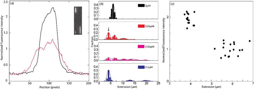
4 Kai Jiang et al.
Fig. 2. (a) Schematic illustration of the nanofluidic chip design (left). The channel system consists of pairs of microchannels, spanned by an array of straight nano-
channels, 500 µm long, 100 nm deep and 150 nm wide. The cartoon (right) shows DNA confined inside a nanochannel. DNA will be partially stretched along the
nanochannel, with an extension R||, shorter than its contour length L. (b) Relative extension R||/L of T7-DNA inside 100 nm × 150 nm channels plotted versus the NC
(1–55) concentration (bottom axis) and NC(1–55) to bp ratio (top axis). DNA concentration is 5 µM base pairs. The dashed line is drawn as an aid to the eye and the
arrow denotes the condensation threshold. The inset is a montage of fluorescence images of T7-DNA molecules at different NC(1–55) concentrations (from left to
right: 0, 0.1, 0.5 and 1 µM).
these concentrations, indicating that the condensation was facili- Beltz et al., 2005; Godet et al., 2006; Vo et al., 2009). Though
tated by the nanoconfinement inside the channels, which has the overhangs are rather short (12 nucleotides), they can accom-
been observed also for other DNA-condensing proteins (Zhang modate two NC(1–55) molecules, because it has been demon-
et al., 2013a; Jiang et al., 2015). At higher concentrations (2 µM strated on a very large number of sequences that the footprint
and above), condensation was observed also in the feeding of NC(1–55) is 5–7 nucleotides (De Guzman, 1998; Fisher
microchannels. et al., 1998; Vuilleumier et al., 1999; Amarasinghe et al., 2001;
Beltz et al., 2005; Avilov et al., 2009; Darlix et al., 2011).
If concatemers form as a result of the NC(1–55)-promoted
NC anneals short complementary ssDNA
annealing of complementary ssDNA overhangs, we expect
To further investigate the binding of NC(1–55) to ssDNA and that circular DNA molecules, resulting from intramolecular
dsDNA, we used λ-DNA, a linear dsDNA, 48.5 kilobase pairs annealing of λ-DNA molecules, may form as well (see sche-
long, where the 5′ -terminal ends protrude as self-complementary matic representation in Fig. 3c). Circular DNAs are character-
single-stranded chains, 12 nucleotides long, due to the circular ized by approximately half the extension and twice the
origin of λ-DNA. These single-stranded ends can anneal to gen- emission intensity compared with linear DNAs, since they are
erate circles or DNA concatemers (Sanger et al., 1982) (see sche- double-folded in the channels (Alizadehheidari et al., 2015;
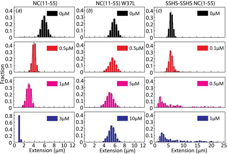
matic representation in Fig. 3c). The distribution in the extension Frykholm et al., 2015). Fig. 4a shows a linear λ-DNA (right
at different concentrations of NC(1–55) with T7-DNA and trace) together with a λ-DNA molecule with a shorter extension
λ-DNA is shown in Figs 3a and b, respectively. The larger exten- (∼60%) and an approximately twofold increased fluorescence inten-
sion R|| of λ-DNA without protein bound agrees well with its lon- sity (left trace), suggesting that it is a circular DNA molecule
ger contour length (L = 16 µm), and for both DNAs used, the (Alizadehheidari et al., 2015).
relative extension (R||/L) without protein is ca 35% of L. With It is difficult to distinguish circular DNA from compacted linear
increasing concentrations of NC(1–55), a decrease in the exten- DNA in Fig. 3. To promote the formation of circular DNA and limit
sion of single DNA molecule was observed also for λ-DNA, the number of concatemers, we decreased the overall DNA and pro-
but no complete condensation was observed even at 1 µM. For tein concentration. Samples at a lower λ-DNA concentration
over-threshold concentrations (>1 µM), condensed DNA aggre- (0.5 µM base pairs, ten times lower DNA concentration) with the
gates were observed in the microchannels (data not shown). same protein to DNA bp ratios as in Fig. 3 were therefore analysed
Interestingly, these aggregates are too large to enter the nanochan- (Fig. 4b). The peak at 5.5 µm, corresponding to single naked λ-DNA
nels, indicating that they consist of more than one DNA molecule. molecules, is split into two when NC(1–55) is added, one still at
A striking difference between λ-DNA and T7-DNA is that for 5.5 µm and one at approximately half that extension (see arrow).
λ-DNA, many DNA molecules have an extension that is much The scatterplot of intensity versus extension (Fig. 4c) shows two
longer than naked DNA. This indicates the formation of DNA clear clusters. The circular form has approximately double the emis-
concatemers in the presence of NC(1–55). With increasing NC sion intensity and half the extension of the linear form
(1–55) concentrations, more λ-DNA concatemers with longer (Alizadehheidari et al., 2015; Frykholm et al., 2015). This indicates
extension were observed (∼10% at 0.1 µM, ∼50% at 0.5 µM and that the fraction of molecules at half extension is due to the forma-
∼60% at 1 µM), but no concatemers were observed at any protein tion of circular DNA molecules. We also observe DNA concatemers
concentration with T7-DNA. Moreover, no DNA concatemers in the presence of NC(1–55) at this lower total concentration. The
were observed for λ-DNA in the absence of NC(1–55). This peaks for circular, linear and concatemers of λ-DNA are observed at
strongly indicates that the formation of concatemers is due to both 0.01 and 0.05 µM. It should be noted that at this lower DNA
the annealing of the λ-DNA single-stranded overhangs, promoted concentration, it is not possible to obtain data that fully represent
by the NC(1–55) protein (Fig. 3c), in full line with the well- the actual fractions of linear, circular and concatemer molecules,
described ability of NC(1–55) to chaperone the annealing of com- as was done in Figs 2 and 3. This is because the molecules are man-
plementary sequences (Beltz et al., 2004; Hargittai et al., 2004; ually selected before they are inserted into the channels, which
Downloaded from https://www.cambridge.org/core. IP address: 46.4.80.155, on 05 Nov 2021 at 16:20:05, subject to the Cambridge Core terms of use, available at https://www.cambridge.org/core/terms.
https://doi.org/10.1017/S0033583518000124Quarterly Reviews of Biophysics 5
Fig. 3. Extension distribution of (a) T7-DNA mole-
cules and (b) λ-DNA molecules at different concen-
trations of NC(1–55). DNA concentration is 5 µM
base pairs. (c) Sketch of the process for forming cir-
cular DNA and DNA concatemers by NC(1–55). NC
binds to the single-stranded ends of λ-DNA and
anneals the complementary ends to form conca-
temers or circles.
Fig. 4. (a) Intensity profile of circular (black) and linear (red) λ-DNA. Inset shows a fluorescence image of circular (left) and linear (right) λ-DNA in the presence of
0.1 µM NC(1–55) and 5 µM DNA bp. The scale bar is 1 µm. (b) Distribution in the extension of λ-DNA molecules with different concentrations of NC(1–55) at 0.5 µM
DNA bp concentration. The arrow (0.01 µM) highlights the emerging peak that is interpreted as circular DNA. (c) Scatterplot of normalized fluorescence intensity
versus DNA extension at 0.05 µM NC(1–55) and 0.5 µM DNA bp, highlighting a fraction of molecules with approximately double intensity and half extension.
might induce a bias. The important message from Fig. 4 is instead was observed with increasing concentrations of NC(11–55).
that we can identify a fraction of DNA molecules that are circular. However, the concentration needed for condensation was 3 µM,
which is ∼3 times higher than for NC(1–55), and agrees with the
lower binding constant for this mutant (Beltz et al., 2005).
Condensation and ssDNA annealing by NC(1–55) mutants
Interestingly, no concatemers or circularization of λ-DNA were
Figures 2–4 clearly demonstrate that nanofluidic channels can be observed in the presence of NC(11–55) (Fig. 5a), suggesting that
used to characterize two of the main functions of the NC protein, the first ten amino acid residues in NC(1–55) are involved in the
i.e. to condense and protect NAs and to promote the formation annealing of ssDNA and concatemer formation. It is also pos-
of their thermodynamically most favoured conformation. To sible that the balance between condensation and formation of
study the contribution of the different domains of NC(1–55) to concatemers is shifted when the first ten amino acids are
DNA condensation and formation of DNA concatemers, several removed, meaning that condensation happens before annealing
mutants were investigated. Since many studies have shown that can occur.
the N-terminal domain is a major factor in the NA binding and For the NC(11–55)W37L mutant, where the tryptophan in the
aggregation activity of NC(1–55) (Stoylov et al., 1997; Fisher second zinc finger is mutated to a leucine residue, a ∼10% decrease
et al., 1998; Vuilleumier et al., 1999; Bernacchi et al., 2002; in the DNA extension was observed at the highest protein concen-
Krishnamoorthy, 2003), we first investigated the DNA condensa- tration (1 µM). No DNA condensation (Fig. 5b) and no evidence
tion and concatemer formation properties of NC(11–55), a mutant for concatemer formation and hence annealing of ssDNA in the
where the N-terminal domain is deleted. Similar to T7-DNA with concentration interval studied were observed, in line with the
NC(1–55), compaction and eventually condensation of λ-DNA much lower binding constant of this mutant (Beltz et al., 2005).
Downloaded from https://www.cambridge.org/core. IP address: 46.4.80.155, on 05 Nov 2021 at 16:20:05, subject to the Cambridge Core terms of use, available at https://www.cambridge.org/core/terms.
https://doi.org/10.1017/S00335835180001246 Kai Jiang et al.
Fig. 5. Distribution in the extension of λ-DNA with
different concentrations of NC(11–55) (a), NC(11–55)
W37L (b) and SSHS-SSHS NC(1–55) (c). DNA concen-
tration is 5 µM base pairs.
Finally, we examined the role of the zinc fingers in DNA con- threshold concentrations, condensed DNA aggregates were
densation and concatemer formation. This mutant has lost its observed also in the microchannels for both Gag and Δ-p6 Gag.
affinity for zinc by replacing the cysteine residues with serines. A very small fraction of DNA formed concatemers for Gag
It should be noted that this mutant has a completely different sec- (Quarterly Reviews of Biophysics 7
Fig. 6. Distribution in the extension of λ-DNA with Gag (a) and Δ-p6 Gag
(b) at increasing protein concentrations. The arrow in (b) (0.05 µM) high-
lights the emerging peak that is interpreted as circular DNA. DNA
concentration is 5 µM base pairs.
2013a; Jiang et al., 2015; Malabirade et al., 2017; Guttula et al., SSHS-SSHS NC(1–55), which has been shown to be an efficient
2018). NA annealer, due to its flexible and highly positively charged
The condensation concentration varies between the different nature (Williams et al., 2001; Godet et al., 2012).
NC mutants and correlates directly with the binding constant The NC protein functions both in its mature form and as a
(Table 1). For example, deleting the first ten amino acids of NC part of the Gag protein in vivo. Both the full-length Gag and
that include several positively charged residues increases the the truncated Δ-p6 Gag condensed DNA at a much lower concen-
condensation concentration by >3-fold, in full line with the tration than NC(1–55). This higher condensation efficiency corre-
∼5-fold change in the reported binding constants (Beltz et al., lates well with their higher affinity as compared with NC(1–55)
2005). Furthermore, NC(11–55)W37L, where the the tryptophan (Cruceanu, 2006; Godet et al., 2012), which is attributed to the
in the second zinc finger is mutated to a leucine, does not con- participation of the matrix domain of Gag in the binding process
dense DNA at the concentrations investigated, in agreement (Alfadhli et al., 2011; Rein et al., 2011). Moreover, as the full-
with its ∼30 times lower affinity (Beltz et al., 2005; Darlix et al., length Gag and its truncated Δ-p6 Gag version show comparable
2011). A zinc-free mutant that is thought to be unfolded was DNA condensation, the p6 domain does not seem to play a key
also investigated. The slightly higher affinity of SSHS-SSHS NC role in the condensation process. In sharp contrast, the DNA
(1–55) compared with NC(1–55) is again in line with the literature annealing capacity is almost completely lost with the full-length
(Beltz et al., 2005) and can be explained by the unfolding of zinc Gag, but partly retained when the p6 region is deleted, suggesting
fingers that allows for additional electrostatic interactions with that the p6 region may hinder the annealing of ssDNA or favour
DNA. DNA condensation over DNA concatemer formation. This hin-
To probe the chaperone ability of NC, we investigated the abil- drance and thus, the lower activity of the NC domain in the full-
ity of different versions of NC to anneal the 12 base pairs ssDNA length Gag may be the result of the previously reported ability of
overhangs of λ-DNA. While long concatemers form efficiently the p6 domain to fold back and interact with the NC zinc fingers
with the wild-type NC(1–55), no concatemers were observed for (Wang et al., 2014). These results are in line with the tasks per-
T7 DNA that has blunt ends, clearly indicating that their forma- formed by the different proteins, where Gag is not involved in
tion results from the annealing of the complementary single- the chaperone activities that mature NC performs. Importantly,
stranded overhangs. Previous studies have shown that NC(1–55) this is the first study of full Gag on the single DNA molecule
can bind to both ssDNA and dsDNA, but there is a preference level and it highlights small but significant differences compared
for ssDNA over dsDNA (Mirambeau et al., 2006). This selectivity with Δ-p6 Gag that has been used in previous single molecule
for ssDNA can explain the formation of concatemers that is studies.
favoured over DNA compaction at low NC(1–55) concentration. It is important to relate the studies done here with other single
We did not observe any concatemers for the N-terminal dele- molecule studies of NC and Gag, in particular by the Williams
tion mutant NC(11–55). This could be due to the key role of group. The use of optical tweezers to stretch single DNA mole-
the N-terminal domain in the annealing process or to a shift in cules one by one gives unprecedented details on parameters like
the competition between the condensation and chaperone activi- binding affinity and how many base pairs each protein covers.
ties, so that the DNA condenses before annealing can occur. The chaperone activity of the protein can be characterized both
Concatemerization was also observed by the zinc-free mutant when it comes to destabilizing dsDNA and promoting the
Downloaded from https://www.cambridge.org/core. IP address: 46.4.80.155, on 05 Nov 2021 at 16:20:05, subject to the Cambridge Core terms of use, available at https://www.cambridge.org/core/terms.
https://doi.org/10.1017/S00335835180001248 Kai Jiang et al.
Table 1. Ability of NC and Gag and their mutants to form λ-DNA concatemers and concentration required to condense λ-DNA
Protein Concatemer Condensation conc. (μM) Binding constant (cTAR DNA) (μM−1)(Beltz et al., 2005)
NC(1–55) Y ∼1 1(±0.2) × 102
NC(11–55) N ∼3 1.7(±0.2) × 10 (NC12–55)
NC(11–55) W37L N N.A. 3.4(±1.0) × 10−1 (NC12–55)
NC(1–55) SSHS Y ∼0.5 >5 × 10²
NC(11–55) SSHS Y ∼3 N.A.
Gag Δ-p6 Y ∼0.2 N.A.
Gag Y ∼0.2 N.A.
formation of thermodynamically favoured structures. The nano- Acknowledgements. Dr. Rajhans Sharma is acknowledged for fruitful dis-
fluidic channels work at a lower force, where DNA is not cussions regarding the manuscript layout and for proof-reading the
stretched to its full length, which in turn means that the DNA manuscript.
is exposed to forces that are more like forces in the cell. The Financial support. This project is funded by grants from the Swedish
DNA extensions studied in this paper relate to forces below Research Council (no. 2015–5062) and the Olle Engqvist Byggmästare founda-
0.1 pN (Bustamante et al., 1994), which means that weak interac- tion to FW. This work was also supported by a grant from the Agence
tions easily interrupted in a tweezers setup are retained. We can Nationale de la Recherche (ANR blanc PICO2) to YM.
directly determine the balance between DNA condensation and
ssDNA annealing on long DNA molecules. We can also investi- Conflict of interest. None.
gate potential intermolecular DNA–DNA interactions since
many DNA molecules are present when the protein is added.
References
The nanofluidic channels also allow measurements at higher
throughput since tens of DNA molecules can be characterized Abd El-Wahab EW, Smyth RP, Mailler E, Bernacchi S, Vivet-Boudou V,
at the same time and hence hundreds of molecules can be inves- Hijnen M, Jossinet F, Mak J, Paillart J-C and Marquet R (2014)
tigated for each experimental condition. Specific recognition of the HIV-1 genomic RNA by the Gag precursor.
Finally, in addition to revealing biologically relevant informa- Nature Communications 5, 4304.
Alfadhli A, McNett H, Tsagli S, Bächinger HP, Peyton DH and Barklis E
tion on the interaction between NC and Gag with DNA, the
(2011) HIV-1 Matrix protein binding to RNA. Journal of Molecular
results obtained in this study highlight an important feature of Biology 410, 653–666.
the nanofluidic setup for studying DNA–protein interactions on Alizadehheidari M, Werner E, Noble C, Reiter-Schad M, Nyberg LK,
single DNA molecules. While many single DNA molecule meth- Fritzsche J, Mehlig B, Tegenfeldt JO, Ambjörnsson T, Persson F and
ods by definition involve only one molecule, we are able to study Westerlund F (2015) Nanoconfined circular and linear DNA: equilibrium
intermolecular DNA–DNA interactions between large DNA mol- conformations and unfolding kinetics. Macromolecules 48, 871–878.
ecules. This is demonstrated by the annealing of the ssDNA over- Amarasinghe GK, Zhou J, Miskimon M, Chancellor KJ, McDonald JA,
hangs by the NC protein that leads to concatemer formation. The Matthews AG, Miller RR, Rouse MD and Summers MF (2001)
same principles also mean that we can directly image processes on Stem-loop SL4 of the HIV-1 Ψ RNA packaging signal exhibits weak affinity
DNA ends and potentially also proteins that diffuse on DNA to for the nucleocapsid protein. Structural studies and implications for genome
recognition. Journal of Molecular Biology 314, 961–970.
find DNA ends. These two scenarios are possible to investigate
Avilov SV, Godet J, Piémont E and Mély Y (2009) Site-Specific characteriza-
since the studies are done on molecules freely suspended in solu- tion of HIV-1 nucleocapsid protein binding to oligonucleotides with two
tion and both ends of the DNA are free. binding sites. Biochemistry 48, 2422–2430.
To conclude, we introduce nanofluidic channels to investigate Bell NM and Lever AML (2013) HIV gag polyprotein: processing and early
the delicate balance between DNA condensation and chaperone viral particle assembly. Trends in Microbiology 21, 136–144.
activity of the NC protein, both in its mature form and as part Beltz H, Piémont E, Schaub E, Ficheux D, Roques B, Darlix JL and Mély Y
of Gag. The first ten amino acids are important for both the chap- (2004) Role of the structure of the top half of HIV-1 cTAR DNA on the
erone activity and DNA condensation of NC. In Gag, the conden- nucleic acid destabilizing activity of the nucleocapsid protein NCp7.
sation is more efficient than for NC alone, while the chaperone Journal of Molecular Biology 338, 711–723.
activity is almost completely lost. When deleting the p6 region Beltz H, Clauss C, Piémont E, Ficheux D, Gorelick RJ, Roques B, Gabus C,
Darlix J-L, de Rocquigny H and Mély Y (2005) Structural determinants of
of Gag, some of the chaperone activity is retained while the con-
HIV-1 nucleocapsid protein for cTAR DNA binding and destabilization,
densation is not affected. Our study is the first that directly com- and correlation with inhibition of self-primed DNA synthesis. Journal of
pares Gag and Δ-p6 Gag on the single DNA molecule level. Apart Molecular Biology 348, 1113–1126.
from revealing important information about the biophysics of NC Bernacchi S, Stoylov S, Piémont E, Ficheux D, Roques BP, Darlix JL and
and Gag when interacting with long DNA, our study also high- Mély Y (2002) HIV-1 nucleocapsid protein activates transient melting of
lights how nanofluidic channels can be used to study reactions least stable parts of the secondary structure of TAR and its complementary
on DNA ends, which is not possible with most competing single sequence. Journal of Molecular Biology 317, 385–399.
DNA molecule techniques. Bernacchi S, Abd El-Wahab EW, Dubois N, Hijnen M, Smyth RP, Mak J,
Marquet R and Paillart J-C (2017) HIV-1 Pr55 Gag binds genomic and spliced
Author ORCIDs. Fredrik Westerlund, 0000-0002-4767-4868 RNAs with different affinity and stoichiometry. RNA Biology 14, 90–103.
Downloaded from https://www.cambridge.org/core. IP address: 46.4.80.155, on 05 Nov 2021 at 16:20:05, subject to the Cambridge Core terms of use, available at https://www.cambridge.org/core/terms.
https://doi.org/10.1017/S0033583518000124Quarterly Reviews of Biophysics 9
Briggs JAG and Kräusslich H-G (2011) The molecular architecture of HIV. Godet J, Boudier C, Humbert N, Ivanyi-Nagy R, Darlix J-L and Mély Y
Journal of Molecular Biology 410, 491–500. (2012) Comparative nucleic acid chaperone properties of the nucleocapsid
Briggs JAG, Riches JD, Glass B, Bartonova V, Zanetti G and protein NCp7 and Tat protein of HIV-1. Virus Research 169, 349–360.
Krausslich H-G (2009) Structure and assembly of immature HIV. Guttula D, Liu F, van Kan JA, Arluison V and van der Maarel JRC (2018)
Proceedings of the National Academy of Sciences 106, 11090–11095. Effect of HU protein on the conformation and compaction of DNA in a
Bustamante C, Marko JF, Siggia ED and Smith S (1994) Entropic elasticity of nanochannel. Soft Matter 14, 2322–2328.
lambda-phage DNA. Science 265, 1599–1600. Hargittai MRS, Gorelick RJ, Rouzina I and Musier-Forsyth K (2004)
Campbell S and Rein A (1999) In vitro assembly properties of human immu- Mechanistic insights into the kinetics of HIV-1 nucleocapsid protein-
nodeficiency virus type 1 Gag protein lacking the p6 domain. Journal of facilitated tRNA annealing to the primer binding site. Journal of
Virology 73, 2270–2279. Molecular Biology 337, 951–968.
Cruceanu M (2006) Nucleic acid binding and chaperone properties of HIV-1 Heath MJ, Derebail SS, Gorelick RJ and DeStefano JJ (2003) Differing roles
Gag and nucleocapsid proteins. Nucleic Acids Research 34, 593–605. of the N- and C-terminal zinc fingers in human immunodeficiency virus
Cruceanu M, Gorelick RJ, Musier-Forsyth K, Rouzina I and Williams MC nucleocapsid protein-enhanced nucleic acid annealing. Journal of
(2006) Rapid kinetics of protein–nucleic acid interaction is a Major compo- Biological Chemistry 278, 30755–30763.
nent of HIV-1 nucleocapsid protein’s nucleic acid chaperone function. Jiang K, Zhang C, Guttula D, Liu F, van Kan JA, Lavelle C, Kubiak K,
Journal of Molecular Biology 363, 867–877. Malabirade A, Lapp A, Arluison V and van der Maarel JRC (2015)
Darlix J-L, Lapadat-Tapolsky M, de Rocquigny H and Roques BP (1995) Effects of Hfq on the conformation and compaction of DNA. Nucleic
First glimpses at structure-function relationships of the nucleocapsid pro- Acids Research 43, 4332–4341.
tein of retroviruses. Journal of Molecular Biology 254, 523–537. Jones CP, Datta SAK, Rein A, Rouzina I and Musier-Forsyth K (2011)
Darlix J, Godet J, Ivanyi-Nagy R, Fossé P, Mauffret O and Mély Y (2011) Matrix domain modulates HIV-1 gag’s nucleic acid chaperone activity via
Flexible nature and specific functions of the HIV-1 nucleocapsid protein. inositol phosphate binding. Journal of Virology 85, 1594–1603.
Journal of Molecular Biology 410, 565–581. Krishnamoorthy G, Roques B, Darlix JL and Mély Y (2003) DNA conden-
De Guzman RN (1998) Structure of the HIV-1 nucleocapsid protein bound to sation by the nucleocapsid protein of HIV-1: a mechanism ensuring DNA
the SL3 -RNA recognition element. Science 279, 384–388. protection. Nucleic Acids Research 31, 5425–5432.
El Meshri SE, Dujardin D, Godet J, Richert L, Boudier C, Darlix JL, Kundukad B, Cong P, Van Der Maarel JRC and Doyle PS (2013)
Didier P, Mély Y and de Rocquigny H (2015) Role of the nucleocapsid Time-dependent bending rigidity and helical twist of DNA by rearrange-
domain in HIV-1 gag oligomerization and trafficking to the plasma mem- ment of bound HU protein. Nucleic Acids Research 41, 8280–8288.
brane: a fluorescence lifetime imaging microscopy investigation. Journal of Levin JG, Guo J, Rouzina I and Musier‐Forsyth K (2005) Nucleic acid
Molecular Biology 427, 1480–1494. chaperone activity of HIV‐1 nucleocapsid protein: critical role in reverse
Fisher RJ, Rein A, Fivash M, Urbaneja MA, Casas-Finet JR, Medaglia M transcription and molecular mechanism. Progress in Nucleic acid Research
and Henderson LE (1998) Sequence-specific binding of human immuno- and Molecular Biology 80, 217–286.
deficiency virus type 1 nucleocapsid protein to short oligonucleotides. Levy SL and Craighead HG (2010) DNA manipulation, sorting, and mapping
Journal of virology 72, 1902–1909. in nanofluidic systems. Chemical Society Reviews 39, 1133.
Fornander LH, Frykholm K, Fritzsche J, Araya J, Nevin P, Werner E, Malabirade A, Jiang K, Kubiak K, Diaz-Mendoza A, Liu F, van Kan JA,
Çakır A, Persson F, Garcin EB, Beuning PJ, Mehlig B, Modesti M and Berret J-F, Arluison V and van der Maarel JRC (2017) Compaction and
Westerlund F (2016) Visualizing the nonhomogeneous structure of condensation of DNA mediated by the C-terminal domain of Hfq.
RAD51 filaments using nanofluidic channels. Langmuir 32, 8403–8412. Nucleic Acids Research 45, 7299–7308.
Freed EO (2015) HIV-1 assembly, release and maturation. Nature Reviews McKinstry WJ, Hijnen M, Tanwar HS, Sparrow LG, Nagarajan S, Pham ST
Microbiology 13, 484–496. and Mak J (2014) Expression and purification of soluble recombinant full
Frykholm K, Alizadehheidari M, Fritzsche J, Wigenius J, Modesti M, length HIV-1 Pr55Gag protein in Escherichia coli. Protein Expression and
Persson F and Westerlund F (2014) Probing physical properties of a Purification 100, 10–18.
DNA-protein complex using nanofluidic channels. Small 10, 884–887. Mirambeau G, Lyonnais S, Coulaud D, Hameau L, Lafosse S, Jeusset J,
Frykholm K, Nyberg LK, Lagerstedt E, Noble C, Fritzsche J, Karami N, Justome A, Delain E, Gorelick RJ and Le Cam E (2006) Transmission
Ambjörnsson T, Sandegren L and Westerlund F (2015) Fast size- electron microscopy reveals an optimal HIV-1 nucleocapsid aggregation
determination of intact bacterial plasmids using nanofluidic channels. with single-stranded nucleic acids and the mature HIV-1 nucleocapsid
Lab on a Chip 15, 2739–2743. protein. Journal of Molecular Biology 364, 496–511.
Frykholm K, Berntsson RP-A, Claesson M, de Battice L, Odegrip R, Nyberg L, Persson F, Åkerman B and Westerlund F (2013) Heterogeneous
Stenmark P and Westerlund F (2016) DNA compaction by the bacterio- staining: a tool for studies of how fluorescent dyes affect the physical prop-
phage protein Cox studied on the single DNA molecule level using nano- erties of DNA. Nucleic Acids Research 41, e184–e184.
fluidic channels. Nucleic Acids Research 44, 7219–7227. Persson F and Tegenfeldt JO (2010) DNA in nanochannels – directly visual-
Frykholm K, Nyberg LK and Westerlund F (2017) Exploring DNA–protein izing genomic information. Chemical Society Reviews 39, 985.
interactions on the single DNA molecule level using nanofluidic tools. Persson F, Fritzsche J, Mir KU, Modesti M, Westerlund F and
Integrative Biology 9, 650–661. Tegenfeldt JO (2012) Lipid-based passivation in nanofluidics. Nano
Ganser-Pornillos BK, Yeager M and Sundquist WI (2008) The structural Letters 12, 2260–2265.
biology of HIV assembly. Current Opinion in Structural Biology 18, 203–217. Rein A (2010) Nucleic acid chaperone activity of retroviral Gag proteins. RNA
Godet J and Mély Y (2010) Biophysical studies of the nucleic acid Biology 7, 700–705.
chaperone properties of the HIV-1 nucleocapsid protein. RNA Biology 7, Rein A, Henderson LE and Levin JG (1998) Nucleic-acid-chaperone activity
687–699. of retroviral nucleocapsid proteins: significance for viral replication. Trends
Godet J, De Rocquigny H, Raja C, Glasser N, Ficheux D, Darlix JL and in Biochemical Sciences 23, 297–301.
Mély Y (2006) During the early phase of HIV-1 DNA synthesis, nucleocap- Rein A, Datta SAK, Jones CP and Musier-Forsyth K (2011) Diverse interac-
sid protein directs hybridization of the TAR complementary sequences tions of retroviral Gag proteins with RNAs. Trends in Biochemical Sciences
via the ends of their double-stranded stem. Journal of Molecular Biology 36, 373–380.
356, 1180–1192. Reisner W, Pedersen JN and Austin RH (2012) DNA confinement in nano-
Godet J, Ramalanjaona N, Sharma KK, Richert L, de Rocquigny H, channels: physics and biological applications. Reports on Progress in Physics
Darlix J-L, Duportail G and Mély Y (2011) Specific implications of the 75, 106601.
HIV-1 nucleocapsid zinc fingers in the annealing of the primer binding Sanger F, Coulson AR, Hong GF, Hill DF and Petersen GB (1982)
site complementary sequences during the obligatory plus strand transfer. Nucleotide sequence of bacteriophage λ DNA. Journal of Molecular
Nucleic Acids Research 39, 6633–6645. Biology 162, 729–773.
Downloaded from https://www.cambridge.org/core. IP address: 46.4.80.155, on 05 Nov 2021 at 16:20:05, subject to the Cambridge Core terms of use, available at https://www.cambridge.org/core/terms.
https://doi.org/10.1017/S003358351800012410 Kai Jiang et al.
Shvadchak V, Sanglier S, Rocle S, Villa P, Haiech J, Hibert M, Van Webb JA, Jones CP, Parent LJ, Rouzina I and Musier-Forsyth K (2013)
Dorsselaer A, Mély Y and de Rocquigny H (2009) Identification by high Distinct binding interactions of HIV-1 Gag to Psi and non-Psi RNAs:
throughput screening of small compounds inhibiting the nucleic acid destabi- implications for viral genomic RNA packaging. RNA 19, 1078–1088.
lization activity of the HIV-1 nucleocapsid protein. Biochimie 91, 916–923. Williams MC, Rouzina I, Wenner JR, Gorelick RJ, Musier-Forsyth K and
Stoylov SP, Vuilleumier C, Stoylova E, De Rocquigny H, Roques BP, Bloomfield VA (2001) Mechanism for nucleic acid chaperone activity of
Gérard D and Mély Y (1997) Ordered aggregation of ribonucleic acids HIV-1 nucleocapsid protein revealed by single molecule stretching.
by the human immunodeficiency virus type 1 nucleocapsid protein. Proceedings of the National Academy of Sciences 98, 6121–6126.
Biopolymers 41, 301–312. Williams MC, Gorelick RJ and Musier-Forsyth K (2002) Specific
Tanwar HS, Khoo KK, Garvey M, Waddington L, Leis A, Hijnen M, zinc-finger architecture required for HIV-1 nucleocapsid protein’s nucleic
Velkov T, Dumsday GJ, McKinstry WJ and Mak J (2017) The thermody- acid chaperone function. Proceedings of the National Academy of Sciences
namics of Pr55Gag-RNA interaction regulate the assembly of HIV. PLoS 99, 8614–8619.
Pathogens 13, 1–24. Wu H, Mitra M, McCauley MJ, Thomas JA, Rouzina I, Musier-Forsyth K,
Tegenfeldt JO, Prinz C, Cao H, Chou S, Reisner WW, Riehn R, Wang YM, Williams MC and Gorelick RJ (2013) Aromatic residue mutations reveal
Cox EC, Sturm JC, Silberzan P and Austin RH (2004) The dynamics of direct correlation between HIV-1 nucleocapsid protein’s nucleic acid chap-
genomic-length DNA molecules in 100-nm channels. Proceedings of the erone activity and retroviral replication. Virus Research 171, 263–277.
National Academy of Sciences 101, 10979–10983. Wu H, Mitra M, Naufer MN, McCauley MJ, Gorelick RJ, Rouzina I,
van der Maarel JRC, Zhang C and van Kan JA (2014) A nanochannel Musier-Forsyth K and Williams MC (2014) Differential contribution of
platform for single DNA studies: from crowding, protein DNA interaction, basic residues to HIV-1 nucleocapsid protein’s nucleic acid chaperone func-
to sequencing of genomic information. Israel Journal of Chemistry 54, tion and retroviral replication. Nucleic Acids Research 42, 2525–2537.
1573–1588. Zhang C, Shao PG, van Kan JA and van der Maarel JRC (2009)
Vo M-N, Barany G, Rouzina I and Musier-Forsyth K (2006) Mechanistic Macromolecular crowding induced elongation and compaction of single
studies of mini-TAR RNA/DNA annealing in the absence and presence DNA molecules confined in a nanochannel. Proceedings of the National
of HIV-1 nucleocapsid protein. Journal of Molecular Biology 363, 244–261. Academy of Sciences 106, 16651–16656.
Vo M-N, Barany G, Rouzina I and Musier-Forsyth K (2009) HIV-1 nucle- Zhang C, Gong Z, Guttula D, Malar PP, van Kan JA, Doyle PS and van der
ocapsid protein switches the pathway of transactivation response element Maarel JRC (2012) Nanouidic compaction of DNA by like-charged protein.
RNA/DNA annealing from loop–loop ‘kissing’ to ‘zipper’. Journal of The Journal of Physical Chemistry B 116, 3031–3036.
Molecular Biology 386, 789–801. Zhang C, Guttula D, Liu F, Malar PP, Ng SY, Dai L, Doyle PS, van Kan JA
Vuilleumier C, Bombarda E, Morellet N, Gérard D, Roques BP and Mély Y and van der Maarel JRC (2013a) Effect of H-NS on the elongation
(1999) Nucleic acid sequence discrimination by the HIV-1 nucleocapsid and compaction of single DNA molecules in a nanospace. Soft Matter 9,
protein NCp7: a fluorescence study †. Biochemistry 38, 16816–16825. 9593.
Wang W, Naiyer N, Mitra M, Li J, Williams MC, Rouzina I, Gorelick RJ, Zhang C, Hernandez-Garcia A, Jiang K, Gong Z, Guttula D, Ng SY,
Wu Z and Musier-Forsyth K (2014) Distinct nucleic acid interaction prop- Malar PP, van Kan JA, Dai L, Doyle PS, Vries Rd and van der
erties of HIV-1 nucleocapsid protein precursor NCp15 explain reduced Maarel JRC (2013b) Amplified stretch of bottlebrush-coated DNA in nano-
viral infectivity. Nucleic Acids Research 42, 7145–7159. fluidic channels. Nucleic Acids Research 41, e189–e189.
Downloaded from https://www.cambridge.org/core. IP address: 46.4.80.155, on 05 Nov 2021 at 16:20:05, subject to the Cambridge Core terms of use, available at https://www.cambridge.org/core/terms.
https://doi.org/10.1017/S0033583518000124You can also read