Isolation and Characterization of Proteins from Chia Seeds (Salvia hispanica L.)
←
→
Page content transcription
If your browser does not render page correctly, please read the page content below
Article

                                                                                                                                          pubs.acs.org/JAFC

Isolation and Characterization of Proteins from Chia Seeds (Salvia
hispanica L.)
María R. Sandoval-Oliveros† and Octavio Paredes-López*,§
†
  Programa de Posgrado en Alimentos del Centro de la República (PROPAC), Universidad Autónoma de Querétaro, Cerro de las
 Campanas s/n, Santiago de Querétaro, Qro., Mexico CP 76010
§
  Centro de Investigación y de Estudios Avanzados del IPN, Unidad Irapuato, Km. 9.6 Libramiento Norte Carr, Irapuato-León,
 Irapuato, Gto., Mexico CP 36821

    ABSTRACT: Chia (Salvia hispanica L.) is a plant that produces seeds rich in some nutraceutical compounds with high protein
    content, but little is known about them; for this reason the aim of this study was to characterize the seed storage proteins. Protein
    fractions were extracted from chia seed flour. The main protein fraction corresponded to globulins (52%). Sedimentation
    coefficient studies showed that the globulin fraction contains mostly 11S and 7S proteins. The molecular sizes of all the reduced
    fractions were about 15−50 kDa. Electrophoretic experiments under native conditions exhibited four bands of globulins in the
    range of 104−628 kDa. The denaturation temperatures of crude albumins, globulins, prolamins, and glutelins were 103, 105,
    85.6, and 91 °C, respectively; albumins and globulins had relatively good thermal stability. Selected globulin peptides by mass
    spectrometry showed homology to sesame proteins. A good balance of essential amino acids was found in the seed flour and
    globulins, especially of methionine+cysteine.
    KEYWORDS: Salvia hispanica, chia seed, proteins, globulins

■    INTRODUCTION
Salvia hispanica L., with the popular name chia, is an annual
                                                                            utilized to elaborate coatings and edible films.14 Although chia
                                                                            is not a well-known food, its global production has increased in
plant of the Lamiaceae family that grows in arid or semiarid                recent years due to its healthy properties and new popularity.
climates. Chia seed is considered a pseudocereal and, because               Chia seeds are also used in the United States, Latin America,
of its high oil content, is also an oilseed native of Mesoamerica,          and Australia as nutritional supplements, as well as in the
exhibiting the greatest genetic diversity in the slope of the               manufacture of bars, breakfast cereals, and cookies.8
Pacific Ocean from central Mexico to northern Guatemala.1                       On the other hand, protein isolates from vegetal sources are
This oilseed and corn, beans, and amaranth were some of the                 of interest due to their increasing use as ingredients with
main crops for the pre-Columbian people.2 Mayans and Aztecs                 functional properties that can also improve the nutritive quality
used it as a medicine and food supplement for energy,                       of foods.15,16 The main proteins in seeds are storage proteins
endurance, and strength needed under extreme conditions.1,3                 accounting for about 60−80% of the total proteins;17 their
Chia has been cultivated in Mexico for thousands of years, and              analysis is greatly complicated by the polypeptides hetero-
recent evaluations have shown that seeds have a large potential             geneity and the different solubility behaviors. Some studies have
to be exploited; their consumption may bring remarkable                     reported that edible seeds generally contain two types of major
beneficial health effects.4 Seed composition appears very                     storage proteins that differ by size; the first group includes
attractive, being a good source of protein, with high amounts               proteins with sedimentation coefficients around 11S, which are
of natural antioxidants such as phenolic compounds including                referred to as “legumin-like” or 11S globulins, and the second
chlorogenic and caffeic acids, quercetin, and kaempferol, as well            group includes proteins with sedimentation coefficients around
as high dietary fiber content (>30% of the total weight).5−8 In              7S, which are classified as “vicilin-like” or 7S globulins. There is
recent years these seeds have become increasingly important                 also another type of proteins in a minor proportion, 2S-like
for nutrition because of their high content of unsaturated fatty            proteins.18−20 Therefore, it is not surprising that the cereal seed
acids; almost 60% is α-linolenic acid (omega-3).9,10 All of these           proteins have been a major topic of research for many years,
mentioned features may provide health benefits effective in                   with the aim of understanding their structures, control of
reducing cardiovascular diseases, obesity, regulation of intestinal         synthesis, and role in the grain utilization as well as their
transit, and cholesterol and triglycerides levels, as well as               functional and nutraceutical properties.17 Up to now the
prevention of diseases such as type II diabetes and some types              storage proteins of chia seed have not been fully characterized.
of cancer.6,7,10−12 On the other hand, protein content in chia              Thus, the aim of the present work was to fractionate and
seeds is higher than most of the traditionally utilized grains;             characterize these proteins, as well as to identify, isolate, and
they contain approximately 19−23%, which is higher than
wheat (14%), corn (14%), rice (8.5%), oats (15.3%), and barley              Received:    August 9, 2012
(9.2%).2,13 Chia seed not only is a nutrient supplying food but             Revised:     November 23, 2012
also has potential as a functional ingredient to be used as a               Accepted:    December 17, 2012
thickener in foods, and the mucilage from the seed has been                 Published:   December 17, 2012

                               © 2012 American Chemical Society       193                    dx.doi.org/10.1021/jf3034978 | J. Agric. Food Chem. 2013, 61, 193−201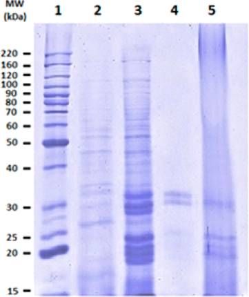
Journal of Agricultural and Food Chemistry                                                                                                                     Article

characterize the main protein fraction from molecular, thermal,                    precipitate this fraction with 20% of trichloroacetic acid (TCA). A total
and nutritional viewpoints with the aim of generating                              of 20 μg of each protein sample was loaded per lane, and approximate
information leading to a wider use of these macromolecules.                        molecular sizes of the proteins were determined by Invitrogen

■
                                                                                   molecular size standards. Electrophoresis was performed at a constant
                                                                                   current of 60 V per gel for approximately 2 h. The gels were stained
     MATERIALS AND METHODS                                                         with Coomassie brilliant blue R.
A single lot of chia seeds (S. hispanica L. var. Chionacalyx) harvested in            Thermal Characterization of Protein Fractions. Triplicate
November 2010 was provided by Ing. Roberto Nahum Amaya                             samples (5 mg of each protein isolate) were suspended in 15 μL of
Zamora, from Colima (Mexico). The chemicals were purchased from                    water and hydrated for 24 h prior to the test. A hermetic DSC pan was
Sigma-Aldrich (St. Louis, MO, USA) and the reagents used for                       used to encapsulate the samples of freeze-dried protein dispersed in
electrophoresis and staining solutions from Bio-Rad (Hercules, CA,                 deionized water. The denaturation temperature of protein fractions
USA). Molecular weight standards were purchased from Invitrogen                    was measured using a Q1000 differential scanning calorimeter (DSC)
(Mexico, DF, Mexico) and amino acid standards from Pierce                          (TA Instruments, New Castle, DE, USA). The sealed pan was placed
(Rockford, IL, USA). All chemicals used were of analytical grade,                  in a calorimeter previously calibrated with indium. The temperature
and deionized water was used.                                                      scan was carried out from 20 to 180 °C at 10 °C/min.15,27 Universal
   Chemical Analyses. Moisture, fat, protein (N × 6.25), ash, and                  Analysis 2010 Software (TA Instruments) was used to analyze the
total dietary fiber contents were determined using standard methods                 thermograms to determine the denaturation peak temperature (Td),
925.10, 920.85, 981.10, 923.03, and 985.29, respectively, reported by              denaturation temperature range (ΔTd), and denaturation enthalpy
AOAC;21 the total nitrogen content of the flour was analyzed using the              (ΔHd).
micro-Kjeldahl procedure to determine the crude protein content.                      Mass Spectrometric Analysis. Tandem Mass Spectrometry (LC/
   Sample Preparation. The seeds were soaked in water (ratio 1:10,                 ESI-MS/MS). To further characterize the isolated globulin proteins,
w/v) during 2 h. The seeds, which are coated with swollen mucilage,                peptide mass fingerprinting was performed for some bands with more
were frozen (−80 °C) overnight and freeze-dried, and the dry                       intensity and clarity, obtained in the SDS-PAGE. Bands from the
mucilage was removed mechanically.5 Mucilage-free seeds were milled                electrophoresis were carefully excised from the gel and washed
into a flour and passed through a 0.5 mm mesh to obtain a uniform                   successively with ultrapure water and 25 mM ammonium bicarbonate
particle size. The flour was defatted with hexane (ratio 1:10, w/v) in a            (NH4HCO3). Gel pieces were dehydrated with acetonitrile (ACN) to
Soxhlet unit at 65−70 °C and dried overnight under a hood at room                  remove contaminants and stain. Samples were reduced with 10 mM
temperature to remove the trace of remaining hexane; then a second                 dithiothreitol (DTT) in 25 mM NH4HCO3 followed by protein
grinding was performed to obtain a smaller particle size (0.18 mm),                alkylation with 55 mM iodoacetamide. The isolated proteins were
and afterward it was stored at 4 °C until use.6                                    digested with modified porcine trypsin (Promega, Madison, WI, USA)
   Protein Extraction and Fractionation Procedure. Fractiona-                      and extracted from the polyacrylamide gel. The pooled supernatants
tion of proteins was carried out according to the Osborne22                        were concentrated, and the peptides were desalted and concentrated
classification using a modification of the method reported by Barba                  to a final volume of 5 μL with Zip-Tip C18 (Millipore, Billerica, MA,
de la Rosa et al.23 All of the suspensions were stirred for 4 h at 4 °C            USA), according to the manufacturer’s protocol.28
and centrifuged at 14000g during 1 h at 4 °C; the first suspension was                 MS analysis was carried out on a 3200 Q TRAP hybrid tandem
flour/water (1:10, w/v), and the resulting supernatant was designated               mass spectrometer (Applied Biosystems/MDS Sciex, Concord, ON,
as crude albumin fraction. The pellet was resuspended in 10 mL of a                Canada), equipped with a nanoelectrospray ion source (NanoSpray II)
50 mM Tris-HCl, pH 8, buffer solution containing 0.5 M NaCl. After                  and a MicroIonSpray II head. The instrument was coupled online to a
centrifugation, the supernatant was separated, and it was referred to as           nano Acquity Ultra Performance LC system (Waters Corp., Milford,
globulin fraction; the pellet was resuspended in 10 mL of a 70%                    MA, USA). Mass calibration of the hybrid triple-quadrupole linear ion
aqueous isopropanol solution and extracted under constant stirring.                trap spectrometer was done with polypropylene glycol standard
The resulting supernatant was now the prolamin fraction, and the                   solutions. The instrument was then tuned and tested using [Glu1]-
pellet was resuspended in 10 mL of a 0.1 M Na2B4O7·10H2O, pH 10,                   fibrinopeptide B (Sigma-Aldrich). Peptides were separated on a BEH,
solution. After centrifugation, the supernatant was separated, the                 C18 UPLC column (1.7 μm, 75 μm × 100 mm, Waters Corp.)
glutelin fraction was obtained, and the pellet was the residue. The                equilibrated with 2% acetonitrile and 0.1% formic acid, using a linear
residue after extraction from each solvent was washed twice using a                gradient of 2−70% acetonitrile, and 0.1% formic acid over a 60 min
small portion of water. The washings and the first extract were                     period, at a flow rate of 0.25 μL/min. Spectra were acquired in
combined for each fraction. Fractions were dialyzed, freeze-dried, and             automated mode using information-dependent acquisition (IDA).
stored at 4 °C for further analysis.                                                  Precursor ions were selected in Q1 using the enhanced MS mode
   Protein Quantification. The protein content of the isolated                      (EMS) as a survey scan. The scan range for EMS was set at m/z 400−
fractions was assessed by micro-Kjeldahl, and the soluble protein                  1500 and 4000 amu/s, with an ion spray voltage of +2.2 kV applied to
content in each fraction was determined by the BCA (Pierce)                        a Picotip emitter FS150-20-10-N (New Objective, Woburn, MA,
method.21                                                                          USA). The interface heater for desolvation was held at 150 °C. The
   Sedimentation Coefficient Determination of Globulin                               precursor ions were fragmented by collisionally activated dissociation
Fraction. The globulin isolate fraction was layered onto a linear                  (CAD) in the Q2 collision cell. Collision voltages were automatically
sucrose density gradient (5−20% in a pH 8 buffer of 50 mM Tris-HCl                  adjusted on the basis of the ion charge state and mass using rolling
+ 0.3 M NaCl) and centrifuged at 218000g during 24 h at 4 °C                       collision energy. Generated fragments ions were captured and their
(Beckman, 15-65, SW 40 ti rotor). Fractions of 1 mL were collected,                masses analyzed in the Q3 linear ion trap.
and protein concentration was determined according to the BCA                         Database Search and Protein Identification. Data interpretation
method.24                                                                          and protein identification were performed with the MS/MS spectral
   Molecular Size Determination. Molecular size was determined                     data sets using the MASCOT search algorithm (version 1.6b9, Matrix
by sodium dodecyl sulfate−polyacrylamide gel electrophoresis (SDS-                 Science, London, UK; available at http://www.matrixscience.com).
PAGE) according to the Laemmli method.25 SDS-PAGE was carried                      Searches were conducted using the SwissProt database of the National
out on a slab gel (5% stacking gel, 12% separating gel), in an SDS−                Center for Biotechnology Information nonredundant database
Tris−glycine discontinuous buffer system. Also, for a better resolution             (NCBInr, http://www.ncbi.nih.gov, and http://genetics.bwh.harvard.
of proteins with low molecular weight, the method of Schagger and                  edu/msblast).
Von Jagow26 was used with polyacrylamide gradient gels of 5−13%.                      Amino Acid Analysis. Amino acid content was determined in
Proteins were prepared in native, reducing, and nonreducing                        triplicate using an RP-HPLC with precolumn derivatized phenyl-
conditions, in buffer solutions with or without 2-mercaptoethanol.                  isothiocyanate, according to previously published procedures.29,30 In
To improve the electrophoretic pattern of prolamins, we have to                    brief, the dried protein samples were hydrolyzed, in triplicate (1 mg

                                                                             194                      dx.doi.org/10.1021/jf3034978 | J. Agric. Food Chem. 2013, 61, 193−201
Journal of Agricultural and Food Chemistry Article
each), in constant-boiling 6 N HCl, and melted crystalline phenol was Table 2. Proportion of the Protein Fractions of Chia Seed
added for aromatic amino acid protection. Hydrolysis was performed
under vacuum in a heating block for 24 h at 110 °C. After cooling at sample g/100 g proteina
room temperature, the samples and mixture of amino acid standards albumins 17.3 ± 0.8
were derivatized by adding 20 μL of solution containing ethanol/ globulins 52.0 ± 1.0
water/triethylamine/phenylisothiocyanate (7:1:1:1, v/v) and incu- prolamins 12.7 ± 0.2
bated at room temperature for 20 min. The samples were dried in a
glutelins 14.5 ± 0.2
vacuum centrifuge, dissolved in 0.2 mL of 50 mM sodium phosphate
insoluble proteins 3.4 ± 0.6
buffer, pH 7.4, and filtered through a 0.22 μm filter, and then the
sample was subjected to reverse-phase chromatography. The phenyl-
a
Values are the mean ± SD of three determinations.
thiocarbamyl derivatives were detected by their absorbance at 254 nm.
After separation, the peaks were integrated and quantified using a
standard curve of peak areas previously obtained from known
concentrations of the amino acid standard mixtures.
Chemical Score. The chemical score (CS) was calculated as
m
CS = EAA × 100
mR
where mEAA is the mass (g) of the essential amino acid in the examined
protein and mR is the corresponding reference amino acid require-
ments. The FAO/WHO/UNU31 pattern of amino acid requirements
for the two extreme age groups (0.5−1 and >18 years) was used as a
reference to calculate amino acid scores and assess the quality of
dietary protein.
In Vitro Digestibility. The in vitro digestibility method used was a
modification of that of Hsu et al.32 Fifty milliliters of an aqueous
protein suspension (6.25 mg protein/mL) was prepared; these
solutions were adjusted to pH 8.0 with 0.1 N HCl or NaOH. On
the other hand, a multienzyme solution was also prepared (1.6 mg
trypsin with 15 units per mg powder; 3.1 mg chymotrypsin with 60
units per mg powder; and 1.3 mg peptidase with 40 units per g
powder/mL) which was adjusted to pH 8.0 and maintained in an ice
bath until use. Five milliliters of the multienzyme solution was added
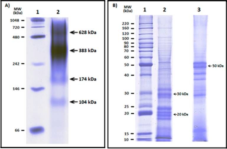
to the protein suspension with agitation, which was then incubated at Figure 1. Electrophoretic patterns of protein fractions from defatted
37 °C in a water bath with stirring during 10 min. A rapid decline in chia flour, in reduced conditions with the presence of mercaptoetha-
pH was produced at 10 min; for this reason the pH drop was recorded nol. Lanes: 1, molecular weight marker; 2, albumins; 3, globulins; 4,
in this period to estimate the in vitro digestibility. prolamins; 5, glutelins.
Statistics. All statistical analyses were performed using SigmaPlot
statistical software (version 11.0). All experiments were conducted at
least in triplicate, and data are expressed as the mean ± standard
deviation (SD).
■ RESULTS AND DISCUSSION
Proximate Analysis. The proximate composition and
dietary fiber of chia seed are summarized in Table 1. The
Table 1. Proximate Composition and Dietary Fiber of Chia
Seed
component amounta (g/100 g dry solids)
moisture 4.5 ± 0.0
lipids 32.5 ± 2.7
protein 22.7 ± 0.7 Figure 2. Sedimentation coefficient profile of globulins from chia
ash 3.7 ± 0.3 seeds: graph of protein concentration versus sedimentation coefficient.
dietary fiber
soluble 8.2 ± 0.8
insoluble 25.4 ± 2.2 (13.4%), wheat (12.6%), and soybean (15%). This confirms
total 33.5 ± 2.7 that chia is an outstanding source of dietary fiber as compared
carbohydrates (by difference) 3.1 to most known sources.34 On the other hand, the oil content
a
Values are the mean ± SD of three determinations. (32.5%) was higher than that of other oilseeds of commercial
importance, such as soybean (24%) and cotton seed (22−
values of all measured parameters are consistent with those 24%).9 The protein content was similar to that of lentil (23%)
reported by Ayerza and Coates2 and Reyes-Caudillo et al.33 The or chickpea (21%) and higher than that of chan (14%), of the
seeds contain low amounts of moisture (4.5%), minerals same family, and other oilseeds.35,36 Thus, chia is an important
(3.7%), and carbohydrates (3.1%), as well as a large amount of source of protein; this, together with the high content of oil rich
total dietary fiber (33.5%), which is superior to traditional in omega-3, makes the potential of this seed, for health and
sources of fiber such as flaxseeds (22.3%), barley (17.3%), corn nutrition, of a very remarkable level.
195 dx.doi.org/10.1021/jf3034978 | J. Agric. Food Chem. 2013, 61, 193−201Journal of Agricultural and Food Chemistry Article
Figure 3. Electrophoretic patterns of the globulin fraction from chia seeds: (A) native conditions (lane 1, molecular weight marker; lane 2, native
globulins); (B) SDS-PAGE of globulins in the presence and absence of mercaptoethanol (lane 1, molecular weight marker; lane 2, presence of
mercaptoethanol; lane 3, absence of mercaptoethanol).
Fractionation and Quantification of Chia Seed chia seed on sucrose density gradient revealed the presence of
Proteins. The total protein content of defatted flour of four protein fractions as shown in Figure 2, confirming that 11S
mucilage-free chia seeds increased to 35.5% as determined by globulin was a major component of this fraction. It also showed
Kjeldahl analysis (data not shown). Hereinafter we continued the presence of 7S-like proteins in a much lesser proportion,
working with defatted flour of mucilage-free seeds. After which is common in dicotyledon seeds. To our knowledge, this
protein extraction and fractionation by solubility, each fraction is the first time that chia seed globulins have been characterized
was quantified by micro-Kjeldahl and BCA method, and the by the sedimentation coefficient; thus, it is not possible to make
proportion obtained was 17.3% of crude albumins, 52% of comparisons with other studies on chia globulins. However, the
globulins, 12.7% of prolamins, and 14.5% of glutelins, whereas sedimentation pattern of chia seeds is somewhat similar to that
3.4% of the protein was not recovered (Table 2). This pattern observed in globulins of amaranth, sesame, barley, and some
of protein composition shows some similarity with other other seeds.39−41 The presence of the globulins 7S and 11S in
important seeds such as peas, lupins, and cotton.37 It is clear food ingredients may confer nutritional, physiological, and
that these seed proteins may vary according to the botanical functional characteristics to the foods that are dependent on
source, type of variety, preparation of the meal, extraction their structural sequence and conformation as well as on their
method, and other factors. However, the higher proportion physicochemical properties; for example, the 7S-like proteins in
found here for the chia globulin fraction is consistent with general exhibit emulsifying properties, the 11S globulins of
previous studies,6 which have reported levels of 64.8%. amaranth have peptides with antihypertensive activity, and the
Electrophoretic Pattern of Osborne Fractions. In the 11S-like proteins from various sources possess good gelling
electrophoretic analysis by SDS-PAGE (Figure 1) we found capacity; in other words, these proteins may act as ingredients
that the fractions of albumins and globulins showed a large providing favorable characteristics to food products.23,39
number of bands with a wide range of molecular sizes; however, Figure 2 also reveals the presence in a low proportion of
in the albumin fraction we did not observe bands with high proteins with unusual sedimentation coefficients such as 6S and
intensity, unlike the globulin fraction, which showed seven 19S; this may be because the subunits of 11S globulins
concentrated bands with increased presence having molecular sometimes form intermediate structures of high molecular
sizes between 18 and 35 kDa. As indicated before, to slightly weight.42 These proteins were also seen in the electrophoretic
improve the electrophoretic pattern of prolamins, it was pattern under native conditions in Figure 3A represented by the
necessary to precipitate the protein with TCA due to the low bands of 104 kDa (6S) and 628 kDa (19S); these last values of
resolution of this fraction; at the end, only three bands between the sediment constants may be due to different aggregation−
25 and 33 kDa were visible. On the other hand, the glutelin disaggregation phenomena during the preparation procedure of
fraction showed four bands with molecular sizes around 20−30 the globulins (i.e., temperature, pH, dialysis, lyophilization),
kDa with a certain similarity to globulins; this is consistent with and especially the pH is involved in the structural changes of
the classification of Fukushima,38 who included these two the globulins, producing association and dissociation of the
fractions in a single one, based on primary structur homology hexamer subunits of this protein fraction.18,43
criteria. Molecular Size Determination of Globulin Fraction.
Determination of Sedimentation Coefficient of Glob- The electrophoretic pattern of the globulin fraction in native
ulins. The sedimentation profile of the globulin fraction from conditions (Figure 3A) showed four bands with molecular sizes
196 dx.doi.org/10.1021/jf3034978 | J. Agric. Food Chem. 2013, 61, 193−201Journal of Agricultural and Food Chemistry Article
Figure 4. DSC thermographs of the four protein fractions of chia seed flour: (A) albumins; (B) globulins; (C) prolamins; (D) glutelins.
Table 3. Denaturation Temperature Range (ΔTd), between 50 and 60 kDa; these monomers under reducing
Denaturation Peak Temperature (Td), and Denaturation conditions with β-mercaptoethanol are resolved into acidic (30
Enthalpy (ΔHd) of Lyophilized Extract of the Protein kDa) and basic (20 kDa) subunits.23,39 These data fit with the
Fractions of Chia Seed results observed in the electrophoretic pattern of globulins in
Figure 3B, results that under reducing and nonreducing
fraction ΔTd (°C) Tda (°C) ΔHda (J/g)
conditions indicate that the globulin fraction contains disulfide
albumins 96.0−118.8 103.6 ± 0.7 12.6 ± 0.8 bonds in their structure, ensuring the abundant presence of 11S
globulins 94.3−116.6 104.7 ± 0.2 4.7 ± 0.9 protein.
prolamins 72.1−93.2 85.6 ± 0.6 2.3 ± 0.2 Thermal Characterization of Protein Fractions. The
glutelins 76.0−104.9 91.3 ± 0.8 6.2 ± 0.1 thermal properties of the protein fractions from chia seed flour
a
Values are the mean ± SD of three determinations. were analyzed by DSC; this is a valuable tool for assessing the
potential of protein isolates as functional ingredients in
between 104 and 628 kDa, of which the major band was that of different food systems requiring heat processing. Because the
383 kDa and represents the 11S protein, thereby confirming functional properties of proteins are greatly influenced by their
that the results obtained in the determination of sedimentation conformation and DSC is a technique highly sensitive to
coefficient and the molecular size are consistent with those conformational changes,44 we applied it to our protein
reported in the literature of approximately 300−400 kDa. This fractions, and the thermograms are shown in Figure 4. There
corresponds to the hexameric conformation typical of the 11S was only a single peak for each of the fractions; peaks for
proteins, which are resolved in denaturing conditions without albumins and prolamins appeared to have a better definition
β-mercaptoethanol into monomers with molecular sizes than those for the other fractions. Table 3 shows thermal
197 dx.doi.org/10.1021/jf3034978 | J. Agric. Food Chem. 2013, 61, 193−201Journal of Agricultural and Food Chemistry Article
the protein structure; the values for albumin, globulin,
prolamin, and glutelin were 12.6, 4.7, 2.3, and 6.2 J/g,
respectively. These results demonstrate how different ionic
strengths may be affecting the stability of chia proteins because
the thermal stability is mainly controlled by the balance of polar
and nonpolar residues in a protein structure, and a higher
content of nonpolar residues means greater thermal stabil-
ity. 24,27 The relatively low enthalpy values and high
denaturation temperatures found for chia proteins deserve
further studies. Due to the thermostability of proteins found in
chia seeds, they may be used in food systems undergoing high
heat treatments.
Mass Spectrometric Analysis. To identify peptides in
globulins of chia seed and to confirm the results obtained by
the analysis of sedimentation coefficient, we used liquid
chromatography electrospray ionization quadrupole time of-
flight tandem mass spectrometry analysis (LC-ESI-Q/TOF
MS/MS). Nine major protein bands from one-dimension
electrophoretic gel (Figure 5) were selected for further
identification by MS/MS. The results of this analysis are
shown in Table 4; this confirmed the presence of peptides
belonging to the 11S protein (G3, G4, G5, G6) and, in less
proportion, peptides of the 7S protein (G1, G2). Only three of
Figure 5. Electrophoretic pattern of globulin fraction from chia seeds
by the Shägger and von Jagow26 method in polyacrylamide gradient
nine analyzed peptides could not be identified by this method
(5−13%). Lanes: 1, molecular weight marker; 2, globulins in the (G7, G8, G9). Protein identification by studying homologous
presence of mercaptoethanol. sequences or comparison of their mass can be carried out on
the basis of the fact that many proteins from plants are highly
properties of the fractions. The onset and end temperatures conserved. Thus, proteins that share sequence similarity are
indicate that the protein starts to denature or unfold and likely to play the same function; for this reason, the databases
completely denatures, respectively, whereas the midpoint of the tend to identify homologous sequences in different species that
peak is considered as the Td, the denaturation temperature of may facilitate the identification and allocation of function. In
the protein; as mentioned before, this is clearer in the this study, all of the proteins that were identified showed
thermograms of albumins and prolamins (Figure 4). The homology to proteins of sesame (Sesamum indicum) with a low
denaturation temperatures of albumins and globulins were rate of coverage; however, this is common in the protein
similar, 103.6 and 104.7 °C, respectively. These denaturation identification of species for which the genome has not been
temperatures were relatively higher than those for several plant sequenced, and even in these cases is difficult to obtain
proteins, such as legumes and cereals, which are mostly lower homologies.20 The fact that all of our identified proteins have
than 100 °C;45 additionally, the Td of the four fractions from homology with sesame proteins leads us to the general belief
amaranth seeds are between 70 and 96 °C.24 The fact that in that the synthesis and behavior of the chia seed proteins may
the thermograms is observed only a single peak of denaturation have, like the sesame seeds proteins, beneficial effects such as
suggests the presence of a single proteinic species. Moreover, lowering blood pressure and improvement of cholesterol
due to the high denaturation temperatures, especially of the profiles.46
albumins and globulins, it is most likely that the conformation Amino Acid Analysis. The amino acid composition of
of the components of these fractions are stabilized by a number defatted flour showed that chia seeds are a good source of
of hydrophobic interactions, which are of endothermic nature sulfur, aspartic, and glutamic amino acids (Table 5). The profile
and therefore require a high amount of energy for of amino acids in chia seeds has been reported previously by
denaturation.24 On the other hand, the ΔH values give Ayerza and Coates,13 which is in general agreement with that of
information about the amount of energy required to denature this study. On the other hand, the composition of amino acids
Table 4. Globulin Proteins Identified by LC-MS/MS
band theor MWa/pIb protein identity organism peptides matched sequence coverage (%) score NCBI accession no.
G1 67027/7.55 7S globulin Sesamum indicum 5 4 71 gi|13183177
G2 67027/7.55 7S globulin Sesamum indicum 6 8 66 gi|13183177
G3 56553/8.57 11S globulin Sesamum indicum 4 5 110 gi|13183173
G4 56553/8.57 11S globulin Sesamum indicum 4 8 112 gi|13183173
G5 56553/8.57 11S globulin Sesamum indicum 6 10 139 gi|13183173
G6 56553/8.57 11S globulin Sesamum indicum 5 10 134 gi|13183173
G7 unidentified
G8 unidentified
G9 unidentified
a
MW, theoretical molecular weight. bpI, theoretical isoelectric point.
198 dx.doi.org/10.1021/jf3034978 | J. Agric. Food Chem. 2013, 61, 193−201Journal of Agricultural and Food Chemistry Article
Table 5. Amino Acid Composition of Chia Defatted Flour and of the Globulin (glob) Fraction and Contribution of Essential
Amino Acids with Respect to the Requirement Patterns for Two Age Groups
contribution (%) of essential amino acids
infants 0.5−1 year adults >18 years
amino acid content (mg/g raw protein) %CRc %CR
amino acid seed flour globulins b
RP seed flour glob RP seed flour glob
Asp 47.3 ± 0.9 72.9 ± 0.4
Glu 70.8 ± 1.1 243.0 ± 1.3
Ser 26.2 ± 0.3 69.3 ± 0.7
Gly 22.8 ± 0.7 73.6 ± 0.6
Arg 42.3 ± 0.4 94.2 ± 1.6
Ala 26.8 ± 0.3 39.4 ± 0.5
Pro 19.9 ± 0.7 106.4 ± 1.0
Hisa 13.7 ± 0.1 40.0 ± 0.6 20 69 200 15 91 267
Thra 18.0 ± 0.2 62.3 ± 0.7 31 58 201 23 78 271
Vala 28.5 ± 0.4 35.9 ± 0.6 43 66 83 39 73 92
Met+Cysa 27.8 ± 0.5 57.5 ± 0.4 28 99 205 22 126 261
Ilea 24.2 ± 0.4 30.1 ± 1.2 32 76 94 30 81 100
Leua 41.5 ± 0.6 44.4 ± 1.7 66 63 67 59 70 75
Phe+Tyra 38.8 ± 0.5 109.3 ± 0.8 52 75 210 38 102 288
Lysa 29.9 ± 0.5 15.4 ± 0.6 57 52 27 45 66 34
a
Essential amino acids. bRP, requirement patterns for the different age groups (mg/g raw protein). c%CR, coverage of requirement for that specific
essential amino acid in percentage.31
Table 6. In Vitro Digestibility of Flour and Globulin fraction isolation (Table 5). It is interesting to point out that
Fraction of Chia Seed the percentage of essential amino acids quantitated in Table 5 is
about 50% (46.5%), which is much higher than the
sample digestibilitya (%)
corresponding values reported for soybean (41.0%) and
globulins 82.5 ± 1.1 safflower (38.1%); this is an important aspect in favor of the
defatted flour 78.9 ± 0.7 quality of the chia seed proteins.47
casein 88.6 ± 1.1 In Vitro Digestibility. The results of in vitro digestibility
a
Values are the mean ± SD of three determinations. analysis are shown in Table 6. The in vitro digestibility of the
globulin fraction (82.5%) was slightly higher than that of the
defatted chia flour (78.9%), but slightly lower than that for
of the isolated globulin fraction has a high content of aromatic
casein (88.6%) used as control. The in vitro digestibility of
and sulfur amino acids as well as threonine and histidine; this
defatted chia flour showed a value similar to that previously
sample also exhibited a high percentage of glutamic and aspartic
reported for chia; these values are around 77.5%,6 and they are
acids, which is typical in seeds with an abundance of globulins.
also similar to those reported for Phaseolus vulgaris (77.5%) and
Low levels of lysine were observed in both samples. The
abundance of sulfur amino acids suggests that they may be higher than those for some cereals such as maize (66.6%), rice
intimately involved in maintaining the tertiary and quaternary (59.4%), sorghum (59.1%), and wheat (52.7%).48,49 There are
structure of the proteins, and the presence of high levels of no reports of antinutritive factors in chia, ruling out the
glutamic acid has been of interest in the food industry due to presence of protease inhibitors that could retard the in vitro
the potential of this amino acid to stimulate the central nervous digestibility.49 This digestibility value is a general indicator of
and immunologic systems in humans.47 The potential of the nutritional quality of proteins, and it may be associated with
aspartic acid rich foods in the hormonal regulation for the their special arrangement, because the tertiary and quaternary
proper functioning of the nervous system has been reported. In structures have different susceptibilities to proteolytic
general, the protein quality of chia has been demonstrated to be enzymes.50
higher than that of some cereals and oilseeds; this may In conclusion, chia seeds show high contents of proteins and
represent an important nutraceutical contribution to foods that fiber, particularly insoluble fiber and lipids. Globulins were by
contain chia seeds and isolated globulins as ingredients.18 far the major fraction with seven intense bands between 18 and
Chemical Score. In the case of the seed flour, the coverage 35 kDa, four of them with some similarity to those of glutelins.
of the amino acid requirement for infants was about 100% Ultracentrifugation experiments showed that globulins contain
satisfactory for the sulfur amino acids; the coverage for the 11S and 7S proteins as major and minor components.
remaining essential amino acids ranged from 52 to 76%. On the Electrophoretic studies under reducing and nonreducing
other hand, the required coverage was much better for adults; conditions confirmed the presence of 11S type of proteins.
the essential amino acids in seed flour varied from 66 to 126%. Thermal stability using DSC showed that albumins and
The globulin fraction exhibited ranges of coverage of globulins have denaturation temperatures above 100 °C, higher
requirements wider than those of seed flour, 27−210% for than those from other plant proteins; these protein fractions
infants and 34−288% for adults; the lowest values corre- may be suitable for certain food products undergoing high heat
sponded to lysine in view of the known limitations of cereals in treatment. Mass spectrometry analysis identified four major
this amino acid and its partial destruction during the protein globulin peptides as belonging to 11S type of proteins and two
199 dx.doi.org/10.1021/jf3034978 | J. Agric. Food Chem. 2013, 61, 193−201Journal of Agricultural and Food Chemistry Article
of them as 7S. The essential amino acids of both seed flour and (8) Martínez, L.; Marín, M.; Salgado, C.; Revol, J.; Penci, M.; Ribotta,
globulin exhibited in general a relatively good balance of them, P. Chia (Salvia hispanica L.) oil extraction: study of processing
especially Met+Cys; globulins are a good source of aromatic parameters. Food Sci. Technol. 2012, 47, 78−82.
amino acids. The in vitro digestibility of seed flour and (9) Ayerza, R. Oil content and fatty acid composition of oil of chia
(Salvia hispanica L.) from five locations in northwestern Argentina. J.
globulins reached better values than those reported for most
Am. Oil Chem. Soc. 1995, 72, 1079−1081.
cereals. In brief, our results support the potential use of chia (10) Jin, F.; Nieman, D. C.; Sha, W.; Guoxiang Xie, G.; Qiu, Y.; Jia,
seeds as a good source of proteins, including fractions with W. Supplementation of milled chia seeds increases plasma ALA and
remarkable thermal stability, and other important nutritious EPA in postmenopausal women. Plant Foods Hum. Nutr. 2012, 67,
and nutraceutical components. 105−110.
■
(11) Bemelmans, W.; Broer, J.; Fesfens, E.; Smit, A.; Muskiet, F.;
AUTHOR INFORMATION Lefrandt, J.; Bom, V.; May, V.; Meyboom-de Jong, B. Effect of an
increased intake of α-linolenic acid and group nutritional education on
Corresponding Author cardiovascular risk factor: the Mediterranean α-linolenic enriched
*Phone: 01 (462) 6239674. E-mail:oparedes@ira.cinvestav.mx. Groningen dietary intervention study. Am. J. Clin. Nutr. 2002, 75,
221−227.
Funding
(12) Poudyal, H.; Panchal, S.; Waanders, J.; Ward, L.; Brown, L.
We thank the Consejo Nacional de Ciencia y Tecnologı ́a- Lipid redistribution by α-linolenic acid-rich chia seed inhibits stearoyl-
México for partial funding of this study and the scholarship to CoA desaturase-1 and induces cardiac and hepatic protection in diet
M.R.S.-O. induced obese rats. J. Nutr. Biochem. 2012, 23, 153−162.
Notes (13) Ayerza, R.; Coates, W. Protein content, oil content and fatty
acid profiles as potential criteria to determine the origin of
The authors declare no competing financial interest.
■
commercially grown chia (Salvia hispanica L.). Ind. Crops Prod.
2011, 34, 1366−1371.
ACKNOWLEDGMENTS (14) Muñoz, L. A.; Cobos, A.; Diaz, O.; Aguilera, J. M. Chia seeds:
microstructure, mucilage extraction and hydration. Int. J. Food Eng.
We thank with pleasure Dr. Guillermo Mendoza-Hernández
2012, 108, 216−224.
(FM, UNAM), Ing. Francisco Garcı ́a Suarez (Ceprobi, IPN), (15) Paredes-López, O.; Ordorica-Falomir, C.; Guevara-Lara, F.;
Dr. Luis E. González de la Vara, and Dra. Marı ́a Elena Valverde Covarrubias, M. M. Las proteinas ́ vegetales: presente y futuro de la
(Cinvestav, Irapuato) for very valuable technical advice and for alimentación. In Perspectiva de la Biotecnologiá en México; Fundación
many critiques and helpful suggestions throughout this work. Javier Barrios Sierra. A.C. CONACYT. R.: Quintero, Mexico, 1985;
■
pp331−350.
ABBREVIATIONS USED (16) Segura-Nieto, M.; Barba de la Rosa, A. P.; Paredes-López, O.
Biochemistry of amaranth proteins. In Amaranth − Biology, Chemistry
BCA, bicinchoninic acid; CAD, collisionally activated dissoci- and Technology; Paredes-López, O., Ed.; CRC Press: Boca Raton, FL,
ation; CS, chemical score; DSC, differential scanning 1999; pp76−95.
calorimeter; DTT, dithiothreitol; EMS, enhanced mass (17) Shewry, P. R. Cereal seed storage proteins: structures, properties
spectrometry; ESI, electrospray ionization; IDA, information- and role in grain utilization. J. Exp. Bot. 2002, 53, 947−958.
dependent acquisition; LC, liquid chromatography; ME, β- (18) Derbyshire, E.; Wright, D. J.; Roulter, D. Legumin and vicilin,
mercaptoethanol; MS, mass spectrometry; MW, molecular storage proteins of legume seeds. Phytochemistry 1976, 15, 3−24.
weight; SD, standard deviation; TCA, trichloroacetic acid (19) Shewry, P. R. Plant storage proteins. Biol. Rev. 1995, 70, 375−
■
426.
(20) Nadal, P.; Canela, P.; Katakis, I.; O’Sullivan, K. Extraction,
REFERENCES isolation, and characterization of globulin proteins from Lupinus albus.
(1) Cahill, J. P. Genetic diversity among varieties of chia (Salvia J. Agric. Food Chem. 2011, 59, 2752−2758.
hispanica L.). Genet. Res. Crop. Evol. 2004, 51, 773−781. (21) Association of Official Analytical Chemists (AOAC). Official
(2) Ayerza, R.; Coates, W. Ground chia seed and chia oil effects on Methods of Analysis, 15th ed.; Washington, DC, 1990.
plasma lipids and fatty acids in the rat. Nutr. Res. (N.Y.) 2005, 25, (22) Osborne, T. B. The Vegetable Proteins; Longmans: Green, NY,
995−1003. 1924.
(3) Bueno, M.; Di Sapio, O.; Barolo, M.; Busilacchi, H.; Quiroga, M.; (23) Barba de la Rosa, A. P.; Herrera, A.; Utsumi, S.; Paredes-López,
Severin, C. Análisis de la calidad de los fruto de Salvia hispanica L. O. Molecular characterization, cloning and structural analysis of a
(Lamiaceae) comercializados en la ciudad de Rosario (Santa Fe, cDNA encoding an amaranth globulin. J. Plant Physiol. 1996, 149,
Argentina). Bol. Latinoam. Caribe Plantas Med. Aromaticas 2010, 9, 527−532.
221−227. (24) Chen, S.; Paredes-López, O. Isolation and characterization of
(4) Weber, C. W.; Gentry, H. S.; Kohlhepp, E. A.; McCrohan, P. R. the 11S globulin from amaranth seeds. J. Food Biochem 1997, 22, 53−
The nutritional and chemical evaluation of chia seeds. Ecol. Food Nutr. 65.
1991, 26, 119−125. (25) Laemmli, U. K. Cleavage of structural proteins during the
(5) Olivos-Lugo, B. L.; Valdivia-López, M.Á .; Tecante, A. Thermal assembly of the head of bacteriophage T4. Nature 1970, 217, 680−
and physicochemical porpieties and nutritional value of the protein 685.
fraction of Mexican chia seed (Salvia hispanica L.). Food Sci. Technol. (26) Shägger; Von Jagow, G. Tricin-sodium dodecyl sulfate-
Int. 2010, 1, 1−8. polyacrylamide gel electrophoresis for separation of protein in the
(6) Vázquez-Ovando, A.; Rosado-Rubio, G.; Chel-Guerrero, L.; range from 1 to 100 kDa. Anal. Biochem. 1987, 166, 368−379.
Betancur-Ancona, D. Dry processing of chia (Salvia hispanica L.) flour: (27) Bello-Perez, L. A.; Agama-Acevedo, E.; Sánchez-Hernández, L.;
chemical characterization of fiber and protein. J. Food 2010, 8, 117− Paredes-López, O. Isolation and partial characterization of banana
127. starches. J. Agric. Food Chem. 1999, 47, 854−857.
(7) Ixtania, V.; Martínez, M.; Sportorno, V.; Mateo, M.; Maestri, D.; (28) Bienvenut, W. V.; Sanchez, J. C.; Karmime, A.; Rougue, V.;
Diehl, B.; Nolasco, S.; Tomás, M. Characterization of chia seed oils by Rose, K.; Binz, P. A.; Hochstrasser, D. F. Toward a clinical molecular
pressing and solvent extraction. J. Food Compos. Anal. 2011, 24, 166− scanner for proteome research: parallel protein chemical processing
174. before and during western blot. Anal. Chem. 1999, 71, 4800−4807.
200 dx.doi.org/10.1021/jf3034978 | J. Agric. Food Chem. 2013, 61, 193−201Journal of Agricultural and Food Chemistry Article
(29) Bidlingmeyer, B. A.; Cohen, S. A.; Tarvin, T. L. Rapid analysis of absorption and fluorescence techniques. Int. J. Pept. Protein Res. 1990,
amino acids using pre-column derivatization. J. Chromatogr. 1984, 336, 35, 25−34.
93−104.
(30) Hendrikson, R. L.; Meridith, S. C. Amino acid analysis by
reverse-phase high performance liquid chromatography: precolumn
derivatization with phenylisothiocyanate. Anal. Biochem. 1984, 136,
65−74.
(31) FAO/WHO/UNU. Protein and amino acids requirements in
human nutrition. WHO Tech. Report Ser. 2008, No. 935, 135−183,
247−248.
(32) Hsu, H.; Vavak, D.; Satterlee, L.; Miller, G. A. Multienzime
technique for estimating protein digestibility. J. Food Sci. 1977, 42,
1269−1279.
(33) Reyes-Caudillo, E.; Tecante, A.; Valdivia-López, M. A. Dietary
fiber content and antioxidant activity of phenolic compounds present
in Mexican chia (Salvia hispanica L.) seeds. Food Chem. 2008, 107,
656−663.
(34) Devinder, D.; Mona, M.; Hradesh, R.; Patil, R. Dietary fiber in
foods: a review. J. Food Sci. Technol. 2011, 49, 63−69.
(35) Mossé, J. Nitrogen to protein conversion factor for ten cereals
and six legumes or oilseeds. A reappraisal of its definition and
determination. Variation according to species and to seed protein
content. J. Agric. Food Chem. 1990, 38, 18−24.
(36) Aguirre, C.; Torres, I.; Mendoza-Hernández, G.; García-Gasca,
T.; Blanco-Labra, A. Analysis of protein fractions and some minerals
present in chan (Hypstys suaveolens L.) seeds. J. Food Sci. 2011, 71,
15−19.
(37) Nikokyris, P. N.; Kandylis, K. Feed protein fractions in various
solvents of ruminant feedstuffs. J. Agric. Food Chem. 1997, 75, 198−
204.
(38) Fukushima, D. Structures of plant storage proteins and their
function. Food Rev. Int. 1991, 7, 353−381.
(39) Plietz, P.; Damaschun, G.; Zirwer, D.; Gast, K.; Schwenke, K.
D.; Prakash, V. Shape and quaternary structure of α-globulin from
sesame (Sesanum indicum L.) seeds as revealed by small angle X-ray
scattering and quasi-elastic light scattering. J. Biol. Chem. 1986, 261,
12686−12691.
(40) Casey, R.; Domoney, C.; Smith, A. M. Biochemistry and
molecular biology of seed products. In Peas: Genetics, Molecular Biology
and Biotechnology; Casey, R., Davies, D. R., Eds.; CAB International:
Wallingford, UK, 1993; pp 121−163.
(41) Romero-Zepeda, H.; Paredes-López, O. Isolation and character-
ization of amarantin, the 11S amaranth seed globulin. J. Food Biochem.
1996, 19, 329−339.
(42) Mori, T.; Utsumi, S. Purification and properties of storage
proteins of broad bean. J. Agric. Food Chem. 1979, 43, 577−583.
(43) Brooks, J. R.; Morr, C. V. Current aspects of soy protein
fractionation and nomenclature. J. Am. Oil Chem. Soc 1985, 62, 1347−
1353.
(44) Harwalkar, V. R.; Ma, C. Study of thermal properties of oat
globulin by differential scanning calorimetry. J. Food Sci. 1987, 52,
394−398.
(45) Scilingo, A. A.; Añon, M. C. Calorimetric study of soybeans
protein isolates: effect of calcium and thermal treatments. J. Agric. Food
Chem. 1996, 44, 3751−3756.
(46) Cheung, S. C. M.; Szeto, Y. T.; Benzie, I. F. F. Antioxidant
protection of edible oils. Plant Foods Hum. Nutr. 2007, 62, 39−42.
(47) Paredes-Lopez, O. Safflower proteins for food use. In
Development in Food Proteins, 7 ed.; Hudson, B. J. F., Ed.; Elsevier
Science: London, UK, 1991; pp 1−33.
(48) Betancur-Ancona, D.; Gallegos-Tintoré, S.; Chel-Gerrero, L.
Wet-fractionation of Phaseolus lunatus seeds: partial characterization of
starch and protein. J. Sci. Food Agric. 2004, 84, 1193−1201.
(49) Siddhuraju, P.; Becker, K. Effect of various domestic processing
methods on anti-nutrients and in vitro protein and starch digestibility
of two indigenous varieties of Indian tribal pulse, Mucuna pruriens val.
utilis. J. Agric. Food Chem. 2001, 49, 3058−3067.
(50) Deshpande, S. S.; Damodaran, S. Conformational characteristics
of legume 7S globulins as revealed by circular dichroic, derivative U.V.
201 dx.doi.org/10.1021/jf3034978 | J. Agric. Food Chem. 2013, 61, 193−201You can also read