Size conservation emerges spontaneously in biomolecular condensates formed by scaffolds and surfactant clients - Nature
←
→
Page content transcription
If your browser does not render page correctly, please read the page content below
www.nature.com/scientificreports
OPEN Size conservation emerges
spontaneously in biomolecular
condensates formed by scaffolds
and surfactant clients
Ignacio Sanchez‑Burgos1,2, Jerelle A. Joseph1,2,3, Rosana Collepardo‑Guevara1,2,3 &
Jorge R. Espinosa1,2*
Biomolecular condensates are liquid-like membraneless compartments that contribute to
the spatiotemporal organization of proteins, RNA, and other biomolecules inside cells. Some
membraneless compartments, such as nucleoli, are dispersed as different condensates that do not
grow beyond a certain size, or do not present coalescence over time. In this work, using a minimal
protein model, we show that phase separation of binary mixtures of scaffolds and low-valency clients
that can act as surfactants—i.e., that significantly reduce the droplet surface tension—can yield either
a single drop or multiple droplets that conserve their sizes on long timescales (herein ‘multidroplet
size-conserved’ scenario’), depending on the scaffold to client ratio. Our simulations demonstrate
that protein connectivity and condensate surface tension regulate the balance between these two
scenarios. The multidroplet size-conserved scenario spontaneously arises at increasing surfactant-to-
scaffold concentrations, when the interfacial penalty for creating small liquid droplets is sufficiently
reduced by the surfactant proteins that are preferentially located at the interface. In contrast, low
surfactant-to-scaffold concentrations enable continuous growth and fusion of droplets without
restrictions. Overall, our work proposes one thermodynamic mechanism to help rationalize how
size-conserved coexisting condensates can persist inside cells—shedding light on the roles of protein
connectivity, binding affinity, and droplet composition in this process.
To fulfill their biological functions, cells must organize their contents into different compartments. One way of
achieving spatiotemporal organization is via the formation of membrane-bound organelles. Another, that does
not rely on membranes, is the self-assembly of proteins and nucleic acids into biomolecular condensates through
the process of liquid–liquid phase separation (LLPS)1–3. In LLPS, a protein solution demixes into a protein-rich
liquid phase—the condensate—in coexistence with a protein-poor liquid matrix. Condensates are ubiquitous
within cells, with some of the most famous examples including stress g ranules4, P granules5, nuclear s peckles6,
and the nucleolus7.
The interest in understanding the molecular grammar8, 9 and biophysical determinants of cellular LLPS has
significantly increased in recent years due, in part, to the realization that condensates take part in a wide-range
of cellular functions—including genome silencing10, 11, signaling12, buffering cellular noise13, and formation
of super-enhancers14, among many o thers15, 16. Furthermore, neurodegenerative diseases such as Alzheimer’s,
Parkinson’s or ALS have been associated with aberrant or misregulated phase transitions17–19.
The chief drivers of biomolecular LLPS are multivalent molecules (i.e., those possessing multiple interaction
sites) known as ‘scaffolds’20. Scaffolds are defined as the molecules—e.g., a single type of protein, a combination
of various cognate proteins, or mixtures of proteins and nucleic a cids21—that are able to sustain LLPS on their
own. Above a critical concentration, scaffolds establish a sufficiently high number of transient attractive scaf-
fold–scaffold interactions per unit of volume (the ‘liquid-network connectivity’22) to compensate for the entropic
loss upon demixing. In addition, scaffolds can establish interactions with biomolecules that are unable to phase
separate on their own, known as ‘clients’, recruiting them to the condensates via scaffold–client interactions. The
1
Maxwell Centre, Cavendish Laboratory, Department of Physics, University of Cambridge, J J Thomson Avenue,
Cambridge CB3 0HE, UK. 2Yusuf Hamied Department of Chemistry, University of Cambridge, Lensfield Road,
Cambridge CB2 1EW, UK. 3Department of Genetics, University of Cambridge, Downing Site, Cambridge CB2 3EH,
UK. *email: jr752@cam.ac.uk
Scientific Reports | (2021) 11:15241 | https://doi.org/10.1038/s41598-021-94309-y 1
Vol.:(0123456789)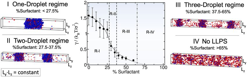
www.nature.com/scientificreports/
recruitment of clients can alter significantly the stability and structural properties of c ondensates22–24. There-
fore, together, scaffolds and clients shape the biophysical properties of the biomolecular condensates that play
fundamental roles within the c ell25–28.
A striking observation is the presence of condensates inside cells that do not grow beyond a certain s ize7, 29–31.
Basic thermodynamics suggest that over time, LLPS should result in the formation of a single large condensate
rather than multiple coexisting small droplets32. The latter case is disfavored because, cummulatively, it yields
a high surface area to volume ratio (i.e., high interfacial free energy penalty), while the former ensures that the
interfacial free energy penalty is minimized. Despite of the thermodynamic preference for single condensates
over multidroplet systems, both types of architectures are present within cells. Indeed, single large condensates
have been observed26, 33, but also diverse coexisting size-restricted droplets have been reported in different in vivo
systems, such as in the amphibian oocyte nucleolus7, in the Saccharomyces cerevisiae cytoplasmic processing
bodies34 and elsewhere35–37. Additionally, size-conserved multidroplet architectures have been found in non-
coalescing ribonucleoprotein c ondensates26 and in multiphase complex coacervates in vitro38. However, the
underlying molecular mechanisms and biophysical driving forces behind the formation of multiple coexisting
droplets, also known as e mulsification39, require further investigation.
Several explanations for why or how emulsions can be thermodynamically stable at biological relevant time-
scales are currently under d ebate40. One possible mechanism is that the presence of active ATP-dependent
processes might conveniently regulate the conditions where droplets grow and c oalesce41–44. Other studies sug-
gest that proteins with various highly distinct interacting domains may form micelle-like condensates31, 45, 46. In
multicomponent mixtures, another possibility could be that specific binding proteins act as powerful surfactants
and, thus, reduce the droplet surface tension penalty leading to multicondensate c oexistence35, 36, 38, 47. Moreover,
a recent alternative explanation suggests that the interplay between protein diffusion and saturation of protein
binding sites can also induce size-conservation in c ondensates29, 30. It is plausible that all these different mecha-
nisms contribute to the size conservation of condensates inside cells under different conditions.
In this work, we use a minimal protein model, which recapitulates the experimentally-observed relationship
between protein valency and critical p arameters23, 48, 49, to investigate the regulation of droplet size in binary
mixtures of multivalent proteins (scaffolds and clients). We show that liquid–liquid phase separation of scaffolds
and clients mixtures, where clients act as surfactants, can give rise to single droplets or multiple size-conserved
droplets (Fig. 1a). Further, we reveal that the transition between the two scenarios can be regulated by the con-
densate scaffold/surfactant client ratio. Our simulations suggest how general molecular features such as protein
connectivity, binding affinity, and droplet composition can critically modulate and stabilize the formation of
size-conserved condensates29, 30, and might have also implications to understanding the phase behavior of mul-
tilayered condensates23, 38, 50.
Results
A minimal protein model for scaffold and client mixtures. Coarse-grained potentials have emerged
as powerful tools for describing the phase behavior of biomolecules, such as proteins and nucleic acids, and
delineating the underlying physicochemical features that drive LLPS51, 52. Various levels of molecular resolu-
tion can be achieved with coarse-grained models; encompassing mean field d escriptions53, 54, lattice-based
55, 56 23, 48, 49, 56–60
approaches , minimal models , and sequence-dependent r epresentations46, 60–65. Here, we employ
our minimal protein model48, which has been previously applied to unveil the role of protein multivalency in
multicomponent condensates49 and multilayered condensate o rganization66, as well as to investigate the role of
RNA in RNA-binding protein nucleation and stability67. In this model, proteins are described by a pseudo hard-
sphere potential68 that accounts for their excluded volume, and by short-range potentials for modeling the differ-
ent protein binding sites, and thereby mimicking protein multivalency48 (Fig. 1b). For computational efficiency,
an implicit solvent is used; therefore, the condensed phase corresponds to a liquid phase, and the diluted phase
to a vapor. In what follows, the unit of distance is σ , the molecular diameter of the proteins (both scaffolds and
clients), and the unit of energy kB T (for further details on the model parameters and the employed reduced units
see the “Methods” section).
Following the framework of Banani et al.20, scaffolds are defined as proteins that can establish both homotypic
interactions and heterotypic interactions with clients, while clients (or hereafter called surfactants) as proteins
that are limited to bind only to scaffolds (i.e., they do not bind to other clients; except where otherwise stated).
Within this scheme, phase separation is driven by scaffolds (high-valency proteins), whereas surfactants (proteins
with lower valency) are recruited to condensates at the expense of depleting LLPS-stabilizing scaffold–scaffold
interactions. We model scaffolds as 4-binding sites particles and surfactants as 3-binding sites particles. (Fig. 1b).
This choice fulfills two important requirements: (1) it ensures that scaffolds have a higher valency than the clients,
and (2) it allows us to easily establish a common simulation temperature for the system at which each type of
patchy particle distinctly behaves as either a scaffold or a surfactant client. The two-phase coexisting densities
as a function of temperature for our model of scaffold proteins (blue), a 50:50 binary mixture of scaffolds and
surfactants (black), and a system composed of just surfactants (red) are depicted in Fig. 1c (Top panel). Note that
while in the 50:50 mixture, surfactant proteins do not interact homotypically, in the pure surfactant system they
do; the latter allows us to compare the effect of protein valency on the critical parameters of pure self-interacting
protein systems. These simulations show that the addition of surfactant proteins that are strong competitors for
the scaffold binding sites significantly hinders the ability of scaffolds to phase separate (i.e., clients lower the
critical temperature)20, 49. Moreover, the presence of surfactant clients drastically reduces the surface tension
of the condensates (black curve) as shown in Fig. 1c (Bottom panel). In the following section, we elucidate the
implications of the client-induced surface tension reduction on the behavior of phase-separated condensates.
Scientific Reports | (2021) 11:15241 | https://doi.org/10.1038/s41598-021-94309-y 2
Vol:.(1234567890)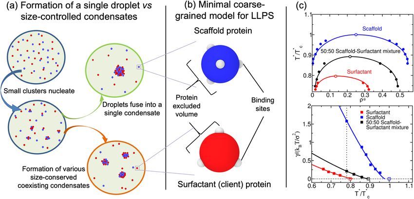
www.nature.com/scientificreports/
Figure 1. (a) Schematic representation of single-droplet formation versus size-conserved protein condensation.
Scaffold proteins are depicted as blue spheres, while surfactant clients are shown as red spheres. (b) Minimal
coarse-grained model for protein LLPS: Blue and red spheres represent the excluded volume of scaffold and
surfactant (client) proteins respectively, while gray patches represent the binding sites of the proteins. Two
different proteins are modeled: scaffold proteins, with 4 promiscuous binding sites in a tetrahedral arrangement,
and surfactant proteins, with 3 binding sites in a planar equidistant arrangement that can only bind to scaffold
binding sites (except where otherwise stated). Details on the model parameters are provided in the “Methods”
section. (c) Top: Phase diagram in the temperature–density plane for a scaffold protein system (blue), a 50:50
scaffold–surfactant mixture (black) and for a hypothetical surfactant system in which client proteins can
self-interact (red). The same self-interacting potential employed for scaffold proteins (blue) is also applied for
surfactant proteins that can hypothetically self-interact (red). This system serves to further illustrate the effect
of multivalency in LLPS. Filled circles indicate the estimated coexisting densities from Direct Coexistence
simulations68–71, and empty ones depict the critical points calculated via the law of rectilinear diameters and
critical exponents72 (see Supporting Information for details on these calculations). Bottom: Surface tension
(γ ) dependence on temperature for the scaffold system (blue), the 50:50 scaffold–surfactant mixture (black),
and the hypothetical system in which surfactant proteins can self interact (red). Filled squares represent direct
estimations of γ , continuous curves depict fits to our data of the following form: γ ∝ (T ∗ − Tc∗ )1.26, and empty
squares show the critical temperature of each system evaluated through the law of rectilinear diameters and
critical exponents72. The vertical dashed line indicates the temperature at which the remainder of our study is
performed. Note that temperature (in reduced units, T ∗) is renormalized by the critical temperature Tc∗ (also in
reduced units) of the scaffold protein system (Tc∗ = 0.12).
Size‑conserved condensates can be regulated by surfactant proteins. Despite their seemingly
subordinate role in condensate formation, client molecules (e.g. low valency proteins or self-avoiding nucleic
acids) can significantly impact the organization of biomolecular condensates20, 38, 73, 74. Remarkable examples
include the multilayered organization of stress g ranules75, 76, the nucleoli26, nuclear speckles77, mixtures of RNA-
binding proteins with RNA78, 79, and in vitro complex coacervates38, 73, 74. While scaffolds mainly contribute to
maximizing the condensate liquid network connectivity20, 49, clients can severely reduce the droplet interfacial
free energy cost of creating an interface23. The proliferation marker protein Ki-67, a component of the mitotic
chromosome periphery, is an important example of a natural surfactant in intracellular compartmentalization35.
In this context, our simulations show that low-valency clients that do not establish homotypic interactions
behave as surfactants within condensates, as they can greatly decrease the condensate surface tension [Fig. 1c
(Bottom)].
To further investigate the role of surfactant clients, we perform two sets of Direct Coexistence (DC)
simulations68–71: (1) for the pure scaffold system, and (2) for the 50:50 binary mixture of scaffold and surfactants
shown in Fig. 1c. At a constant temperature of T ∗ /Tc∗ = 0.75 and a global (system) density of ρ ∗ = 0.136 (both
in reduced units; see “Methods” section for further details on the employed reduced magnitudes), we create
three different simulation box geometries with the dimensions summarized in Table 1. Using this approach, we
can effectively modulate the surface/volume ratio of the condensed phase (hereafter called droplet): where the
droplet surface is S = 2 ∗ Ly ∗ Lz and its volume is V = Ly ∗ Lz ∗ Lx,slab ; with Lx,slab representing the width of
the condensate in the x direction, and Lx , Ly and Lz being the different sides of the simulation box. Figure 2a–c,
summarizes the phase behavior of the pure scaffold system (Top panels) and the 50:50 binary mixture (Mid-
dle panels) along the three designed simulation box geometries. The pure scaffold condensate exhibits distinct
surface/volume ratios depending on the box geometry (see Table 1), while maintaining the same droplet density
on all three cases. In other words, the scaffold condensate would be able to continuously grow as a single droplet
at expense of the diluted phase until reaching equilibrium. In contrast, the scaffold–surfactant mixture yields
various coexisting equilibrium droplets with a roughly constant surface/volume ratio of 0.21(2) σ −1 in all systems
Scientific Reports | (2021) 11:15241 | https://doi.org/10.1038/s41598-021-94309-y 3
Vol.:(0123456789)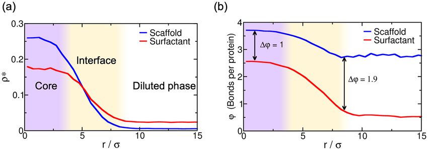
www.nature.com/scientificreports/
Box geometry (a) (b) (c)
Lx/σ 43.7 68.1 124.8
(Ly = Lz)/σ 17.5 13.7 10.3
Scaffold S/V ratio / σ −1 0.14(2) 0.12(2) 0.09(2)
50:50 mixture S/V ratio / σ −1 0.21(2) 0.21(2) 0.20(2)
Table 1. Simulation box dimensions and condensate surface/volume ratios (S/V) of the three box geometries
represented in Fig. 2 for the pure scaffold system and the 50:50 binary mixture of scaffold and surfactant
proteins. Geometries (a), (b) and (c) account for the (a), (b) and (c) panels shown in Fig. 2, respectively.
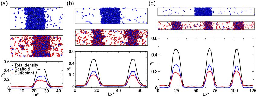
Figure 2. Direct Coexistence simulations for a scaffold protein system (Top panels) and a 50:50 binary mixture
of scaffold and surfactant proteins (Middle panels) with different simulation box geometries (see Table 1
for the different simulation box sides employed in a, b and c geometries). All simulations were performed
at T ∗ /Tc∗ = 0.75 and a global density of ρ ∗ = 0.136. (a)–(c) (Top and Middle panels): Scaffold proteins are
colored in blue and surfactant clients in red; the protein binding sites of both protein types are colored in
gray. (a)–(c) (Bottom panels): Density profiles along the long side (x) of each simulation box Lx for the 50:50
scaffold–surfactant mixture. The (scaffold+surfactant) total density profile is depicted in black, the surfactant
client density profile is shown in red, and that of scaffolds in blue. Densities of the different scaffold–surfactant
coexisting droplets, as well as the scaffold molar fraction in the condensed and diluted phase, for each geometry
are given in Table 2. The density of the pure scaffold condensates in all geometries is 0.54(1).
(Table 1). This result strongly suggests how size-conserved multidroplet phase behavior can be simply induced
by the presence of low valency clients, which actively contribute to lowering the surface tension of the droplets
[Fig. 1c (Bottom)]—thereby serving as natural s urfactants35, 36.
We next analyze the composition of the different coexisting scaffold–surfactant condensates along the distinct
box geometries [Fig. 2 (Bottom panels)]. In all cases, including the pure scaffold condensate, the properties of the
droplets are remarkably similar; i.e., the density of all droplets, as well as their composition and surface tension
are roughly constant (Table 2). Notably, we find that the surfactant density profile becomes higher than that of
scaffolds at the droplet interface, showing how the partition coefficient of surfactants is greater than that of scaf-
folds in the outer region. Previous works suggest that accumulation of surfactants at the interface is preferable
as it minimizes the condensate surface tension35, 38, 66. We also verify that the presence of multiple coexisting
droplets is the thermodynamically stable state, rather than just metastable, by simulating over sufficiently long
timescales to allow for multiple droplet fusion events and variations in droplet composition. Importantly, these
tests reveal that even when in contact the droplets coexist without coalescing or altering their equilibrium com-
position. The multidroplet behavior of size-conserved condensates (in our case of ∼ 0.21 σ −1 surface/volume
ratio) is a consequence of the thermodynamic conditions of our system (i.e., mixture composition, temperature,
and density). Note that droplet curvature effects such as Laplace internal pressure80 or surface tension depend-
ence on droplet c urvature81 have not been considered in our simulations, since we do not expect them to play
an important role at biologically relevant droplet size scales (O µm)40. Those effects are only expected to be
dominant in the nanometer scale (i.e., up to droplet radii of tens of nanometers)81–84.
The presence of surfactant clients within the condensate substantially lowers the liquid network connectivity49,
85, 86
and, therefore, reduces the enthalpic gain sustaining LLPS. Consequently, the system minimizes its free
energy by optimizing the number of surfactants that are incorporated into the condensed phase; i.e., by creat-
ing higher surface/volume ratios, where surfactants are preferentially located towards the interface rather than
in the core. Such free energy optimization yields multiple coexisting condensates of a certain size, rather than
a single-condensate system. Moreover, the emergence of multiple coexisting droplets stabilised by surfactant
Scientific Reports | (2021) 11:15241 | https://doi.org/10.1038/s41598-021-94309-y 4
Vol:.(1234567890)www.nature.com/scientificreports/
Box geometry (a), 1 droplet (b), 2 droplets (c), 3 droplets
γ /(kB T/σ 2) 0.26(4) 0.23(4) 0.23(4)
Scaffold molar fraction in condensate 0.585(5) 0.576(5) 0.583(5)
Scaffold molar fraction in diluted phase 0.041(2) 0.041(2) 0.043(2)
∗
ρcondensate 0.47(1) 0.47(1) 0.46(1)
∗
ρdiluted phase 0.030(3) 0.027(3) 0.029(3)
Table 2. Properties of systems presented in Fig. 2 containing 50:50 scaffold–surfactant compositions in
different simulation box geometries. In all cases, 6 independent simulations (with different initial velocity
distributions), each starting from a pre-equilibrated configuration, were performed. For the geometries with
more than one droplet, the values are averaged over the different droplets, although the variance between
distinct droplets is significantly small as shown in Fig. 2 (Bottom panels).
proteins instead of a single condensate can also contribute to increase the entropy of the system, and thus,
lowering its free energy. Nonetheless, this behavior is only thermodynamically favorable when the condensate
surface tension is very low, as in the case of the 50:50 scaffold–surfactant condensates with a surface tension of
γ = 0.23 kB T σ −2—almost 7 times lower than that of the pure scaffold condensate (γ = 1.58 kB T σ −2) at the
same conditions (T ∗ /Tc∗ = 0.75).
Surfactant concentration critically modulates droplet size. As discussed in the previous section,
our minimal protein model shows that for a given composition of scaffold and surfactant clients, independent
of the imposed box geometry, droplets can only grow to a certain size. This size-restricted growth is, in turn,
determined by the optimal surface/volume ratio that minimizes the free energy of the coexisting liquid phases.
Therefore, larger system sizes lead to higher number of coexisting size-conserved droplets. On the other hand,
when the condensate is only composed of scaffold proteins, as the system size increases, the size of the conden-
sate simply grows instead of yielding new multiple size-conserved droplets (Fig. 2). These results illustrate how a
simple model for scaffold and surfactant proteins, merely controlled by protein valency and binding affinity, can
recapitulate mesoscale features of in vivo and in vitro condensates that exhibit size-conserved g rowth7, 26, 34–38.
Since this phase behavior only arises when the concentration of surfactants is not negligible, we now investigate
how condensates can switch between both scenarios, and how their surface/volume ratio is modulated by their
relative scaffold–surfactant composition.
By gradually increasing the surfactant client concentration of the scaffold–surfactant mixture (at a constant
temperature and system density), the condensate size progressively decreases to accommodate the equilib-
rium droplet surface/volume ratio to the simulation box geometry; i.e., the condensate splits into two and,
subsequently, into three coexisting liquid droplets (see Fig. 3). We note that the composition of the different
coexisting droplets at a given surfactant concentration is remarkably similar, highlighting that all droplets are
in equilibrium. In parallel, we evaluate the surface tension of the different coexisting droplets as a function of
surfactant concentration. We find that γ monotonically decreases (but not linearly) as the surfactant concentra-
tion increases (Fig. 3). This result is not surprising (see Fig. 1c) given that one of the key molecular driving forces
behind size-conserved multidroplet formation is the reduction of γ by surfactants coating the droplet surface.
Above a certain surfactant concentration—exceeding 65 % for our system and at the given conditions—LLPS is
inhibited. Beyond this limit, the condensate liquid network connectivity sustained by scaffold proteins can no
longer compensate the mixing entropy of the system. We also analyze the surface/volume ratio of the droplets
as a function of surfactant concentration. At infinitely low surfactant concentration, the condensate displays a
ratio of ∼ 0.09 σ −1. However, such a ratio is fully determined by the total number of proteins in the system and
the box geometry, since as shown in Fig. 2, scaffold condensates can reach any droplet size when surfactants are
absent. At low surfactant concentrations (i.e., % surfactant < 27.5%), the maximum droplet size corresponds
to a surface/volume ratio of ∼ 0.11 σ −1. Beyond that threshold concentration, the condensate shrinks, and to
achieve the equilibrium surface/volume ratio, it splits into smaller coexisting droplets. The maximum equilib-
rium droplet size in the two-droplet regime is that corresponding to ratios of ∼ 0.19 σ −1. Finally, for surfactant
compositions higher than 38%, three coexisting condensates emerge. The maximum surface/volume ratio that
droplets can achieve is ∼ 0.27 σ −1 at 60% client composition, which is only possible due to the extreme reduc-
tion in the surface tension (γ = 0.15 kB T σ −2)—more than one order of magnitude lower than that of the pure
scaffold condensate (γ = 1.58 kB T σ −2) at the same temperature and system density.
Previous studies have highlighted the challenges associated with measuring condensate surface tensions due
to the small size of protein d roplets3, 87. Nonetheless, there are available estimates of this magnitude for ribonu-
cleoprotein condensates, and these measurements demonstrate that surfactant proteins can reduce γ by orders
of magnitude26. With our minimal model, we qualitatively observe such behavior when surfactant proteins are
recruited to the condensate, giving rise to emulsions of multiple coexisting droplets with very low surface tension.
Surfactant proteins can lead to the formation of multidroplet emulsions by inducing multilayered condensate
architectures26, 38. Diverse biomolecular organelles, either in the cell nucleus, such as the n ucleoli26, or nuclear
speckles77, as well as in the cytosol, such as stress granules,75, 76 exhibit this type of organization. Moreover, dif-
ferent in vitro complex c oacervates38, 50, 73, 74, bioengineered ribonucleoprotein condensates in living c ells88 and
mixtures of RNA-binding proteins and RNA m olecules78, 79 are also known to show multilayered assemblies. In
Fig. 4a, we analyze the droplet architecture of a protein condensate with a 50:50 scaffold–surfactant composition
Scientific Reports | (2021) 11:15241 | https://doi.org/10.1038/s41598-021-94309-y 5
Vol.:(0123456789)www.nature.com/scientificreports/
Figure 3. Surface tension (γ ) dependence on the scaffold–surfactant composition (in %) at T ∗ /Tc∗ = 0.75 and
system (global) density of ρ ∗ = 0.136. Vertical dashed lines indicate the maximum surfactant concentration
which allows LLPS for a given number-droplet regime in our system. Note that the maximum droplet size
varies continuously with surfactant concentration even within the same number-droplet regime. The maximum
condensate size in terms of surface/volume ratio along the different number-droplet regimes are: ∼ 0.11 σ −1
for the one-droplet regime, ∼ 0.19 σ −1 for the two-droplet regime, and ∼ 0.27 σ −1 for the three-droplet
regime. At surfactant client compositions exceeding 65% there is no LLPS. For each computed composition,
the global density of the system and the simulation box cross-section (Lz Ly ) are kept constant. Snapshots of
the DC simulations along different surfactant composition droplet regimes are included. Please note that in
the multidroplet regime, the different coexisting droplets (at the same surfactant concentration) exhibit similar
compositions and densities.
Figure 4. (a) Droplet density profile (in reduced units) for scaffold (blue) and surfactant proteins (red) from
the droplet center of mass towards the surrounding diluted phase for the 50:50 binary mixture within the
simulation box geometry shown in Fig. 2a at T ∗ /Tc∗ = 0.75. (b) Average number of engaged binding sites per
protein (ϕ) as a function of distance from the droplet center of mass for scaffold (blue) and surfactant proteins
(red). One binding site is considered to be engaged to another if the distance between them is less than 0.145 σ
(i.e., the maximum bond length interaction between distinct protein binding sites; for further details on these
calculations see Supporting Information).
[i.e., the system in Fig. 2a (Middle panel)]. We find that in the droplet core, the scaffold to surfactant ratio is
much higher than along the interfacial region (Fig. 4a), where it drops to almost half that of the surfactant pro-
teins. Nonetheless, the surfactant concentration within the droplet core is still remarkably high considering its
destabilizing role in the condensate liquid network c onnectivity49. The observed non-homogeneous condensate
organization stems from the higher valency of scaffold proteins, which allows them to establish higher molecular
connectivity within the core condensate and, thus, induce higher enthalpic gain upon multilayered assembly.
Finally, to gain further insight on the droplet liquid network connectivity, we evaluate the average number
of engaged binding sites per protein (ϕ ) as a function of distance from the center of mass of the condensates
(Fig. 4b). Scaffold proteins present a significantly higher amount of molecular connections per protein than sur-
factants (i.e., ϕ ∼ 3.7 and ϕ ∼ 2.5 for scaffolds and surfactants, respectively), at the droplet core. This observation
highlights how surfactants negatively contribute to the stability of the condensate. However at the interface, such
diminished connectivity of surfactant proteins (ϕ ∼ 1) with respect to that of scaffolds (ϕ ∼ 3) substantially
reduces the condensate surface tension—by decreasing the enthalpic cost ( hi ) of creating an interface. This
energetically favorable protein arrangement, controlled in our model just by the variance in protein valency of
the components, is expected to be contributed also by changes in relative binding affinities among proteins, and
Scientific Reports | (2021) 11:15241 | https://doi.org/10.1038/s41598-021-94309-y 6
Vol:.(1234567890)www.nature.com/scientificreports/
be modulated by post-translational modifications in vivo7, 34. Furthermore, such variations can be also relevant to
understand the physical parameters controlling multilayered condensate organization and, ultimately, regulation
of the formation of size-conserved multidroplet e mulsions29–31.
Conclusions
In this work, we employ our minimal protein model48 to demonstrate how biomolecular multidroplet emulsifica-
tion can be controlled by the subtle balance between liquid network connectivity and droplet surface tension, and
how general molecular features such as protein valency and binding affinity can critically regulate this behavior.
By using a binary mixture of scaffold and client proteins that act as surfactants (following the original definition
proposed by Banani et al.20), we design a set of Direct Coexistence simulations in which we can conveniently
modulate the simulation box geometry to assess the propensity of the condensates to accommodate different sur-
face/volume ratios. The ability (or disability) of these mixtures to adopt different surface/volume ratios, imposed
by the box geometry, can be regarded as an indirect measurement of the droplet propensity to grow beyond
a certain size. We find that pure component scaffold condensates can easily adapt to distinct surface/volume
ratios; in support of their ability to grow and fuse into a single droplet. However, 50:50 binary scaffold–surfactant
mixtures stabilize instead several coexisting liquid condensates with roughly constant surface/volume ratios to
accommodate the imposed system size geometry. Such behavior is a clear signature of size-conserved multidro-
plet emulsification, as found in the n ucleolus7, ribonucleoprotein c ondensates26, 88, micelle-like c ondensates31,
45
, and in vitro complex coacervates38.
We also elucidate the role of surfactant concentration in size-conserved droplet growth. By gradually decreas-
ing the scaffold–surfactant ratio in our mixtures, we observe that the maximum droplet size is reduced, while
simultaneously increasing the number of coexisting condensates. This trend continues until a sufficiently high
surfactant concentration is reached, where LLPS is no longer possible. Moreover, as clients are added, the droplet
surface tension dramatically decreases, facilitating the formation of multiple coexisting small liquid droplets at
low interfacial energetic cost. Client proteins, besides decreasing the stability of the c ondensates49, can effec-
tively behave as natural droplet s urfactants35, 36. Due to their considerably lower molecular connectivity com-
pared to that of scaffolds, surfactants preferentially migrate towards the droplet interface; thereby, minimizing
the enthalpic cost of creating an i nterface23. Heterogeneous molecular organizations of condensates have been
observed in stress g ranules75, 76, the n ucleoli26 and nuclear s peckles77. We find that such heterogeneity enable
the maximization of the condensate liquid network connectivity through scaffold–scaffold protein interactions
within the droplet core.
Rationalizing the underlying mechanisms employed by cells to precisely regulate the size of their diverse
membraneless compartments and processing b odies7, 34, 36 represents a crucial step towards understanding intra-
cellular spatiotemporal cell organization. Taken together, our coarse-grained simulations help to elucidate the
relationship between single-droplet phase formation and size-conserved multidroplet architecture, and put for-
ward general molecular features such as valency and binding affinity as chief drivers in these scenarios.
Methods
We model our coarse-grained multivalent proteins using the MD-Patchy potential proposed in Ref.48, which is
composed by two different set of potentials: a Pseudo Hard-Sphere (PHS) potential89 to continuously describe
the repulsive interaction and excluded volume between different protein replicas, and a continuous square-well
(CSW)90 potential to describe the patch-patch interactions among different protein binding sites. The uPHS
potential is described by the following expression:
� � �� �
r a ε � � � � �
σ r
r a R r − σr a + εR ; if r < ar σ
uPHS = � � (1)
0; if r ≥ r σ a
where a = 49 and r = 50 are the exponents of the attractive and repulsive terms respectively, εR accounts for
the energy shift of the PHS interaction, σ is the molecular diameter (and our unit of length) and r is the center-to-
center distance between different PHS particles. For the patch–patch interaction we use the following expression:
1 r − rw
uCSW = − εCSW 1 − tanh (2)
2 α
where εCSW is the depth of the potential energy well, rw the radius of the attractive well, and α controls the
steepness of the well. We choose α = 0.005σ and rw = 0.12σ so that each patch can only interact with another
single patch.
The mass of each patch is a 5% of the central PHS particle mass, which is set to 3.32 × 10⁻²⁶ kg, despite being
this choice irrelevant for equilibrium simulations. This 5% ratio fixes the moment of inertia of the patchy particles
(our minimal proteins). The molecular diameter of the proteins, both scaffold and clients, is σ = 0.3405 nm,
and the value of εR /kB is 119.81K. All our results are presented in reduced units: reduced temperature is defined
as T ∗ = kB T/ε CSW , reduced density as ρ = (N/V )σ , reduced pressure as p = pσ /(kB T), and the reduced
∗ 3 ∗ 3
unit of time as σ 2 m/(kB T) . In order to keep the PHS interaction as similar as possible to a pure HS interac-
tion, we fix kB T/εR at a value of 1.5 as suggested in Ref.89 (fixing T = 179.71K). We then control the effective
strength of the binding protein attraction by varying εCSW such that the reduced temperature, T ∗ = kB T/εCSW ,
is of the order of O (0.1).
Scientific Reports | (2021) 11:15241 | https://doi.org/10.1038/s41598-021-94309-y 7
Vol.:(0123456789)www.nature.com/scientificreports/
Since both uPHS and uCSW potentials are continuous and differentiable, we perform all our simulations using
the LAMMPS Molecular Dynamics p ackage91. Periodic boundary conditions are used in the three directions of
space. The timestep chosen for the Verlet integration of the equations of motion is t ∗ = 3.7 × 10⁻⁴. The cut-off
radius of the interactions of both potentials is set to 1.17σ . We use a Nosé–Hoover thermostat92, 93 for the NVT
simulations with a relaxation time of 0.074 in reduced units. For NpT simulations, a Nosé–Hoover barostat is
employed with the same relaxation t ime94.
The methodological details of the calculation of the phase diagram, surface tension and engaged binding sites
per protein through a local order parameter, are provided in the Supporting Information document.
Received: 30 April 2021; Accepted: 6 July 2021
References
1. Hyman, A. A., Weber, C. A. & Jülicher, F. Liquid–liquid phase separation in biology. Annu. Rev. Cell Dev. Biol. 30, 39–58 (2014).
2. Shin, Y. & Brangwynne, C. P. Liquid phase condensation in cell physiology and disease (2017).
3. Brangwynne, C. P. et al. Germline p granules are liquid droplets that localize by controlled dissolution/condensation. Science 324,
1729–1732 (2009).
4. Molliex, A. et al. Phase separation by low complexity domains promotes stress granule assembly and drives pathological fibrilliza-
tion. Cell 163, 123–133 (2015).
5. Marnik, E. A. & Updike, D. L. Membraneless organelles: P granules in caenorhabditis elegans. Traffic 20, 373–379 (2019).
6. Lu, H. et al. Phase-separation mechanism for c-terminal hyperphosphorylation of RNA polymerase II. Nature 558, 318–323 (2018).
7. Brangwynne, C. P., Mitchison, T. J. & Hyman, A. A. Active liquid-like behavior of nucleoli determines their size and shape in
Xenopus laevis oocytes. Proc. Natl. Acad. Sci. 108, 4334–4339 (2011).
8. Wang, J. et al. A molecular grammar governing the driving forces for phase separation of prion-like RNA binding proteins. Cell
174, 688–699 (2018).
9. Krainer, G. et al. Reentrant liquid condensate phase of proteins is stabilized by hydrophobic and non-ionic interactions. Nat.
Commun. 12, 1–14 (2021).
10. Strom, A. R. et al. Phase separation drives heterochromatin domain formation. Nature 547, 241–245 (2017).
11. Larson, A. G. et al. Liquid droplet formation by HP1α suggests a role for phase separation in heterochromatin. Nature 547, 236–240
(2017).
12. Su, X. et al. Phase separation of signaling molecules promotes T cell receptor signal transduction. Science 352, 595–599 (2016).
13. Yoo, H., Triandafillou, C. & Drummond, D. A. Cellular sensing by phase separation: Using the process, not just the products. J.
Biol. Chem. 294, 7151–7159 (2019).
14. Sabari, B. R. et al. Coactivator condensation at super-enhancers links phase separation and gene control. Science 361 (2018).
15. Kroschwald, S. et al. Different material states of Pub1 condensates define distinct modes of stress adaptation and recovery. Cell
Rep. 23, 3327–3339 (2018).
16. Bouchard, J. J. et al. Cancer mutations of the tumor suppressor SPOP disrupt the formation of active, phase-separated compart-
ments. Mol. Cell 72, 19–36 (2018).
17. Brundin, P. et al. Prion-like transmission of protein aggregates in neurodegenerative diseases. Nat. Rev. Mol. Cell Biol. 11, 301–307
(2010).
18. Ambadipudi, S., Biernat, J., Riedel, D., Mandelkow, E. & Zweckstetter, M. Liquid–liquid phase separation of the microtubule-
binding repeats of the Alzheimer-related protein tau. Nat. Commun. 8, 1–13 (2017).
19. Robberecht, W. & Philips, T. The changing scene of amyotrophic lateral sclerosis. Nat. Rev. Neurosci. 14, 248–264 (2013).
20. Banani, S. F. et al. Compositional control of phase-separated cellular bodies. Cell 166, 651–663 (2016). http://www.sciencedirect.
com/science/article/pii/S0092867416307395.
21. Banani, S. F., Lee, H. O., Hyman, A. A. & Rosen, M. K. Biomolecular condensates: Organizers of cellular biochemistry. Nat. Rev.
Mol. Cell Biol. 18, 285–298 (2017).
22. Espinosa, J. R. et al. Liquid-network connectivity regulates the stability and composition of biomolecular condensates with many
components.
23. Sanchez-Burgos, I., Espinosa, J. R., Joseph, J. A. & Collepardo-Guevara, R. Valency and binding affinity variations can regulate the
multilayered organization of protein condensates with many components. Biomolecules 11, 278 (2021).
24. Ditlev, J. A., Case, L. B. & Rosen, M. K. Who’s in and who’s out–compositional control of biomolecular condensates (2018).
25. Shin, Y. et al. Liquid nuclear condensates mechanically sense and restructure the genome. Cell 175, 1481–1491 (2018).
26. Feric, M. et al. Coexisting liquid phases underlie nucleolar subcompartments. Cell 165, 1686–1697 (2016). http://www.sciencedir
ect.com/science/article/pii/S0092867416304925.
27. Liu, X. et al. Phase separation drives decision making in cell division. J. Biol. Chem. 295, 15172 (2020).
28. Zhang, Y., Li, Z., Chen, N., Huang, Y. & Huang, S. Phase separation of arabidopsis EMB1579 controls transcription, mRNA splic-
ing, and development. PLoS Biol. 18, e3000782 (2020).
29. Ranganathan, S. & Shakhnovich, E. I. Dynamic metastable long-living droplets formed by sticker-spacer proteins. Elife 9, e56159
(2020).
30. Dar, F. & Pappu, R. Phase separation: Restricting the sizes of condensates. Elife 9, e59663 (2020).
31. Nakamura, H., DeRose, R. & Inoue, T. Harnessing biomolecular condensates in living cells. J. Biochem. 166, 13–27 (2019).
32. Binder, K., Block, B. J., Virnau, P. & Tröster, A. Beyond the van der Waals loop: What can be learned from simulating Lennard–Jones
fluids inside the region of phase coexistence. Am. J. Phys. 80, 1099–1109 (2012).
33. Zhang, H. et al. RNA controls polyQ protein phase transitions. Mol. Cell 60, 220–230 (2015).
34. Kilchert, C., Weidner, J., Prescianotto-Baschong, C. & Spang, A. Defects in the secretory pathway and high Ca2+ induce multiple
p-bodies. Mol. Biol. Cell 21, 2624–2638 (2010).
35. Cuylen, S. et al. Ki-67 acts as a biological surfactant to disperse mitotic chromosomes. Nature 535, 308–312 (2016).
36. Navarro, M.G.-J. et al. RNA is a critical element for the sizing and the composition of phase-separated RNA-protein condensates.
Nat. Commun. 10, 1–13 (2019).
37. Wegmann, S. et al. Tau protein liquid–liquid phase separation can initiate tau aggregation. EMBO J. 37, e98049 (2018).
38. Lu, T. & Spruijt, E. Multiphase complex coacervate droplets. J. Am. Chem. Soc. 142, 2905–2914 (2020).
39. Lovell, P. A. & Schork, F. J. Fundamentals of emulsion polymerization. Biomacromolecules 21, 4396–4441 (2020).
40. Alberti, S., Gladfelter, A. & Mittag, T. Considerations and challenges in studying liquid–liquid phase separation and biomolecular
condensates. Cell 176, 419–434 (2019).
41. Wurtz, J. D. & Lee, C. F. Chemical-reaction-controlled phase separated drops: Formation, size selection, and coarsening. Phys.
Rev. Lett. 120, 078102 (2018).
Scientific Reports | (2021) 11:15241 | https://doi.org/10.1038/s41598-021-94309-y 8
Vol:.(1234567890)www.nature.com/scientificreports/
42. Zwicker, D., Seyboldt, R., Weber, C. A., Hyman, A. A. & Jülicher, F. Growth and division of active droplets provides a model for
protocells. Nat. Phys. 13, 408–413 (2017).
43. Wurtz, J. D. & Lee, C. F. Stress granule formation via ATP depletion-triggered phase separation. N. J. Phys. 20, 045008 (2018).
44. Kirschbaum, J., & Zwickerhttps, D. Controlling biomolecular condensates via chemical reactions. J. R. Soc. Interface. 18, 20210255.
https://royalsocietypublishing.org/doi/full/10.1098/rsif.2021.0255 (2021).
45. Ruff, K. M., Harmon, T. S. & Pappu, R. V. Camelot: A machine learning approach for coarse-grained simulations of aggregation
of block-copolymeric protein sequences. J. Chem. Phys. 143, 12B607 (2015).
46. Tejedor, A. R., Garaizar, A., Ramírez, J. & Espinosa, J. R. Dual RNA modulation of protein mobility and stability within phase-
separated condensates. bioRxiv (2021).
47. Kelley, F. M., Favetta, B., Regy, R. M., Mittal, J. & Schuster, B. S. Amphiphilic proteins coassemble into multiphasic condensates
and act as biomolecular surfactants. bioRxiv (2021).
48. Espinosa, J. R., Garaizar, A., Vega, C., Frenkel, D. & Collepardo-Guevara, R. Breakdown of the law of rectilinear diameter and
related surprises in the liquid-vapor coexistence in systems of patchy particles. J. Chem. Phys 150, 224510 (2019).
49. Espinosa, J. R. et al. Liquid network connectivity regulates the stability and composition of biomolecular condensates with many
components. Proc. Natl. Acad. Sci. 117, 13238–13247 (2020).
50. Fisher, R. S. & Elbaum-Garfinkle, S. Tunable multiphase dynamics of arginine and lysine liquid condensates. Nat. Commun. 11,
1–10 (2020).
51. Dignon, G. L., Best, R. B. & Mittal, J. Biomolecular phase separation: From molecular driving forces to macroscopic properties.
Annu. Rev. Phys. Chem. 71, 53–75 (2020).
52. Choi, J.-M., Holehouse, A. S. & Pappu, R. V. Physical principles underlying the complex biology of intracellular phase transitions.
Annu. Rev. Biophys. 49, 107–133 (2020).
53. Lee, J., Popov, Y. O. & Fredrickson, G. H. Complex coacervation: A field theoretic simulation study of polyelectrolyte complexation.
J. Chem. Phys. 128, 224908 (2008).
54. Weber, C. A., Zwicker, D., Jülicher, F. & Lee, C. F. Physics of active emulsions. Rep. Prog. Phys. 82, 064601 (2019).
55. Harmon, T. S., Holehouse, A. S. & Pappu, R. V. Differential solvation of intrinsically disordered linkers drives the formation of
spatially organized droplets in ternary systems of linear multivalent proteins. N. J. Phys. 20, 045002 (2018).
56. Choi, J.-M., Dar, F. & Pappu, R. V. Lassi: A lattice model for simulating phase transitions of multivalent proteins. PLOS Comput.
Biol. 15, e1007028 (2019).
57. Statt, A., Casademunt, H., Brangwynne, C. P. & Panagiotopoulos, A. Z. Model for disordered proteins with strongly sequence-
dependent liquid phase behavior. J. Chem. Phys. 152, 075101 (2020).
58. Jacobs, W. M. & Frenkel, D. Phase transitions in biological systems with many components. Biophys. J. 112, 683–691 (2017).
59. Russo, J., Tartaglia, P. & Sciortino, F. Reversible gels of patchy particles: Role of the valence. J. Chem. Phys. 131, 014504 (2009).
60. Blas, F. J., MacDowell, L. G., De Miguel, E. & Jackson, G. Vapor–liquid interfacial properties of fully flexible Lennard–Jones chains.
J. Chem. Phys. 129, 144703 (2008).
61. Dignon, G. L., Zheng, W., Kim, Y. C., Best, R. B. & Mittal, J. Sequence determinants of protein phase behavior from a coarse-grained
model. PLoS Comput. Biol. 14, e1005941 (2018).
62. Das, S., Lin, Y.-H., Vernon, R. M., Forman-Kay, J. D. & Chan, H. S. Comparative roles of charge, π , and hydrophobic interactions
in sequence-dependent phase separation of intrinsically disordered proteins. Proc. Natl. Acad. Sci. 117, 28795–28805 (2020).
63. Alshareedah, I., Moosa, M. M., Raju, M., Potoyan, D. A. & Banerjee, P. R. Phase transition of RNA–protein complexes into ordered
hollow condensates. Proc. Natl. Acad. Sci. 117, 15650–15658 (2020).
64. Latham, A. P. & Zhang, B. Consistent force field captures homolog resolved hp1 phase separation. bioRxiv (2021).
65. Regy, R. M., Dignon, G. L., Zheng, W., Kim, Y. C. & Mittal, J. Sequence dependent phase separation of protein–polynucleotide
mixtures elucidated using molecular simulations. Nucleic Acids Res. 48, 12593–12603 (2020).
66. Sanchez-Burgos, I., Espinosa, J. R., Joseph, J. A. & Collepardo-Guevara, R. Valency and binding affinity variations can regulate
the multilayered organization of protein condensates with many components. Biomolecules 11 (2021). https://www.mdpi.com/
2218-273X/11/2/278.
67. Joseph, J. A. et al. Thermodynamics and kinetics of phase separation of protein–RNA mixtures by a minimal model. Biophys. J.
120, 1–12 (2021).
68. Jover, J., Haslam, A., Galindo, A., Jackson, G. & Müller, E. Pseudo hard-sphere potential for use in continuous molecular-dynamics
simulation of spherical and chain molecules. J. Chem. Phys. 137, 144505 (2012).
69. Ladd, A. J. & Woodcock, L. V. Triple-point coexistence properties of the Lennard–Jones system. Chem. Phys. Lett. 51, 155–159
(1977).
70. García Fernández, R., Abascal, J. L. & Vega, C. The melting point of ice ih for common water models calculated from direct coexist-
ence of the solid–liquid interface. J. Chem. Phys. 124, 144506 (2006).
71. Espinosa, J. R., Sanz, E., Valeriani, C. & Vega, C. On fluid-solid direct coexistence simulations: The pseudo-hard sphere model. J.
Chem. Phys. 139, 144502 (2013).
72. Rowlinson, J. S. & Widom, B. Molecular Theory of Capillarity (Courier Corporation, 2013).
73. Mountain, G. A. & Keating, C. D. Formation of multiphase complex coacervates and partitioning of biomolecules within them.
Biomacromolecules (2019).
74. Boeynaems, S. et al. Spontaneous driving forces give rise to protein–RNA condensates with coexisting phases and complex material
properties. Proc. Natl. Acad. Sci. 116, 7889–7898 (2019).
75. Jain, S. et al. ATPase-modulated stress granules contain a diverse proteome and substructure. Cell 164, 487–498 (2016).
76. Guillén-Boixet, J. et al. RNA-induced conformational switching and clustering of G3BP drive stress granule assembly by condensa-
tion. Cell 181, 346–361 (2020).
77. Fei, J. et al. Quantitative analysis of multilayer organization of proteins and RNA in nuclear speckles at super resolution. J. Cell Sci.
130, 4180–4192 (2017).
78. Sanders, D. W. et al. Competing protein–RNA interaction networks control multiphase intracellular organization. Cell 181, 306–324
(2020).
79. Kaur, T. et al. Sequence-encoded and composition-dependent protein–RNA interactions control multiphasic condensate topolo-
gies. bioRxiv (2020).
80. Liu, H. & Cao, G. Effectiveness of the Young–Laplace equation at nanoscale. Sci. Rep. 6, 1–10 (2016).
81. Tolman, R. C. The effect of droplet size on surface tension. J. Chem. Phys. 17, 333–337 (1949).
82. Espinosa, J. R., Vega, C. & Sanz, E. Homogeneous ice nucleation rate in water droplets. J. Phys. Chem. C 122, 22892–22896 (2018).
83. Montero de Hijes, P., Espinosa, J. R., Sanz, E. & Vega, C. Interfacial free energy of a liquid-solid interface: Its change with curvature.
J. Chem. Phys. 151, 144501 (2019).
84. Sanchez-Burgos, I., de Hijes, P. M., Rosales-Pelaez, P., Vega, C. & Sanz, E. Equivalence between condensation and boiling in a
Lennard–Jones fluid. Phys. Rev. E 102, 062609 (2020).
85. Bianchi, E., Largo, J., Tartaglia, P., Zaccarelli, E. & Sciortino, F. Phase diagram of patchy colloids: Towards empty liquids. Phys. Rev.
Lett. 97, 168301 (2006).
86. Mazarakos, K. P., Ghosh, A., Zhang, X., Nguemaha, V. & Zhou, H.-X. Affinity between macromolecular regulators leads to precise
control of liquid–liquid phase separation. Biophys. J. 118, 202a (2020).
Scientific Reports | (2021) 11:15241 | https://doi.org/10.1038/s41598-021-94309-y 9
Vol.:(0123456789)www.nature.com/scientificreports/
87. Alshareedah, I., Thurston, G. M. & Banerjee, P. R. Quantifying viscosity and surface tension of multicomponent protein-nucleic
acid condensates. Biophys. J. (2021).
88. Cochard, A. et al. RNA at the surface of phase-separated condensates impacts their size and number (2021).
89. Jover, J., Haslam, A. J., Galindo, A., Jackson, G. & Müller, E. A. Pseudo hard-sphere potential for use in continuous molecular-
dynamics simulation of spherical and chain molecules. J. Chem. Phys. 137, 144505 (2012).
90. Espinosa, J., Vega, C. & Sanz, E. The mold integration method for the calculation of the crystal-fluid interfacial free energy from
simulations. J. Chem. Phys. 141, 134709 (2014).
91. Plimpton, S. Fast parallel algorithms for short-range molecular dynamics. J. Comput. Phys. 117, 1–19 (1995).
92. Nosé, S. A unified formulation of the constant temperature molecular dynamics methods. J. Chem. Phys. 81, 511–519 (1984).
93. Hoover, W. G. Canonical dynamics: Equilibrium phase-space distributions. Phys. Rev. A 31, 1695–1697. https://doi.org/10.1103/
PhysRevA.31.1695 (1985).
94. Hoover, W. G. Constant-pressure equations of motion. Phys. Rev. A 34, 2499 (1986).
Acknowledgements
This project has received funding from the European Research Council (ERC) under the European Union
Horizon 2020 research and innovation programme (grant agreement No 803326). J. R. E. acknowledges fund-
ing from the Oppenheimer Fellowship and from Emmanuel College Roger Ekins Research Fellowship. I. S. B.
acknowledges funding from the Oppenheimer Fellowship, EPRSC Doctoral Programme Training number EP/
T517847/1 and Derek Brewer Emmanuel College scholarship. J. A. J. is a Junior Research Fellow at Kings Col-
lege. This work has been performed using resources provided by the Cambridge Tier-2 system operated by the
University of Cambridge Research Computing Service (http://www.hpc.cam.ac.uk) funded by EPSRC Tier-2
capital grant EP/P020259/1.
Author contributions
I.S.B. and J.R.E. conceived the project. I.S.B. conducted the simulations. I.S.B. and J.R.E. analyzed the results.
J.A.J. and R.C.G. contextualized the results. I.S.B. and J.R.E. wrote the initial version of the manuscript. All the
authors reviewed and edited the manuscript.
Competing interests
The authors declare no competing interests.
Additional information
Supplementary Information The online version contains supplementary material available athttps://doi.org/10.
1038/s41598-021-94309-y.
Correspondence and requests for materials should be addressed to J.R.E.
Reprints and permissions information is available at www.nature.com/reprints.
Publisher’s note Springer Nature remains neutral with regard to jurisdictional claims in published maps and
institutional affiliations.
Open Access This article is licensed under a Creative Commons Attribution 4.0 International
License, which permits use, sharing, adaptation, distribution and reproduction in any medium or
format, as long as you give appropriate credit to the original author(s) and the source, provide a link to the
Creative Commons licence, and indicate if changes were made. The images or other third party material in this
article are included in the article’s Creative Commons licence, unless indicated otherwise in a credit line to the
material. If material is not included in the article’s Creative Commons licence and your intended use is not
permitted by statutory regulation or exceeds the permitted use, you will need to obtain permission directly from
the copyright holder. To view a copy of this licence, visit http://creativecommons.org/licenses/by/4.0/.
© The Author(s) 2021
Scientific Reports | (2021) 11:15241 | https://doi.org/10.1038/s41598-021-94309-y 10
Vol:.(1234567890)You can also read