Archaeal Ribosomal Proteins Possess Nuclear Localization Signal-Type Motifs: Implications for the Origin of the Cell Nucleus - Melnikov Lab
←
→
Page content transcription
If your browser does not render page correctly, please read the page content below
Archaeal Ribosomal Proteins Possess Nuclear Localization
          Signal-Type Motifs: Implications for the Origin of the Cell
          Nucleus
          Sergey Melnikov ,*,1 Hui-Si Kwok,†,1 Kasidet Manakongtreecheep,‡,1 Antonia van den Elzen,2
          Carson C. Thoreen,2 and Dieter Söll*,1,3
          1
           Department of Molecular Biophysics and Biochemistry, Yale University, New Haven, CT
          2
           Department of Cellular and Molecular Physiology, Yale University, New Haven, CT
          3
           Department of Chemistry, Yale University, New Haven, CT
          †
           Present address: Department of Chemistry and Chemical Biology, Harvard University, Cambridge, MA
          ‡
           Present address: Broad Institute, Cambridge, MA
          *Corresponding authors: E-mails: serguey.v.melnikov@gmail.com; dieter.soll@yale.edu.

                                                                                                                                                                  Downloaded from https://academic.oup.com/mbe/article-abstract/37/1/124/5566250 by guest on 02 March 2020
          Associate editor: Claus Wilke

          Abstract
          Eukaryotic cells are divided into the nucleus and the cytosol, and, to enter the nucleus, proteins typically possess short
          signal sequences, known as nuclear localization signals (NLSs). Although NLSs have long been considered as features
          unique to eukaryotic proteins, we show here that similar or identical protein segments are present in ribosomal proteins
          from the Archaea. Specifically, the ribosomal proteins uL3, uL15, uL18, and uS12 possess NLS-type motifs that are
          conserved across all major branches of the Archaea, including the most ancient groups Microarchaeota and
          Diapherotrites, pointing to the ancient origin of NLS-type motifs in the Archaea. Furthermore, by using fluorescence
          microscopy, we show that the archaeal NLS-type motifs can functionally substitute eukaryotic NLSs and direct the
          transport of ribosomal proteins into the nuclei of human cells. Collectively, these findings illustrate that the origin of
          NLSs preceded the origin of the cell nucleus, suggesting that the initial function of NLSs was not related to intracellular
          trafficking, but possibly was to improve recognition of nucleic acids by cellular proteins. Overall, our study reveals rare
          evolutionary intermediates among archaeal cells that can help elucidate the sequence of events that led to the origin of
          the eukaryotic cell.
          Key words: ribosomal proteins, translation, origin of the nucleus, nuclear localization signals.

          Introduction                                                                          channels, the nuclear pores (Brocks et al. 1999; Rasmussen
Article

          Our understanding of past evolutionary events mainly relies                           et al. 2008). To help them pass via the nuclear pores from the
          on the discovery of transitional forms. Classical examples of                         cytoplasm into the nucleus, cellular proteins have evolved
          these transitional forms include the fossilized bird-like dino-                       specialized signal sequences, the nuclear localization signals
          saur Archaeopteryx lithographica and the crawling fish                                (NLSs) (Dingwall et al. 1982). A typical NLS is a short and
          Tiktaalik roseae, whose bone structures illuminated many                              surface-exposed stretch of basic residues that is recognized by
          aspects of how, between the Late Jurassic and the Late                                specialized receptors, karyopherins (Gorlich et al. 1994).
          Devonian, the fins of ancestral fish were transformed into                            Karyopherins transfer NLS-containing proteins across the nu-
          the legs of terrestrial animals and subsequently into the feath-                      clear pores (Gorlich et al. 1994). Although the nuclear–
          ered wings of birds (Chiappe 1999; Norell and Xu 2005;                                cytoplasmic trafficking machinery includes more than 100
          Daeschler et al. 2006). However, the further we delve into                            different components, including karyopherins, nuclear pore
          the past, the more we find ourselves limited in our archeo-                           components, and other proteins, the order in which these
          logical record, so that the most ancient events in history of life                    proteins evolved and the factors that drove the transition
          on Earth are known in sketchy outline or remain enigmatic.                            from prokaryotic to eukaryotic cell structures are currently
             One such enigmatic event is related to the question how                            unknown. In the absence of evidence of a single transitional
          the cell nucleus originated. Fossil records indicate that, be-                        form between prokaryotes and eukaryotes, there are at least
          tween 1.7 and 2.7 billion years ago (Bya), a group of ancestral                       twelve alternative hypotheses of how the system of nuclear–
          prokaryotic cells were transformed into what we now refer to                          cytoplasmic trafficking might have emerged (Martin et al.
          as eukaryotes: they acquired a DNA-storage compartment—                               2015).
          the nucleus—that was separated from the cytoplasm by a                                   Although the origin of the nucleus remains a matter of
          nuclear membrane equipped with selectively penetrable                                 debate, we have some general understanding of the preceding

          ß The Author(s) 2019. Published by Oxford University Press on behalf of the Society for Molecular Biology and Evolution.
          All rights reserved. For permissions, please e-mail: journals.permissions@oup.com

          124                        Mol. Biol. Evol. 37(1):124–133 doi:10.1093/molbev/msz207 Advance Access publication September 10, 2019
NLS-Motifs in Archaeal R-Proteins . doi:10.1093/molbev/msz207                                                          MBE
events based on the available fossil record and molecular          sequence alignments of eukaryotic ribosomal proteins
clock analyses. According to the existing evidence, life origi-    (from 482 species) and their homologs in the Bacteria
nated on Earth between 3.9 and 4.3 Bya (Dodd et al. 2017);         (2,951 species) and Archaea (402 species) (supplementary
and about 2–2.7 Bya the life forms were split into two major       data 1, Supplementary Material online).
lineages—the lineage that gave rise to modern Bacteria, and            We found that six NLS-motifs—including those in ribo-
the lineage that gave rise to modern Archaea and Eukarya           somal proteins uS3, uS4, uS8, uL13, uL23, and uL29—are
(Eme et al. 2018). Before Eukarya have emerged as a separate       highly conserved among the Eukarya but absent in Bacteria
branch on the tree of life, their ancestors belong to the same     and Archaea (table 1). This finding was consistent with stud-
branch of life as modern Archaea (Eme et al. 2018). It is          ies of other proteins, such as histones, illustrating that NLSs
therefore not surprising that presently living Archaea are         are found only in eukaryotic ribosomal proteins (Jenkinson
studied to gain insights into the origin of eukaryotes.            and Chong 2003; Henneman et al. 2018). However, four pro-
   Also, previous studies have shown that many aspects of          teins—uL3, uL15, uL18, and uS12—were found to have NLS-

                                                                                                                                      Downloaded from https://academic.oup.com/mbe/article-abstract/37/1/124/5566250 by guest on 02 March 2020
the early evolution of life on Earth can be understood through     type motifs not only in the Eukarya but also in the Archaea
analysis of the macromolecules of the ancient origin of a living   (fig. 1, supplementary data 1, Supplementary Material online).
cell, such as ribosomes. Because of the ancient origin of ribo-        The ribosomal structure has been determined for several
somes, their structure has been used as a living molecular         archaeal species, including Haloarcula marismortui (Ban et al.
fossil to gain an understanding of such evolutionary enigmas       2000) and Pyrococcus furiosus (Armache et al. 2013), so we
as the origin of catalytic RNA (Krupkin et al. 2011; Noller        therefore next investigated whether archaeal and eukaryotic
2012), the evolution of protein folding (Klein et al. 2004;        NLS-type motifs have conserved structures. The NLS-type
Hsiao et al. 2009), the origin of the genetic code (Johnson        motifs in proteins uL3, uL15, uL18, and uS12 turned out to
and Wang 2010; Hartman and Smith 2014), and the reason             have conserved secondary and tertiary structures between
for the stereospecific structure of proteins (Melnikov et al.      Eukarya and Archaea (fig. 1). Thus, our analysis revealed
2019), among others (Bokov and Steinberg 2009; Petrov et al.       that NLS-motifs are not limited to eukaryotic proteins but
2015; Melnikov, Manakongtreecheep, et al. 2018).                   can also be found in their archaeal homologs.
Additionally, eukaryotic ribosomes have been shown to be
adapted to the nuclear–cytoplasmic separation of eukaryotic
cells because eukaryotic ribosomal proteins, unlike their bac-     NLS-Type Motifs Are Conserved in All Groups of
terial counterparts, have evolved NLSs that allow ribosomal        Archaea, Including the Most Ancient Archaeal
proteins to enter the cell nucleus where they are subsequently     Branches
incorporated into nascent ribosomes (Melnikov et al. 2015).        Having found NLSs-type segments in archaeal ribosomal pro-
This finding suggested that further investigation into ribo-       teins, we next asked whether these motifs are preserved in all
somes’ structures may lead to a better understanding of            branches of the Archaea or just in a subset of archaeal species.
the origin of the nuclear–cytoplasmic trafficking (Melnikov        To answer this, we analyzed the conservation of NLS-motifs in
et al. 2015).                                                      the archaeal proteins uL3, uL15, uL18, and uS12 (fig. 2, sup-
   In this study we have analyzed ribosome structures from         plementary data 2, Supplementary Material online). To date,
the three domains of life to investigate origins of NLSs in        archaeal species have been divided into four large lineages,
eukaryotic proteins. By comparing homologous ribosomal             including DPANN (a superphylum named after
proteins from each of the three domains of life, we found          Diapherotrites,       Parvarchaeota,        Aenigmarchaeota,
that protein segments that were described as NLSs in eukary-       Nanoarchaeota, and Nanohaloarchaea lineages), TACK (a
otic ribosomal proteins were also present in homologous            superphylum named after Thaumarchaeota, Aigarchaeota,
proteins from Archaea. This finding indicates that at least        Crenarchaeota, and Korarchaeota lineages), Euryarchaeota,
some NLSs evolved in proteins considerably earlier than the        and Asgard superphyla (fig. 2) (Eme et al. 2018). We antici-
event when cells separated into the nucleus and the cyto-          pated that NLS-type motifs would be present only in the
plasm. This finding reveals a group of rare evolutionary           most recently evolved and eukaryote-like branches of ar-
intermediates—NLS-type motifs in archaeal ribosomal pro-           chaeal species, such as Asgard. Indeed, we found that se-
teins—which may shed light on the sequence of events that          quence similarity between eukaryotic NLSs and archaeal
eventually resulted in the origin of the nuclear–cytoplasmic       NLS-type motifs increases with the transition from ancient
separation of eukaryotic cells.                                    archaeal branches (DPANN) to more recently emerged
                                                                   branches (Asgard) (supplementary data 2, Supplementary
                                                                   Material online). However, even in the DPANN superphylum
Results                                                            the sequence similarity remains above 50% for each of the
Archaeal Ribosomal Proteins Possess NLS-Motifs                     four NLS-type motifs (fig. 2), with some DPANN species (e.g.,
Seeking to better understand when NLS-motifs might have            Mancarchaeum acidiphilum, Nanoarchaeota archaeon, and
emerged in ribosomal proteins, we assessed their conserva-         Aenigmarchaeota archaeon) having only a single substitution
tion among ribosomal proteins from the three domains of life.      in their NLS-type motifs (typically lysine-to-arginine) com-
To date, NLS-motifs have been characterized in ten ribosomal       pared with eukaryotic ribosomal proteins (supplementary
proteins from several eukaryotic species (table 1). We assessed    data 2, Supplementary Material online). Thus, contrary to
the conservation of these NLS-motifs by using multiple             our expectations, we found that NLS-type motifs are
                                                                                                                               125
Melnikov et al. . doi:10.1093/molbev/msz207 MBE
Presence in
conserved across all the archaeal branches, including the most
Archaea
ancient superphylum, DPANN.
2
2
2
1
1
2
1
1
2
2
Notably, many archaeal NLS-type motifs have precisely
same sets of hydrophobic and basic residues as the NLS-
motifs of eukaryotic ribosomal proteins, which is particularly
common among species from the eukaryote-like Asgard
Tag Used to Monitor the Nucleolar
b-galactosidase at the N-terminus
b-galactosidase at the C-terminus
b-galactosidase at the C-terminus
b-galactosidase at the C-terminus
HA-tag, position is not specified
superphylum (supplementary data 2, Supplementary
eGFP at the N- and C-termini Material online). For instance, the NLS of the ribosomal pro-
tein uL18 in the eukaryote Saccharomyces cerevisiae has a set
Accumulation
eGFP at the C-terminus
eGFP at the C-terminus
eGFP at the C-terminus
GST at the C-terminus
of hydrophobic and basic residues identical to those in 89
archaeal species, including species from Pyrococcus
(Euryarchaeota), Acidianus (TACK), and Lokiarchaeum
(S. cerevisiae)
(S. cerevisiae)
Downloaded from https://academic.oup.com/mbe/article-abstract/37/1/124/5566250 by guest on 02 March 2020
(Asgard) genera (supplementary data 2, Supplementary
Material online). Similarly, an identical set of hydrophobic
and basic residues can be found in the NLS of S. cerevisiae
protein uL3 and in the corresponding NLS-type sequences
from 27 archaeal species (supplementary data 2,
[Underwood and Fried
Supplementary Material online). Furthermore, in three of
[Moreland et al. 1985]
[Melnikov et al. 2015]
[Timmers et al. 1999]
[Rosorius et al. 2000]
[Schaap et al. 1991]
these archaeal species uL3 has a preserved adjacent serine
[Chen et al. 2008]
[Koch et al. 2012]
[Lindstrom 2012]
[Das et al. 2013]
residue (Ser24 in S. cerevisiae), phosphorylation of which in
yeast is thought to regulate uL3 intracellular transport and
Reference
ribosome biogenesis (Chi et al. 2007). Overall, this analysis
1990]
revealed that even the most ancient lineages of archaeal spe-
cies carry highly conserved protein segments that closely re-
semble eukaryotic NLSs.
2-GKCRGLRTARKLRSHRRDQKWHDKQYKKAHLGTALKANPF-41
NLS-Type Motifs Have Evolved Independently of
Changes in Ribosomal RNA
Finding NLS-type motifs in archaeal proteins was surprising as
it raised the question: why do organisms that lack a cell nu-
cleus have conserved NLS-type motifs? What advantage
71-KGKKYQPKDLRAKKTRALRRALTKF-95
would be conferred by having these signal sequences in ar-
chaeal cells? Many segments in ribosomal proteins have pre-
Sequence
2-SHRKYEAPRHGHLGFLPRKRA-21
viously been shown to have coevolved with novel segments in
21-RRRREGKTDYYARKRLV-37
rRNA (Ben-Shem et al. 2011; Klinge et al. 2011; Rabl et al. 2011;
171-GRVKRKNAKKGQ-182
Melnikov et al. 2012), so we sought to determine whether the
255-KKPKKEVKKKR-265
Table 1. Previously Determined NLSs in Eukaryotic Ribosomal Proteins.
emergence of NLSs in ribosomal proteins was somehow re-
18-TNGKKALKVRT-28
lated to the evolution of ribosomal RNA. To answer this
20-GKRQVLIRP-28
11-KKAVVKG-17
23-KHRKHPG-29
question, we analyzed NLSs’ contacts with rRNA (fig. 3).
6-KTRKHRG-13
Previously, we showed that NLSs in ribosomal proteins are
59-RRK-61
3-KKRK-7
buried within the ribosome interior, and explained how these
signals become inactivated during ribosome biogenesis to
prevent nuclear import of mature ribosomes (Melnikov
et al. 2015). We also showed that, within the ribosomal struc-
ture, NLSs of ribosomal proteins interact with helical junc-
S. cerevisiae,
S. cerevisiae
S. cerevisiae
S. cerevisiae
S. cerevisiae
Organism
tions, suggesting that NLSs may facilitate rRNA-folding during
H. sapiens
H. sapiens
H. sapiens
H. sapiens
H. sapiens
H. sapiens
H. sapiens
X. laevis
ribosome biogenesis (Melnikov et al. 2015). Here, comparing
ribosome structures from the archaeon P. furiosus and the
bacterium E. coli, we found that the NLS-type motifs of ar-
chaeal ribosomal proteins bind conserved rRNA segments
Old Name
L23a, L25
that have overall conserved secondary and tertiary structure
in Archaea and Bacteria (fig. 3). For instance, in archaeal
L13a
S22
S23
L29
L35
S3
S9
L3
L5
ribosomes, the NLS-type segment of protein uL3 interacts
with 23S rRNA helices H99 and H100, and these helices
show only local conformational changes compared with the
Protein
corresponding 23S rRNA segments in bacterial ribosomes
uS12
uL13
uL15
uL18
uL23
uL29
uS3
uS4
uS8
uL3
(fig. 3). Furthermore, these local rearrangements may be
126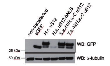
NLS-Motifs in Archaeal R-Proteins . doi:10.1093/molbev/msz207                                                                           MBE

                                                                                                                                                        Downloaded from https://academic.oup.com/mbe/article-abstract/37/1/124/5566250 by guest on 02 March 2020
FIG. 1. Archaeal ribosomal proteins have segments that are similar or identical to NLSs. This figure compares the crystal structures and sequences of
homologous ribosomal proteins from Archaea, Bacteria, and Eukarya. NLSs in eukaryotic proteins and corresponding segments in homologous
archaeal proteins are highlighted in red. This figure illustrates that NLSs of eukaryotic proteins are absent in homologous bacterial ribosomal
proteins, but present in homologous archaeal ribosomal proteins. To compare structural similarity between eukaryotic NLSs and corresponding
protein segments in bacterial and archaeal proteins, the RMSD values are shown for superposition of Ca-atoms of NLS segment in eukaryotic
protein and a corresponding protein segment in archaeal and bacterial homologs.

caused in part by structural divergence of the adjacent helices               H36/H46 and H38/H39 that have conserved tertiary structure
H1 and H98 (fig. 3).                                                          in bacterial and archaeal 23S rRNA, even though in the vicin-
   Similarly, the NLS-type motif of protein uS12 binds 16S                    ity of this NLS-type segment there are additional Archaea/
rRNA segments that have conserved secondary and tertiary                      Eukaryote-specific protein features, including extensions in
structures between Archaea and Bacteria, even though these                    proteins uL4 and eL18 that are missing in bacteria. Overall,
rRNA segments may differ in sequence (e.g., nucleotides 23,                   this structural analysis indicates that NLSs emerged in ribo-
303, 304, 503, 504, and 906 between T. thermophilus and                       somal proteins independently of large structural changes in
P. furiosus 23S rRNA, according to the E. coli numbering).                    rRNA (such as expansion segments) and was accompanied
Similar structural conservation with a moderate degree of                     with relatively minor local conformation and/or sequence
sequence variability can be observed in the rRNA that con-                    variations in 5S, 16S, and 23S rRNA.
tacts the NLS-type segment of uL18 (fig. 3). The NLS-type
segment of uL18, together with another Archaea/                               Archaeal NLS-Type Motifs Can Substitute Eukaryotic
Eukaryote-specific protein segment of uL18, binds 5S rRNA                     NLSs to Direct Intracellular Transport of Ribosomal
and mediates its interaction with 23S rRNA in the central                     Proteins in Eukaryotic Cells
protuberance region of the large ribosomal subunit.                           Finally, we explored whether archaeal NLS-type motifs can
Compared with bacterial 5S rRNA, archaeal 5S rRNA shows                       functionally substitute NLSs of eukaryotic ribosomal proteins.
sequence variations and local base-pairing change in the vi-                  NLSs in ribosomal proteins were described as proteins seg-
cinity of the NLS-type segment, but retains its overall second-               ments comprising 10–20 residues, but none of these
ary and tertiary structure (fig. 3). Similarly, the NLS-type                  residues were studied by point mutagenesis to elucidate
segment of protein uL15 binds 23S helical junctions                           their individual contribution to NLS activity (Table 1,
                                                                                                                                                127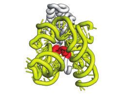
Melnikov et al. . doi:10.1093/molbev/msz207                                                                                                 MBE
Supplementary Material online). However, studies of NLSs in                           proteins, meaning that many archaeal species have fully intact
other eukaryotic proteins revealed that, commonly, NLS ac-                            NLSs segments in proteins uL3 and uL18 (supplementary data
tivity relies on patches of basic and hydrophobic residues that                       2, Supplementary Material online). In ribosomal protein uL15,
mediate NLS interactions with karyopherines (Rout et al.                              Archaea have only one of the two NLS-segments that are
1997; Lee et al. 2006). Are these functional NLS residues pre-                        required for the nuclear import of eukaryotic uL15, indicating
sent in the archaeal NLS-type motifs?                                                 that the complete NLS in eukaryotic uL15 was possibly formed
    As noted above, many archaeal species have identical                              only after the Archaea/Eukarya separation. However, in ribo-
sequences of the NLS-type segments in proteins uL3 and                                somal protein uS12, the 3D-structure of the NLS-containing
uL18 compared with NLS-segments in homologous eukaryotic                              segment is conserved between Archaea and Eukarya (fig. 1),
                                                                                      but the patches of basic and hydrophobic residues are not
                                         uL3      uL15       uL18     uS12            fully preserved in any of the archaeal lineages (supplementary
 (more ancient)                                                                       data 2, Supplementary Material online).
                   DAPNN                 +3.5         +2.1    +4.1   +11.2

                                                                                                                                                           Downloaded from https://academic.oup.com/mbe/article-abstract/37/1/124/5566250 by guest on 02 March 2020
                                                                                          To test if this NLS-like motif in archaeal uS12 can fulfil the
                                                                                      NLS function, we have replaced the NLS in human uS12 with
                   Euryarchaeota         +5.3         +2.1    +6.2    +8.3
                                                                                      the corresponding NLS-type segment of archaeal uS12 from
                                                                                      Sulfolobus solfataricus or Thermoplasma acidophilum. We
                   TACK                  +6.1         +3.1    +5.2   +13.2
                                                                                      then expressed these chimeric proteins as eGFP-fusions in
                                                                                      human cell lines and tested if these proteins can be efficiently
                   Asgard                +6.4         +3.1    +4.2   +12.2
                                                                                      delivered to the nucleoli, a site of ribosome biogenesis in the
  (more recently evolved)
                                                                                      cell nucleus (fig. 4A–K). Notably, as overexpressed ribosomal
                                                                                      proteins are being rapidly degraded in eukaryotic cells (Lam
            Sequence similarity to eukaryotic NLSs (%)
 0                                                                   100              et al. 2007; Sung et al. 2016), we observed that all our expres-
                                                                                      sion constructs (including wild-type human uS12-eGFP fu-
FIG. 2. The NLS-type motifs are conserved in ribosomal proteins from                  sion) have generated not only uS12-eGFP fusion but also
all known branches of the Archaea. A phylogenetic tree of life, as                    some residual quantities of eGFP, which possibly represents
according to Eme et al. (2018), showing the major branches of ar-                     an intermediate product of uS12-eGFP degradation (fig. 4A).
chaeal species and a diagram illustrating high sequence conservation
                                                                                      We then observed that deletion of NLS-segment in uS12
among NLS-type motifs in archaeal ribosomal proteins of the major
groups of archaeal species, from the most ancient branches (DAPNN
                                                                                      resulted in loss of protein stability and loss of fluorescent
superphylum) to the most recently emerged eukaryote-like branch                       signal from human nucleoli (fig. 4A, G, and K). However,
(Asgard superphylum). The numbers in each circle (e.g., þ3.5 in uL3                   uS12 chimeras with archaeal NLS-type motifs had similar ex-
from DAPNN) show an average net charge of an NLS-type segment in                      pression levels and nucleoli localization as the wild-type hu-
a corresponding clade of Archaea.                                                     man uS12, illustrating that the NLS-type motif of archaeal

                                                       uL3                 uL15                  uL18             uS12

                                                                     H36/H46
                                                                                                         5S               h3/h19
                                                                                                               h11

                             Bacteria       H73                                   H37
                             (E. coli)

                                                H92          H91                                                 h3
                                                                                           H85                             h19
                                                                           H38/H39

                                                                     H36/H46
                                                                                                         5S               h3/h19
                                                                                                               h11
                              Archaea
                                                                                     H37
                            (P. furiosus) H73

                                                H92          H91                           H85                   h3
                                                                                                                           h19
                                                                           H38/H39

FIG. 3. NLS-type motifs have emerged independently of changes in ribosomal RNA. The panels show the interior of bacterial (E. coli, PDB ID: 6by1)
and archaeal (P. furiosus, PDB ID: 4v6u) ribosomes where the NLS-type motifs of ribosomal proteins (highlighted in red) and corresponding
segments in bacterial ribosomal proteins interact with rRNA. The figure illustrates that Archaea/Eukarya-specific NLS-type motifs are buried in the
ribosome interior where they bind highly conserved rRNA segments. In the archaeal ribosome structure, the NLS-type motifs bind rRNA helical
junctions, suggesting that the NLS-motifs may help to recognize rRNA or govern rRNA-folding during ribosome biogenesis. Overall, this figure
illustrates that the rRNA structure before and after the emergence of NLS-type motifs remains conserved, illustrating that NLS-type motifs in
archaeal ribosomal proteins have evolved independently of changes in rRNA.

128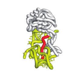
NLS-Motifs in Archaeal R-Proteins . doi:10.1093/molbev/msz207                                                                                          MBE
                                                            eGFP             H.s. uS12          H.s. uS12-∆NLS       S.s.-N/H.s.-C uS12       T.a.-N/H.s.-C uS12
                                                          (control)       (wild-type NLS)        (deleted NLS)         (replaced NLS)           (replaced NLS)

                                                    B                 D                     F                    H                        J
       A

                                                    C                 E                     G                    I                        K

                                                                                                                                                                         Downloaded from https://academic.oup.com/mbe/article-abstract/37/1/124/5566250 by guest on 02 March 2020
FIG. 4. Archaeal NLS-type motifs can functionally substitute NLS-signals of the eukaryotic ribosomal protein uS12 in human cells. (A) Western blot
of HEK293T cells extracts to evaluate expression of uS12-eGFP fusions: the labels correspond to eGFP and uS12-eGFP fusions where H.s. stands for
Human sapiens, S.s.—Sulfolobus solfataricus, and T.a.—Thermoplasma acidophilum. (B–K) The panels show microscopic snapshots of eGFP
fluorescence (green, top panels) and fluorescence of the DNA-staining agent DAPI (blue, bottom panels) in the human cell line HEK293T. Red
arrows point to the location of human cell nucleoli. Cells are expressing the following eGFP fusions: (B, C) eGFP alone (as a negative control) shows
both cytoplasmic and nuclear localization; (D, E) eGFP fusion with human uS12 accumulates in nucleoli; (F, G) eGFP fusion with human uS12
lacking the NLS-containing N-terminus (residues 1–41) is no longer localized in cell nucleoli; (H, I) eGFP fusion with human uS12 in which the N-
terminus (residues 1–41) is replaced by the N-terminus of uS12 from the archaeon S. solfataricus; (J, K) eGFP fusion with human uS12 in which the
N-terminus (residues 1–41) is replaced by the N-terminus of uS12 from the archaeon T. acidophilum. The panels H–K illustrate that N-terminal
segments of the archaeal ribosomal protein uS12 (either from S. solfataricus or T. acidophilum) are able to functionally substitute the NLS-
containing N-terminal segment of human uS12, to direct uS12 accumulation in human cell nucleoli.

ribosomal protein uS12 is not only similar in sequence and                     could not support flight. Thus, the advantage of flight could
structure to eukaryotic NLSs, but can also complements the                     not drive the positive selection of species with “germinal,”
activity of eukaryotic NLSs (fig. 4A and H–K). Overall, these                  “premature” wings. Thus, what was the advantage of “half a
observations suggest that—in some cellular proteins—                           wing”? Studies of modern birds suggest that wings could ini-
protein segments, which can fulfil biological activity of eu-                  tially have emerged to facilitate gliding or accelerated climb-
karyotic NLSs, preexisted the origin of the nucleus and the                    ing—two forms of motion related to flying that are preserved
split of archaeal and eukaryotic lineages of life.                             in the behavior of modern birds (Padian and Chiappe 1998;
                                                                               Zhou 2004; Dial et al. 2008).
                                                                                   Similar to the origin of wings, it is possible that NLSs could
Discussion                                                                     initially have emerged to fulfil similar, yet not identical, or
In this study, we have identified a group of rare evolutionary                 incomplete functions of modern NLSs. In this regard, it is
intermediates that can shed light on the sequence of events                    important to note that NLSs are known to fulfil three biolog-
that led to the emergence of the nuclear–cytoplasmic trans-                    ical activities: 1) they participate in the recognition of nucleic
port system. Our finding of NLS-type sequences in the ribo-                    acids, because most known NLSs reside within DNA- or RNA-
somal proteins uS12, uL3, uL15, and uL18 suggests that at                      binding protein domains (LaCasse and Lefebvre 1995); 2) they
least some cellular proteins evolved NLS-motifs long before                    mediate protein transport across nuclear pores by recruiting
the nucleus emerged, indicating that the initial function of                   karyopherins (Dingwall et al. 1982; Gorlich et al. 1994); and 3)
NLSs could have been distinct from directing protein trans-                    karyopherin-binding to NLSs shields their electrostatic
port. Why the NLS-motifs emerged in ribosomal proteins in                      charges and prevents nonspecific interactions of nucleic
the Archaea—in species that have neither cell nucleus nor                      acid-binding domains(Jakel et al. 2002). In other words, kar-
karyopherin proteins to recognize the NLS-type motifs—                         yopherins that bind NLSs not only fulfil the role of transport
remains unclear (Koonin and Aravind 2009; Field and Rout                       factors but also serve as chaperones for highly charged pro-
2019), as does what advantage might have been conferred by                     teins in eukaryotic cells (Jakel et al. 2002).
having these signals in the absence of nuclear–cytoplasmic                         Based on our findings, it is tempting to suggest an order in
transport. Furthermore, the initial function of NLS-type                       which these three biological activities of NLSs may have
sequences in cells in the absence of the nuclear envelope                      evolved. High conservation of NLS-type motifs in archaeal
and the nuclear–cytoplasmic trafficking system is unknown.                     ribosomal proteins and their abundant contacts with rRNA
   These questions resonate with several classical studies in                  suggest that, at least in some proteins, the NLS-type sequen-
evolutionary biology, such as studies on the origin of birds                   ces could have evolved as nucleic acid-binding domains that
(Dial et al. 2006). In the origin of birds theory, there is a                  were required to increase the specificity of interactions in the
famous question “of what use is half a wing?” This refers to                   increasingly complex macromolecular content of evolving
the fact that it took multiple millions of years to transform                  cells (fig. 5A). Later, these cells could have evolved a DNA/
the limbs of ancestral reptiles into the wings of ancestral birds.             RNA-mimicking chaperone that would recognize NLS-type
Yet, for most of this time, the transitional forms of a wing                   sequences in the way chaperones for hydrophobic proteins
                                                                                                                                                                   129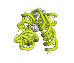
Melnikov et al. . doi:10.1093/molbev/msz207                                                                                                MBE

                                                                                                                                                           Downloaded from https://academic.oup.com/mbe/article-abstract/37/1/124/5566250 by guest on 02 March 2020
FIG. 5. A hypothetical order of NLS evolution in eukaryotic proteins. In the modern eukaryotic cell, NLSs fulfil three biological activities: 1) they
serve as signal peptides to direct protein transport into the nucleus; 2) during this protein transport, they recruit trafficking factors (karyopherins)
that shield high positive charge of nucleic acid-binding domains (which prevents nonspecific interactions of NLS-containing proteins with other
molecules in a cell); and 3) typically, NLSs reside within DNA- or RNA-binding domains of proteins and, therefore, NLSs mediate specific
recognition of nucleic acids. Our model suggests that NLSs may have originally emerged as nucleic acid-binding domains in a cell lacking the
nuclear–cytoplasmic separation (Stage A). Later, a chaperone emerged that shielded the highly positively charged nucleic acid-binding domains
(Stage B). Finally, when cells got separated into the nucleus and the cytoplasm, this chaperone turned into a karyopherin when it acquired the
additional capacity to mediate the long-distance trafficking of proteins across nuclear pores (Stage C). Thus, NLSs could initially emerge to help
cellular proteins to recognize nucleic acids, and only later NLSs were endowed with an additional function of trafficking signals.

recognize hydrophobic protein segments to prevent nonspe-                      NLS-type motifs described in this study precisely overlap
cific interactions and protein aggregation (fig. 5B). Only later,              with the N-terminal AE-segments in ribosomal proteins
when this chaperone for charged proteins had evolved the                       uS12, uL3, uL15, and uL18 (fig. 1). Given that NLSs have
ability to bind to the nuclear pores and direct protein trans-                 been experimentally identified only in about a quarter of
port to the nucleus, did the NLSs eventually become signals of                 ribosomal proteins, it is tempting to suggest that some of
intracellular transport (fig. 5C). If this hypothetical order of               the AE segments may point to the location of NLS and NLS-
events is correct, then the system of nuclear–cytoplasmic                      type motifs in eukaryotic and archaeal ribosomal proteins for
trafficking could originally have evolved as a system of chap-                 which location of NLSs is currently unknown.
erones for highly charged proteins, to prevent nucleic acid-                      Our study leaves unanswered the question: why do the
binding domains from nonspecific interactions in the increas-                  nonglobular extensions of ribosomal proteins have different
ingly complex environment of ancestral eukaryotic cells.                       structures in Bacteria and Archaea, even though these exten-
    Noteworthy, we showed earlier that many ribosomal pro-                     sions bind invariable, universally conserved rRNA segments?
teins undergo local structural remodeling in Archaea and                       Previously, it was suggested that these structural differences
Eukarya compared with Bacteria. We termed the locally al-                      stemmed from an independent evolutionary origin for non-
tered protein segments as “AE”-segments, where “AE” indi-                      globular segments in bacterial and eukaryotic ribosomal pro-
cates conservation of their 3D structures in Eukarya and                       teins (Klein et al. 2004). Another possible reason may be
Archaea, but not in Bacteria. Curiously, all the four                          related to differences in ribosome biogenesis pathways

130
NLS-Motifs in Archaeal R-Proteins . doi:10.1093/molbev/msz207                                                          MBE
between Bacteria (in which ribosome biogenesis largely              sequences of the eukaryotic NLSs. The NLS-consensus
occurs via self-assembly and with the help of protein-              sequences were defined as follows: 11-RKLxxxRRxxRWxxxx-
guided rRNA modification machinery) and Archaea/                    YKKRxxxxxxKxxP40 for protein uS12 (residue numbers indi-
Eukarya (in which ribosome biogenesis requires archaea/             cate their position in uS12 from S. cerevisiae, “x” designates
eukaryote-specific biogenesis factors and RNA-guided ma-            any amino acid); 3-HRKxxxPRHxxxxxxPRKR21 for protein
chinery for rRNA modification) (Hage and Tollervey 2004;            uL3;     4-
                                                                               RxxKxRKxR12      for     protein      uL15;   and
Blombach et al. 2011; Yip et al. 2013; de la Cruz et al. 2015).     20-
                                                                       FRRRRxxKxxY30 for protein uL18. To estimate an average
   We also cannot exclude the possibility that these differ-        net charge of the NLS-type segments, the net charge was
ences may be related not to the problem of ribosome evolu-          calculated for each NLS segment (as they are defined in ta-
tion and biogenesis but to a more general problem of specific       ble 1) from each archaeal species shown in supplementary
macromolecular interactions in a cell. When life first emerged      data 1, Supplementary Material online by using the Prot pi
on our planet, the earliest life forms were likely made of a very   Protein Tool of Zurich University of Applied Sciences (https://

                                                                                                                                      Downloaded from https://academic.oup.com/mbe/article-abstract/37/1/124/5566250 by guest on 02 March 2020
limited number of molecules. Therefore, it was relatively easy      www.protpi.ch/Calculator/ProteinTool;        last     accessed
for these molecules to find one specific partner among all the      September 16, 2019) with default values (pH 7.4), and then
other molecules in a living cell. However, as cells grew in size    average net-charge values were shown for proteins uL3, uL15,
and complexity, it is possible, even probable, that the old rules   uL18, and uS12 from each of the archaeal clades in fig. 2.
of specific interactions between cellular molecules had to be       Sequence similarity shown in fig. 2 was calculated according
gradually redefined in order to help cellular proteins and nu-      to the AMAS definitions for the hierarchical analysis of resi-
cleic acids to find their specific partners more easily in the      due conservation (Livingstone and Barton 1993) using Jalview
complex environment of a complex cell. Given that all the           (Troshin et al. 2011) (supplementary data 2, Supplementary
NLS-type sequences identified in this study create a new            Material online).
RNA/protein interface, it is possible that evolving these
sequences gave ribosomal proteins the advantage of more             Comparison of Ribosome Structures
specific recognition of nucleic acid and thereby gave cells a       The ribosome structures were retrieved from the protein
chance to further expand diversity of their proteomes and           databank (https://www.rcsb.org; last accessed September 16,
nucleic acids without a risk of nonspecific interactions. In        2019) and were visualized and inspected by using PyMOL
other words, it is possible that NLS-type sequences could           Molecular Graphics System (Version 2.0 Schrödinger, LLC.).
originally emerge to increase specific molecular interactions       Homologous ribosomal proteins were aligned by using “align”
and only later were repurposed into the signals of intracellular    command to superpose Ca-atoms of conserved globular
trafficking.                                                        domains of homologous proteins or “pair_fit” command to
                                                                    superpose phosphate atoms of conserved rRNA segments in
                                                                    bacterial and archaeal ribosomes. To measure RMSD values
Materials and Methods                                               between NLSs in eukaryotic ribosomal proteins and corre-
Comparison of Protein Sequences                                     sponding segments in bacterial and archaeal ribosomal pro-
The sequences of ribosomal proteins were retrieved from the         teins, we used “pair_fit” command in the PyMOL Molecular
Uniprot protein databank (https://www.uniprot.org; last             Graphics System (Version 2.0 Schrödinger, LLC.) to align cor-
accessed September 16, 2019) by using a script described in         responding Ca-atoms as defined in the multiple sequence
Melnikov, van den Elzen, et al. (2018). For species containing      alignment in fig. 1.
numerous copies of ribosomal proteins’ genes (for instance, S.
cerevisiae or Arabidopsis thaliana) only one isoform (isoform       Cloning of Ribosomal Proteins
A or isoform 1) was used per species. For bacterial species in      The cDNA of human protein uS12 (NCBI Gene ID: 6228 and
which several ribosomal proteins are encoded by two genes           Uniport ID: P62266) and its N-terminally truncated mutant
(corresponding to Zn-coordinating and Zn-free isoforms of           (deleted residues 1–41) were cloned into pEGFP-N1 vector
ribosomal proteins), we only used the genes coding for Zn-          (Promega) between KpnI and XhoI sites as described in
coordinating isoforms. All the retrieved sequences are listed in    (Melnikov et al. 2015). To create chimeras between human
supplementary data 1, Supplementary Material online. To             uS12 and homologous proteins from T. acidophilum and S.
create multiple sequence alignments, we used the                    solfataricus, the genomic sequence that corresponds to resi-
precompiled package of MAFFT (MAFFT-7.427) with default             dues 1–41 of human uS12 was replaced by codon-optimized
settings (Nakamura et al. 2018).                                    sequences from T. acidophilum (NCBI Gene ID: 1455748) and
   To assess the conservation of NLS-type motifs in archaeal        S. solfataricus (NCBI Gene ID: 38467843) corresponding to
proteins, we calculated consensus sequences of NLS-type             residues 1–37 of uS12 from T. acidophilum (Uniprot ID:
motifs for each of the four ribosomal proteins (uS12, uL3,          Q9HLY2) or residues 1–42 from of uS12 from S. solfataricus
uL15, and uL18) in each of the major archaeal branches              (Uniport ID: P39573). The codon optimization for protein
(DAPNN, Euryarchaeota, TACK, and Asgard) by using multi-            expression in human cells was done by using the codon op-
ple sequence alignments (MAFFT with default settings) of            timization algorithm from the Integrated DNA Technologies
archaeal protein sequences listed in supplementary data 1,          (https://www.idtdna.com; last accessed September 16, 2019).
Supplementary Material online. These consensus sequences            The DNA fragments corresponding to the N-termini of ar-
were then compared with the corresponding consensus                 chaeal uS12 were purchased from GenScript (https://www.
                                                                                                                               131
Melnikov et al. . doi:10.1093/molbev/msz207 MBE
genscript.com; last accessed September 16, 2019) and cloned Molecular Biology Organization (ASTF 434-2014 to S.M.) and
into the H. sapiens uS12-pEGFP-N1 construct by using In- from the National Institute of General Medical Sciences
Fusion HD Cloning Plus (Takara). (R35GM122560 to D.S.).
Cell Lines, Transfection, Confocal Microscopy, and References
Western Blot Analysis Armache JP, Anger AM, Marquez V, Franckenberg S, Frohlich T, Villa E,
Nucleolar accumulation of eGFP-fused human ribosomal pro- Berninghausen O, Thomm M, Arnold GJ, Beckmann R. 2013.
tein uS12 and its variants were examined in the human cell Promiscuous behaviour of archaeal ribosomal proteins: implications
for eukaryotic ribosome evolution. Nucleic Acids Res.
line HEK293T (ATCC, CCL11268). HEK293T cells were main- 41(2):1284–1293.
tained in DMEM (Gibco) supplemented with 10% FBS Ban N, Nissen P, Hansen J, Moore PB, Steitz TA. 2000. The complete
(Gibco). Cells were plated on poly-L-lysine-coated glass cover atomic structure of the large ribosomal subunit at 2.4 A resolution.
slips at 70–80% confluence, and transfected with the respec- Science 289(5481):905–920.
Downloaded from https://academic.oup.com/mbe/article-abstract/37/1/124/5566250 by guest on 02 March 2020
tive plasmids encoding uS12 and its variants using Ben-Shem A, Garreau de Loubresse N, Melnikov S, Jenner L, Yusupova G,
Yusupov M. 2011. The structure of the eukaryotic ribosome at 3.0 A
Lipofectamine 2000 (Invitrogen), according to the manufac- resolution. Science 334(6062):1524–1529.
turers’ protocol. Blombach F, Brouns SJ, van der Oost J. 2011. Assembling the archaeal
Confocal fluorescent images were obtained by a Zeiss LSM ribosome: roles for translation-factor-related GTPases. Biochem Soc
880 Airyscan NLO/FCS confocal microscope 48 h after trans- Trans. 39(1):45–50.
fection. Prior to imaging, cell samples were fixed with 4% Bokov K, Steinberg SV. 2009. A hierarchical model for evolution of 23S
ribosomal RNA. Nature 457(7232):977–980.
paraformaldehyde, permeabilized with 0.1% triton X-100 Brocks JJ, Logan GA, Buick R, Summons RE. 1999. Archean molecular
R
and mounted using with ProLongV Diamond Antifade fossils and the early rise of eukaryotes. Science 285(5430):1033–1036.
Mountant (Invitrogen) with DAPI. For each eGFP-uS12 vari- Chen IJ, Wang IA, Tai LR, Lin A. 2008. The role of expansion segment of
ant (and also for eGFP protein as a negative control), the human ribosomal protein L35 in nuclear entry, translation activity,
snapshots were taken for six nonoverlapping fields at 63x— and endoplasmic reticulum docking. Biochem Cell Biol 86:271–217.
Chi A, Huttenhower C, Geer LY, Coon JJ, Syka JE, Bai DL, Shabanowitz J,
for better data redundancy (supplementary fig. S1, Burke DJ, Troyanskaya OG, Hunt DF. 2007. Analysis of phosphory-
Supplementary Material online). For each genetic construct, lation sites on proteins from Saccharomyces cerevisiae by electron
experiments were repeated three times. transfer dissociation (ETD) mass spectrometry. Proc Natl Acad Sci U
To carry out the Western blot analysis, HEK293T cells were S A. 104(7):2193–2198.
transfected with the constructs encoding eGFP-fused human Chiappe P. 1999. The wing of Archaeopteryx as a primary thrust gener-
ator. Nature 399:60–62.
ribosomal protein uS12 and its variants using Fugene HD Daeschler EB, Shubin NH, Jenkins FA Jr. 2006. A Devonian tetrapod-like
(Promega). The cells were lysed in radioimmunoprecipitation fish and the evolution of the tetrapod body plan. Nature
(RIPA) lysis buffer, supplemented with 1x Halt TM protease 440(7085):757–763.
inhibitor cocktail (Thermo Scientific), for 15 min on ice. Das P, Basu A, Biswas A, Poddar D, Andrews J, Barik S, Komar AA,
Lysates were cleared at 15,000 g for 15 min at 4 C. Mazumder B. 2013. Insights into the mechanism of ribosomal in-
corporation of mammalian L13a protein during ribosome biogene-
Protein concentrations were quantified with Quick Start sis. Mol Cell Biol. 33:2829–2842.
TM Bradford 1x dye reagent (Bio-Rad). Samples were resolved de la Cruz J, Karbstein K, Woolford JL Jr. 2015. Functions of ribosomal
on 10% SDS-PAGE gels. Immunoblots were incubated with proteins in assembly of eukaryotic ribosomes in vivo. Annu Rev
anti-GFP (Cell Signaling Technology, #2956), or anti-a-tubulin Biochem. 84:93–129.
(Cell Signaling Technology, #3873) overnight at 4 C, followed Dial KP, Jackson BE, Segre P. 2008. A fundamental avian wing-stroke
provides a new perspective on the evolution of flight. Nature
by a secondary horseradish peroxidase HRP-conjugated anti- 451(7181):985–989.
body for 1 h at room temperature. Dial KP, Randall RJ, Dial TR. 2006. What use is half a wing in the ecology
and evolution of birds? BioScience 56(5):437–445.
Supplementary Material Dingwall C, Sharnick SV, Laskey RA. 1982. A polypeptide domain that
specifies migration of nucleoplasmin into the nucleus. Cell
Supplementary data are available at Molecular Biology and 30(2):449–458.
Evolution online. Dodd MS, Papineau D, Grenne T, Slack JF, Rittner M, Pirajno F, O’Neil J,
Little CTS. 2017. Evidence for early life in Earth’s oldest hydrothermal
Acknowledgments vent precipitates. Nature 543(7643):60–64.
Eme L, Spang A, Lombard J, Stairs CW, Ettema T. 2018. Archaea and the
We thank members of Dieter Söll, Michael Rout, and Marat origin of eukaryotes. Nat Rev Microbiol. 16(2):120.
Yusupov laboratories for valuable discussions at the early Field MC, Rout MP. 2019. Pore timing: the evolutionary origins of the
stage of this project. We also thank Richard Prum and nucleus and nuclear pore complex. F1000 Res. 8:369.
Gorlich D, Prehn S, Laskey RA, Hartmann E. 1994. Isolation of a protein
Günter Wagner (Department of Ecology and Evolutionary that is essential for the first step of nuclear protein import. Cell
Biology at Yale University) for critical feedback, stimulating 79(5):767–778.
discussions and inspirational comments during preparation Hage AE, Tollervey D. 2004. A surfeit of factors: why is ribosome assembly
of the manuscript, Adam Bodley for editing, and Vladimir so much more complicated in eukaryotes than bacteria? RNA Biol.
Roudko (Icahn School of Medicine at Mount Sinai), and 1(1):10–15.
Hartman H, Smith TF. 2014. The evolution of the ribosome and the
anonymous reviewers for thorough reading, thoughtful com- genetic code. Life 4(2):227–249.
ments, and valuable suggestions that greatly improved our Henneman B, van Emmerik C, van Ingen H, Dame RT. 2018. Structure
work. This work was supported by grants from the European and function of archaeal histones. PLoS Genet. 14(9):e1007582.
132NLS-Motifs in Archaeal R-Proteins . doi:10.1093/molbev/msz207 MBE
Hsiao C, Mohan S, Kalahar BK, Williams LD. 2009. Peeling the onion: Melnikov S, Manakongtreecheep K, Soll D. 2018. Revising the structural
ribosomes are ancient molecular fossils. Mol Biol Evol. diversity of ribosomal proteins across the three domains of life. Mol
26(11):2415–2425. Biol Evol. 35(7):1588–1598.
Jakel S, Mingot JM, Schwarzmaier P, Hartmann E, Gorlich D. 2002. Melnikov SV, Khabibullina NF, Mairhofer E, Vargas-Rodriguez O,
Importins fulfil a dual function as nuclear import receptors and Reynolds NM, Micura R, Soll D, Polikanov YS. 2019. Mechanistic
cytoplasmic chaperones for exposed basic domains. EMBO J. insights into the slow peptide bond formation with D-amino acids
21(3):377–386. in the ribosomal active site. Nucleic Acids Res. 47(4):2089–2100.
Jenkinson ER, Chong JP. 2003. Initiation of archaeal DNA replication. Melnikov SV, van den Elzen A, Stevens DL, Thoreen CC, Soll D. 2018. Loss
Biochem Soc Trans. 31(Pt 3):669–673. of protein synthesis quality control in host-restricted organisms. Proc
Johnson DB, Wang L. 2010. Imprints of the genetic code in the ribosome. Natl Acad Sci U S A. 115(49):E11505–E11512.
Proc Natl Acad Sci U S A. 107(18):8298–8303. Moreland RB, Nam HG, Hereford LM and Fried HM. 1985. Identification
Padian K, Chiappe LM. 1998. The origin and early evolution of birds. Biol of a nuclear localization signal of a yeast ribosomal protein. Proc Natl
Rev. 73(1):1–42. Acad Sci U S A 82:6561–6565.
Klein DJ, Moore PB, Steitz TA. 2004. The roles of ribosomal proteins in Nakamura T, Yamada KD, Tomii K, Katoh K. 2018. Parallelization of
the structure assembly, and evolution of the large ribosomal subunit. MAFFT for large-scale multiple sequence alignments.
Downloaded from https://academic.oup.com/mbe/article-abstract/37/1/124/5566250 by guest on 02 March 2020
J Mol Biol. 340(1):141–177. Bioinformatics 34(14):2490–2492.
Klinge S, Voigts-Hoffmann F, Leibundgut M, Arpagaus S, Ban N. 2011. Noller HF. 2012. Evolution of protein synthesis from an RNA world. Cold
Crystal structure of the eukaryotic 60S ribosomal subunit in complex Spring Harb Perspect Biol. 4(4):a003681.
with initiation factor 6. Science 334(6058):941–948. Petrov AS, Gulen B, Norris AM, Kovacs NA, Bernier CR, Lanier KA, Fox
Koch B, Mitterer V, Niederhauser J, Stanborough T, Murat G, Rechberger GE, Harvey SC, Wartell RM, Hud NV, et al. 2015. History of the
G, Bergler H, Kressler D, Pertschy B. 2012. Yar1 protects the ribo- ribosome and the origin of translation. Proc Natl Acad Sci U S A.
somal protein Rps3 from aggregation. J Biol Chem. 287:21806–21815. 112(50):15396–15401.
Koonin EV, Aravind L. 2009. Comparative genomics, evolution and ori- Rabl J, Leibundgut M, Ataide SF, Haag A, Ban N. 2011. Crystal structure of
gins of the nuclear envelope and nuclear pore complex. Cell Cycle the eukaryotic 40S ribosomal subunit in complex with initiation
8(13):1984–1985. factor 1. Science 331(6018):730–736.
Krupkin M, Matzov D, Tang H, Metz M, Kalaora R, Belousoff MJ, Rasmussen B, Fletcher IR, Brocks JJ, Kilburn MR. 2008. Reassessing the
Zimmerman E, Bashan A, Yonath A. 2011. A vestige of a prebiotic first appearance of eukaryotes and cyanobacteria. Nature
bonding machine is functioning within the contemporary ribosome. 455(7216):1101–1104.
Philos Trans R Soc Lond, B Biol Sci. 366(1580):2972–2978. Rosorius O, Fries B, Stauber RH, Hirschmann N, Bevec D, Hauber J. 2000.
LaCasse EC, Lefebvre YA. 1995. Nuclear localization signals overlap DNA- Human ribosomal protein L5 contains defined nuclear localization
or RNA-binding domains in nucleic acid-binding proteins. Nucleic and export signals. J Biol Chem. 275:12061–12068.
Acids Res. 23(10):1647–1656. Rout MP, Blobel G, Aitchison JD. 1997. A distinct nuclear import path-
Lam YW, Lamond AI, Mann M, Andersen JS. 2007. Analysis of nucleolar way used by ribosomal proteins. Cell 89(5):715–725.
protein dynamics reveals the nuclear degradation of ribosomal pro- Schaap PJ, van’t Riet J, Woldringh CL, Raue HA. 1991. Identification and
teins. Curr Biol. 17(9):749–760. functional analysis of the nuclear localization signals of ribosomal
Lee BJ, Cansizoglu AE, Suel KE, Louis TH, Zhang Z, Chook YM. 2006. Rules protein L25 from Saccharomyces cerevisiae. J Mol Biol. 221:225–237.
for nuclear localization sequence recognition by karyopherin beta 2. Sung MK, Reitsma JM, Sweredoski MJ, Hess S, Deshaies RJ. 2016.
Cell 126(3):543–558. Ribosomal proteins produced in excess are degraded by the
Lindstrom MS. 2012. Elucidation of motifs in ribosomal protein S9 that ubiquitin-proteasome system. Mol Biol Cell. 27(17):2642–2652.
mediate its nucleolar localization and binding to NPM1/nucleophos- Timmers AC, Stuger R, Schaap PJ, van ’t Riet J, Raue HA. 1999. Nuclear
min. PLoS One 7:e52476. and nucleolar localization of Saccharomyces cerevisiae ribosomal
Livingstone CD, Barton GJ. 1993. Protein sequence alignments: a strategy proteins S22 and S25. FEBS Lett. 452:335–340.
for the hierarchical analysis of residue conservation. Comput Appl Troshin PV, Procter JB, Barton GJ. 2011. Java bioinformatics analysis web
Biosci. 9(6):745–756. services for multiple sequence alignment–JABAWS: MSA.
Norell MA, Xu X. 2005. Feathered dinosaurs. Annu Rev Earth Planet Sci. Bioinformatics 27(14):2001–2002.
33(1):277–299. Underwood MR, Fried HM. 1990. Characterization of nuclear localizing
Martin WF, Garg S, Zimorski V. 2015. Endosymbiotic theories for eu- sequences derived from yeast ribosomal protein L29. EMBO J.
karyote origin. Philos Trans R Soc Lond. B Biol Sci. 9:91–99.
370(1678):20140330. Yip WS, Vincent NG, Baserga SJ. 2013. Ribonucleoproteins in archaeal
Melnikov S, Ben-Shem A, Garreau de Loubresse N, Jenner L, Yusupova G, pre-rRNA processing and modification. Archaea 2013:614735.
Yusupov M. 2012. One core, two shells: bacterial and eukaryotic Zhou Z. 2004. The origin and early evolution of birds: discoveries, dis-
ribosomes. Nat Struct Mol Biol. 19(6):560–567. putes, and perspectives from fossil evidence. Naturwissenschaften
Melnikov S, Ben-Shem A, Yusupova G, Yusupov M. 2015. Insights into 91(10):455–471.
the origin of the nuclear localization signals in conserved ribosomal
proteins. Nat Commun. 6:7382.
133You can also read