Hyperbaric oxygen protects against myocardial ischemia reperfusion injury through inhibiting mitochondria dysfunction and autophagy - Spandidos ...
←
→
Page content transcription
If your browser does not render page correctly, please read the page content below
4254                                       Molecular Medicine REPORTS 22: 4254-4264, 2020

                      Hyperbaric oxygen protects against myocardial
                      ischemia‑reperfusion injury through inhibiting
                         mitochondria dysfunction and autophagy
       WAN CHEN1*, LIWEN LV1*, ZHIHUAN NONG2, XIAOYU CHEN2, XIAORONG PAN3 and CHUNXIA CHEN3

                     Departments of 1Emergency, 2Pharmacy and 3Hyperbaric Oxygen, The People's Hospital of
                           Guangxi Zhuang Autonomous Region, Nanning, Guangxi 530021, P.R. China

                                           Received May 22, 2019; Accepted June 22, 2020

                                                    DOI: 10.3892/mmr.2020.11497

Abstract. Our previous study demonstrated that hyperbaric              Introduction
oxygen (HBO) improves heart function predominantly through
reducing oxygen stress, modulating energy metabolism and               Coronary artery ischemic disease is prevalent worldwide (1),
inhibiting cell apoptosis. The present study aimed to investigate      it affects >15 million adults in the United States (2). A lack
the protective effects of HBO on mitochondrial function and            of coronary blood supply caused by thrombosis or the acute
autophagy using rats with a ligated left anterior descending artery.   alteration of coronary atherosclerotic plaques contributes to
The cardioprotective effects of HBO were mainly evaluated using        myocardial ischemia (MI) (3). The early restoration of blood
ELISA, fluorescent probes, transmission electron microscopy            flow is a common treatment strategy (4); however, it can
and reverse transcription‑quantitative PCR (RT‑qPCR). HBO              also cause further severe cardiac damage, which is referred
pretreatment for 14 days (once a day) using a 0.25 MPa chamber         to as myocardial ischemia‑reperfusion injury (MIRI) (5,6).
improved mitochondrial morphology and decreased the number             Therefore, it is of great importance to further investigate safe
of autophagic vesicles, as observed using a transmission electron      and effective novel therapeutic treatments to prevent MIRI and
microscope. HBO pretreatment significantly increased the levels        improve the clinical outcomes of acute MI.
of ATP, ADP, energy charge and the opening of the mitochondrial            It is well established that mitochondria are the powerhouses
permeability transition pore, but decreased the levels of AMP,         of the cell due to their crucial role in generating ATP (7), but
cytochrome c and reactive oxygen species. Moreover, HBO                they are also important regulators of programmed cell death
pretreatment significantly increased the gene or protein expres-       pathways (8). Properties of mitochondrial dysfunction include
sion levels of eIF4E‑binding protein 1, mammalian target of            a reduction in energy charge (EC), the opening of the mito-
rapamycin (mTOR), mitochondrial DNA, NADH dehydrogenase                chondrial permeability transition pore (mPTP), the release
subunit 1, mitofusin 1 and mitofusin 2, whereas it decreased the       of cytochrome c and Ca 2+ overload (9). These properties
gene or protein expression levels of autophagy‑related 5 (Atg5),       subsequently lead to mitochondrial membrane depolarization,
cytochrome c, dynamin‑related protein 1 and p53, as determined         the homeostatic imbalance between apoptotic proteins, and
using RT‑qPCR or immunohistochemistry. In conclusion, HBO              ultimately cardiomyocyte death (10). Reactive oxygen species
treatment was observed to protect cardiomyocytes during                (ROS), of which mitochondria are the predominant source,
myocardial ischemia‑reperfusion injury (MIRI) by preventing            induce MIRI through mitochondrial DNA (mtDNA) damage,
mitochondrial dysfunction and inhibiting autophagy. Thus, these        reducing energy production and inhibiting protein synthesis
results provide novel evidence to support the use of HBO as a          through a vicious circle of mitochondrial damage (11,12). In
potential agent for the mitigation of MIRI.                            addition, mitochondria are dynamic organelles that continu-
                                                                       ally alter their morphology by undergoing routine fission and
                                                                       fusion events (13,14), which requires specifically controlled
                                                                       proteins; for example, dynamin‑related protein 1 (Drp 1), which
                                                                       is a cytosolic GTPase that serves a fundamental role in mito-
Correspondence to: Dr Chunxia Chen, Department of Hyperbaric           chondrial fission by translocating to the outer mitochondrial
Oxygen, The People's Hospital of Guangxi Zhuang Autonomous
                                                                       membrane to generate the force necessary for mitochondrial
Region, 6 Taoyuan Road, Nanning, Guangxi 530021, P.R. China
E‑mail: chunxia251401@126.com                                          fission (15,16), and mitofusin 1 (Mfn1) and mitofusin 2 (Mfn2),
                                                                       which are required for the membrane remodeling processes
*
    Contributed equally                                                necessary for mitochondrial fusion (17,18).
                                                                           Autophagy is an evolutionarily conserved, lyso-
Key words: hyperbaric oxygen, myocardial ischemia‑reperfusion          some‑dependent degradation process that is activated during
injury, mitochondria function, autophagy                               MIRI (19). It has been reported that autophagy serves a dual
                                                                       role in MIRI:A slight induction of autophagy promotes cell
                                                                       survival during ischemia, whilst a significant increase induces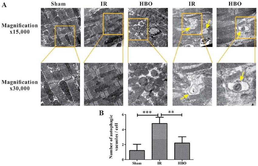
CHEN et al: HYPERBARIC OXYGEN PROTECTS AGAINST MYOCARDIAL ISCHEMIA-REPERFUSION INJURY                           4255

cell death during reperfusion (20,21). Although a small            Experimental design and groupings. According to previous
amount of autophagy can maintain cell function, excessive          guidelines (41), following 1 week of acclimatization to the labo-
autophagy was found to promote myocardial injury due to the        ratory conditions, the 60 rats were divided into the following
consumption of cellular constituents (22). Mammalian target        three groups (20 rats/group) using the random number table
of rapamycin (mTOR) kinase is an important regulator of the        method: i) Sham group; ii) ischemia‑reperfusion (IR) group
classical autophagy pathway (23,24), and autophagy related 5       (MIRI model); and iii) HBO group.
(Atg5) and microtubule‑associated protein 1A/1B‑light                  MIRI was inflicted by the occlusion of the LAD coro-
chain 3 (LC3) conjugation systems are required for the forma-      nary artery followed by reperfusion, according to a previous
tion of autophagic vesicles (25). Eukaryotic initiation factor     study (40). Briefly, rats were anaesthetized with 30 mg/kg
4E‑binding protein 1 (4E‑BP1) binds to eukaryotic initia-          sodium pentobarbital (i.p.) and mechanically ventilated using
tion factor 4E (eIF4E) (26). The phosphorylation of 4E‑BP1         an animal respirator (respiration rate, 70 breaths/min; tidal
disrupts the assembly of the eIF4E/4E‑BP1 complex, which           volume, 6‑8 ml/kg; Shanghai Alcott Biotech Co., Ltd.). The
initiates eIF4E‑dependent translation, and thereby the activa-     chest was then opened and the heart was exposed. The LAD
tion of cap‑dependent mRNA translation (27). In addition           artery was ligated using a 5‑0 silk suture for 30 min and
to regulating cell cycle checkpoints and apoptosis (28), p53       released to allow reperfusion for 1 h.
has also been demonstrated to mediate the transactivation of           Rats in the IR and HBO groups were subjected to a 30 min
autophagy inducers (29,30).                                        LAD ligation followed by reperfusion for 1 h, whilst the sham
    Hyperbaric oxygen (HBO) therapy is the clinical applica-       group received encircling of the LAD artery with a suture, but
tion of pure oxygen at a higher pressure (usually 2‑3 times        no ligation. Before the surgical procedure, rats in the HBO
atmospheric pressure) in a chamber to treat ischemia‑ or           group were pretreated with HBO for 1 h (0.25 MPa) for 14 days
hypoxia‑associated diseases, such as coronary heart disease,       (once daily) in a hyperbaric chamber (Yantai Hongyuan Co.,
cerebral infarction and carbon monoxide poisoning (31‑34).         Ltd.), as previously described (38).
HBO therapy has been widely agreed by major hyperbaric                 At the end of the reperfusion, all rats were immediately
craft groups, such as the Undersea and Hyperbaric Medical          anesthetized using 30 mg/kg sodium pentobarbital (i.p.) and
Society or the U.S. Food and Drug Administration (35). In          were subsequently euthanized through exsanguination by
addition, data from our previous studies demonstrated that         collecting 8 ml blood from the abdominal aorta. Death was
HBO exerted neuroprotective effects in certain animal              confirmed following the detection of a still heartbeat, and
models; for example, HBO combined with Madopar protected           when breathing had stopped. Blood samples were centrifuged
against 6‑hydroxydopamine‑induced Parkinson's disease              at 302 x g for 10 min at 4˚C, and the supernatant was collected
in rats (36) and HBO treatment also alleviated the with-           and stored at ‑80˚C for use in the biochemical assays. The
drawal symptoms induced by morphine dependence (37).               heart was promptly removed from the rats and the infarct was
Moreover, HBO prevented the cognitive impairments                  isolated on an ice box for further measurements.
induced by D‑galactose (38,39). The protective effects of
HBO are mainly associated with the increased induction of          Determination of ATP, ADP, AMP and cytochrome c levels
antioxidant enzymes and ischemic tolerance, as well as the         in MI tissue. Myocardial samples (n=5) were homogenized
inhibition of cell apoptosis and the modulation of neurotrans-     in ice‑cold physiological saline (10%, w/v) and centrifuged at
mitters (36‑39). Previously, our group discovered that HBO         3,354 x g for 10 min at 4˚C to collect the supernatant. ATP
preconditioning protected against MI and improved cardiac          and ADP levels were determined using ATP and ADP assay
function (40), of which the underlying mechanisms were             kits, respectively (colorimetric method; cat. nos. ab83355
associated with the reduction of oxygen stress, the correc-        and ab83359; Abcam), according to the manufacturer's
tion of energy metabolism and the inhibition of apoptosis.         protocols. AMP and cytochrome c levels were determined
However, the effect of HBO treatment on mitochondria,              using an AMP ELISA kit (cat. no. tw045885; Shanghai
and the interaction between mitochondrial dysfunction and          Tongwei Biological Technology, Co., Ltd.) and a cytochrome c
autophagy, remain unclear. The present study aimed to inves-       ELISA kit (cat. no. ab210575; Abcam), respectively, and a
tigate the effects of HBO treatment in a rat model of MIRI,        SpectraMax Plus 384 microplate reader (Molecular Devices,
established by the ligation of the left anterior descending        LLC). The EC was calculated using the following formula:
(LAD) artery, through analyzing mitochondrial function and         (ATP+0.5 ADP)/(ATP+ADP+AMP).
the mTOR‑mediated autophagy pathway.
                                                                   Measurement of intracellular ROS levels and opening of the
Materials and methods                                              mPTP. Intracellular ROS and mPTP openings were detected
                                                                   using dihydroethidium (DHE; 10 µM) and calcein‑AM
Animal studies. All animal experimental procedures were            fluorescent probes as permeabilization reagents, respectively
approved by the Animal Ethical Committee of Guangxi                (n=5). PBS solution (37˚C, 1 min) was used as blocking
Medical University. A total of 60 healthy Sprague‑Dawley rats      reagent. Briefly, fresh frozen myocardial specimens were cut
of both sexes (ratio, 1:1; weight, 180‑220 g; age, 6 weeks) were   into 10‑µm sections using a Leica CM1950 frozen section
obtained from the Experimental Animal Centre of Guangxi            machine (Leica Microsystems GmbH). According to the
Medical University (Guangxi, China). Animals were housed           manufacturer's protocol, sections were incubated with 10 µM
under controlled conditions at a temperature of 25±2˚C and         DHE (cat. no. GMS10111.2; Genmed Scientific, Inc.) and
a relative humidity of 60±10%, with a 12‑h light/dark cycle.       10 µM Calcein‑AM (cat. no. GMS12705, Genmed Scientific,
Food and water were available ad libitum.                          Inc.) for ~30 min at 37˚C and subsequently washed with
4256 Molecular Medicine REPORTS 22: 4254-4264, 2020
Table I. Primer sequences for reverse transcription‑quantitative PCR.
Gene Primer sequence (5'→3')
NADH dehydrogenase subunit 1 F: CGGCTCCTTCTCCCTACAA
R: ATGGTCCTGCGGCGTATT
Cytochrome c F: CCCCTGCTATAACCCAATACA
R: CCAAACCCTGGAAGAATTAAGA
GAPDH F: TGTTGCTGTAGCCATATTCATTGT
R: CCATTCTTCCACCTTTGATGCT
Dynamin‑related protein 1 F: CGTAGTGGGAACTCAGAGCA
R: TGGACCAGCTGCAGAATAAG
Mitofusin 1 F: GCTGCATACAGACAGACAGCCT
R: GGTAATGACCTGTCTCAGGGCT
Mitofusin 2 F: GAACTTGTGTCTTGCATTTGGC
R: TGCAGGCCTAACTCCTCCCAC
β‑actin F: CCTCTATGCCAACACAGTGC
R: ATACTCCTGCTTGCTGATCC
mTOR F: GTGTGGCAAGAGCGGCAGAC
R: TGTTGGCAGAGGATGGTCAAGTTG
p53 F: GTCACCTCCACACCTCCACCTG
R: TGCCTGTCGTCCAGATACTCAGC
GAPDH F: GGAGAAGGAGCAGGAGAATC
R: GAGACAGACAGGAGGTGATG
F, forward; R, reverse.
0.1 mol/l PBS solution. Stained sections were visualized solution (pH 6.0) for ~15 min at 100˚C for antigen retrieval
using a fluorescence microscope at a magnification of x200 and then blocked in 10% goat serum (cat. no. 701323A;
(Olympus Corporation). The average fluorescence intensity Beijing Zhongsan Jinqiao Biotechnology Co., Ltd.) for 15 min
was calculated using Image‑ProPlus version 6.0 software at room temperature. Tissue sections were incubated with
(Media Cybernetics, Inc.). primary antibodies against 4E‑BP1 (1:100; cat. no. ab131453;
Abcam), Atg5 (1:100; cat. no. ab227084; Abcam) and mTOR
Transmission electron microscopy. Myocardial infarct tissue (1:100; cat. no. ab32028; Abcam) overnight at 4˚C. Following
(~1 mm3; n=5) was fixed in 2.5% glutaraldehyde overnight the primary incubation, sections were incubated with a
at 4˚C, rinsed with PBS (pH 7.2) and subsequently fixed in 1% biotinylated goat anti‑rabbit immunoglobulin G secondary
osmium tetroxide for 3 h at 4˚C. Samples were dehydrated in antibody (100 µl per section) for 25 min at 37˚C and then a
an increasing concentration ethanol series, then embedded in streptavidin‑biotin complex (100 µl per section) for 15 min
epoxy resin overnight at 60˚C. Embedded sections were cut at 37˚C (both from the same kit; cat. no. SAP‑9100; Beijing
into 70‑nm thick slices and stained with 3% uranium acetate Zhongsan Jinqiao Biotechnology Co., Ltd.) for 20 min at
and lead citrate for 15 min at 37˚C. The ultrastructures of room temperature. The slides were subsequently stained with
myocardial cells, including the mitochondria, intercalated 3,3'‑diaminobenzidine for 15 min and 4E‑BP1‑, Atg5‑ and
discs, myofilaments and autophagosomes, were observed mTOR‑positive cells were observed under a light microscope
using a Hitachi H‑7650 transmission electron microscope (magnification, x200; Olympus Corporation). In total, five
at magnifications of x15,000 and x30,000 (Hitachi fields of each section were captured using a Leica DM6000
High‑Technologies Corporation) and analyzed using digital camera (Leica Microsystems GmbH).
RADIUS 2.0 (EMSIS GmbH).
Measurement of mtDNA copy number. DNA was extracted
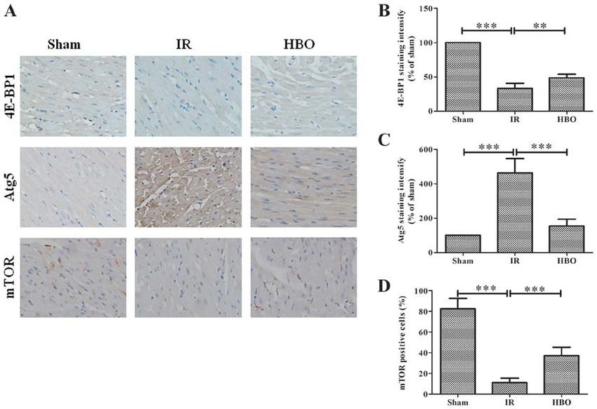
Immunohistochemistry of 4E‑BP1, Atg5and mTOR expression from ischemic myocardial tissue using a mitochondrial DNA
levels. At the end of reperfusion, myocardial tissue (n=5) was isolation kit (Abnova) (7). PCR amplification (TaKaRa Ex Taq
fixed in 4% buffered paraformaldehyde solution overnight at HSasDNA polymerase; Takara Bio, Inc.) and analysis were
4˚C. After processing with routine histological procedures performed using a 7500 Real‑Time PCR system equipped
(dehydration, transparent, dipped wax and embedding), with SDS software v2.0 (Applied Biosystems; Thermo Fisher
paraffin‑embedded tissues were cut into 4 µm‑thick sections. Scientific, Inc.). The thermal cycling conditions were: Initial
The tissue sections were subsequently deparaffinized with denaturation at 95˚C for 30 sec, followed by 40 cycles of 95˚C
xylene at 37˚C and rehydrated in a descending series of alcohol. for 5 sec, 55˚C for 30 sec and 72˚C for 30 sec. The following
Sections were incubated in 0.01 M sodium citrate buffer primer pairs were used for the PCR: mtDNA (238 bp) forward,CHEN et al: HYPERBARIC OXYGEN PROTECTS AGAINST MYOCARDIAL ISCHEMIA-REPERFUSION INJURY 4257 Figure 1. Effect of HBO pretreatment on ATP, ADP, AMP and cytochrome c expression levels and EC in myocardial infarction tissue. Expression levels of (A) ATP and (B) ADP were determined using specific assay kits in the sham, IR and HBO groups. (C) AMP expression levels were detected using an ELISA kit in the sham, IR and HBO groups. (D) EC was determined in the sham, IR and HBO groups. (E) Cytochrome c expression levels were determined using an ELISA in the sham, IR and HBO groups. Data are presented as the mean ± SD (n=5). *P
4258                                  Molecular Medicine REPORTS 22: 4254-4264, 2020

Results

Effect of HBO pretreatment on mitochondrial function. The
levels of AMP in the MI tissue were significantly increased
by 164% in the IR group compared with the sham group
(105.8±13.16 vs. 64.4±6.88; F=24.280; P
CHEN et al: HYPERBARIC OXYGEN PROTECTS AGAINST MYOCARDIAL ISCHEMIA-REPERFUSION INJURY                                              4259

Figure 3. Effect of HBO pretreatment on myocardial ultrastructure. (A) Representative micrograph of the myocardial tissue in the sham, IR and HBO
group. Normal mitochondria and no autophagic vacuoles were observed in the sham group. Significant damage to the mitochondria and increased numbers
of autophagic vacuoles were observed in the IR group, compared with the sham group. Only slight damage to the mitochondria and decreased numbers of
autophagic vacuoles were observed (concentric circle changes) in the HBO group, compared with the IR group. Magnification, x15,000 and x30,000. The
yellow arrow indicates autophagic vacuoles. (B) Semi‑quantification of the number of autophagic vacuoles from sham group, IR group and HBO group (n=5).
**
  P
4260                                       Molecular Medicine REPORTS 22: 4254-4264, 2020

                                                                          previous study demonstrated that oxidative stress parameters,
                                                                          such as superoxide dismutase, malondialdehyde and gluta-
                                                                          thione peroxidase, were activated in MIRI model rats (40).
                                                                          Consistent with our previous study, in the present study MIRI
                                                                          promoted higher relative fluorescence levels of ROS in the
                                                                          myocardial tissue. In addition, in IR rats, ATP and ADP levels
                                                                          were decreased, whereas AMP levels were increased, and the
                                                                          EC was also decreased in IR rats. Notably, HBO pretreatment
                                                                          was observed to significantly reverse all of these changes.
Figure 5. Effect of HBO pretreatment on mtDNA copy number. Data were           Due to energy homeostasis dysfunction, the loss of ATP
normalized to the sham group and presented as percentages. Data are       leads to the disruption of ionic pumps systems, such as
presented as the mean ± SD (n=5). ***P
CHEN et al: HYPERBARIC OXYGEN PROTECTS AGAINST MYOCARDIAL ISCHEMIA-REPERFUSION INJURY 4261 Figure 6. Statistical analysis of reverse transcription‑quantitative PCR results. (A‑E) Effect of HBO pretreatment on the mRNA expression levels of mitochon- drial dynamics‑related genes: (A) ND1, (B) cytochrome c, (C) Drp1, (D) Mfn1 and (E) Mfn2 in the sham, IR and HBO groups. Effect of HBO pretreatment on the expression levels of autophagy‑related genes: (F) mTOR and (G) p53 in the sham, IR and HBO groups. Data are presented as the mean ± SD (n=5). * P
4262                                   Molecular Medicine REPORTS 22: 4254-4264, 2020

autophagy by directly activating ULK1 through phosphorylation      Authors' contributions
on Ser317/777 under the conditions of glucose starvation (65).
The current study suggested that HBO pretreatment may inhibit      WC and LL wrote the manuscript. WC, LL, XC, XP, and
MIRI‑induced autophagy through regulating the mTOR‑medi-           CC performed the experiments. ZN collected the data and
ated autophagy pathway, by increasing the expression levels of     analyzed the data. CC designed the study, revised the manu-
mTOR and 4E‑BP1and decreasing the expression levels of Atg5        script and funded the research. All authors read and approved
and p53. Notably, numerous studies have demonstrated that          the final manuscript.
crosstalk between autophagy and apoptosis exists (66,67). Our
previous study has demonstrated that the Bax family members,       Ethics approval and consent to participate
caspase cascade, and cardiomyocyte apoptosis were inhibited
following HBO pretreatment (40). Thus, these results suggested     All animal experimental procedures were approved by the
that the cardioprotective mechanism of HBO may be involved         Animal Ethical Committee of Guangxi Medical University.
in inhibiting mTOR‑mediated autophagy.
    Nonetheless, the present study has numerous limitations.       Patient consent for publication
It was observed that mitochondrial dysfunction and excessive
autophagy occurred in the MIRI model following 30 min of           Not applicable.
ischemia and 1 h of reperfusion, as evidenced by transmis-
sion electron microscopy, immunohistochemistry and the             Competing interests
analysis of mRNA expression levels. These findings have also
been reported in numerous previous studies (20,21,68,69).          The authors declare that they have no competing interests.
In the present study, HBO pretreatment was found to restore
mitochondrial function and inhibit cardiomyocyte autophagy;        References
therefore, it was hypothesized that the protective effect of HBO
pretreatment may be related to the modulation of mitochondrial      1. Nabel EG and Braunwald E: A tale of coronary artery disease
function and cell autophagy. However, the mechanism by which           and myocardial infarction. N Engl J Med 366: 54‑63, 2012.
                                                                    2. Writing Group Members, Mozaffarian D, Benjamin EJ, Go AS,
HBO pretreatment affects the cardiomyocyte mitochondria and            Arnett DK, Blaha MJ, Cushman M, Das SR, Ferranti SD,
autophagy remains to be further investigated. Secondly, several        Després JP, et al: Executive summary: Heart disease and stroke
experimental methods were used to analyze the mitochondrial            statistics‑2016 update: A report from the American Heart
                                                                       Association. Circulation 133: 447‑454, 2016.
function, including EC, cytochrome c levels, intracellular ROS      3. Buja LM: Myocardial ischemia and reperfusion injury.
production, the opening of the mPTP, mitochondrial ultrastruc-         Cardiovasc Pathol 14: 170‑175, 2005.
ture, mtDNA copy number and the mRNA expression levels of           4. Levisman J and Price MJ: Update on the guidelines for the
                                                                       management of ST‑elevation myocardial infarction. Am
Drp 1, Mfn1 and Mfn2. In future experiments, experimental              J Cardiol 115 (Suppl 5): A3‑A9, 2015.
methods investigating the bioenergetics and the status of oxida-    5. Yellon DM and Hausenloy DJ: Myocardial reperfusion injury.
tive phosphorylation will be used to further determine the             N Engl J Med 357: 1121‑1135, 2007.
                                                                    6. Ibáñez B, Heusch G, Ovize M and Van de Werf F: Evolving
mitochondrial function.                                                therapies for myocardial ischemia/reperfusion injury. J Am Coll
    In conclusion, the present study demonstrated that HBO             Cardiol 65: 1454‑1471, 2015.
pretreatment effectively protected rat hearts from MIRI. This       7. Lee MS: Role of mitochondrial function in cell death and body
                                                                       metabolism. Front Biosci (Landmark Ed) 21: 1233‑1244, 2016.
effect may be related to the restoration of mitochondrial func-     8. Shen YQ, Guerra‑Librero A, Fernandez‑Gil BI, Florido J,
tion and the inhibition of cardiomyocyte autophagy. Thus, these        García‑López S, Martinez‑Ruiz L, Mendivil‑Perez M,
findings suggested that HBO treatment may be a useful agent            Soto‑Mercado V, Acuña‑Castroviejo Dario, Ortega‑Arellano H,
                                                                       et al: Combination of melatonin and rapamycin for head and
for the mitigation of MIRI.                                            neck cancer therapy: Suppression of AKT/mTOR pathway
                                                                       activation, and activation of mitophagy and apoptosis via
Acknowledgements                                                       mitochondrial function regulation. J Pineal Res 64: e12461,
                                                                       2017.
                                                                    9. Zhou H, Hu SY, Jin QH, Shi C, Zhang Y, Zhu PJ, Ma Q, Tian F
The authors would like to thank Dr Jianquan Li, from the               and Chen YD: Mff‑dependent mitochondrial fission contributes
Guangxi Medical University, China, for his technical support in        to the pathogenesis of cardiac microvasculature ischemia/reper-
                                                                       fusion injury via induction of mROS‑Mmediated cardiolipin
immunohistochemistry.                                                  oxidation and HK2/VDAC1 disassociation‑involved mPTP
                                                                       opening. J Am Heart Assoc 6: e005328, 2017.
Funding                                                            10. Shires SE and Gustafsson AB: Mitophagy and heart failure.
                                                                       J Mol Med (Berl) 93: 253‑262, 2015.
                                                                   11. Saito T and Sadoshima J: Molecular mechanisms of mitochon-
This present study was supported by the National Natural               drial autophagy/mitophagy in the heart. Circ Res 116: 1477‑1490,
Science Foundation of China (grant nos. 81701089                       2015.
                                                                   12. Scarffe LA, Stevens DA, Dawson VL and Dawson TM: Parkin
and 81960246), The Guangxi Natural Science Foundation                  and PINK1: Much more than mitophagy. Trends Neurosci 37:
(grant no. 2017GXNSFBA198010) and the Guangxi Sanitation               315‑324, 2014.
Research Project (grant nos. Z2016582 and Z20201096).              13. Hausenloy DJ and Scorrano L: Targeting cell death. Clin
                                                                       Pharmacol Ther 82: 370‑373, 2007.
                                                                   14. Liesa M, Palacin M and Zorzano A: Mitochondrial dynamics in
Availability of data and materials                                     mammalian health and disease. Physiol Rev 89: 799‑845, 2009.
                                                                   15. Ingerman E, Perkins EM, Marino M, Mears JA, McCaffery JM,
                                                                       Hinshaw JE and Nunnari J: Dnm1 forms spirals that are
The datasets used and/or analyzed during the current study are         structurally tailored to fit mitochondria. J Cell Biol 170:
available from the corresponding author on reasonable request          1021‑1027, 2005.
CHEN et al: HYPERBARIC OXYGEN PROTECTS AGAINST MYOCARDIAL ISCHEMIA-REPERFUSION INJURY 4263
16. Smirnova E, Griparic L, Shurland DL and van der Bliek AM: 37. Chen CX, Fan QP, Nong ZH, Chen W, Li YX, Huang LY,
Dynamin‑related protein Drp1 is required for mitochondrial Feng DR, Pan XR and Lan SY: Hyperbaric oxygen attenuates
division in mammalian cells. Mol Biol Cell 12: 2245‑2256, withdrawal symptoms by regulating monoaminergic neurotrans-
2001. mitters and NO signaling pathway at nucleus accumbens in
17. Rapaport D, Brunner M, Neupert W and Westermann B: Fzo1p is morphine‑dependent rats. Neurochem Res 43: 531‑539, 2018.
a mitochondrial outer membrane protein essential for the biogen- 38. Chen CX, Huang LY, Nong ZH, Li YX, Chen W, Huang JP,
esis of functional mitochondria in Saccharomyces cerevisiae. Pan XR, Wu GW and Lin YZ: Hyperbaric oxygen prevents
J Biol Chem 273: 20150‑20155, 1998. cognitive impairments in mice induced by D‑galactose by
18. Santel A and Fuller MT: Control of mitochondrial morphology improving cholinergic and anti‑apoptotic functions. Neurochem
by a human mitofusin. J Cell Sci 114: 867‑874, 2001. Res 42: 1240‑1253, 2017.
19. Hao M, Zhu S, Hu L, Zhu H, Wu X and Li Q: Myocardial isch- 39. Chen X, Li Y, Chen W, Nong Z, Huang J and Chen C: Protective
emic postconditioning promotes autophagy against ischemia effect of hyperbaric oxygen on cognitive impairment induced by
reperfusion injury via the activation of the nNOS/AMPK/mTOR D‑galactose in mice. Neurochem Res 41: 3032‑3041, 2016.
pathway. Int J Mol Sci 18: 614, 2017. 40. Chen C, Chen W, Nong Z, Ma Y, Qiu S and Wu G: Cardioprotective
20. Jian J, Xuan F, Qin F and Huang R: Bauhinia championii flavone effects of combined therapy with hyperbaric oxygen and dilti-
inhibits apoptosis and autophagy via the PI3K/Akt pathway in azem pretreatment on myocardial ischemia‑reperfusion injury in
myocardial ischemia/reperfusion injury in rats. Drug Des Devel rats. Cell Physiol Biochem 38: 2015‑2029, 2016.
Ther 9: 5933‑5945, 2015. 41. Bøtker HE, Hausenloy D, Andreadou I, Antonucci S, Boengler K,
21. Xuan F and Jian J: Epigallocatechin gallate exerts protective Davidson SM, Deshwal S, Devaux Y, Lisa FD, Sante MD, et al:
effects against myocardial ischemia/reperfusion injury through Practical guidelines for rigor and reproducibility in preclinical
the PI3K/Akt pathway‑mediated inhibition of apoptosis and the and clinical studies on cardioprotection. Basic Res Cardiol 113:
restoration of the autophagic flux. Int J Mol Med 38: 328‑336, 018‑0696, 2018.
2016. 42. Livak KJ and Schmittgen TD: Analysis of relative gene expres-
22. Hariharan N, Zhai P and Sadoshima J: Oxidative stress stimu- sion data using real‑time quantitative PCR and the 2(‑Delta Delta
lates autophagic flux during ischemia/reperfusion. Antioxid C(T)) method. Methods 25: 402‑408, 2001.
Redox Signal 14: 2179‑2190, 2011. 43. Qiao X, Jia S, Ye J, Fang X, Zhang C, Cao Y, Xu C, Zhao L,
23. Kang R, Zeh HJ, Lotze MT and Tang D: The Beclin 1 network Zhu Y, Wang L and Zheng M: PTPIP51 regulates mouse cardiac
regulates autophagy and apoptosis. Cell Death Differ 18: 571‑580, ischemia/reperfusion through mediating the mitochondria‑SR
2011. junction. Sci Rep 7: 45379, 2017.
24. Yu L, McPhee CK, Zheng LX, Mardones GA, Rong YG, Peng JY, 44. Cook SA, Sugden PH and Clerk A: Regulation of bcl‑2 family
Mi N, Zhao Y, Liu ZH and Wan FY: Termination of autophagy proteins during development and in response to oxidative stress
and reformation of lysosomes regulated by mTOR. Nature 465: in cardiac myocytes: Association with changes in mitochondrial
942‑946, 2010. membrane potential. Circ Res 85: 940‑949, 1999.
25. Han YF, Zhao YB, Li J, Li L, Li YG, Li SP and Li ZD: 45. Farber JL: Mechanisms of cell injury by activated oxygen
Stat3‑Atg5 signal axis inducing autophagy to alleviate hepatic species. Environ Health Perspect 10 (Suppl 10): S17‑S24, 1994.
ischemia‑reperfusion injury. J Cell Biochem 119: 3440‑3450, 46. Kalogeris T, Baines CP, Krenz M and Korthuis RJ: Cell biology
2018. of ischemia/reperfusion injury. Int Rev Cell Mol Biol 298:
26. Pópulo H, Lopes JM and Soares P: The mTOR signalling 229‑317, 2012.
pathway in human cancer. Int J Mol Sci 13: 1886‑1918, 2012. 47. Zorov DB, Juhaszova M and Sollott SJ: Mitochondrial reactive
27. Bramham CR, Jensen KB and Proud CG: Tuning specific oxygen species (ROS) and ROS‑induced ROS release. Physiol
translation in cancer metastasis and synaptic memory: Control Rev 94: 909‑950, 2014.
at the MNK‑eIF4E axis. Trends Biochem Sci 41: 847‑858, 48. Zhang T, Zhang Y, Cui MY, Jin L, Wang YM, Lv FX, Liu YL,
2016. Zheng W, Shang HB, Zhang J, et al: CaMKII is a RIP3 substrate
28. Foster SS, De S, Johnson LK, Petrini JH and Stracker TH: Cell mediating ischemia‑ and oxidative stress‑induced myocardial
cycle‑ and DNA repair pathway‑specific effects of apoptosis on necroptosis. Nat Med 22: 175‑182, 2016.
tumor suppression. Proc Natl Acad Sci USA 109: 9953‑9958, 49. Twig G, Elorza A, Molina AJA, Mohamed H, Wikstrom JD,
2012. Walzer G, Stiles L, Haigh SE, Katz S, Las G, et al: Fission and
29. Tasdemir E, Maiuri MC, Galluzzi L, Vitale I, Djavaheri‑Mergny M, selective fusion govern mitochondrial segregation and elimina-
D'Amelio M, Criollo A, Morselli E, Zhu C, Harper F, et al: tion by autophagy. EMBO J 27: 433‑446, 2008.
Regulation of autophagy by cytoplasmic p53. Nat Cell Biol 10: 50. Brooks C, Wei Q, Cho SG and Dong Z: Regulation of mitochon-
676‑687, 2008. drial dynamics in acute kidney injury in cell culture and rodent
30. Crighton D, Wilkinson S, O'Prey J, Syed N, Smith P, Harrison PR, models. J Clin Invest 119: 1275‑1285, 2009.
Gasco M, Garrone O, Crook T and Ryan KM: DRAM, a 51. Estaquier J and Arnoult D: Inhibiting Drp1‑mediated mitochon-
p53‑induced modulator of autophagy, is critical for apoptosis. drial fission selectively prevents the release of cytochrome c
Cell 126: 121‑134, 2006. during apoptosis. Cell Death Differ 14: 1086‑1094, 2007.
31. Dekleva M, Neskovic A, Vlahovic A, Putnikovic B, Beleslin B 52. Lander ES and Lodish H: Mitochondrial diseases: Gene mapping
and Ostojic M: Adjunctive effect of hyperbaric oxygen treatment and gene therapy. Cell 61: 925‑926, 1990.
after thrombolysis on left ventricular function in patients with 53. Borst P and Grivell LA: The mitochondrial genome of yeast.
acute myocardial infarction. Am Heart J 148: 031, 2004. Cell 15: 705‑723, 1978.
32. Bennett MH, Lehm JP and Jepson N: Hyperbaric oxygen therapy 54. Haendeler J, Dröse S, Büchner N, Jakob S, Altschmid J, Goy C,
for acute coronary syndrome. Cochrane Database Syst Rev 7: Spyridopoulos L, Zeiher AM, Brandt U and Dimmeler S:
CD004818, 2015. Mitochondrial telomerase reverse transcriptase binds to and
33. Rusyniak DE, Kirk MA, May JD, Kao LW, Brizendine EJ, protects mitochondrial DNA and function from damage.
Welch JL, Cordell WH and Alonso RJ: Hyperbaric oxygen Arterioscler Thromb Vasc Biol 29: 929‑935, 2009.
therapy in acute ischemic stroke: Results of the hyperbaric 55. Ong SB, Subrayan S, Lim SY, Yellon DM, Davidson SM and
oxygen in acute ischemic stroke trial pilot study. Stroke 34: Hausenloy DJ: Inhibiting mitochondrial fission protects the heart
571‑574, 2003. against ischemia/reperfusion injury. Circulation 121: 2012‑2022,
34. Liu WC, Yang SN, Wu CW, Chen LW and Chan JY: Hyperbaric 2010.
oxygen therapy alleviates carbon monoxide poisoning‑induced 56. Ong SB, Hall AR and Hausenloy DJ: Mitochondrial dynamics
delayed memory impairment by preserving brain‑derived neuro- in cardiovascular health and disease. Antioxid Redox Signal 19:
trophic factor‑dependent hippocampal neurogenesis. Crit Care 400‑414, 2013.
Med 44: e25‑39, 2016. 57. Zhang Y, Zhang L, Zhang Y, Fan X, Yang WW, Yu BY, Kou JP
35. Fife CE, Eckert KA and Workman WT: Ethical issues, stan- and Li F: YiQiFuMai powder injection attenuates coronary artery
dards, and quality control in the practice of hyperbaric medicine. ligation‑induced heart failure through improving mitochondrial
Springer, Switzerland. Textbook of Hyperbaric Medicine function via regulating ROS generation and CaMKII signaling
pp.597‑608, 2016. pathways. Front Pharmacol 10: 381, 2019.
36. Pan X, Chen C, Huang J, Wei H and Fan Q: Neuroprotective 58. Ma H, Guo R, Yu L, Zhang Y and Ren J: Aldehyde dehydro-
effect of combined therapy with hyperbaric oxygen and madopar genase 2 (ALDH2) rescues myocardial ischaemia/reperfusion
on 6‑hydroxydopamine‑induced Parkinson's disease in rats. injury: Role of autophagy paradox and toxic aldehyde. Eur Heart
Neurosci Lett 600: 220‑225, 2015. J 32: 1025‑1038, 2011.4264 Molecular Medicine REPORTS 22: 4254-4264, 2020
59. Ma X, Liu H, Foyil SR, Godar RJ, Weinheimer CJ and Diwan A: 66. Luo S and Rubinsztein DC: Apoptosis blocks Beclin 1‑dependent
Autophagy is impaired in cardiac ischemia‑reperfusion injury. autophagosome synthesis: An effect rescued by Bcl‑xL. Cell
Autophagy 8: 1394‑1396, 2012. Death Differ 17: 268‑277, 2010.
60. Dong W, Yang R, Yang J, Ding J, Wu H and Zhang J: Resveratrol 67. Yang B and Zhao S: Polydatin regulates proliferation, apoptosis
pretreatment protects rat hearts from ischemia/reperfusion and autophagy in multiple myeloma cells through mTOR/p70s6k
injury partly via a NALP3 inflammasome pathway. Int J Clin pathway. Onco Targets Ther 10: 935‑944, 2017.
Exp Pathol 8: 8731‑8741, 2015. 68. Liu CY, Zhang YH, Li RB, Zhou LY, An Tao, Zhang RC, Zhai M,
61. Rabanal‑Ruiz Y, Otten EG and Korolchuk VI: mTORC1 as the Huang Y, Yan KW, Dong YH, et al: lncRNA CAIF inhibits
main gateway to autophagy. Essays Biochem 61: 565‑584, 2017. autophagy and attenuates myocardial infarction by blocking
62. Yang Z and Klionsky DJ: Mammalian autophagy: Core molec- p53‑mediated myocardin transcription. Nat Commun 9: 29, 2018.
ular machinery and signaling regulation. Curr Opin Cell Biol 22: 69. Guo X, Jiang H, Yang J, Chen J, Yang J, Ding JW, Li S, Wu H
124‑131, 2010. and Ding HS: Radioprotective 105 kDa protein attenuates isch-
63. Dan E, Joungmok K, Shaw RJ and Kun‑Liang G: The autophagy emia/reperfusion‑induced myocardial apoptosis and autophagy
initiating kinase ULK1 is regulated via opposing phosphoryla- by inhibiting the activation of the TLR4/NF‑κ B signaling
tion by AMPK and mTOR. Autophagy 7: 643‑644, 2011. pathway in rats. Int J Mol Med 38: 885‑893, 2016.
64. Hardie DG: AMP‑activated/SNF1 protein kinases: Conserved
guardians of cellular energy. Nat Rev Mol Cell Biol 8: 774‑785, This work is licensed under a Creative Commons
2007. Attribution-NonCommercial-NoDerivatives 4.0
65. Kim J, Kundu M, Viollet B and Guan KL: AMPK and mTOR International (CC BY-NC-ND 4.0) License.
regulate autophagy through direct phosphorylation of Ulk1. Nat
Cell Biol 13: 132‑141, 2011.You can also read