How I Treat How I treat mixed-phenotype acute leukemia
←
→
Page content transcription
If your browser does not render page correctly, please read the page content below
From www.bloodjournal.org by guest on September 29, 2016. For personal use only.

How I Treat

How I treat mixed-phenotype acute leukemia
Ofir Wolach and Richard M. Stone
Department of Medical Oncology, Dana-Farber Cancer Institute, Boston, MA

Mixed-phenotype acute leukemia (MPAL)              monograph on classification of tumors of           regimen followed by allogeneic stem-cell
encompasses a heterogeneous group of rare          hematopoietic and lymphoid tissues. In adult       transplant may be advisable; addition of a
leukemias in which assigning a single line-        patients, MPAL is characterized by relative        tyrosine kinase inhibitor in patients with
age of origin is not possible. A variety of        therapeutic resistance that may be attributed      t(9;22) translocation is recommended. The role
different terms and classification systems         in part to the high proportion of patients with    of immunophenotypic and genetic markers
have been used historically to describe this       adverse cytogenetic abnormalities. No pro-         in guiding chemotherapy choice and postre-
entity. MPAL is currently defined by a limited     spective, controlled trials exist to guide         mission strategy, as well as the utility of
set of lineage-specific markers proposed           therapy. The limited available data suggest        targeted therapies in non–Ph-positive MPALs
in the 2008 World Health Organization              that an “acute lymphoblastic leukemia–like”        is unknown. (Blood. 2015;125(16):2477-2485)

Introduction
The absence of essentially any useful prospectively collected data on        derived disease (ALL). Sometimes the immature cells display cyto-
how to treat mixed-phenotypic acute leukemia (MPAL) in adults                chemical and/or immunophenotypic features of both lineages (biphe-
both simplifies and complicates any discussion of this topic. Given           notypic) or there are different populations of leukemia cells (bilineal).
the availability of little truly useful information, we have derived         The distinction between bilineal and biphenotypic leukemias is often
an approach based on data in the literature that makes logical sense         blurred, especially because 2 “populations” of cells perhaps represent
and can be adhered to: once MPAL is definitely identified, patients            subclones derived from a unique stem cell. Accordingly, this distinction
should be treated according to an “acute lymphoid leukemia”–type in-         does not generally affect our diagnostic or therapeutic approach.
duction regimen followed by allogeneic stem-cell transplant (alloSCT)            Two important recent algorithms have been used to define this entity.
in responding patients if feasible.                                          In the first of these (1995), the European Group for Immunological
                                                                             Characterization of Acute Leukemias (EGIL) developed a scoring
                                                                             algorithm in which a point system determined whether a patient
                                                                             had enough immunophenotypic variety to qualify as biphenotypic
Case presentation: part 1                                                    (Figure 2A).1,2 The second and most recent 2008 World Health
                                                                             Organization (WHO) monograph on classification of tumors of hema-
A 51-year-old physician complained of 2 weeks of dyspnea upon
                                                                             topoietic and lymphoid tissues includes a helpful chapter on acute
exertion. White blood cell (WBC) count was 280 3 109/L (76% blasts),
                                                                             leukemias of ambiguous lineage: “leukemias that show no clear evi-
platelet count was 91 3 109/L, and hemoglobin was 9 g/dL. Bone-
                                                                             dence of differentiation along a single lineage.”3 These encompass
marrow aspirate (Figure 1A) revealed 2 distinct morphologic/
                                                                             MPAL, the primary topic of this review, and acute undifferentiated
cytochemical populations of blasts that were positive for myeloperox-
                                                                             leukemia (AUL), wherein the malignant cells do not express lineage-
idase (MPO) and periodic acid-Schiff (PAS) stains, respectively. Flow
                                                                             specific antigens. This classification (Figure 2B) tries to minimize the
cytometry (Figure 1B) also demonstrated 2 atypical blast populations
                                                                             difficult distinction between bilineal and biphenotypic leukemia (BAL)
with distinct CD45 expression associated with (1) uniform expression of
                                                                             and subclassifies these promiscuously-derived cells as usually either
B-lymphoid markers CD19 and CD10, uniform stem-cell marker CD34,
                                                                             B-myeloid or T-myeloid. MPALs that harbor Philadelphia chromosome
and variable myeloid marker CD33; and (2) uniform expression of CD33,
                                                                             (Ph1) or MLL rearrangements are considered a distinct diagnostic
small subset CD34, variable CD19, and lack of CD10 expression.
                                                                             subgroup (Figure 2B). An important point is that AML-defining
   All 20 metaphases assessed contained a t(9;22)(q34;q11.2) trans-
                                                                             balanced translocations such as t(8;21), a type of favorable prognosis
location associated with loss of the 7p and 16q arms. Molecular studies
                                                                             AML that frequently expresses multiple B-cell markers,4 are not
demonstrated the e1a2 BCR-ABL transcript (p190) but no additional
                                                                             considered biphenotypic. It also excludes secondary leukemias (arising
recurrent mutations associated with acute leukemia (95-gene panel).
                                                                             after prior cancer therapy or myelodysplasia), leukemias with FGFR1
                                                                             mutations that have features of both T-lymphoid and myeloid
                                                                             differentiation, and chronic myeloid leukemia (CML) in blast crisis,
What is mixed-phenotype acute leukemia?                                      which can present with a variety of lineages. The latter is sometimes
                                                                             difficult to separate from Ph1 MPAL (that may actually represent
Patients diagnosed with acute leukemia (.20% blasts in blood or              transformation from a previously undiagnosed chronic-phase CML).
marrow, or fewer in the case of certain chromosomal translocations or            The essential feature of MPAL (Figure 2B) is that cells express
an extramedullary presentation) can generally be classified as having         lineage-specific myeloid markers as well as lineage-specific T- or
either myeloid lineage–derived disease (AML) or lymphoid lineage–            B-lymphoid markers. Although there are caveats (see legend

Submitted October 30, 2014; accepted January 7, 2015. Prepublished online    © 2015 by The American Society of Hematology
as Blood First Edition paper, January 20, 2015; DOI 10.1182/blood-2014-10-
551465.

BLOOD, 16 APRIL 2015 x VOLUME 125, NUMBER 16                                                                                                     2477
From www.bloodjournal.org by guest on September 29, 2016. For personal use only.
2478 WOLACH and STONE BLOOD, 16 APRIL 2015 x VOLUME 125, NUMBER 16
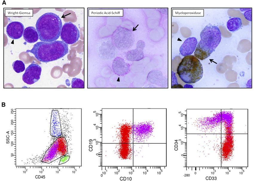
Figure 1. Diagnostic pathology of clinical case. (A) Two atypical blast populations are seen on bone marrow aspirate smear. One population (arrowhead) is composed of
small cells with round nuclei, slightly condensed chromatin, distinct nucleoli, and scant cytoplasm that shows cytoplasmic reactivity with periodic acid-Schiff (PAS) in a blocklike
pattern and lacks reactivity with myeloperoxidase (MPO). The other population (arrow) is composed of large cells with irregular nuclei, dispersed chromatin, variably distinct
nucleoli, and small-to-moderate amounts of blue-gray cytoplasm that shows cytoplasmic reactivity with MPO and lacks reactivity with PAS. (B) Flow cytometric analysis of this
bone marrow aspirate reveals 2 atypical blast populations (one highlighted in purple, one highlighted in red) with distinct CD45 expression and variable antigen expression
profiles. The CD45(dim) purple population exhibits uniform expression of B-lymphoid markers CD19 and CD10, uniform stem-cell marker CD34, and variable myeloid marker
CD33. The red population shows brighter CD45 expression, exhibits uniform expression of CD33, small subset CD34, variable CD19, and lacking CD10 expression.
to Figure 2B), CD3 expression equals T-lymphoid development, and transformation occurs at that stage.7 The term “lineage infidelity” de-
CD19 plus 1 or 2 other markers suggests B-lymphoid origin. Myeloid noted an alternative hypothesis involving oncogenetically-driven mis-
origin can be determined with a set of monocytic markers, or more programming of the leukemic cell, resulting in multilineage-expressing
commonly by MPO expression. Although various thresholds for flow- blasts.8,9 Significant strides have been made since then in our under-
based MPO positivity were introduced over the years (eg, 10% of blast standing of the normal and pathologic pathways that drive lineage fate.
population1,5), no specific threshold has been acknowledged in the Maturation and differentiation of blood cells during the process
2008 WHO monograph.3,6 of hematopoiesis is associated with the expression of specific sets
Compared with the EGIL classification, the 2008 WHO classification of markers that define lineage. This is a tightly regulated, multistep,
uses a more limited set of lineage markers that can be more consistently hierarchical process driven by a network of transcription factors. Al-
applied. In 2015, the 2008 WHO classification still remains the most though some transcription factors are thought to have primary roles in
practical means to define and subclassify MPAL, but it is hoped that driving hematopoietic progenitors toward a specific lineage (eg, C/EBPa
advances in deciphering the molecular pathogenesis of acute leukemia in myeloid cells or PAX-5 in B lymphocytes), this relationship in vivo is
will soon lead to a more robust approach to the diagnosis of these entities. far more complex, context dependent, and regulated at multiple cellular
levels.10-12 Early hematopoietic multipotential progenitors were pre-
viously shown to express markers of multiple lines, with the specific fate
selection relying on complex interactions that both promote a specific
What drives biphenotypic expression lineage phenotype and also suppress alternative programming (so called
in leukemia? “lineage priming”).13 The timing and level of expression of a specific
transcription factor may affect lineage determination. Competing
During the 1980s, 2 leading hypotheses were raised to explain bi- transcription factors interact to antagonize each other’s functions to
phenotypic expression in leukemia. The Greaves hypothesis suggested promote the expression of one lineage over the other.11 For example,
“lineage promiscuity,” in which hematopoietic progenitor cells possess a high level of PAX5 expression is critical for development of common
multilineage potential that is preserved as a relic if leukemic lymphoid progenitors along the B-cell pathway, whereas low levelsFrom www.bloodjournal.org by guest on September 29, 2016. For personal use only.
BLOOD, 16 APRIL 2015 x VOLUME 125, NUMBER 16 HOW I TREAT MIXED-PHENOTYPE ACUTE LEUKEMIA 2479
result in a mixed phenoptype14; C/EBPa suppression of PAX-5 drives cytogenetic information, 20% had a Ph1 and 8% had MLL gene
common lymphoid progenitor cells toward myeloid phenotypes.15 In (11q23) rearrangements. Thirty-two percent had complex karyotype
zebrafish PU.1 and GATA-1 exert mutually antagonistic effects with the (CK) that was commonly associated with deletion of the long arm of
balance driving myeloid vs erythroid differentiation.16 The fate of early chromosome 6, abnormalities involving the long arm of chromosome 7,
T-cell lineage progenitors is dependent on the Notch receptor–signaling or abnormalities in the long arm of chromosome 5. Normal karyotype
pathway, without which myeloid differentiation may occur.17-19 was demonstrated in 13%. Although early deaths were seen, most of the
Dysregulation and aberrant expression of transcription factors that patients died of their disease, with an overall median survival of 18
govern cell differentiation occur on the basis of the genomic and months and a 37% overall 5-year survival. Age, Ph1, and the type of
epigenetic alterations seen in acute leukemia.11,20 Gene-expression induction therapy were significant predictors for survival, with children
profiling in a large group of patients with AML correlated T/myeloid surviving 139 months vs 11 months for adults, 8 months for Ph1 vs 139
phenotype with a distinct expression profile that included C/EBPa months for those with normal karyotype, and 28 months for those with
promoter hypermethylation/gene silencing and upregulation of T cell– other abnormalities. Yan et al27 reported on 117 patients with WHO
lineage pathways, via aberrantly activated NOTCH1 signaling.21 Ac- 2008–defined MPAL. Median patient age was 35 years (range 14-81)
tivating mutations in NOTCH1 were previously described in the with a slight male predominance (51.3%) and a median WBC count
context of lineage switch from AML to T-ALL, suggesting the poten- of 5.4 3 109/L (range 0.8-278.7) at diagnosis. Thirty-four percent
tial role of mutations in transcription factors on lineage-specific cell of patients demonstrated AML morphology (primarily FAB M1 and
reprogramming.22 Indeed, mutations may trump phenotype. Early FAB M5), 44% were believed to have ALL (FAB L1), and 22% were
T-cell precursor (ETP) ALL is associated with recurrent mutations unclassifiable. B-myeloid immunophenotype was seen in 55% and
typically seen in myeloid tumors such as DNMT3A, IDH1, and T-myeloid phenotype in 33%. Of 92 patients assessed, 64% presented
IDH223-25 and is transcriptionally related to myeloid progenitors. with cytogenetic abnormalities; CK was the most prevalent aberration
found in 24% of patients, followed by Ph1 chromosome in 15% (all
B-myeloid phenotype) and translocations involving MLL gene at 11q23
in 4.3% of patients. Monosomy 7, polysomy 21, and trisomy 8 were
Natural history of MPAL also noted in a significant minority of patients.
Cytogenetic abnormalities in MPAL and BAL were reported
Incidence in a recent systematic review to be present in 59% to 91% of patients.30
The prevalence of Ph1 and complex karyotype increases with age.
The frequency, clinical features, and outcome of patients with am-
The frequency of translocations involving 11q23 (usually MLL-AF4 or
biguous lineage expression are largely dependent on the classification
MLL-ENL fusions) decreases with age and is quite uncommon in
system used at the time of report. The WHO 2008 classification is less
adults with MPAL.30 One could argue that leukemia with CK or other
inclusive than the preceding EGIL system, resulting in a lower reported
myelodysplastic-specific cytogenetic abnormalities should be clas-
prevalence. Weinberg and Arber retrospectively reviewed series en-
sified as AML with myelodysplasia-related changes rather than
compassing 7627 pediatric and adult patients with acute leukemia
MPAL.
and determined that 2.8% had BAL and 1.6% had MPAL using the
EGIL and WHO 2008 systems, respectively.26 A more recent Chinese
study reported MPAL in 2.4% of 4780 patients with acute leukemia Genetic alterations in MPAL
(ages 14-81 years).27 In 517 pediatric and adult Dutch patients with
Rubnitz et al31 analyzed gene expression patterns in 13 pediatric
acute leukemia, 30 patients (5.8%) would be considered as having BAL
patients with EGIL-defined BAL and found that, although 5 patients
based on EGIL criteria, and 8 cases (1.5%) were consistent with MPAL
made up a group with known AML expression patterns, 8 patients
using the WHO 2008 classification; only 6 patients (1.1%) would
displayed gene expression patterns that were different from AML and
qualify as both BAL and MPAL, suggesting that these classification
ALL, suggesting that some cases of BAL may be a biologically distinct
systems may select different patients.28
entity. In contrast, microRNA profiling studies suggest that MPAL
Characteristics of MPAL does not appear to be a distinct entity. de Leeuw et al32 analyzed 16
cases of acute leukemia of ambiguous lineage and demonstrated that all
Matutes et al29 presented a review of 100 patients, mostly from the cases had microRNA expression profiles that clustered with AML
United Kingdom and Austria, with MPAL based on the WHO defi- or ALL. Heesch et al 33 noted a higher expression of BAALC and
nition. Of the 62 men and 38 women (32% under the age of 16), ERG—adverse prognostic characteristics in AML—in 26 cases of
39 displayed ALL, 38 had AML, and 13 cases were defined as acute EGIL-defined BAL compared with other cases of AML.
undifferentiated leukemia by morphologic assessment (10 were not Information regarding the mutational landscape of MPAL is
analyzed). Immunophenotyping showed that 58% of the cases had a based on small patient numbers. Yan et al27 analyzed 31 patients with
B-myeloid and 36% had a T-myeloid phenotype. Combined B1T and MPAL for 18 leukemia-related mutations and reported that 12
trilineage (myeloid1B1T) immunophenotypes were rare (n 5 6) and patients (39%) were found to harbor a mutation, including IKZF1
all of these had ALL morphology. Expression of stem cell–like markers deletion in 4 patients (all B-myeloid phenotype), EZH2 in 3 (B- or
was common and included terminal deoxynucleotidyl transferase in T-myeloid), ASXL1 in 2 (both B-myeloid), TET2 in one (B-myeloid),
89% of the cases, human leukocyte antigen–D-related in 92% and CD34 and ETV6 and NOTCH1 in 1 patient each (both T-myeloid). A high
in 74%. Among cases with myeloid commitment, MPO was expressed in rate of DNMT3A mutations was reported in adults with T-myeloid
at least in 5% of the blasts in 98% of cases, and in .20% of the blasts in MPAL (10/18 patients; mostly biallelic mutations).34 Whole-exome se-
76% of the cases. All except 9 cases expressed MPO, as well as CD33 quencing in 19 adult patients with MPAL (12 T-myeloid, 6 B-myeloid,
and/or CD13. By definition, cytoplasmic CD3 was expressed in all 35 and 1 B/T) demonstrated that 63% of patients had mutations in epige-
cases with T-myeloid phenotype and CD19 was present in 93% of cases netic regulatory genes. DNMT3A was the most common mutation
with the B-myeloid phenotype and was always associated with CD10, (n 5 6) followed by EZH2, IDH1/2, TET1, and TET3. Other recurrent
cytCD22, and/or cytCD79a expression. In the 76 patients with mutations included PRPF40B (n 5 6), TP53 (n 5 5), BRAF (n 5 4),From www.bloodjournal.org by guest on September 29, 2016. For personal use only.
2480 WOLACH and STONE BLOOD, 16 APRIL 2015 x VOLUME 125, NUMBER 16
Figure 2. Diagnostic criteria for BAL and MPAL. (A) EGIL
criteria for the diagnosis of biphenotypic acute leukemia.a (B)
2008 WHO criteria. Leukemias that fail to demonstrate differen-
tiation along a single lineage are defined as acute leukemias
of ambiguous lineage (ALAL) and are further subdivided into
diagnostic subgroups. A practical approach for the diagnosis of
MPAL is presented.
and NOTCH1 (n 5 4).35 Some of these mutations (eg, IDH1/2, BRAF, IKZF1, MEF2C, BCOR, EBF1, KRAS, LEF1, MBNL1, PBX3, and
NOTCH1, FLT3) could be theoretically targeted by available agents or RUNX1.27
those in current clinical trials. In another series, clustering of FLT3 ITD
and TKD mutations was reported in patients with T-myeloid MPAL. Risk factors and outcomes
Seven of 15 patients (47%) were positive for FLT3 mutations (mostly
ITD), all of which were CD1171.36 Array-based comparative genomic The reasons underlying resistance to therapy in this heterogeneous
hybridization analysis in 12 patients with MPAL demonstrated that all group are not clear but may be related to the high prevalence of
patients had at least 1 abnormality, including deletions of CDKN2A, drug efflux pump expression 37-39 and the high proportion withFrom www.bloodjournal.org by guest on September 29, 2016. For personal use only.
BLOOD, 16 APRIL 2015 x VOLUME 125, NUMBER 16 HOW I TREAT MIXED-PHENOTYPE ACUTE LEUKEMIA 2481
Figure 2. (Continued).
cytogenetic abnormalities.30 Whatever classification is used, there physicians, because there was no widespread treatment policy. The few
appears to be a uniformly poor outcome in MPAL (or BAL) that is studies that retrospectively garnered MPAL cases from cooperative
inferior to the outcome in more typical AML or ALL. Based on adult group trials used more homogeneous treatments, but inevitably
and mixed pediatric/adult series, patients with EGIL-defined BAL excluded cases from eligibility based on ambiguity. Neither is there
have complete remission (CR) rates of 30% to 80.6%,33,37,40-46 with much guidance in the National Comprehensive Cancer Network
median disease-free survival (DFS) and overall survival (OS) of 5 to (NCCN) guidelines. The precise definition of which cases should be
12 months37,44-46 and 6.5 to 30.3 months,33,37,40,41,44-46 respectively. treated according to the ALL vs AML guidelines is sidestepped. The
In the few larger retrospective series of WHO 2008–defined MPAL, reader is advised to consult a center or individual with experience in
CR rates are reported at 61.5% to 85.2%,27,29,47,48 and median OS is diagnosing these entities.51 As someone who serves on the AML
reported to be 14.8 to 18 months.29,47 Factors associated with out- NCCN guidelines committee, I (R.M.S.) readily admit personal
come in these analyses include age,29,31,37,40,44,48-50 WBC count at uncertainty, which can be blamed on the lack of evidence.
diagnosis,41,44 Ph1 status,29,40 CK or MLL rearrangement,45 base- Caveats aside, the dilemma when considering a patient who has
line creatinine and uric acid levels,47 extra medullary involvement at a bona fide case of MPAL can be lessened by (1) gathering information,
diagnosis,44 immunophenotype (T-myeloid being worse),41 failing (2) considering pathophysiology, and (3) consulting the literature. The
to respond to induction therapy,37,44,48 type of induction therapy optimum information would include the patient’s age, past medical
(favoring non-AML),29,44,48 and type of postremission therapy history/comorbidities, blast morphology (including cytochemistry), a
(favoring transplant27,33 and more intensive conditioning48). complete immunophenotype, cytogenetics, and molecular studies. Older
infirm patients with multiple comorbidities are not good candidates for
standard ALL or AML induction therapy. The presence of Auer rods,
degree of MPO1 on flow cytometry, and cytochemistry could be the
Treatment of MPAL basis for a therapeutic decision, albeit without any supporting data.
If they are rapidly available, cytogenetic studies could categorize the
There are no prospective trials that point to an optimal strategy. Beyond patient. Patients with MPAL and 11q23 rearrangement are considered
one’s own experience, we are left with heterogeneous case series that a separate entity in the 2008 WHO schema, although their initial treat-
describe outcomes retrospectively. Further complicating data inter- ment may not be different from that for most MPAL patients. However,
pretation is the inclusion of patients with well-defined AML syn- it is critical to define the Ph1 patient as rapidly as possible because such
dromes in previous classifications (such as core-binding-factor patients should have a tyrosine kinase inhibitor (TKI) added to their
leukemias) that may bias those reports toward the use of AML-type treatment. Finally, although the molecular biology has not been studied
therapy. Case studies from individual centers or countries tend to at any depth in MPAL, it makes sense to assess for the presence of mu-
examine all cases of acute leukemia and describe MPAL in 2% to 3% tations with prognostic and/or therapeutic relevance in leukemia and
of the population. Although these studies probably reflect the true perhaps to rule out the presence of Ph-like signature, which could have
incidence of the entity fairly accurately, treatment decisions are eventual therapeutic implications in ALL.52 If nothing else, collecting
haphazard and are subject to unknown bias regarding individual the data may be retrospectively useful in learning about MPAL.From www.bloodjournal.org by guest on September 29, 2016. For personal use only.
2482 WOLACH and STONE BLOOD, 16 APRIL 2015 x VOLUME 125, NUMBER 16
Because this disease is believed to emanate from a proximal, compared characteristics and outcomes of 13 Ph1 MPAL patients
presumed long-lived stem cell in the hematopoietic hierarchy (high with 27 patients with Ph1 ALL and demonstrated comparable CR
level of CD34 expression, capable of lineage switch or infidelity), rates among the 2 groups (100% vs 85%) as well as similar 5-year OS
one could surmise that chemotherapy alone would be insufficient to (55% vs 53%) and DFS (46% vs 42%).65
eradicate the disease. Ph1 ALL and MDS-associated and/or adverse
chromosome AML are historically incurable without a stem-cell Non-Ph1 MPAL
transplant; the same likely applies to MPAL. Moreover, the inclusion
of more chemotherapeutic agents in up-front therapy used in an ALL What is the best approach for the non-t(9;22) MPAL patient? We treat
or combined regimen would seem more logical than an AML reg- with an ALL regimen and consolidate with an alloSCT if a donor is
imen, especially the latter’s use of cytarabine, less useful against a available. Most of the retrospective case series suggest that the CR rate
slowly dividing primitive stem cell. Ideally, one could inhibit the is higher with ALL therapy or an ALL/AML combined regimen than
gene product of a “founder” mutation present early in disease de- with AML-type therapy. Matutes et al29 noted a CR rate of 85%
velopment and throughout the course. This serendipitous situation compared with 41% for AML-type therapy. It is presumed that many of
appears to be the case for Ph1 MPAL. the patients who had morphologic AML (42%) received AML-type
therapy; the inferior CR rate with this therapy may have been a
Ph1 and MLL rearranged MPAL manifestation of intrinsic resistance in this subset. Whether these
“AML-like” patients would fare better with ALL-type therapy is
The only special cases within the MPAL WHO framework are patients unknown. Other studies, albeit with smaller patient numbers, showed
with (9;22) or 11q23 cytogenetic abnormalities. Currently, treatment similar findings regarding ALL-type vs AML-type CR rates: 75% vs
considerations for those with cytogenetic rearrangements at 11q23 are 28%46 and 64% vs 33%, respectively.45
not different from those for MPAL with any non-Philadelphia Although the preponderant thought has been to use ALL-type
cytogenetic abnormal or normal karyotype. However, the 11q23- therapy, the situation is far from straightforward. Are there any patients
rearranged patients should be considered for a pathophysiologically- in whom it makes sense to use AML-type therapy? Although we have
based clinical trial if chemotherapy and alloSCT fail or if the patient tended to use 317 in MPAL patients who express MPO cytochemi-
is not a candidate for aggressive chemotherapy. Such therapy could cally, there is little support for this common sense approach. One
include a histone-modifying–enzyme inhibitor or a bromodomain prospective clinical trial used AML therapy in 7 MPAL patients who
inhibitor based on the primary molecular abnormality53,54 or could had .20% expression of MPO by flow but noted only 2 patients who
target downstream activation of Hox genes via glycogen synthase achieved CR.46 We could find no data applying the use in MPO or
kinase 3 or b-catenin inhibitors.55 Sudan black positivity to assign therapy. The use of combination
Ph1 MPAL demand a specific approach involving the use of a AML1ALL regimens has some appeal (eg, the VAPA 10 approach66).
TKI. This entity is usually a combination of B-lymphoid and myeloid These combined regimens vary among studies but generally add ALL-
markers; it accounts for about 25% of all MPAL.30 Essentially, all case active agents such as steroids and vincristine to the AML anthracycline/
series describing this entity mention the adverse prognosis engendered cytarabine backbone. Remission rates with these combined regimens
by this molecular lesion.29,40 However, in the TKI era, the situation are largely comparable with those achieved with established ALL
may be changing. protocols,27,40,41,44-46 and some reports found these regimens to be
One can reasonably look to the Ph1 ALL literature for guidance. rather toxic.40,44 In a few reports from adult and pediatric BAL series,
Ph1 ALL was historically considered a poor prognostic entity, but high rates of remission with ALL-directed salvage therapy were
prospective studies in which imatinib56,57 or dasatinib58 have been reported after AML induction failure31,46 and vice versa.27
combined with standard multiagent chemotherapy depict a long- Emerging data suggest that pediatric-type regimens lead to a 50%
term DFS of 40% to 60%, approaching that seen with Ph– ALL in to 60% 3-year survival in adults ages 18 to 40 with ALL,64,67
adults. Although Ph1 ALL patients achieving remission with TKI compared with a 30% to 40% rate with legacy regimens such as
plus chemotherapy-based therapy conventionally should be consol- CALGB 9111 and hyperCVAD.61,68 Although MPALs were not
idated with alloSCT if feasible, emerging data suggest that au- specifically included in the few reports, it does make sense to use
tologous transplant for patients with Ph1 ALL in remission59 and/or such therapy in patients up to age 40. Children with MPAL seem
ongoing TKI as maintenance56 may be associated with long-term to fare better with ALL regimens, although they do have inferior
remissions, calling into question the obligate need for alloSCT in this outcome compared with other children with non-MPAL ALL.50
MPAL subtype. For older adults, excellent short-term results have Although patients with MPAL who achieve CR and have an alloSCT
been obtained with dasatinib plus steroids and intrathecal chemo- are a select group, every study suggests superior outcomes in adult
therpy60; one wonders about the need for aggressive chemotherapy patients who receive alloSCT compared with those who receive only
even in younger patients given the potent antileukemic efficacy chemotherapy in the postremission setting.27,33 Moreover, alloSCT
of TKIs. Therefore, we treat all MPAL t(9;22) patients with age- is better than chemo in essentially all high risk leukemias.69 For
specific ALL chemotherapy in combination with a TKI (CALGB example,33 12 of 34 patients with ALAL underwent SCT. There was
9111,61 hyperCVAD,56 MRC-ECOG 299362 for middle-aged patients, a 5-year OS rate of 70% compared with 19% for those who received
and the Foa regimen60 for older patients) followed by alloSCT chemotherapy only. Liu et al48 focused on 59 patients with ALAL
if feasible. In some pediatric and adult series, biphenotypic ex- who underwent alloSCT and noted a 5-year OS likelihood of 55%
pression was reported to be associated with a high frequency of with an intensive preparative regimen and 24% with a standard
central nervous system (CNS) involvement at presentation44,49,50,63; preparation. Although using multiparameter flow cytometry to de-
thus we try to adhere to CNS-directed therapy according to the spe- fine minimal residual disease (MRD) in MPAL cases can be chal-
cific ALL protocol that is chosen. Whether to use highly effective lenging, one could speculate that such patients who are clearly
pediatric-type chemotherapy64 plus TKI for patients under 40 with MRD– early in their course could be treated with consolidation
t(9;22) MPAL is unclear, although limited pediatric experience chemotherapy rather than alloSCT in a similar fashion to Ph1 ALL
suggests this may be possible. A recent retrospective analysis that becomes molecularly negative after therapy.From www.bloodjournal.org by guest on September 29, 2016. For personal use only.
BLOOD, 16 APRIL 2015 x VOLUME 125, NUMBER 16 HOW I TREAT MIXED-PHENOTYPE ACUTE LEUKEMIA 2483
Figure 3. The approach to therapy in patients with
mixed-phenotype acute leukemia (MPAL). Targeted
therapy based on a patient’s specific genetic profile
should be considered in refractory/relapse patients (eg,
MLL rearrangements, FLT3-ITD, IDH1, IDH2).
patient received dasatinib and CNS prophylaxis followed by sibling-
Summary matched alloSCT with myeloablative conditioning. Three months after
transplant, he remains in remission.
As shown in Figure 3, a reasonable approach to a patient with MPAL
is to first determine whether the disease is driven by BCR-ABL1. If
so, age-appropriate ALL therapy plus a TKI followed by SCT is
reasonable. If a patient is BCR-ABL1-negative, age-appropriate ALL
Acknowledgments
therapy followed by SCT after remission is an acceptable strategy.
Important areas for further study are: (1) whether the degree of MPO Dr Elizabeth Morgan from Brigham and Women’s Hospital,
positivity by immunophenotype /cytochemistry should influence the Department of Pathology, Harvard Medical School supplied and
choice of therapy, (2) whether the presence of myeloid-specific muta- prepared the representative pathology images for Figure 1.
tions or other genetic and molecular markers should be considered, (3)
can the pathophysiology of 11q23 leukemia be successfully exploited,
and (4) will alloSCT be needed for MPAL t(9;22) patients who respond
very well to chemotherapy plus TKI?
Authorship
Case presentation: part 2
Contribution: O.W. and R.M.S. wrote the paper.
Based on immunophenotype and cytogenetic information, the di- Conflict-of-interest disclosure: The authors declare no competing
agnosis of MPAL with t(9;22)(q34;q11.2);BCR-ABL1 was made. The financial interests.
patient was treated with a CALGB9111-type induction regimen plus Correspondence: Richard M. Stone, Dana-Farber Cancer Insti-
dasatinib and promptly entered into CR; no immunophenotype-based tute, 450 Brookline Ave, Room D2053, Boston, MA 02115; e-mail:
MRD was noted, but BCR-ABL1 transcripts remained detectable. The rstone@partners.org.From www.bloodjournal.org by guest on September 29, 2016. For personal use only.
2484 WOLACH and STONE BLOOD, 16 APRIL 2015 x VOLUME 125, NUMBER 16
References
1. Bene MC, Castoldi G, Knapp W, et al; European 19. Wada H, Masuda K, Satoh R, et al. Adult T-cell leukemia and T/myeloid acute leukemia. Am J
Group for the Immunological Characterization progenitors retain myeloid potential. Nature. 2008; Clin Pathol. 2012;137(2):213-219.
of Leukemias (EGIL). Proposals for the 452(7188):768-772.
37. Legrand O, Perrot JY, Simonin G, et al. Adult
immunological classification of acute leukemias.
20. Lee TI, Young RA. Transcriptional regulation and biphenotypic acute leukaemia: an entity with poor
Leukemia. 1995;9(10):1783-1786.
its misregulation in disease. Cell. 2013;152(6): prognosis which is related to unfavourable
2. Bene MC, Bernier M, Casasnovas RO, et al; 1237-1251. cytogenetics and P-glycoprotein over-expression.
The European Group for the Immunological Br J Haematol. 1998;100(1):147-155.
21. Wouters BJ, Jordà MA, Keeshan K, et al. Distinct
Classification of Leukemias (EGIL). The reliability
gene expression profiles of acute myeloid/T- 38. Nakagawa Y, Hasegawa M, Kurata M, et al.
and specificity of c-kit for the diagnosis of acute
lymphoid leukemia with silenced CEBPA and Expression of IAP-family proteins in adult acute
myeloid leukemias and undifferentiated
mutations in NOTCH1. Blood. 2007;110(10): mixed lineage leukemia (AMLL). Am J Hematol.
leukemias. Blood. 1998;92(2):596-599.
3706-3714. 2005;78(3):173-180.
3. Borowitz MJ, Bene MC, Harris NL, Porwit A,
22. Palomero T, McKenna K, O-Neil J, et al. 39. Tiribelli M, Damiani D, Masolini P, Candoni A,
Matutes E. Acute leukemias of ambiguous
Activating mutations in NOTCH1 in acute myeloid Calistri E, Fanin R. Biological and clinical features
lineage. In: Swerdlow SH, Campo E, Harris NL,
leukemia and lineage switch leukemias. of T-biphenotypic acute leukaemia: report from
et al, eds. WHO Classification of Tumors of
Leukemia. 2006;20(11):1963-1966. a single centre. Br J Haematol. 2004;125(6):
Haematopoietic and Lymphoid Tissues. Lyon:
814-815.
IARC Press; 2008:150-155. 23. Coustan-Smith E, Mullighan CG, Onciu M, et al.
4. Kita K, Nakase K, Miwa H, et al. Phenotypical Early T-cell precursor leukaemia: a subtype of 40. Killick S, Matutes E, Powles RL, et al. Outcome of
characteristics of acute myelocytic leukemia very high-risk acute lymphoblastic leukaemia. biphenotypic acute leukemia. Haematologica.
associated with the t(8;21)(q22;q22) Lancet Oncol. 2009;10(2):147-156. 1999;84(8):699-706.
chromosomal abnormality: frequent expression of 24. Van Vlierberghe P, Ambesi-Impiombato A, Perez- 41. Lee JH, Min YH, Chung CW, et al; Korean Society
immature B-cell antigen CD19 together with stem Garcia A, et al. ETV6 mutations in early immature of Hematology AML/MDS Working Party.
cell antigen CD34. Blood. 1992;80(2):470-477. human T cell leukemias. J Exp Med. 2011; Prognostic implications of the immunophenotype
5. van den Ancker W, Westers TM, de Leeuw DC, 208(13):2571-2579. in biphenotypic acute leukemia. Leuk Lymphoma.
et al. A threshold of 10% for myeloperoxidase by 2008;49(4):700-709.
25. Zhang J, Ding L, Holmfeldt L, et al. The genetic
flow cytometry is valid to classify acute leukemia basis of early T-cell precursor acute lymphoblastic 42. Rubio MT, Dhedin N, Boucheix C, et al. Adult
of ambiguous and myeloid origin. Cytometry B leukaemia. Nature. 2012;481(7380):157-163. T-biphenotypic acute leukaemia: clinical and
Clin Cytom. 2013;84(2):114-118. biological features and outcome. Br J Haematol.
26. Weinberg OK, Arber DA. Mixed-phenotype acute
6. Borowitz MJ. Mixed phenotype acute leukemia. 2003;123(5):842-849.
leukemia: historical overview and a new definition.
Cytometry B Clin Cytom. 2014;86(3):152-153. Leukemia. 2010;24(11):1844-1851. 43. Weir EG, Ali Ansari-Lari M, Batista DA, et al.
7. Greaves MF, Chan LC, Furley AJ, Watt SM, Acute bilineal leukemia: a rare disease with poor
27. Yan L, Ping N, Zhu M, et al. Clinical,
Molgaard HV. Lineage promiscuity in hemopoietic outcome. Leukemia. 2007;21(11):2264-2270.
immunophenotypic, cytogenetic, and molecular
differentiation and leukemia. Blood. 1986;67(1): genetic features in 117 adult patients with mixed- 44. Xu XQ, Wang JM, Lü SQ, et al. Clinical and
1-11. phenotype acute leukemia defined by WHO-2008 biological characteristics of adult biphenotypic
8. Smith LJ, Curtis JE, Messner HA, Senn JS, classification. Haematologica. 2012;97(11): acute leukemia in comparison with that of acute
Furthmayr H, McCulloch EA. Lineage infidelity in 1708-1712. myeloid leukemia and acute lymphoblastic
acute leukemia. Blood. 1983;61(6):1138-1145. leukemia: a case series of a Chinese population.
28. van den Ancker W, Terwijn M, Westers TM, et al.
Haematologica. 2009;94(7):919-927.
9. Smith LJ, McCulloch EA. Lineage infidelity Acute leukemias of ambiguous lineage: diagnostic
following exposure of T lymphoblasts (MOLT-3 consequences of the WHO2008 classification. 45. Zhang Y, Wu D, Sun A, et al. Clinical
cells) to 5-azacytidine. Blood. 1984;63(6): Leukemia. 2010;24(7):1392-1396. characteristics, biological profile, and outcome of
1324-1330. biphenotypic acute leukemia: a case series. Acta
29. Matutes E, Pickl WF, Van’t Veer M, et al. Mixed-
Haematol. 2011;125(4):210-218.
10. Borghesi L. Hematopoiesis in steady-state versus phenotype acute leukemia: clinical and laboratory
stress: self-renewal, lineage fate choice, and the features and outcome in 100 patients defined 46. Zheng C, Wu J, Liu X, Ding K, Cai X, Zhu W.
conversion of danger signals into cytokine signals according to the WHO 2008 classification. Blood. What is the optimal treatment for biphenotypic
in hematopoietic stem cells. J Immunol. 2014; 2011;117(11):3163-3171. acute leukemia? Haematologica. 2009;94(12):
193(5):2053-2058. 1778-1780; author reply 1780.
30. Manola KN. Cytogenetic abnormalities in acute
11. Orkin SH, Zon LI. Hematopoiesis: an evolving leukaemia of ambiguous lineage: an overview. 47. Deffis-Court M, Alvarado-Ibarra M, Ruiz-Argüelles
paradigm for stem cell biology. Cell. 2008;132(4): Br J Haematol. 2013;163(1):24-39. GJ, et al. Diagnosing and treating mixed
631-644. phenotype acute leukemia: a multicenter 10-year
31. Rubnitz JE, Onciu M, Pounds S, et al. Acute
12. Regalo G, Leutz A. Hacking cell differentiation: experience in México. Ann Hematol. 2014;93(4):
mixed lineage leukemia in children: the
transcriptional rerouting in reprogramming, 595-601.
experience of St Jude Children’s Research
lineage infidelity and metaplasia. EMBO Mol Med. Hospital. Blood. 2009;113(21):5083-5089. 48. Liu QF, Fan ZP, Wu MQ, et al. Allo-HSCT for
2013;5(8):1154-1164. acute leukemia of ambiguous lineage in adults:
32. de Leeuw DC, van den Ancker W, Denkers F,
13. Orkin SH. Priming the hematopoietic pump. the comparison between standard conditioning
et al. MicroRNA profiling can classify acute
Immunity. 2003;19(5):633-634. and intensified conditioning regimens. Ann
leukemias of ambiguous lineage as either acute
Hematol. 2013;92(5):679-687.
14. Simmons S, Knoll M, Drewell C, et al. myeloid leukemia or acute lymphoid leukemia.
Biphenotypic B-lymphoid/myeloid cells expressing Clin Cancer Res. 2013;19(8):2187-2196. 49. Al-Seraihy AS, Owaidah TM, Ayas M, et al.
low levels of Pax5: potential targets of BAL Clinical characteristics and outcome of children
33. Heesch S, Neumann M, Schwartz S, et al. Acute
development. Blood. 2012;120(18):3688-3698. with biphenotypic acute leukemia.
leukemias of ambiguous lineage in adults:
Haematologica. 2009;94(12):1682-1690.
15. Hsu CL, King-Fleischman AG, Lai AY, Matsumoto molecular and clinical characterization. Ann
Y, Weissman IL, Kondo M. Antagonistic effect of Hematol. 2013;92(6):747-758. 50. Gerr H, Zimmermann M, Schrappe M, et al. Acute
CCAAT enhancer-binding protein-alpha and Pax5 leukaemias of ambiguous lineage in children:
34. Kern W, Grossmann V, Roller A, et al. Mixed
in myeloid or lymphoid lineage choice in common characterization, prognosis and therapy
Phenotype Acute Leukemia, T/Myeloid, NOS
lymphoid progenitors. Proc Natl Acad Sci USA. recommendations. Br J Haematol. 2010;149(1):
(MPAL-TM) has a high DNMT3A mutation
2006;103(3):672-677. 84-92.
frequency and carries further genetic features of
16. Rhodes J, Hagen A, Hsu K, et al. Interplay of pu.1 both AML and T-ALL: results of a comprehensive 51. O’Donnell MR, Abboud CN, Altman J, et al. Acute
and gata1 determines myelo-erythroid progenitor next-generation sequencing study analyzing 32 myeloid leukemia. J Natl Compr Canc Netw.
cell fate in zebrafish. Dev Cell. 2005;8(1):97-108. genes. [abstract] Blood. 2012;120:403. 2012;10(8):984-1021.
17. De Obaldia ME, Bell JJ, Wang X, et al. T cell 35. Eckstein OS, Wang L, Punia JN, et al. Mixed 52. Roberts KG, Li Y, Payne-Turner D, et al.
development requires constraint of the myeloid Phenotype Acute Leukemia (MPAL) has a high Targetable kinase-activating lesions in Ph-like
regulator C/EBP-a by the Notch target and frequency of mutations in epigenetic regulatory acute lymphoblastic leukemia. N Engl J Med.
transcriptional repressor Hes1. Nat Immunol. genes: results from whole exome sequencing. 2014;371(11):1005-1015.
2013;14(12):1277-1284. [abstract] Blood. 2014;124(21):3560.
53. Daigle SR, Olhava EJ, Therkelsen CA, et al.
18. Sultana DA, Bell JJ, Zlotoff DA, De Obaldia ME, 36. Hoehn D, Medeiros LJ, Chen SS, et al. CD117 Selective killing of mixed lineage leukemia cells by
Bhandoola A. Eliciting the T cell fate with Notch. expression is a sensitive but nonspecific predictor a potent small-molecule DOT1L inhibitor. Cancer
Semin Immunol. 2010;22(5):254-260. of FLT3 mutation in T acute lymphoblastic Cell. 2011;20(1):53-65.From www.bloodjournal.org by guest on September 29, 2016. For personal use only.
BLOOD, 16 APRIL 2015 x VOLUME 125, NUMBER 16 HOW I TREAT MIXED-PHENOTYPE ACUTE LEUKEMIA 2485
54. Dawson MA, Prinjha RK, Dittmann A, et al. positive acute lymphoblastic leukemia achieves regimen used for adults aged 18-50 years with
Inhibition of BET recruitment to chromatin as an outcomes similar to allogeneic transplantation: newly diagnosed acute lymphoblastic leukemia.
effective treatment for MLL-fusion leukaemia. results of CALGB Study 10001 (Alliance). Leukemia. 2014. [Epub ahead of print].
Nature. 2011;478(7370):529-533. Haematologica. 2014;99(1):111-115.
65. Shimizu H, Yokohama A, Hatsumi N, et al.
55. Fung TK, Gandillet A, So CW. Selective treatment 60. Foà R, Vitale A, Vignetti M, et al; GIMEMA Acute Philadelphia chromosome-positive mixed
of mixed-lineage leukemia leukemic stem cells Leukemia Working Party. Dasatinib as first-line phenotype acute leukemia in the imatinib era. Eur
through targeting glycogen synthase kinase 3 and treatment for adult patients with Philadelphia J Haematol. 2014;93(4):297-301.
the canonical Wnt/b-catenin pathway. Curr Opin chromosome-positive acute lymphoblastic
Hematol. 2012;19(4):280-286. leukemia. Blood. 2011;118(25):6521-6528. 66. Weinstein HJ, Mayer RJ, Rosenthal DS, et al.
Treatment of acute myelogenous leukemia in
56. Thomas DA, Faderl S, Cortes J, et al. Treatment 61. Larson RA, Dodge RK, Linker CA, et al. children and adults. N Engl J Med. 1980;303(9):
of Philadelphia chromosome-positive acute A randomized controlled trial of filgrastim 473-478.
lymphocytic leukemia with hyper-CVAD and during remission induction and consolidation
imatinib mesylate. Blood. 2004;103(12): chemotherapy for adults with acute lymphoblastic 67. Huguet F, Leguay T, Raffoux E, et al. Pediatric-
4396-4407. leukemia: CALGB study 9111. Blood. 1998;92(5): inspired therapy in adults with Philadelphia
57. Wassmann B, Pfeifer H, Goekbuget N, et al. 1556-1564. chromosome-negative acute lymphoblastic
Alternating versus concurrent schedules of 62. Rowe JM, Buck G, Burnett AK, et al; ECOG; leukemia: the GRAALL-2003 study. J Clin Oncol.
imatinib and chemotherapy as front-line therapy MRC/NCRI Adult Leukemia Working Party. 2009;27(6):911-918.
for Philadelphia-positive acute lymphoblastic Induction therapy for adults with acute 68. Ram R, Wolach O, Vidal L, Gafter-Gvili A,
leukemia (Ph1 ALL). Blood. 2006;108(5): lymphoblastic leukemia: results of more than 1500 Shpilberg O, Raanani P. Adolescents and young
1469-1477. patients from the international ALL trial: MRC adults with acute lymphoblastic leukemia have
58. Ravandi F, O’Brien S, Thomas D, et al. First UKALL XII/ECOG E2993. Blood. 2005;106(12): a better outcome when treated with pediatric-
report of phase 2 study of dasatinib with hyper- 3760-3767. inspired regimens: systematic review and meta-
CVAD for the frontline treatment of patients with 63. Park JA, Ghim TT, Bae K, et al. Stem cell analysis. Am J Hematol. 2012;87(5):472-478.
Philadelphia chromosome-positive (Ph1) acute transplant in the treatment of childhood 69. Koreth J, Schlenk R, Kopecky KJ, et al. Allogeneic
lymphoblastic leukemia. Blood. 2010;116(12): biphenotypic acute leukemia. Pediatr Blood stem cell transplantation for acute myeloid
2070-2077. Cancer. 2009;53(3):444-452. leukemia in first complete remission: systematic
59. Wetzler M, Watson D, Stock W, et al. Autologous 64. DeAngelo DJ, Stevenson KE, Dahlberg SE, et al. review and meta-analysis of prospective clinical
transplantation for Philadelphia chromosome- Long-term outcome of a pediatric-inspired trials. JAMA. 2009;301(22):2349-2361.From www.bloodjournal.org by guest on September 29, 2016. For personal use only.
2015 125: 2477-2485
doi:10.1182/blood-2014-10-551465 originally published
online January 20, 2015
How I treat mixed-phenotype acute leukemia
Ofir Wolach and Richard M. Stone
Updated information and services can be found at:
http://www.bloodjournal.org/content/125/16/2477.full.html
Articles on similar topics can be found in the following Blood collections
Clinical Trials and Observations (4392 articles)
Free Research Articles (4070 articles)
How I Treat (182 articles)
Lymphoid Neoplasia (2378 articles)
Myeloid Neoplasia (1543 articles)
Information about reproducing this article in parts or in its entirety may be found online at:
http://www.bloodjournal.org/site/misc/rights.xhtml#repub_requests
Information about ordering reprints may be found online at:
http://www.bloodjournal.org/site/misc/rights.xhtml#reprints
Information about subscriptions and ASH membership may be found online at:
http://www.bloodjournal.org/site/subscriptions/index.xhtml
Blood (print ISSN 0006-4971, online ISSN 1528-0020), is published weekly by the American Society
of Hematology, 2021 L St, NW, Suite 900, Washington DC 20036.
Copyright 2011 by The American Society of Hematology; all rights reserved.You can also read