Autoantibodies detection in anti-N-methyl-D-aspartate receptor encephalitis
←
→
Page content transcription
If your browser does not render page correctly, please read the page content below
Review Article
                                                                                                                                          Page 1 of 11

Autoantibodies detection in anti-N-methyl-D-aspartate receptor
encephalitis
Jiayu Li1,2,3, Qi Wang2, Honghao Wang1
1
    Department of Neurology, Nanfang Hospital, Southern Medical University, Guangzhou, China; 2Science and Technology Innovation Center,
Guangzhou University of Chinese Medicine, Guangzhou, China; 3Guangdong Provincial Hospital of Chinese Medicine, Guangzhou, China
Contributions: (I) Conception and design: H Wang, Q Wang; (II) Administrative support: H Wang, Q Wang; (III) Provision of study materials or
patients: J Li; (IV) Collection and assembly of data: J Li; (V) Data analysis and interpretation: J Li; (VI) Manuscript writing: All authors; (VII) Final
approval of manuscript: All authors.
Correspondence to: Honghao Wang. Department of Neurology, Nanfang Hospital, Southern Medical University, 1838 North Guangzhou Avenue,
Guangzhou, Guangdong 510515, China. Email: wang_whh@163.com.

                  Abstract: Autoimmune encephalitis (AE) covers a group of neurological diseases caused by autoantibodies.
                  AE is severe but treatable. It has attracted more and more attention currently. Anti-N-methyl-D-aspartate
                  receptor (NMDAR) encephalitis is the most common AE characterized by specific autoantibody mainly
                  against NMDAR subunit 1. Cell-based assays (CBA) on human embryonic kidney 293 (HEK293) cells and
                  immunohistochemistry (IHC) on rat brain tissue have been widely used to detect antibody in patients with
                  AE. However, few studies focused on the overview of these assays detecting autoantibodies in AE. Here we
                  reviewed the detection assays in AE and compared the sensitivity and specificity of CBA and IHC. It’s found
                  that IHC got a higher positive rate than CBA in both serum and CSF when screening potential AE, while
                  CBA was more specific. Besides, more positive samples were found in CSF than in serum by either IHC or
                  CBA. Hence, both serum and CSF should be sent to detect antibodies by two assays to avoid misdiagnosis.
                  CSF antibody titers were believed more clinically relevant. When positive results were shown in IHC but
                  negative in CBA, other kinds of antibodies associated AE instead of anti-NMDAR encephalitis should be
                  taken into account. Further studies should pay attention to serum testing for diagnosis or assessment of the
                  disease, as CSF testing is invasive and not always available.

                  Keywords: Anti-N-methyl-D-aspartate receptor (NMDAR) encephalitis; antibody detection; cell-based assay
                  (CBA); immunohistochemistry (IHC)

                  Submitted Mar 07, 2020. Accepted for publication Jun 18, 2021.
                  doi: 10.21037/atm-20-2279
                  View this article at: https://dx.doi.org/10.21037/atm-20-2279

Introduction                                                                   against cell surface antigens can directly influence the
                                                                               neurotransmission and excitability by targeting molecules
Autoimmune encephalitis (AE) covers a group of central
                                                                               including encephalitis: anti-N-methyl-D-aspartate
nervous system diseases with clinical manifestations as                        (NMDA) receptor and α-amino-3-hydroxy-5-methyl-4-
neurological and/or psychiatric symptoms. AE is severe                         isoxazolepropionic acid (AMPA) receptors via changing the
but treatable. It has gained increasing attention currently.                   function of the target protein (2,3). Antibodies may act as
The body’s immune function can be disturbed under                              either agonist or antagonist on receptors (4), interfere with
certain conditions such as tumor and infection, producing                      adjacent molecular interactions or reduce the expression
antibodies directed against neuronal autoantigens. Anti-                       of receptors on cell surface by altering the localization of
neuronal antibodies include antibodies against cell surface,                   membrane receptors or causing receptor internalization
synaptic and intraneuronal antigens (1,2). Antibodies                          (i.e., anti-NMDAR antibodies) (5,6). Moreover, they can

© Annals of Translational Medicine. All rights reserved.                              Ann Transl Med 2021 | https://dx.doi.org/10.21037/atm-20-2279
Page 2 of 11 Li et al. Autoantibodies detection in anti-NMDAR encephalitis
lead to the opening of transmembrane ion channels or cell requires the detection of pathogenic anti-NMDAR
death because of complement deposition and activation antibodies. Some anti-NMDAR encephalitis patients could
of natural killer cells. Antibodies against synaptic antigens be misdiagnosed as mentally ill and missed early effective
are believed to alter the release or responsiveness of immunosuppressive therapy if they showed isolated
neurotransmitters (3). In contrast, antibodies against psychiatric episodes in the early days. However, once
intraneuronal antigens (i.e., anti-Hu, anti-Yo and anti- these misdiagnosed patients received the individualized
Ma) are most likely not directly pathogenic, probably an immunotherapy (first-line immunotherapy including
epiphenomenon of T-cell-mediated immune response corticosteroids, intravenous immunoglobulins or plasma
and classified as paraneoplastic neurological syndrome- exchange; or second-line immunotherapy such as rituximab
related onconeural antibodies (7,8). Further discoveries or cyclophosphamide, or both, or long-term immunotherapy
showed that these antibodies caused cellular dysfunction or (mycophenolate mofetil or azathioprine >1 year),
injury through multiple effector mechanisms. Intracellular their symptoms could be gradually cured (18). Thus,
antigens were not accessible to immune attack in situ; improving the sensitivity and specificity of autoantibody
but upregulated major histocompatibility complex class diagnostic bioassay in NMDAR antibodies is meaningful.
I molecules in a pro-inflammatory cytokine milieu after Clinicians generally make the diagnosis according to the
proteasomal degradation, and then they were accessible to standard proposed by Graus et al. in 2016 (19), which is
peptide-specific cytotoxic T cells (3). dependent on clinical symptoms and the presence of anti-
Back in 2005, a case that the condition of one NMDAR antibodies in serum and CSF. A definite diagnosis
patient with paraneoplastic encephalitis was severe and can be confirmed when CSF anti-NMDAR antibodies are
potentially fatal, but the treatment-was effective (9). detected. However, on the contrary, some asymptomatic
Two years later, Dalmau et al. used rat tissue, neuronal individuals were found with positive antibodies (20), which
cultures, and human embryonic kidney 293 (HEK293) did not meet the diagnostic criteria.
cells expressing subunits of the NMDAR to analyze This study aimed to provide an overview on detection
serum/cerebrospinal fluid (CSF) antibodies (10). They assays of NMDAR antibodies, focusing on their clinical
discovered that the autoantigen expressed on the neuronal significance, sensitivity and specificity to increase the
membrane was the NMDAR, and, for the first time, awareness for this previously under-recognized disease.
proposed the pathological role of anti-NMDAR antibody
in this encephalitis in detail (10). Since then, many new
Antibody detection assays for AE
antibodies associated with AE have been discovered, such
as α-amino-3-hydroxy-5-methyl-4-isoxazolepropionic The clinical manifestations of AE are varied (15,21).
acid receptor (AMPAR) antibodies (11), γ-aminobutyric Routine serological, CSF, electroencephalograms (22)
ac i d t yp e B r e c e p to r (GABABR ) a nti b o d i es (12) , and imaging examinations (23) are not conclusive to the
antibodies against glutamic acid decarboxylase (GAD) (13) diagnosis of anti-NMDAR encephalitis. If anti-NMDAR
and leucine-rich, glioma-inactivated 1 (LGI1) (14). antibodies are detected in CSF, the diagnosis can be
Anti-NMDAR encephalitis is the most common and confirmed owing to its high specificity (16). Four different
thoroughly studied AE (15). Anti-NMDAR encephalitis techniques are used to detect antibodies in anti-NMDAR
commonly presents with symptoms such as psychosis, encephalitis: tissue-based assay (TBA) on the brain tissue of
epilepsy, dysfunction of the autonomic nervous system rodents using indirect immunohistochemical (IHC) analysis
and various disturbances in movement (15). Although or- indirect immunofluorescence, culture of dissociated
tumors such as teratoma were often found in patients hippocampal neurons from rats, and cell-based assay (CBA)
with anti-NMDAR encephalitis, they were not the with HEK293 cells (22,24).
indispensable factor inducing disease, because a significant At present, immunofluorescence has been widely used
proportion of patients still did not have tumors (16). Early in laboratories. A positive group showed specific binding
application of immunotherapy or tumor resection was with fluorescent markers after incubation with anti-human
effective, which depended on timely diagnosis (17). Anti- immunoglobulin G (IgG) with fluorescent labeling. The
NMDAR encephalitis is distinguished by the presence of steps and results of IHC analysis were similar to those of
autoantibodies primarily against NMDAR subunit 1 (NR1) immunofluorescence (25). The IHC analysis could detect
in CSF and/or serum (10). Clinically, a definitive diagnosis the presence of most antibodies, but lacks specificity.
© Annals of Translational Medicine. All rights reserved. Ann Transl Med 2021 | https://dx.doi.org/10.21037/atm-20-2279Annals of Translational Medicine, 2021 Page 3 of 11
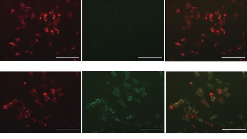
A B C
100 μm 100 μm 100 μm
HEK293 cells transfected with cDNA Anti-human IgG Merge (negative control)
D E F
100 μm 100 μm 100 μm
HEK293 cells transfected with cDNA Anti-human IgG Merge (antibody positive)
Figure 1 Detection and characterization of NMDAR antibodies in CBA. (A,D) Human embryonic kidney (HEK293) cells transfected with
cDNA (red); (B,E) surface binding of patient CSF (anti-IgG, green); (C,F) fluorescence overlaps show antigen-antibody binding (100×
magnification); (A-C were results from the control group, D-F mean antibody positive). (All figures were from the Laboratory of Neurology,
Nanfang Hospital, Southern Medical University).
Hippocampal neurons were cultured from embryonic rats transfected with NR1 complementary (c) DNA are used.
as previously described (26). They were applied to patients IgG antibodies against the subunit NR1 of NMDAR are
with anti-NMDAR encephalitis to examine the effects of highly specific to anti-NMDAR encephalitis and have
antibodies on neurons rather than to prove the presence been demonstrated as an indicator of this disease (16,22).
of anti-NMDAR antibodies. The neurons were incubated HEK293 cells could be transfected with plasmids expressing
with antibodies against NR1 followed by the appropriate both NR1 and NR2B cDNA in a certain ratio to enhance
fluorescence-conjugated secondary antibodies. Imaging and the antigen–antibody reaction as much as possible (16,28).
quantification were conducted to determine the amount of An enhanced green fluorescent protein expression vector
immunolabeling of NMDA receptors by patients’ antibodies was co-transfected with cells to visualize the existence of
(10,22). These antibodies decreased the number of NMDA- cDNAs. After transfection, the cells were incubated with
receptor clusters in postsynaptic dendrites selectively and patients’ serum or CSF with subsequent fixation using 4%
reversibly. In addition, neurons incubated with IgG isolated paraformaldehyde (live CBA) (28). Alternatively, the cells
from the serum of patients or control showed that the were fixed before being incubated with sera or CSF (pre-
amount of patients’ IgG decreased the cell-surface fraction fixed CBA) (29). A secondary antibody - anti-human IgG
of NMDA receptors. The correlation coefficient between with fluorescent labeling could cause double fluorescent
the patients’ antibody titers and the reduction in the labeling and further reduce the possibility of false positives.
number of receptors on the cell surface was positive (22). The negative group showed no binding (Figure 1A,B,C),
There different subunits (NR1-3) were involved in while the positive group showed specific binding to
the formation of NMDAR as a heterotetramer. NR1, an fluorescent markers in CBA (Figure 1D,E,F). The binding
essential subunit of a fully functional NMDAR on the cell of antibodies to antigens on the cell surface or tissue are
surface, binds to glycine, while NR2 bind glutamate (27). visualized using a fluorescence microscope to obtain a semi-
The anti-NMDAR antibody belongs to the immunoglobulin quantitative result (30) and scored visually on a scale from 0
G (IgG) subclass, it is directed against NR1 and NR2B (no binding) to 4 (very strong binding). A score of 0–0.5 was
subunits, but mainly binds to NR1 by amino acid 369 considered as normal. Otherwise, the result was antibody
in the N-terminal epitope (16). In CBA, HEK293 cells positive (31). A low CSF antibody titer was considered as 1:1
© Annals of Translational Medicine. All rights reserved. Ann Transl Med 2021 | https://dx.doi.org/10.21037/atm-20-2279Page 4 of 11 Li et al. Autoantibodies detection in anti-NMDAR encephalitis
or 1:3.2, while a high titer as 1:10 or 1:32 (32). The three-phase gold standard is rarely used for routine
inspections at present. Whether it is too strict to produce a
false-negative result remains unknown.
Application in clinical scenarios
Autoantibody testing is primarily performed in patients
Clinically, a CSF NMDAR antibody titer of 1:10 is suspected with AE. CBA is the most widely used method
generally considered positive. Pre-fixed CBA and IHC due to its high specificity. Since CSF obtained by lumbar
analysis were frequently used for antibody detection. The puncture was traumatic, some medical institutions tended
former focused on the antigen antibody reaction between to test antibodies only in serum (28,29,39). When both
antibodies in samples and fixed cells overexpressing CSF and serum were examined, about 15% of serum results
NMDAR; the latter targeted antibodies against cell surface were false positive (16,40,41); however, these patients
or synaptic proteins using frozen sections of a rat brain seemed to be elderly with milder neurologic symptoms
(22,33). The CBA could detect antibodies against neuronal and a low rate of tumors (42). In addition, other studies
surface antigens. Combined with TBA or IHC analysis, reported that antibodies were found only in CSF in some
CBA could further confirm the regions of the brain where patients with anti-NMDAR encephalitis (43-45). In the
the autoimmune response occurred. Live CBA was reported largest study including 412 patients (paired serum and
to have lower sensitivity compared with pre-fixed CBA (16). CSF), no patient was found to have antibodies only in
Actually, live cells might be destroyed after incubation with serum. The antibodies in CSF correlated better with
high antibody titers, resulting in a lower antibody detection this disease than those in serum (3,38). However, if the
rate. However, whether it was fixed or live cells, false- diagnosis is delayed or patients are treated with plasma
positive or false-negative results were achieved without exchange or intravenous immunoglobulin, antibodies may
confirmatory tests (e.g., IHC) (16,34,35), suggesting be detected only in CSF. NMDAR antibody testing is also
suggested that multiple methods should be used to ensure applied in patients diagnosed with postpartum psychosis
the accuracy of antibody detection results. Besides, anti- and first-episode psychosis (29,46). Some of these patients
NMDAR antibodies in patients’ serum and CSF could show showed positive results and were identified as having anti-
reactivity with live neurons. The IHC analysis on cultured NMDAR encephalitis subsequently. NMDAR antibodies
hippocampus also revealed the presence of anti-NMDAR were reported to be detectable in Creutzfeldt-Jakob disease
antibodies (22). However, the distribution of NMDA (CJD) (47), schizophrenia (31), or neurodegenerative
receptors on the surface of cultured neurons was influenced diseases (48). In these cases, serum was tested using CBA
by patients’ antibodies, leading to the false-negative and revealed anti-NMDAR antibodies, whereas CSF was
results in the IHC analysis. Patients’ antibody mediated not tested or was negative in anti-NMDAR antibodies.
NMDA receptors endocytosis, which decreased the These findings were not reproduced in studies using CBA
number of NMDA receptor on the surface of neurons (22). and IHC analysis in combination to detect antibodies in
These methods usually took several days to obtain the both serum and CSF (22,37,49,50). The serum antibody
experimental results. Thus, cultured neurons were rarely titer from a patient diagnosed with CJD was 1:80, but the
used for a routine inspection to prove the presence of patient failed to respond to immunotherapy and eventually
antibodies but were used for basic researches focusing on died (47). Despite the high specificity of CBA, false
the effects of antibodies on neurons (36). Nevertheless, positives should be taken into account, especially when
Dalmau who discovered the anti-NMDAR antibody testing with serum only. If the autoantibody was detectable
suggested that antibody tests should include three methods only with IHC analysis but not with CBA, or the antibody
(IHC, CBA, and cultured neurons). If one’s serum antibody was detectable in serum but not in CSF, the result might be
was weakly positive in TBA, verification using another considered as false positive. Under this condition, clinical
assay or analyzing CSF by CBA was needed to avoid missed symptoms and differential diagnoses play an important role.
diagnosis and false positives. When using this three-stage On comparing CBA with IHC analysis, a higher antibody
methodology in patients with schizophrenia, no antibody- - positive rate and a greater antigen detection range were
positive case was found. The positive result only in CBA found in IHC. This result was not surprising because
was possibly false positive according to Dalmau’s standard CBA focused on the specific antigen – antibody reaction
(37,38). Subsequently, many improved versions of CBA with while IHC analysis targeted antibodies against cell surface
higher sensitivity and specificity were developed (28,33). proteins or synaptic proteins throughout the rodent’s brain
© Annals of Translational Medicine. All rights reserved. Ann Transl Med 2021 | https://dx.doi.org/10.21037/atm-20-2279Annals of Translational Medicine, 2021 Page 5 of 11
(22,33). Thus, CBA would present a higher specificity to colleagues intended to find out whether flow cytometry
autoantibodies, while positive results detected by IHC [(Fluorescence-activated cell sorting (FACS)]—based assay
analysis could reveal the existence of antibodies other than was more objective and reliable to prove the presence of
known antibodies. Hence, IHC analysis helped confirm the antibodies (54). FACS is a type of flow cytometry, cells need
presence of autoantibodies, while CBA with high specificity to stay alive; FACS was compared with live CBA. Human
helped confirm the presence of specific antibodies. NMDAR antibodies were transfected into HEK293A cells
Clinically, some patients who were antibody negative to bind to the antibodies in serum, and the hCD2-EmGFP
in CBA but positive in IHC analysis shared common fusion protein was transfected to determine unspecific
symptoms with AE but failed to find out a definite cause. binding of serum antibodies. After transfection, the cells
These patients might be classified as having other kinds were detached by trypsin from tissue culture test plates and
of antibodies associated with AE but not anti-NMDAR incubated with patients’ serum. Bound serum antibodies
encephalitis, such as AMPAR, GAD and LGI1, which were detected by the allophycocyanin-conjugated AffiniPure
required further detection of related antibodies. Despite goat anti-human IgG antibody. Washing buffer containing
failing to find the etiology in unexplained AE, positive 7-amino-actinomycin D was used for incubation to exclude
results in IHC analysis still provided some reference for dead cells. The analysis was performed on a flow cytometer.
clinicians to make decisions. In anti-NMDAR encephalitis, The results showed that the specificity of FACS based assay
the concrete epidemiology and optimal immunotherapy was as high as that of CBA. However, FACS based assay had
remains to be determined. However, early diagnosis and a lower sensitivity and high inter-assay variation. The high
early treatment can bring better efficacy (22,51). Hence, it inter-assay variation might be due to the reason that not all
is extremely important to get a timely and correct diagnosis. samples were analyzed with the same batch of transfected
Patients with delayed diagnosis, long course of illness, or and trypsinized cells. Hence, CBA is still a more reliable
persistent symptoms may have negative serum antibodies, detection method.
but CSF titers may continue to rise (52). Combining CBA In addition, single nanoparticle imaging in hippocampal
and IHC analysis to test both serum and CSF could increase neurons was reported to detect patients with low antibody
the “positive rate” (for autoimmune cause), and also lower titers, implying its high sensitivity. Single nanoparticle
the rate of missed diagnosis. However, the evidence for imaging relied on the binding of individual antibodies
treatment based on IHC-positivity is lacking. to their target and was therefore independent of the
sample titer, contrary to other methods dependent on a
sufficient amount of antibodies to provide a detectable
Optimization of detection assays in practice
signal. It is worth noting that unspecific trajectories were
Many factors account for the inconsistent positivity of rarely detected without anti-NMDAR IgG (46). In this
antibody detection between different laboratories, such method, sensitivity may be higher than that of CBA in
as the process of preparing HEK 293 cells and slices of a larger- sample study (27). Single nanoparticle imaging
the brain, artificial visual judgment of the results and even in hippocampal neurons would play an important role
different equipment in the laboratory. Multiple antibodies in clinical diagnosis because of its high sensitivity and
can be found in some laboratory tests; we should repeatedly specificity, however, it is technically demanding and time
verify to prevent false positives. On the contrary, NMDAR consuming.
antibodies can also be found in some patients with herpes Nan-Chang Chiu and his colleges found that replacing
simplex encephalitis (HSE), a transient and subclinical reporter fluorophore fluorescein isothiocyanate in the
synthesis of neuronal antibodies occurs, which becomes commercial kits with Alexa Fluor 488 could obtain better
undetectable several months after the infection (53). Only a detection result, especially in samples with low titers (18)
longer follow-up of patients HSE can clarify whether they because the fluorescence from Alexa Fluor 488 was brighter
have a propensity to develop AE. Some researchers try to and more stable (55,56). A more sophisticated optic/imaging
improve and optimize the detection methods to improve device is recommended because it allows even weak signals
the detection rate of positive samples. to be displayed from samples with low titers. A double
The evaluation of cell surface staining by fluorescence labeling approach, widely used at present, with a mouse
microscopy is strongly dependent on the experience of anti-NR1 mAb and biotinylated goat anti-human IgG could
investigators in CBA. Hence, Melanie Ramberger and his avoid false-positive results.
© Annals of Translational Medicine. All rights reserved. Ann Transl Med 2021 | https://dx.doi.org/10.21037/atm-20-2279Page 6 of 11 Li et al. Autoantibodies detection in anti-NMDAR encephalitis
Clinical implications for antibody testing NMDAR encephalitis (60). Recent studies demonstrated
that 7% of patients with HSE were positive for IgG
As NMDAR antibodies target neuronal receptors and
NMDAR antibodies (61). Moreover, a child with post-HSE
weaken the glutamatergic transmission, mental and
choreoathetosis was found to have NMDAR antibodies, who
cognitive impairment manifestations are apparent in patients
did not improve with antiviral therapy but recovered after
with anti-NMDAR encephalitis. Some patients with anti-
immunotherapy. The findings indicated that a subgroup
NMDAR encephalitis were misdiagnosed with psychosis
of post-HSE represented a separate disease entity, which
because only neuropsychiatric symptoms were observed in
actually was anti-NMDAR encephalitis. Patients with
the early stage (46). In contrast, patients with anti NMDAR
relapsing HSE or prolonged atypical symptoms should be
encephalitis with chronic mental illness usually did not
tested for NMDAR IgG antibodies in CSF and serum when
develop anti-NMDAR encephalitis (38,40). Additionally,
they have negative CSF polymerase chain reaction for HSV.
overlapping autoimmune antibodies were found in some
Combining CBA and IHC analysis to screen suspected AE
patients (41). Thus, solving these diagnostic challenges is
can reduce the rate of missed diagnosis and pick out more
more complex. The treatment of anti-NMDAR encephalitis
potential patients.
focuses on improving prominent psychiatric symptoms and
Modified Rankin score (mRS) was used to access
executive dysfunction. First-line therapies were effective
patients’ disease severity and outcomes in the acute phase
with good outcomes, although some patients even required
and follow-up period. Anti-NMDAR antibody titers
intensive care (15,57,58). Hence, patients showing first-
correlated with mRS scores and clinical improvement (28).
episode psychosis or neuropsychiatric symptoms chiefly
Patients with good prognosis had significantly lower
orofacial dyskinesias and/or autonomic dysfunction should
anti-NMDAR antibody levels and mRS scores, whereas
be screened for NMDAR antibodies to help them get an
patients with unimproved symptoms or poor outcomes and
accurate diagnosis and receive appropriate treatment.
deaths had high antibody levels, although they received
For both CBA and TBA to detect antibodies, clinical
immunotherapy. Whatever the outcomes, antibody titers
symptoms are more important in diagnosing anti-
declined over time (16,22). Hence, spontaneous clinical
NMDAR encephalitis. One patient with a serum antibody
improvement and antibody disappearance in some cases
titer of 1:320 could not be diagnosed with anti-NMDAR
were probably due to slow spontaneous regression of the
encephalitis, because CSF antibody was not found and his
immune response (62). Additionally, immunotherapy led to
clinical symptoms did not support the diagnosis of anti-
a decline in antibody titers. Patients with better outcomes
NMDAR encephalitis (59). If one patient is positive for
had declining levels of CSF antibodies early in the disease,
serum NMDAR antibodies, is negative for CSF, and has
while patients with clinical recurrence and deterioration
no neuropsychiatric symptoms, this patient cannot be
showed increased CSF antibody titers. This correlation
diagnosed with anti-NMDAR encephalitis. Or, in some
was not obvious in serum titers. Dalmau and colleagues
patients with multiple core symptoms, IHC analysis reveals
found that the change in NMDAR antibody concentrations
unknown antibodies while the CBA for an NMDAR
in CSF was more closely related to clinical relapses
antibody is negative. Immunosuppression treatment may be
compared with those in serum (16). To note, the treatment
effective for those showing multiple core symptoms without
(plasma exchange or intravenous immunoglobulin) could
an NMDAR antibody; however, reliable evidence is lacking.
temporarily decreased the levels of antibodies in serum
Besides, HSE caused by herpes simplex virus (HSV)
but not in CSF (63). The difference in titers between
infection is most closely related to anti-NMDAR
patients with poor prognosis and those with good prognosis
encephalitis. Virus infection causes the destruction of
might be greater than statistical results suggested, as
neurons. Then, the exposure of neuronal surface antigen and
more intensive immunotherapy might be administered
the breaking of immune tolerance trigger the autoimmune
to those with poor outcomes (16). However, Irani and his
response. It is also possible that B cell activation (nonspecific)
colleagues found that though the intrathecal synthesis of
or molecular mimicry is involved. This is because HSV and
anti-NMDAR antibody was obvious, the absolute level of
NMDAR share the same antigen epitopes. Therefore, it is
antibody in serum was 13.5 times higher than that in CSF
speculated that viral infection causes B cell activation and
on average, although the intrathecal synthesis of the anti-
production of antibodies against the virus, which cross-
NMDAR antibody was obvious (28), serum antibody titers
reacts with NMDAR, leading to the occurrence of anti-
were also considered to be correlated with the improvement
© Annals of Translational Medicine. All rights reserved. Ann Transl Med 2021 | https://dx.doi.org/10.21037/atm-20-2279Annals of Translational Medicine, 2021 Page 7 of 11
of clinical symptoms (44). The heterogeneity of testing clinical researches in the Chinese population revealed
methods between different laboratories might account for that anti-NMDAR encephalitis had unpredictable MRI
the inconsistent conclusion. Besides, each CBA had its findings that easily obscured its diagnosis and caused serious
specificity in terms of techniques used, such as fixed or live sequelae (69). An NMDAR antibody test is required for a
cell, transfecting with different NR subunit plasmids, and timely diagnosis and immunotherapy (17). Most patients
ratios. Also, the NMDAR conformation in the rodent brain with severe anti-NMDAR encephalitis would eventually
or HEK cells might not be the same as that in humans. achieve good long-term prognoses after receiving early,
Relapse means the occurrence of new symptoms or the positive, and unremitting combined immunotherapy and
aggravation of original symptoms after more than 2 months life support (70). Significantly, the largest Chinese anti-
of the convalescence period. Patients with undetected or NMDAR encephalitis cohort so far, which recruited
recurrent tumors (22) or non-paraneoplastic cases as well 220 patients, showed that the primary clinical presentations
as those did not receive timely immunotherapy were more were psychosis and seizures (71). Tumors were not frequent,
likely to relapse (64). Clinical recovery does not mean the incidence of ovarian teratoma in women was the
the disappearance of serum and CSF antibodies, some maximum, and only one man had lung cancer. Most patients
patients could still show positive antibodies in serum and (99.5%) received first-line therapy, and only 7.3% received
CSF (16,65,66). Hence, clinical decisions should be based second-line immunotherapy. More than half of the patients
on a combination of symptoms, antibody detection results were administered long-term immunotherapy. During the
and other auxiliary examination (19). These patients are first year of follow-up, a large majority of patients (94.1%)
supposed to attend the immunological follow-up and tumor achieved a good outcome, 2.3% of patients died, and 17.3%
screening (15). The determination of baseline serum and experienced relapses. First-line immunotherapy is effective
CSF titers after recovery is potentially helpful in classifying in managing anti-NMDAR encephalitis in the acute phase.
new-onset symptoms as possible relapses, predicting disease Although relapse is relatively common, most patients
risk, and managing the disease (according to the rise in the reached favorable outcomes with combined first-line and
titer) (16). Early immunotherapy and tumors removal could long-term immunotherapy (71).
effectively improve outcomes, reduce antibody levels and
prevent relapses (28).
Conclusions
In China, the earliest case diagnosed with anti-NMDAR
encephalitis was in 2010. Subsequently, more centers AE is a treatable immune-mediated disorder that it can
gradually started clinical research on anti-NMDAR be diagnosed by the specific autoantibody detection.
encephalitis. The clinical research scope covered biomarkers, The diagnosis of AE should be based on antibody testing
genetics, and antibody testing, but only a limited number combined with clinical symptoms. IHC analysis involves
of studies focused on the detection assay of AE in China. could pick out more potential patients than CBA.
For example, Liu and his colleagues used TBA to detect Antibodies should be detected in both serum and CSF using
antibodies in CSF in 739 patients. Of these, 37 patients had IHC analysis and CBA to avoid misdiagnosis. CSF antibody
a neuronal antibody pattern. In another study, 17 patients titers are believed to be more clinically relevant in anti-
were detected with NMDAR antibodies. The conclusion NMDAR encephalitis. However, prospective studies are
of this study was similar to that of another previous needed to determine the prognostic value of antibody titers
study (35). These results showed the advantages of TBA in because some clinically cured patients still have detectable
discovering the AE of unknown antigen, especially in CSF. NMDAR antibodies detectable. A long -term follow-up
Commercially available CBA is frequently used for anti- is recommended. Further studies should pay attention to
NMDAR antibody detection in China. The results also potential biomarkers in serum for diagnosis or assessment of
showed that the positive rate of CSF was higher than that of this disease because CSF testing is invasive and not always
serum (67,68). However, the shortcoming lay in the small available.
sample size of these studies.
Other aspects, such as clinical characteristics, magnetic
Acknowledgments
resonance imaging (MRI), immunotherapy regimens
and long-term outcomes of patients with anti-NMDAR Funding: This work was funded by the National Natural
encephalitis in China have also been described. These Science Foundation of China (81673950), Guangdong
© Annals of Translational Medicine. All rights reserved. Ann Transl Med 2021 | https://dx.doi.org/10.21037/atm-20-2279Page 8 of 11 Li et al. Autoantibodies detection in anti-NMDAR encephalitis
Provincial Science and Technology plan projects Paraneoplastic cerebellar ataxia due to autoantibodies
(2017A020215182), and Natural Science Foundation of against a glutamate receptor. N Engl J Med
Guangdong Province (2019A1515011434). 2000;342:21-7.
5. Mikasova L, De Rossi P, Bouchet D, et al. Disrupted
surface cross-talk between NMDA and Ephrin-B2
Footnote
receptors in anti-NMDA encephalitis. Brain
Provenance and Peer Review: This article was commissioned 2012;135:1606-21.
by the Guest Editors (Dr. Hai-feng Li and Dr. Xiangjun 6. Boyse EA, Stockert E, Old LJ. Modification of the
Chen) for the series “Laboratory investigations in antigenic structure of the cell membrane by thymus-
neuroimmunological diseases and their clinical significance” leukemia (TL) antibody. Proc Natl Acad Sci U S A
published in Annals of Translational Medicine. The article has 1967;58:954-7.
undergone external peer review. 7. Albert ML, Austin LM, Darnell RB. Detection and
treatment of activated T cells in the cerebrospinal fluid of
Conflicts of Interest: All authors have completed the ICMJE patients with paraneoplastic cerebellar degeneration. Ann
uniform disclosure form (available at https://dx.doi. Neurol 2000;47:9-17.
org/10.21037/atm-20-2279). The series “Laboratory 8. Roberts WK, Deluca IJ, Thomas A, et al. Patients
investigations in neuroimmunological diseases and their with lung cancer and paraneoplastic Hu syndrome
clinical significance” was commissioned by the editorial harbor HuD-specific type 2 CD8+ T cells. J Clin Invest
office without any funding or sponsorship. The authors 2009;119:2042-51.
have no conflicts of interest to declare. 9. Vitaliani R, Mason W, Ances B, et al. Paraneoplastic
encephalitis, psychiatric symptoms, and hypoventilation in
Ethical Statement: The authors are accountable for all ovarian teratoma. Ann Neurol 2005;58:594-604.
aspects of the work in ensuring that questions related 10. Dalmau J, Tüzün E, Wu HY, et al. Paraneoplastic anti-N-
to the accuracy or integrity of any part of the work are methyl-D-aspartate receptor encephalitis associated with
appropriately investigated and resolved. ovarian teratoma. Ann Neurol 2007;61:25-36.
11. Lai M, Hughes EG, Peng X, et al. AMPA receptor
Open Access Statement: This is an Open Access article antibodies in limbic encephalitis alter synaptic receptor
distributed in accordance with the Creative Commons location. Ann Neurol 2009;65:424-34.
Attribution-NonCommercial-NoDerivs 4.0 International 12. Lancaster E, Lai M, Peng X, et al. Antibodies to the
License (CC BY-NC-ND 4.0), which permits the non- GABA(B) receptor in limbic encephalitis with seizures:
commercial replication and distribution of the article with case series and characterisation of the antigen. Lancet
the strict proviso that no changes or edits are made and the Neurol 2010;9:67-76.
original work is properly cited (including links to both the 13. Boronat A, Sabater L, Saiz A, et al. GABA(B) receptor
formal publication through the relevant DOI and the license). antibodies in limbic encephalitis and anti-GAD-associated
See: https://creativecommons.org/licenses/by-nc-nd/4.0/. neurologic disorders. Neurology 2011;76:795-800.
14. Lai M, Huijbers MG, Lancaster E, et al. Investigation
of LGI1 as the antigen in limbic encephalitis previously
References
attributed to potassium channels: a case series. Lancet
1. Lancaster E, Dalmau J. Neuronal autoantigens-- Neurol 2010;9:776-85.
pathogenesis, associated disorders and antibody testing. 15. Dalmau J, Lancaster E, Martinez-Hernandez E, et al.
Nat Rev Neurol 2012;8:380-90. Clinical experience and laboratory investigations in
2. van Coevorden-Hameete MH, de Graaff E, Titulaer patients with anti-NMDAR encephalitis. Lancet Neurol
MJ, et al. Molecular and cellular mechanisms underlying 2011;10:63-74.
anti-neuronal antibody mediated disorders of the central 16. Gresa-Arribas N, Titulaer MJ, Torrents A, et al. Antibody
nervous system. Autoimmun Rev 2014;13:299-312. titres at diagnosis and during follow-up of anti-NMDA
3. Iorio R, Lennon VA. Neural antigen-specific autoimmune receptor encephalitis: a retrospective study. Lancet Neurol
disorders. Immunol Rev 2012;248:104-21. 2014;13:167-77.
4. Sillevis Smitt P, Kinoshita A, De Leeuw B, et al. 17. Huang X, Fan C, Wu J, et al. Clinical analysis on anti-N-
© Annals of Translational Medicine. All rights reserved. Ann Transl Med 2021 | https://dx.doi.org/10.21037/atm-20-2279Annals of Translational Medicine, 2021 Page 9 of 11
methyl-D-aspartate receptor encephalitis cases: Chinese Fluid Antibody Titers in Patients With anti-NMDAR
experience. Int J Clin Exp Med 2015;8:18927-35. Encephalitis. Clin EEG Neurosci 2019;50:56-62.
18. Chiu NC, Lin YJ, Tzang RF, et al. Optimization of an 32. Wandinger KP, Saschenbrecker S, Stoecker W, et al.
Anti-NMDA Receptor Autoantibody Diagnostic Bioassay. Anti-NMDA-receptor encephalitis: a severe, multistage,
Front Neurol 2018;9:661. treatable disorder presenting with psychosis. J
19. Graus F, Titulaer MJ, Balu R, et al. A clinical approach Neuroimmunol 2011;231:86-91.
to diagnosis of autoimmune encephalitis. Lancet Neurol 33. Hara M, Martinez-Hernandez E, Ariño H, et al. Clinical
2016;15:391-404. and pathogenic significance of IgG, IgA, and IgM
20. Armangue T, Titulaer MJ, Sabater L, et al. A novel antibodies against the NMDA receptor. Neurology
treatment-responsive encephalitis with frequent 2018;90:e1386-94.
opsoclonus and teratoma. Ann Neurol 2014;75:435-41. 34. Zandi MS, Paterson RW, Ellul MA, et al. Clinical
21. Guan HZ, Ren HT, Cui LY. Autoimmune Encephalitis: relevance of serum antibodies to extracellular N-methyl-
An Expanding Frontier of Neuroimmunology. Chin Med J D-aspartate receptor epitopes. J Neurol Neurosurg
(Engl) 2016;129:1122-7. Psychiatry 2015;86:708-13.
22. Dalmau J, Gleichman AJ, Hughes EG, et al. Anti-NMDA- 35. Hughes EG, Peng X, Gleichman AJ, et al. Cellular and
receptor encephalitis: case series and analysis of the effects synaptic mechanisms of anti-NMDA receptor encephalitis.
of antibodies. Lancet Neurol 2008;7:1091-8. J Neurosci 2010;30:5866-75.
23. Gastaldi M, Waters P, Vincent A. Detection of 36. Masdeu JC, González-Pinto A, Matute C, et al. Serum
NMDARs Antibodies in Encephalitis. Methods Mol Biol IgG antibodies against the NR1 subunit of the NMDA
2017;1677:117-26. receptor not detected in schizophrenia. Am J Psychiatry
24. Chen QM, Qu HD, Qian WD, et al. Correlation between 2012;169:1120-1.
neuronal antibodies and limbic encephalitis in Chinese 37. Pollak TA, McCormack R, Peakman M, et al. Prevalence
Han subjects. Genet Mol Res 2015;14:2312-21. of anti-N-methyl-D-aspartate (NMDA) receptor corrected
25. Elmariah SB, Oh EJ, Hughes EG, et al. Astrocytes antibodies in patients with schizophrenia and related
regulate inhibitory synapse formation via Trk-mediated psychoses: a systematic review and meta-analysis. Psychol
modulation of postsynaptic GABAA receptors. J Neurosci Med 2014;44:2475-87.
2005;25:3638-50. 38. Davies G, Irani SR, Coltart C, et al. Anti-N-methyl-D-
26. Lynch DR, Anegawa NJ, Verdoorn T, et al. aspartate receptor antibodies: a potentially treatable cause
N-methyl-D-aspartate receptors: different subunit of encephalitis in the intensive care unit. Crit Care Med
requirements for binding of glutamate antagonists, 2010;38:679-82.
glycine antagonists, and channel-blocking agents. Mol 39. Titulaer MJ, McCracken L, Gabilondo I, et al. Treatment
Pharmacol 1994;45:540-5. and prognostic factors for long-term outcome in patients
27. Irani SR, Bera K, Waters P, et al. N-methyl-D-aspartate with anti-NMDA receptor encephalitis: an observational
antibody encephalitis: temporal progression of clinical cohort study. Lancet Neurol 2013;12:157-65.
and paraclinical observations in a predominantly 40. Titulaer MJ, Höftberger R, Iizuka T, et al. Overlapping
non-paraneoplastic disorder of both sexes. Brain demyelinating syndromes and anti–N-methyl-
2010;133:1655-67. D-aspartate receptor encephalitis. Ann Neurol
28. Bergink V, Armangue T, Titulaer MJ, et al. Autoimmune 2014;75:411-28.
Encephalitis in Postpartum Psychosis. Am J Psychiatry 41. Guasp M, Módena Y, Armangue T, et al. Clinical
2015;172:901-8. features of seronegative, but CSF antibody-positive, anti-
29. Leite MI, Jacob S, Viegas S, et al. IgG1 antibodies to NMDA receptor encephalitis. Neurol Neuroimmunol
acetylcholine receptors in 'seronegative' myasthenia gravis. Neuroinflamm 2020;7:e659.
Brain 2008;131:1940-52. 42. Punja M, Pomerleau AC, Devlin JJ, et al. Anti-N-methyl-
30. Zandi MS, Irani SR, Lang B, et al. Disease-relevant D-aspartate receptor (anti-NMDAR) encephalitis: an
autoantibodies in first episode schizophrenia. J Neurol etiology worth considering in the differential diagnosis of
2011;258:686-8. delirium. Clin Toxicol (Phila) 2013;51:794-7.
31. Miao A, Du M, Wang L, et al. Analysis of Relation 43. Suh-Lailam BB, Haven TR, Copple SS, et al. Anti-
Between Electroclinical Features and Cerebrospinal NMDA-receptor antibody encephalitis: performance
© Annals of Translational Medicine. All rights reserved. Ann Transl Med 2021 | https://dx.doi.org/10.21037/atm-20-2279Page 10 of 11 Li et al. Autoantibodies detection in anti-NMDAR encephalitis
evaluation and laboratory experience with the Isothiocyanate- conjugated Antibody. Cell J
anti-NMDA-receptor IgG assay. Clin Chim Acta 2011;13:169-72.
2013;421:1-6. 56. Finke C, Kopp UA, Prüss H, et al. Cognitive deficits
44. Greiner H, Leach JL, Lee KH, et al. Anti-NMDA following anti-NMDA receptor encephalitis. J Neurol
receptor encephalitis presenting with imaging findings and Neurosurg Psychiatry 2012;83:195-8.
clinical features mimicking Rasmussen syndrome. Seizure 57. Kayser MS, Titulaer MJ, Gresa-Arribas N, et al. Frequency
2011;20:266-70. and characteristics of isolated psychiatric episodes in anti–
45. Jézéquel J, Rogemond V, Pollak T, et al. Cell- and Single N-methyl-d-aspartate receptor encephalitis. JAMA Neurol
Molecule-Based Methods to Detect Anti-N-Methyl-D- 2013;70:1133-9.
Aspartate Receptor Autoantibodies in Patients With First- 58. Mantere O, Saarela M, Kieseppä T, et al. Anti-neuronal
Episode Psychosis From the OPTiMiSE Project. Biol anti-bodies in patients with early psychosis. Schizophr Res
Psychiatry 2017;82:766-72. 2018;192:404-7.
46. Mackay G, Ahmad K, Stone J, et al. NMDA receptor 59. Nosadini M, Mohammad SS, Corazza F, et al. Herpes
autoantibodies in sporadic Creutzfeldt-Jakob disease. J simplex virus-induced anti-N-methyl-d-aspartate
Neurol 2012;259:1979-81. receptor encephalitis: a systematic literature review
47. Deakin J, Lennox BR, Zandi MS. Antibodies to the with analysis of 43 cases. Dev Med Child Neurol
N-methyl-D-aspartate receptor and other synaptic proteins 2017;59:796-805.
in psychosis. Biol Psychiatry 2014;75:284-91. 60. Prüss H, Finke C, Höltje M, et al. N-methyl-D-aspartate
48. Grant I, Atkinson JH, Gouaux B. Research on medical receptor antibodies in herpes simplex encephalitis. Ann
marijuana. Am J Psychiatry 2012;169:1119-20; author Neurol 2012;72:902-11.
reply 1120. 61. Iizuka T, Sakai F, Ide T, et al. Anti-NMDA receptor
49. Grau-Rivera O, Sánchez-Valle R, Saiz A, et al. encephalitis in Japan: long-term outcome without tumor
Determination of neuronal antibodies in suspected removal. Neurology 2008;70:504-11.
and definite Creutzfeldt-Jakob disease. JAMA Neurol 62. Furneaux HF, Reich L, Posner JB. Autoantibody
2014;71:74-8. synthesis in the central nervous system of patients with
50. Seki M, Suzuki S, Iizuka T, et al. Neurological paraneoplastic syndromes. Neurology 1990;40:1085-91.
response to early removal of ovarian teratoma in anti- 63. Gabilondo I, Saiz A, Galán L, et al. Analysis of
NMDAR encephalitis. J Neurol Neurosurg Psychiatry relapses in anti-NMDAR encephalitis. Neurology
2008;79:324-6. 2011;77:996-9.
51. Miya K, Takahashi Y, Mori H. Anti-NMDAR autoimmune 64. Hansen HC, Klingbeil C, Dalmau J, et al. Persistent
encephalitis. Brain Dev 2014;36:645-52. intrathecal antibody synthesis 15 years after recovering
52. Armangue T, Spatola M, Vlagea A, et al. Frequency, from anti-N-methyl-D-aspartate receptor encephalitis.
symptoms, risk factors, and outcomes of autoimmune JAMA Neurol 2013;70:117-9.
encephalitis after herpes simplex encephalitis: a prospective 65. Alexopoulos H, Kosmidis ML, Dalmau J, et al.
observational study and retrospective analysis. Lancet Paraneoplastic anti-NMDAR encephalitis: long term
Neurol 2018;17:760-72. follow-up reveals persistent serum antibodies. J Neurol
53. Ramberger M, Peschl P, Schanda K, et al. Comparison 2011;258:1568-70.
of diagnostic accuracy of microscopy and flow cytometry 66. Wang XH, Fang F, Ding CH, et al. Anti-N-methyl-D-
in evaluating N-methyl-D-aspartate receptor antibodies aspartate receptor encephalitis in seven children. Zhonghua
in serum using a live cell-based assay. PLoS One Er Ke Za Zhi 2012;50:885-9.
2015;10:e0122037. 67. Wu Y, Shi Y, Zhang M, et al. Anti-N-methyl-D-aspartate
54. Panchuk-Voloshina N, Haugland RP, Bishop-Stewart J, et receptor encephalitis:clinical analysis of 11 cases. Zhonghua
al. Alexa dyes, a series of new fluorescent dyes that yield Yi Xue Za Zhi 2015;95:2857-60.
exceptionally bright, photostable conjugates. J Histochem 68. Zhang W, Cui L, Wang W, et al. Early identification of
Cytochem 1999;47:1179-88. anti-NMDA receptor encephalitis presenting cerebral
55. Mahmoudian J, Hadavi R, Jeddi-Tehrani M, et al. lesions in unconventional locations on magnetic resonance
Comparison of the Photobleaching and Photostability imaging. J Neuroimmunol 2018;320:101-6.
Traits of Alexa Fluor 568- and Fluorescein 69. Zhang Y, Liu G, Jiang M, et al. Clinical Characteristics
© Annals of Translational Medicine. All rights reserved. Ann Transl Med 2021 | https://dx.doi.org/10.21037/atm-20-2279Annals of Translational Medicine, 2021 Page 11 of 11
and Prognosis of Severe Anti-N-methyl-D-aspartate 70. Xu X, Lu Q, Huang Y, et al. Anti-NMDAR encephalitis:
Receptor Encephalitis Patients. Neurocrit Care A single-center, longitudinal study in China. Neurol
2018;29:264-72. Neuroimmunol Neuroinflamm 2020;7:e633.
Cite this article as: Li J, Wang Q, Wang H. Autoantibodies
detection in anti-N-methyl-D-aspartate receptor encephalitis.
Ann Transl Med 2021. doi: 10.21037/atm-20-2279
© Annals of Translational Medicine. All rights reserved. Ann Transl Med 2021 | https://dx.doi.org/10.21037/atm-20-2279You can also read