Original Article Dynamic change of SGK expression and its role in neuron apoptosis after traumatic brain injury
←
→
Page content transcription
If your browser does not render page correctly, please read the page content below
Int J Clin Exp Pathol 2013;6(7):1282-1293
www.ijcep.com /ISSN:1936-2625/IJCEP1304023

Original Article
Dynamic change of SGK expression and its role in
neuron apoptosis after traumatic brain injury
Xinmin Wu1*, Hui Mao2*, Jiao Liu3, Jian Xu4, Jianhua Cao4, Xingxing Gu5, Gang Cui6
1
 Department of Neurology, Affiliated Hospital of Nantong University, Nantong, Jiangsu Province 226001, People’s
Republic of China; 2Department of Neurosurgery, Affiliated Hospital of Nantong University, Nantong, Jiangsu Prov-
ince 226001, People’s Republic of China; 3Department of Public Health, Nantong University, Nantong, Jiangsu
Province 226001, People’s Republic of China; 4Affiliated mental health centers of Nantong University, Nantong,
Jiangsu Province 226001, People’s Republic of China; 5Key Laboratory of Neuroscience, Nantong University, Nan-
tong 226001, People’s Republic of China; 6Department of Neurosurgery, The First Affiliated Hospital of Soochow
University, Suzhou 215006, People’s Republic of China. *Equal contributors.
Received April 15, 2013; Accepted May 20, 2013; Epub June 15, 2013; Published July 1, 2013

Abstract: Aims: Activation of specific signaling pathways in response to mechanical trauma causes delayed neuro-
nal apoptosis; GSK-3β/β-catenin signaling plays a critical role in the apoptosis of neurons in CNS diseases, SGK was
discovered as a regulator of GSK-3β/β-catenin pathway, The goal of this study was to determine if the mechanism of
cell death or survival mediated by the SGK/GSK-3β/β-catenin pathway is involved in a rat model of TBI. Main meth-
ods: Here, an acute traumatic brain injury model was applied to investigate the expression change and possible
roles of SGK, Expression of SGK, and total-GSK-3β, phospho-GSK3β on ser-9, beta-catenin, and caspase-3 were
examined by immunohistochemistry and Western blot analysis. Double immunofluorescent staining was used to
observe the SGK localizations. Si-RNA was performed to identify whether SGK regulates neuron apoptosis via GSK-
3β/β-catenin pathway, ultimately inhibit caspase-3 activation. Key findings: Temporally, SGK expression showed an
increase pattern after TBI and reached a peak at day 3. Spatially, SGK was widely expressed in the neuron, rarely
in astrocytes and oligodendrocytes; in addition, the expression patterns of active caspase-3 and phospho-GSK3β
were parallel with that of SGK, at the same time, the expression of β-catenin shows similarity with SGK. In vitro, to
further investigate the function of SGK, a neuronal cell line PC12 was employed to establish an apoptosis model.
We analyzed the association of SGK with apoptosis on PC12 cells by western blot, immunofluorescent labeling and
siRNA. Significance: the results implied that SGK plays an important role in neuron apoptosis via the regulation of
GSK3β/β-catenin signaling pathway; ultimately inhibit caspase-3 activation. Taken together, we inferred traumatic
brain injury induced an upregulation of SGK in the central nervous system, which show a protective role in neuron
apoptosis.

Keywords: SGK, GSK3β/β-catenin signaling pathway, traumatic brain injury (TBI), neuron apoptosis

Introduction                                                   rhage; Secondary damage includes processes
                                                               that are initiated at the time of insult, but do not
Traumatic brain injury (TBI) is one of the leading             appear clinically for hours or even days after
causes of death and disability worldwide,                      injury. It can subsequently trigger astrocyte pro-
including the developing world [1]. Traumatic                  liferation, microglia activation and neuronal cell
brain injury is an insult to the brain caused by               death [4]. Although large amount of clinical and
an external physical force, resulting in function-             basic researches focus on TBI, there are limited
al disability [2]. Both clinical and experimental              methods for improving the outcome of patients
studies have shown that the pathophysiology of                 suffering from brain trauma [5, 6]. Both anti-
traumatic brain injury (TBI) is complex and                    apoptotic and pro-apoptotic signaling cascades
involves both primary and secondary injuries                   are activated in secondary tissue injury [7]. In
[3]. Primary damage occurs at the moment of                    addition to causing direct mechanical injury,
insult and includes contusion and laceration,                  trauma initiates secondary cascades of bio-
diffuse axonal injury, and intracranial hemor-                 chemical and cellular changes that substantial-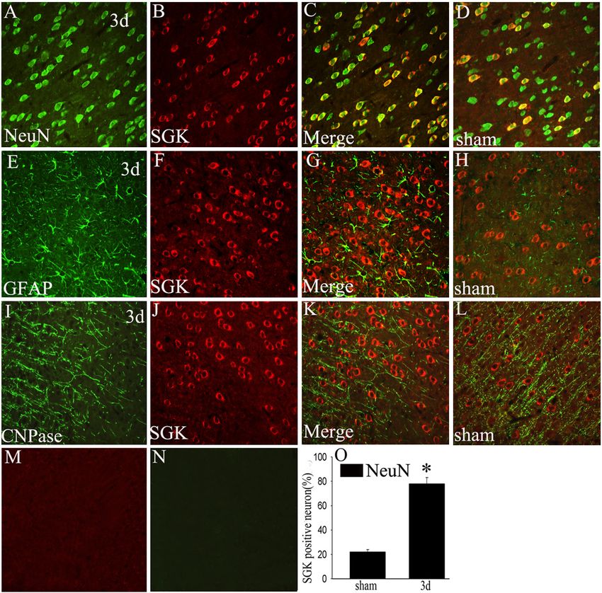
SGK expression in neuron apoptosis after TBI

ly contribute to subsequent tissue damage and        In addition, Many reports have shown that SGK
related neurological deficits.                       mRNA and protein level upregulated after trau-
                                                     matic brain injury which particularly abundant
Activation of specific signaling pathways in         in the central nervous system and neuron-spe-
response to mechanical trauma causes delayed         cific, and play a protective role in the regulation
neuronal apoptosis [8]. One pathway with a           of neuronal function [17-19]. But the inherent
prominent role in neurotrauma is the signaling       role is still little known.
pathway in which the enzyme glycogen syn-
thase kinase 3β (GSK3β) is a key component.          Given the roles of GSK-3β/β-catenin pathway
Glycogen synthase kinase-3β (GSK3β) activa-          and SGK in central nervous system especially
tion promotes cell death [9-12] and inhibits cell    in neuron apoptosis and the mutant relation-
proliferation, a growing body of evidence sug-       ship between them, accordingly, we speculated
gests that GSK3β is an important modulator in        whether the SGK is involved in the neuronal
central nervous system diseases, including           survival via GSK-3β/β-catenin signaling and its
traumatic brain injury and AD (Alzheimer’s dis-      action through the downstream targets, espe-
ease) [13], Phosphorylation GSK3β on serine-9        cially β-catenin, after TBI.
to render it inactive, a mechanism by which
neurons become resistant to apoptotic stimuli        Thus the present study was designed to investi-
[14], which makes it becomes the focus for its       gate the changes of SGK, phospho-GSK3β/β-
role in neuron protection, furthermore, Neuro-       catenin and their roles in the regulation of cas-
protective stimuli lead to an inactivation of        pase-3 (the apoptosis marker) in a TBI model
GSK3β. Prominent in this latter category is the      after 3 days. Moreover, the siRNA was used to
PI3K/Akt pathway. Thus, GSK3β activity               confirm the possible roles of SGK regulate neu-
appears to correlate inversely with neuronal         ron apoptosis via GSK3β/β-catenin signaling
viability [15]. β-catenin, which is central to the   pathway.
Wnt signaling pathway involved in many stages
                                                     Materials and methods
of development, is a GSK3β substrate. GSK3β-
mediated phosphorylation enhances the prote-         Models of TBI
asome-dependent degradation of β-catenin.
The inhibition of GSK-3β caused dramatic ele-        All protocols using animals were conducted in
vations in the level of β-catenin and stimulated     accordance with the guidelines published in
β-catenin-dependent gene transcription that          the NIH Guide for the care and use of laborato-
regulate neuronal homeostasis and support            ry animals and the principles presented in the
neuron cell survival.                                Guidelines for the use of animals in neurosci-
                                                     ence research by the Society for Neuroscience
GSK-3β/β-catenin pathway can be regulated by         and approved by Nantong University Animal
many signaling pathways and proteins, Wnt and        Care Ethics Committee. Male Sprague-Dawley
Akt (also called protein kinase B) are two major     Rats (n=48) with an average body weight of
signaling pathways that have been shown to           250 g (220 ± 275 g) were used in this experi-
regulate GSK-3β activity via distinct mecha-         ment. Unilateral controlled cortical injury was
nisms. Additionally, the recent studies have         performed as previously described [20, 21]. In
shown the Serum- and glucocorticoid-regulated        brief, adult male were anesthetized with
kinase (SGK), known as SGK1, was discovered          Ketamine (90 mg/kg)/xylazine (10 mg/kg), and
as a regulator of GSK-3β/β-catenin pathway,          surgery was performed under aseptic condi-
which is involved in the pathophysiology pro-        tions. An anteroposterior surgical incision
cess of tumor growth, fibrosing disease, isch-       (5-mm-long, 3-mm-deep, and 1-mm-wide) was
emia, neurodegeneration, and traumatic brain         made by inserting a microknife into the right
injury [16]. Previous investigations have dem-       cortex.
onstrated that SGK phosphorylates GSK3β on
serine-9 and then controls β-catenin dynamics,       3 mm lateral from the midline (n=42). Controlled
further takes part in the process of tight junc-     rats (n=6) underwent identical procedures to
tion formation in mammary epithelial tumor           experimental animals, but did not receive brain
cells and in the regulation of L-selectin and per-   injury. Ketoprofen (5 mg/kg) was administered
forin expression as well as activation induced       to minimize postsurgical pain and discomfort.
cell death of T-lymphocytes.                         The overlying muscles and skin were closed in

1283                                                     Int J Clin Exp Pathol 2013;6(7):1282-1293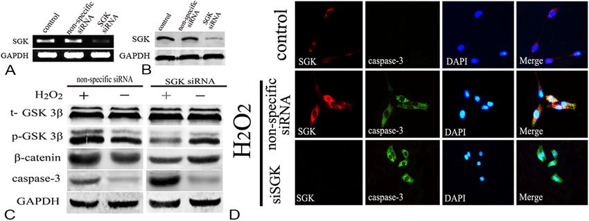
SGK expression in neuron apoptosis after TBI

layers with 4-0 silk sutures and staples, and         mmol/l PMSF, 10 μg/ml aprotinin, and 1 μg/ml
the animals were allowed to recover on a 30°C         leupeptin) and clarified by centrifuging for 20
heating pad. Animals were individually housed         min in a microcentrifuge at 4°C. After determi-
in cages and kept in a temperature-controlled         nation of its protein concentration with the
environment (21°C) on a 12-hr light-dark cycle,       Bradford assay (Bio-Rad), the resulting super-
with access to food and water ad libitum. ani-        natant (50 μg of protein) was subjected to SDS-
mals were killed at 12 h, 1 d, 3 d, 5 d, 7 d, 14 d,   polyacrylamide gel electrophoresis (PAGE). The
and 28 d after injury, and sham-operated rats         separated proteins were transferred to a polyvi-
(n=3) were sacrificed at 3 days. All efforts were     nylidene difluoride membrane (Millipore) by a
made to minimize the number of animals used           transfer apparatus at 350 mA for 1.5 h. The
and their suffering.                                  membrane was then blocked with 5% nonfat
                                                      milk and incubated with primary antibody
Cell cultures and treatment                           against SGK (anti-rabbit, 1:500; Santa Cruz),
                                                      p-GSK3β, β-catenin (anti-mouse, 1:1,000; Cell
PC12 cells were cultured in Dulbecco’s modi-          Signaling), or GAPDH (anti-rabbit, 1:1,000;
fied Eagle’s medium (DMEM) with 10% (v/v)             Santa Cruz), GFAP (anti-mouse, 1:1,000; Cell
fetal bovine serum, 5% donor horse serum and          Signaling; anti-rabbit, 1:1,000; Santa Cruz).
antibiotics at 37°C under 5% CO2 in humidified        After incubating with an anti-rabbit horseradish
air. The cells were passed every 3-4 days. In         peroxidase-conjugated secondary antibody,
order to study apoptosis, cells were seeded           protein was visualized using an enhanced che-
onto a poly-l-lysine-coated 60 mm dishes and          miluminescence system (ECL, Pierce Company,
incubated in a low concentration of serum (1%         USA).
horse serum) for 24 hours prior to treatment
with H2O2 (300 nmol/L) for different time.            Immunofluorescence staining

siRNA and transfection                                After defined survival times, rats were termi-
                                                      nally anesthetized and perfused through the
Primer pairs for the SGK (NM_001292567.1)             ascending aorta with saline, followed by 4%
siRNA expression vector was target the                paraformaldehyde at different survival times
sequence: 5’-CAAGGACCUAGCCGCACAA-3’. For              (n=3 per time point). The brains were removed
transient transfection, the SGK siRNA vector,         and postfixed in the same fixative for 3 hours
and the non-specific vector were carried out          and then replaced with 20% sucrose for 2-3
using lipofectamine 2,000 (Invitrogen) and plus       days, following 30% sucrose for 2-3 days. After
reagent in OptiMEM (Invitrogen) as suggested          treatment with sucrose solution, the tissues
by the manufacturer. Transfected cells were           were embedded in OCT compound. Then,
used for the subsequent experiments 48 h              10-µm frozen cross-sections were prepared
after transfection.                                   and examined. All sections were first blocked
                                                      with 10% normal serum blocking solution-spe-
Western blot analysis                                 cies the same as the secondary antibody, con-
                                                      taining 3% (w/v) bovine serum albumin (BSA)
Western blots were prepared from normal brain         and 0.1% Triton X-100 and 0.05% Tween-20 2 h
cortex or from injured cortex at 12 h-28 days, to     at room temperature in order to avoid unspe-
obtain samples for Western blotting, rats were        cific staining. Then the sections were incubated
sacrificed at different time points post-opera-       with both rabbit polyclonal primary antibodies
tively (n=3 for each time point), the brain tissue    for anti-SGK (1:200; Santa Cruz), goat poly-
surrounding the wound (extending 2 mm to the          clonal primary antibodies for anti-caspase-3
lesion site, weighing 70-90 mg) as well as an         (1:200; Cell Signaling) and mouse monoclonal
equal part of the contralateral, unoperated cor-      primary antibodies anti-GFAP (a marker of
tex were dissected out and immediately frozen         astrocytes, 1:200; Sigma), anti-NeuN (a marker
at -70°C until use. To prepare lysates, frozen        of neuron, 1:600; Chemicon); Briefly, sections
brain tissue samples were minced with eye             were incubated with both primary antibodies
scissors in ice. The samples were then homog-         overnight at 4°C, followed by a mixture of FITC-
enized in lysis buffer (1% NP-40, 50 mmol/l           and TRITC-conjugated secondary antibodies for
Tris, and pH 7.5, 5 mmol/l EDTA, 1% SDS, 1%           2 h at 4°C. In sections from each specimen, the
sodium deoxycholate, 1% Triton X-100, 1               primary antibody was omitted to assess for

1284                                                      Int J Clin Exp Pathol 2013;6(7):1282-1293
SGK expression in neuron apoptosis after TBI
nonspecific binding of the secondary antibody. ble labeled for SGK with NeuN. To identify the
The stained sections were examined with a proportion of NeuN positive cells expressing
Leica fluorescence microscope (Germany). SGK, a minimum of 200 NeuN positive cells
were counted adjacent to the wound in each
Immunohistochemistry section. Then double labeled cells for SGK and
After the sections were prepared, they were NeuN were recorded. Two or three adjacent
blocked with 10% goat serum with 0.3% Triton sections per animal were sampled.
X-100 and 1% BSA for 2 h at room temperature Statistical analysis
and incubated overnight at 4°C with anti-SGK
antibody (rabbit, 1:100; Santa Cruz), followed All data were analyzed with Stata 7.0 statistical
by incubation in biotinylated secondary anti- software. All values are expressed as means ±
body (Vector Laboratories, Burlingame, CA). SEM. The statistical significance of differences
Sections were rinsed again for 5 min (three between groups was determined by one-way
times) and incubated in the complex avidin-bio- analysis of variance (ANOVA) followed by
tin-peroxidase (ABC Kit, Vector Laboratories, Turkey’s post-hoc multiple comparison tests.
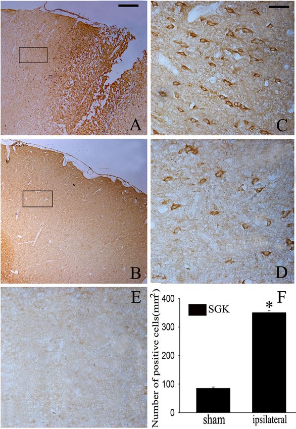
Burlingame, CA, USA) for 40 min at 37°C. PSGK expression in neuron apoptosis after TBI Figure 1. Detection of mRNA level and protein level of SGK change after TBI. Western blot was performed to study the protein level of SGK in the cortex surrounding the wound and sham-unoperated cortex at various survival times after TBI. A: Time courses of SGK expression in ipsilateral brain cortex after TBI. B: Quantification graphs (relative optical density) of the intensity of staining of SGK to GAPDH at each time point. GAPDH was used to confirm equal amount of protein was run on gel. The data are means ± SEM. (n=3, *P
SGK expression in neuron apoptosis after TBI

                                                                       functional effects of RNAi-mediated silenc-
                                                                       ing of SGK on neuron death processes.
                                                                       Among apoptosis pathway, caspase-3 is a
                                                                       key effecter. In this experiment, following
                                                                       administration of siRNA-SGK, phospho-
                                                                       GSK3β and β-catenin was significantly
                                                                       reduced in the siRNA group which markedly
                                                                       different from that in the non-specific group
                                                                       (P
SGK expression in neuron apoptosis after TBI Figure 3. Co-localization of SGK and different phenotype-specific markers in brain cortex. In the adult rat brain cor- tex within 5 mm distance from the lesion site at day 3 after TBI, horizontal sections labeled with SGK (red, B, F, J) and different cell markers, such as neuron marker (green, A, NeuN), astrocyte marker (green, E, GFAP), oligodendrocyte marker (green, I, CNPase), The yellow color visualized in the merged images represented co-localization of SGK with different phenotype-specific markers (C, G, K), co-localizations of SGK with different phenotype-specific markers in the sham-operated group are shown in the brain cortex (D, H, L). M, N: Negative controls, (O) Quantitative analysis of NeuN-positive cells expressing SGK (%) in the sham-operated group and day 3 after injury. *indicate significant difference at P
SGK expression in neuron apoptosis after TBI

                                                                    β-catenin expression after TBI, which show
                                                                    similarity with previous reports.

                                                                      GSK3β/β-catenin pathway can be regulat-
                                                                      ed by lots of serine/threonine protein kinas-
                                                                      es, such as Akt, SGK [31, 32]. The serine-
                                                                      threonine kinase, Akt, plays an important
                                                                      role in the cell death/survival pathway [33].
                                                                      Zhao S et al. have illustrated activation of
                                                                      Akt/GSK-3β/β-catenin signaling pathway is
                                                                      involved in survival of neurons after trau-
                                                                      matic brain injury in rats [27]. Interestingly,
                                                                      SGK contains a catalytic domain, which is
                                                                      most similar to Akt (also known as protein
                                                                      kinase B or PKB) [34, 35], was originally
                                                                      identified as serum- and glucocorticoid-
                                                                      inducible kinase, and plays an important
                                                                      role in central nervous system diseases [17,
                                                                      36], previous study has shown that SGK
                                                                      mRNA was upregulated after brain injury
                                                                      [18], but it is how to play a role in the brain
                                                                      injury is still not clear. Additionally, Akt and
Figure 4. Phosphorylation of GSK3β on serine-9 and of                 SGK always cooperate in controlling GSK3β
β-catenin regulation after TBI. A: Western blot analysis showed
that phospho-GSK3β (serine-9) expression increased signifi-
                                                                      phosphorylation on serine-9 and β-catenin
cantly at 1 day and peaked at 3 days in the cerebral cortex.          dynamics [37, 38], in the light of the role of
There was no prominent change in total GSK3β (t-GSK3β)                Akt/GSK3β/β-catenin signaling pathway in
(P>0.05). Results of the β-catenin analysis are shown here, it        survival of neurons after traumatic brain
gradually increased at 1 day and also peaked at 3 days after          injury in rats and the similarity of catalytic
TBI, GAPDH are shown as an internal control. B: The bar chart
shows the ratio of protein level to GAPDH, *indicate significant      domain between them. So we speculate
difference (P
SGK expression in neuron apoptosis after TBI Figure 5. Detection of SGK mRNA and protein, total-GSK3β, phospho-GSK3β, β-catenin level in H2O2-induced apoptosis model in PC12. RT-PCR analyses of SGK mRNA expression, it is upregulated at 6 h and peaked at 9 h (*P0.05). B, D: Quantitative analysis, *indicate significant difference (P
SGK expression in neuron apoptosis after TBI

and pho-GSK3β on ser-9, but whether the SGK          Nantong 226001, People’s Republic of China; Gang
is a pro-apoptotic and anti-apoptotic function       Cui, Department of Neurosurgery, The First Affiliated
after TBI? siRNA was employed to confirmed it,       Hospital of Soochow University, Suzhou 215006,
interestingly, after interfering the SGK expres-     People’s Republic of China. E-mail: cuigang@suda.
sion, we discovered phospho-GSK3β on ser-9           edu.cn
decreased, phosphorylation of β-catenin and
caspase-3 upregulated, moreover, immunofluo-         Reference
rescent staining suggested knocking down             [1]  Reilly P. The impact of neurotrauma on society:
SGK along with the rise of caspase-3,                     an international perspective. Prog Brain Res
Additionally, DAPI staining indicated that silenc-        2007; 161: 3-9.
ing SGK promoted nuclear condensation and            [2] Weber JT. Altered calcium signaling following
perinuclear apoptotic bodies after H2O2 treat-            traumatic brain injury. Front Pharmacol 2012;
ment in PC12 cells than H2O2 stimulated group             3: 60.
and control group. Based on our data, we con-        [3] Kochanek PM, Clark RS, Ruppel RA, Adelson
cluded that SGK upregulation can be induced               PD, Bell MJ, Whalen MJ, Robertson CL, Satch-
                                                          ell MA, Seidberg NA, Marion DW and Jenkins
by traumatic brain injury at mRNA and protein
                                                          LW. Biochemical, cellular, and molecular
level, which shows a protective role on neuron
                                                          mechanisms in the evolution of secondary
apoptosis via GSK3β/β-catenin signaling                   damage after severe traumatic brain injury in
relates to caspase-3 inhibition.                          infants and children: Lessons learned from the
                                                          bedside. Pediatr Crit Care Med 2000; 1: 4-19.
This study aimed to define the role of SGK-          [4] Loane DJ and Faden AI. Neuroprotection for
dependent regulation of GSK3β/β-catenin                   traumatic brain injury: translational challenges
pathway in the control of neuron apoptosis                and emerging therapeutic strategies. Trends
after TBI. Collectively, SGK is upregulated at            Pharmacol Sci 2010; 31: 596-604.
mRNA and protein level after TBI and induced         [5] Hartings JA, Bullock MR, Okonkwo DO, Murray
the phosphorylation of GSK3β on ser-9 and sta-            LS, Murray GD, Fabricius M, Maas AI, Woitzik J,
bilized the β-catenin, siRNA-SGK leads to cas-            Sakowitz O, Mathern B, Roozenbeek B, Lings-
pase-3 increase and show an anti-apoptotic                ma H, Dreier JP, Puccio AM, Shutter LA, Pahl C
                                                          and Strong AJ. Spreading depolarisations and
role on neuron after brain injury. Our results
                                                          outcome after traumatic brain injury: a pro-
have shown that SGK and GSK3β (Ser9) was
                                                          spective observational study. Lancet Neurol
accelerated in the injured cortex, and involved           2011; 10: 1058-1064.
in neuronal survival after TBI. Moreover, neuro-     [6] Andriessen TM, Horn J, Franschman G, van der
protection of β-catenin against TBI was partly            Naalt J, Haitsma I, Jacobs B, Steyerberg EW
mediated by enhanced and persistent activa-               and Vos PE. Epidemiology, severity classifica-
tion of the SGK/GSK3β signaling pathway.                  tion, and outcome of moderate and severe
These findings suggest that activation of SGK is          traumatic brain injury: a prospective multi-
able to control GSK3β/β-catenin signaling                 center study. J Neurotrauma 2011; 28: 2019-
pathway and finally inactivate the caspase-3 in           2031.
neuron apoptosis.                                    [7] Rink A, Fung KM, Trojanowski JQ, Lee VM,
                                                          Neugebauer E and McIntosh TK. Evidence of
Acknowledgements                                          apoptotic cell death after experimental trau-
                                                          matic brain injury in the rat. Am J Pathol 1995;
                                                          147: 1575-1583.
This work was supported by the Natural Science
                                                     [8] Hilton GD, Stoica BA, Byrnes KR and Faden AI.
Foundation of China (81070992); and A Project
                                                          Roscovitine reduces neuronal loss, glial activa-
Funded by the Priority Academic Program                   tion, and neurologic deficits after brain trau-
Development of Jiangsu Higher Education                   ma. J Cereb Blood Flow Metab 2008; 28:
Institutions (PAPD).                                      1845-1859.
                                                     [9] Carmichael J, Sugars KL, Bao YP and Rubinsz-
Disclosure of conflict of interest                        tein DC. Glycogen synthase kinase-3beta in-
                                                          hibitors prevent cellular polyglutamine toxicity
The authors declare no conflict of interest asso-         caused by the Huntington’s disease mutation.
ciated with this work.                                    J Biol Chem 2002; 277: 33791-33798.
                                                     [10] Jin Y, Sui HJ, Dong Y, Ding Q, Qu WH, Yu SX and
Address correspondence to: Xingxing Gu, Key               Jin YX. Atorvastatin enhances neurite out-
Laboratory of Neuroscience, Nantong University,           growth in cortical neurons in vitro via up-regu-

1291                                                       Int J Clin Exp Pathol 2013;6(7):1282-1293
SGK expression in neuron apoptosis after TBI
lating the Akt/mTOR and Akt/GSK-3beta sig- adult brain injury: implications for glial prolif-
naling pathways. Acta Pharmacol Sin 2012; eration. J Neurotrauma 2010; 27: 361-371.
33: 861-872. [22] Kaytor MD and Orr HT. The GSK3 beta signal-
[11] Maggirwar SB, Tong N, Ramirez S, Gelbard HA ing cascade and neurodegenerative disease.
and Dewhurst S. HIV-1 Tat-mediated activation Curr Opin Neurobiol 2002; 12: 275-278.
of glycogen synthase kinase-3beta contributes [23] Frame S and Cohen P. GSK3 takes centre
to Tat-mediated neurotoxicity. J Neurochem stage more than 20 years after its discovery.
1999; 73: 578-586. Biochem J 2001; 359: 1-16.
[12] Tong N, Sanchez JF, Maggirwar SB, Ramirez [24] Hartigan JA and Johnson GV. Transient increas-
SH, Guo H, Dewhurst S and Gelbard HA. Activa- es in intracellular calcium result in prolonged
tion of glycogen synthase kinase 3 beta (GSK- site-selective increases in Tau phosphorylation
3beta) by platelet activating factor mediates through a glycogen synthase kinase 3beta-de-
migration and cell death in cerebellar granule pendent pathway. J Biol Chem 1999; 274:
neurons. Eur J Neurosci 2001; 13: 1913-1922. 21395-21401.
[13] Hu S, Begum AN, Jones MR, Oh MS, Beech [25] Jope RS and Johnson GV. The glamour and
WK, Beech BH, Yang F, Chen P, Ubeda OJ, Kim gloom of glycogen synthase kinase-3. Trends
PC, Davies P, Ma Q, Cole GM and Frautschy SA. Biochem Sci 2004; 29: 95-102.
GSK3 inhibitors show benefits in an Alzheim- [26] Bhat RV, Shanley J, Correll MP, Fieles WE, Keith
er’s disease (AD) model of neurodegeneration RA, Scott CW and Lee CM. Regulation and lo-
but adverse effects in control animals. Neuro- calization of tyrosine216 phosphorylation of
biol Dis 2009; 33: 193-206. glycogen synthase kinase-3beta in cellular and
[14] Endo H, Nito C, Kamada H, Yu F and Chan PH. animal models of neuronal degeneration. Proc
Akt/GSK3beta survival signaling is involved in Natl Acad Sci U S A 2000; 97: 11074-11079.
acute brain injury after subarachnoid hemor- [27] Zhao S, Fu J, Liu X, Wang T, Zhang J and Zhao
rhage in rats. Stroke 2006; 37: 2140-2146. Y. Activation of Akt/GSK-3beta/beta-catenin
[15] Dudek H, Datta SR, Franke TF, Birnbaum MJ, signaling pathway is involved in survival of neu-
Yao R, Cooper GM, Segal RA, Kaplan DR and rons after traumatic brain injury in rats. Neurol
Greenberg ME. Regulation of neuronal survival Res 2012; 34: 400-407.
by the serine-threonine protein kinase Akt. Sci- [28] Zhu D, Kang Q, Huang PY, He TC and Xie P.
ence 1997; 275: 661-665. Neurogenesis-related genes expression profil-
[16] BelAiba RS, Djordjevic T, Bonello S, Artunc F, ing of mouse fibroblastic stem cells induced by
Lang F, Hess J and Gorlach A. The serum- and Wnt signaling. Neurol Res 2009; 31: 200-203.
glucocorticoid-inducible kinase Sgk-1 is in- [29] Polakis P. The oncogenic activation of beta-
volved in pulmonary vascular remodeling: role catenin. Curr Opin Genet Dev 1999; 9: 15-21.
in redox-sensitive regulation of tissue factor by [30] Yuan J, Zhang J, Wong BW, Si X, Wong J, Yang D
thrombin. Circ Res 2006; 98: 828-836. and Luo H. Inhibition of glycogen synthase ki-
[17] Lang F, Strutz-Seebohm N, Seebohm G and nase 3beta suppresses coxsackievirus-in-
Lang UE. Significance of SGK1 in the regula- duced cytopathic effect and apoptosis via sta-
tion of neuronal function. J Physiol 2010; 588: bilization of beta-catenin. Cell Death Differ
3349-3354. 2005; 12: 1097-1106.
[18] Imaizumi K, Tsuda M, Wanaka A, Tohyama M [31] Bhavsar SK, Merches K, Bobbala D and Lang
and Takagi T. Differential expression of sgk F. AKT/SGK-sensitive phosphorylation of GSK3
mRNA, a member of the Ser/Thr protein ki- in the regulation of L-selectin and perforin ex-
nase gene family, in rat brain after CNS injury. pression as well as activation induced cell
Brain Res Mol Brain Res 1994; 26: 189-196. death of T-lymphocytes. Biochem Biophys Res
[19] Brunet A, Park J, Tran H, Hu LS, Hemmings BA Commun 2012; 425: 6-12.
and Greenberg ME. Protein kinase SGK medi- [32] Sakoda H, Gotoh Y, Katagiri H, Kurokawa M,
ates survival signals by phosphorylating the Ono H, Onishi Y, Anai M, Ogihara T, Fujishiro M,
forkhead transcription factor FKHRL1 Fukushima Y, Abe M, Shojima N, Kikuchi M,
(FOXO3a). Mol Cell Biol 2001; 21: 952-965. Oka Y, Hirai H and Asano T. Differing roles of
[20] Logan A, Frautschy SA, Gonzalez AM and Baird Akt and serum- and glucocorticoid-regulated
A. A time course for the focal elevation of syn- kinase in glucose metabolism, DNA synthesis,
thesis of basic fibroblast growth factor and one and oncogenic activity. J Biol Chem 2003; 278:
of its high-affinity receptors (flg) following a lo- 25802-25807.
calized cortical brain injury. J Neurosci 1992; [33] Datta SR, Dudek H, Tao X, Masters S, Fu H,
12: 3828-3837. Gotoh Y and Greenberg ME. Akt phosphoryla-
[21] Liu Y, Wang Y, Cheng C, Chen Y, Shi S, Qin J, tion of BAD couples survival signals to the cell-
Xiao F, Zhou D, Lu M, Lu Q and Shen A. A rela- intrinsic death machinery. Cell 1997; 91: 231-
tionship between p27(kip1) and Skp2 after 241.
1292 Int J Clin Exp Pathol 2013;6(7):1282-1293SGK expression in neuron apoptosis after TBI
[34] Park J, Leong ML, Buse P, Maiyar AC, Firestone mann TF, Judenhofer MS, Pichler BJ, Biber J,
GL and Hemmings BA. Serum and glucocorti- Wagner CA and Lang F. PKB/SGK-resistant
coid-inducible kinase (SGK) is a target of the PI GSK3 enhances phosphaturia and calciuria. J
3-kinase-stimulated signaling pathway. EMBO Am Soc Nephrol 2011; 22: 873-880.
J 1999; 18: 3024-3033. [39] Beurel E and Jope RS. The paradoxical pro-
[35] Leong ML, Maiyar AC, Kim B, O’Keeffe BA and and anti-apoptotic actions of GSK3 in the in-
Firestone GL. Expression of the serum- and trinsic and extrinsic apoptosis signaling path-
glucocorticoid-inducible protein kinase, Sgk, is ways. Prog Neurobiol 2006; 79: 173-189.
a cell survival response to multiple types of en- [40] Hetman M, Cavanaugh JE, Kimelman D and
vironmental stress stimuli in mammary epithe- Xia Z. Role of glycogen synthase kinase-3beta
lial cells. J Biol Chem 2003; 278: 5871-5882. in neuronal apoptosis induced by trophic with-
[36] Schoenebeck B, Bader V, Zhu XR, Schmitz B, drawal. J Neurosci 2000; 20: 2567-2574.
Lubbert H and Stichel CC. Sgk1, a cell survival [41] Cho JH and Johnson GV. Glycogen synthase ki-
response in neurodegenerative diseases. Mol nase 3 beta induces caspase-cleaved tau ag-
Cell Neurosci 2005; 30: 249-264. gregation in situ. J Biol Chem 2004; 279:
[37] Siraskar B, Volkl J, Ahmed MS, Hierlmeier M, 54716-54723.
Gu S, Schmid E, Leibrock C, Foller M, Lang UE [42] Chen J, Mao H, Zou H, Jin W, Ni L, Ke K, Cao M
and Lang F. Enhanced catecholamine release and Shi W. Up-regulation of ski-interacting pro-
in mice expressing PKB/SGK-resistant GSK3. tein in rat brain cortex after traumatic brain
Pflugers Arch 2011; 462: 811-819. injury. J Mol Histol 2013; 44: 1-10.
[38] Foller M, Kempe DS, Boini KM, Pathare G, Sir-
askar B, Capuano P, Alesutan I, Sopjani M,
Stange G, Mohebbi N, Bhandaru M, Acker-
1293 Int J Clin Exp Pathol 2013;6(7):1282-1293You can also read