RORβ suppresses the stemness of gastric cancer cells by downregulating the activity of the Wnt signaling pathway
←
→
Page content transcription
If your browser does not render page correctly, please read the page content below
ONCOLOGY REPORTS 46: 180, 2021

          RORβ suppresses the stemness of gastric cancer cells by
         downregulating the activity of the Wnt signaling pathway
                       ZHENZHEN WEN1, MING CHEN1, WENHAO GUO2, KE GUO1, PING DU1,
                                YANFEI FANG1, MIN GAO1 and QIANG WANG3

  Departments of 1Gastroenterology and 2Pathology, Sir Run Run Shaw Hospital, School of Medicine, Zhejiang University,
      Hangzhou, Zhejiang 310016; 3Department of Hepatopancreatobiliary Surgery and Minimally Invasive Surgery,
        Zhejiang Provincial People's Hospital, Hangzhou Medical College, Hangzhou, Zhejiang 310014, P.R. China

                                       Received December 8, 2020; Accepted April 27, 2021

                                                      DOI: 10.3892/or.2021.8131

Abstract. Gastric cancer (GC) is the third leading cause of           the expression levels of the pro‑apoptotic gene, Bcl‑2 like
cancer‑related mortality and the fifth most common type of            protein 11, which subsequently inhibited the viability and
cancer worldwide. GC stem cells (GCSCs) have been reported            promoted the apoptosis of GC cells. In addition, ROR β
to be responsible for the malignant behavior of GC. However,          decreased the sphere forming ability, and downregulated the
the key molecular mechanism controlling GCSC function                 expression levels of iPS cell‑ and EMT‑related factors. In vivo,
remains unclear. The present study aimed to investigate the           RORβ suppressed the tumorigenic capacity and stemness of
function of retinoic acid‑related orphan receptor β (RORβ) in         GC cells. Mechanistically, RORβ was revealed to decrease the
GC. The expression levels of RORβ in GC cells and clinical GC         activity of the Wnt/β‑catenin signaling pathway in GCSCs. In
tissues were analyzed using western blotting, reverse transcrip‑      conclusion, the findings of the present study identified RORβ
tion‑quantitative PCR (RT‑qPCR) and immunohistochemistry.             as a novel suppressor of GCSCs and highlighted the prospect
The association between RORβ expression levels and GCSC               of RORβ as a novel candidate target for stem cell‑based GC
markers was analyzed using Gene Set Enrichment Analysis,              therapy.
and GeneChip was performed to identify differentially
expressed genes between control and RORβ‑overexpressing               Introduction
GC cells. CCK‑8 and flow cytometric assays were used to
evaluate the effect of RORβ on cell viability and apoptosis,          Gastric cancer (GC) is the fifth most common type of cancer
respectively. The effect of RORβ on the self‑renewal capacity         worldwide and the third leading cause of cancer‑related
of GCSCs was measured using a sphere formation assay, the             mortality, with approximately 951,600 cases and 723,100
expression levels of induced pluripotent stem (iPS) factors and       GC‑related deaths in 2012 (1). Common therapeutic strategies
epithelial‑mesenchymal transition (EMT)‑related factors were          for GC include surgical resection, chemotherapy, radiotherapy
measured by RT‑qPCR and western blotting, and the tumori‑             and anti‑angiogenic therapy (2). However, the efficacy of
genic capacity was measured by an in vivo mouse model.                current treatment regimens for GC are hindered by multiple
Finally, the impact of RORβ on the Wnt signaling pathway was          factors such as chemotherapy and radiotherapy resistance,
determined using western blotting and a TOP/FOP flash assay.          and tumor relapse (3). At present, cancer stem cells (CSCs)
The results revealed that the expression levels of RORβ were          are regarded as a crucial population contributing to the irre‑
downregulated in GC tissues compared with para‑carcinoma              sponsiveness to GC treatment and the poor prognosis (4).
tissues, and were inversely associated with the expression levels     CSCs can initiate tumor formation, and promote self‑renewal,
of GCSC markers. The overexpression of RORβ upregulated               therapy resistance, metastasis and tumor recurrence (5,6). As a
                                                                      result, it is of great importance to determine the key molecules
                                                                      controlling the malignant properties of gastric CSCs (GCSCs).
                                                                          GCSCs were first identified in 2007 by Yang et al (7).
                                                                      CD44, CD24/CD44 and aldehyde dehydrogenase (ALDH)1
Correspondence      to: Dr Qiang Wang, Department of                  have been identified as GCSCs markers (8,9). Numerous
Hepatopancreatobiliary Surgery and Minimally Invasive Surgery,
                                                                      previous studies have revealed the therapeutic value of
Zhejiang Provincial People's Hospital, Hangzhou Medical College,
158 Shang Tang Road, Hangzhou, Zhejiang 310014, P.R. China
                                                                      targeting CSC markers for GC intervention; for example,
E‑mail: wangqiang@hmc.edu.cn                                          Gong et al (10) reported that leucine rich repeat containing
                                                                      G protein‑coupled receptor 5 (LGR5) antibody conjugates
Key words: gastric cancer, gastric cancer stem cells, self‑renewal,   induced cytotoxicity in GC cells overexpressing LGR5. In
Wnt signaling pathway, retinoic acid‑related orphan receptor β,       addition, all‑trans retinoic acid treatment inhibited GC
epithelial‑mesenchymal transition                                     progression in mouse xenograft models by downregu‑
                                                                      lating the expression levels of CD44 and ALDH1 (11).
                                                                      Recently, an increasing number of therapeutics targeting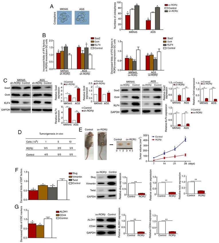
2                            WEN et al: RORβ SUPPRESSES THE STEMNESS OF GASTRIC CANCER CELLS

stemness‑associated functions have been designed to                 Scientific, Inc.) supplemented with 10% FBS. All GC cells
specifically eradicate GCSCs, including those that inhibit          were incubated at 37˚C and 5% CO2. GCSCs were cultured
stemness‑associated genes, block self‑renewal signaling path‑       from GC cells at a density of 1x10⁵ cells/ml in serum‑free
ways and microenvironment‑based anti‑GCSC therapies (12).           medium. EGF (20 g/l; Invitrogen; Thermo Fisher Scientific,
One of the most characterized pathways contributing to the          Inc.), bFGF (20 g/l; Invitrogen; Thermo Fisher Scientific,
function of CSCs is the Wnt signaling pathway, and the aber‑        Inc.), B27 (2%; Invitrogen; Thermo Fisher Scientific, Inc.),
rant activation of the Wnt signaling pathway has been revealed      BSA (0.4%; Roche Diagnostics), insulin (4 mg/l; Invitrogen;
to promote stem cell characteristics of CSCs and initiate the       Thermo Fisher Scientific, Inc.) and gentamicin (200 IU/ml;,
epithelial‑mesenchymal transition (EMT) process (13). Due           Sangon Biotech, Co., Ltd.) were added to DMEM/F‑12 (Gibco;
to the pivotal role of CSCs in tumor progression and metas‑         Thermo Fisher Scientific, Inc.) in 4‑well low adhesion culture
tasis, an improved understanding of the regulatory elements         plates in an incubator at 37˚C with 5% CO2 for 7‑10 days.
that control the malignant behaviors of GCSCs may lead to
the development of effective therapies for patients with GC.        Screening and identification of stable RORβ‑overexpression
    Retinoic acid‑related orphan receptor β (RORβ) is a member      GC cells. The ROR β gene coding sequence from
of the orphan nuclear receptor family (14). RORβ was origi‑         RORβ/pReceiver plasmid (GeneCopoeia, Inc.) was inserted
nally considered to be expressed solely in the central nervous      into pEGFP‑C1 vector at the BamHI site and identified by
system (CNS), mainly in the regions modulating the circadian        DNA sequencing (Sangon Biotech Co., Ltd.). GC cells were
rhythm (15). However, RORβ has since been demonstrated to           seeded into 6‑well plates at a density of 1x10 6 cells/well
be expressed in other regions of the body, including bone tissue,   at 37˚C with 5% CO2 for 24 h. RORβ/pEGFP‑C1 (4 µg/µl)
pancreatic cancer tissue and colorectal cancer tissue (16,17).      and pEGFP‑C1 vector (4 µg/µl) were transfected into GC
Risinger et al reported that the expression levels of RORβ were     cells using Lipofectamine® 3000 reagent (Invitrogen; Thermo
upregulated in women with endometrial cancer compared with          Fisher Scientific, Inc.). After 2 days, the cells were cultured in
healthy women (18). However, despite the reported expressional      the presence of 500 µg/ml G418 (Invitrogen; Thermo Fisher
changes of RORβ, the pathological significance of RORβ              Scientific, Inc.) for 2 weeks to obtain stably transfected GC
remains largely unknown. Therefore, the present study aimed         cells. RORβ‑overexpression GC cells were further verified by
to investigate the expression levels and function of RORβ in        western blotting.
GC, and identified RORβ as a novel suppressor of GCSCs.
These findings may help to propose a valuable target for the        Screening stable RORβ‑knockdown GC cells. GC cells were
stem cell‑based therapy of GC in the future.                        transfected with 4 µg/µl RORβ ‑short hairpin (shRNA) and
                                                                    4 µg/µl control shRNA (both from Santa Cruz Biotechnology,
Materials and methods                                               Inc.) using Lipofectamine® 3000 reagent (Invitrogen; Thermo
                                                                    Fisher Scientific, Inc.) for 2 days and then the cells were
Patient studies. A total of 32 patients with GC at the              screened with puromycin (5 µg/ml) for 14 days to obtain the
General Surgery Department of Sir Run Run Shaw Hospital             stable RORβ ‑knockdown cells further verified by western
(Hangzhou, China) were selected from January 2019 to                blotting.
December 2019. There were 18 males and 14 females
with an age range of 34‑76 years (average age 61.5 years            Nuclear/cytosolic fractionation. Ice‑precooled CER I (200 µl)
old). The fresh operative GC tissues and corresponding              was added to GC cells and the cells were incubated on ice for
para‑cancerous tissues were collected. The pathological             10 min after vortex oscillation for 15 sec using nuclear and
stage was determined according to the American Joint                cytoplasmic extraction reagents (NE‑PER) (Pierce; Thermo
Committee on Cancer (AJCC) and the International Union              Fisher Scientific, Inc.). Ice‑precooled CER II (11 µl) was added
against Cancer (UICC) seventh edition TNM staging                   and incubated, then swirled and shaken for 5 sec, and incubated
system (19). The inclusion criteria were as follows: i) radical     on ice for 1 min. The supernatant (cytoplasm) was transferred
gastrectomy or palliative surgery for GC and pathological           to a clean ice‑precooled centrifuge tube for preservation on ice.
diagnosis was GC; ii) preoperative radiotherapy, and                The sediment was suspended with ice‑precooled NER 100 µl,
chemotherapy were not performed; iii) complete clinico‑             incubated on ice for 10 min, and vortex oscillated 4 times for
pathological data were available. The exclusion criteria            15 sec. After centrifugation at 16,000 x g for 10 min at 4˚C, the
were as follows: i) gastric cancer recurrence or residual           supernatant was transferred to a clean ice‑precooled centrifuge
gastric cancer; ii) combined with organ dysfunction or other        tube (cell nucleus), and then western blotting was performed.
tumors. All patients provided written informed consent prior
to participation and the present study was approved by the          Sphere formation assay. ROR β ‑overexpression and
Institutional Review Board from Sir Run Run Shaw Hospital           RORβ‑knockdown GC cells and control cells were digested
(approval no. 20210429‑30).                                         and cultured in 24‑well plates at a density of 200 cells/well
                                                                    in serum‑free medium incubated at 37˚C with 5% CO2 for
Cell culture. GC cell lines, AGS and MKN45, were purchased          7 days. The medium was replaced every 3‑4 days. Following
from The Cell Bank of Type Culture Collection of the Chinese        the incubation, the spheres were counted manually using a
Academy of Sciences. AGS cells were cultured in DMEM                light microscope with a magnification of x20.
(Gibco; Thermo Fisher Scientific, Inc.) supplemented with
10% FBS (Thermo Fisher Scientific, Inc.), while MKN45 cells         CCK‑8 assay. RORβ‑overexpression and RORβ‑knockdown
were cultured in RPMI‑1640 medium (Gibco; Thermo Fisher             cells and control cells were seeded into 96‑well plates at a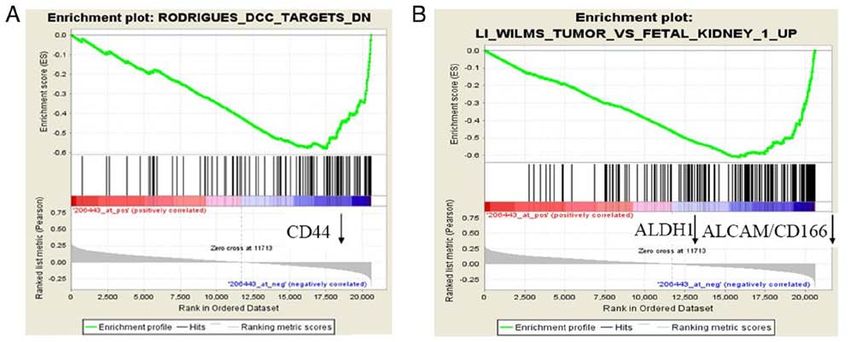
ONCOLOGY REPORTS 46: 180, 2021 3
density of 1x10 4 at 37˚C with 5% CO2 for 24 h. Following Wound healing assay. MKN45 and AGS cells were plated
incubation, 10 µl CCK‑8 reagent (Abcam) was added/well and into 96‑well plates and incubated overnight. Approximately
incubated for 4 h. Subsequently, the absorbance value was 1x10⁴ cells in each well were serum‑starved overnight, and then
measured with a microplate reader at a wavelength of 460 nm. the head of the pipette tip was used to produce a scratch in the
cell monolayer. The cells were then washed with PBS 3 times
Flow cytometric analysis of apoptosis. RORβ‑overexpression to remove the unattached cells and subsequently incubated in
GC cells and control cells were trypsinized, and 1x10⁶/ml cells fresh culture medium without FBS for indicated time‑points
were collected by centrifugation (300 x g, 5 min at 4˚C). Cells at 37˚C. Images of the wound were captured at 0, 12 and 24 h
were washed with PBS twice and incubated in 500 µl binding using a light microscope (magnification, x20). The percentage
buffer, 5 µl Annexin V‑EGFP and 10 µl propidium iodide of migration area was calculated using ImageJ software (1.52v;
at room temperature in the dark for 15 min using Annexin National Institutes of Health).
V‑FITC/PI Apoptosis Detection Kit (Beyotime Institute of
Biotechnology) according to the manufacturer's instructions. F‑actin polymerization experiment. AGC cells grown
Apoptotic cells were visualized and quantified using FACSAria on cover‑slips inside a petri dish were transfected with
(BD Biosciences) with FlowJo software (version 7.6.1; Tree RORβ/pReceiver plasmid and control and were incubated
Star, Inc.). at 37˚C for 2 days using Lipofectamine ® 3000 reagent
(Invitrogen; Thermo Fisher Scientific, Inc.). The cells were
Nude mouse xenograft model. All animal experiments were washed once with PBS. Then, the cells were incubated with
approved by the Institutional Animal Care and Use Committee 3‑4% formaldehyde in PBS at room temperature for 30 min.
of Zhejiang University (Hangzhou, China) and complied with The staining solution was aspirated carefully and the fixed cells
the Animal Welfare Act (20). BALB/c nude mice (30 females; were washed 2‑3 times in PBS. Conjugate working solution
4 weeks old; body mass 17‑20 g) were purchased from 1X Phalloidin‑iFluor™ 488 (AAT Bioquest, Inc.) was added to
Shanghai Laboratory Animal Center. The mice were bred at the fixed cells. Subsequently, the cells were incubated at room
the Laboratory Animal Research Center at Sir Run Run Shaw temperature for 60 min. DAPI staining solution (BIOSS) was
Hospital in a barrier environment, specific pathogen‑free (SPF) then added and the cells were incubated at room temperature
grade, with humidity approximately 30 to 50% at 22˚C. In addi‑ for 5 min. The cells were rinsed gently 2‑3 times with PBS
tion, water and food was provided ad libitum. A total of 0.2 ml to remove excess phalloidin conjugate. Mounting medium
RORβ‑overexpression GC cells and control cells (at the density was added and sealed. The cells were observed at a magni‑
of 1x10⁵, 5x10⁵ and 1x10⁶) were subcutaneously inoculated into fication of x400 under a fluorescence microscope (Olympus
the backs of nude mice. There were five mice in each group. Corporation) with an FITC filter set.
Tumor growth was observed weekly, and rats were sacrificed
by cervical spinal cord transection after 4 weeks, and the tumor Reverse transcription‑quantitative PCR (RT‑qPCR).
body was measured and analyzed. The tumor volumes were Total RNA from GC cells was extracted using RNeasy
assessed according to the diameters of the tumors. Mini Kit (cat. no. 74104; Qiagen GmbH) and then reverse
transcribed into cDNA using PrimeScript RT reagent kit
Immunohistochemistry (IHC). All GC tissue specimens were (cat. no. RR037A; Takara Bio, Inc.) according to the manu‑
fixed with 10% neutral formaldehyde solution for 4‑6 h at facturer's protocol. RT‑qPCR was performed using Premix Ex
room temperature, and were routinely dehydrated, waxed and Taq (cat. no. RR420A; Takara Bio, Inc.). The thermocycling
wrapped. Then, the tissues were cut into 3‑µm sections. The conditions were as follows: Initial denaturation at 95˚C for
tissue sections were baked at 60˚C overnight. Then, the tissue 5 min; annealing and elongation at 95˚C for 30 sec, 95˚C
sections were subsequently deparaffinized by the following for 5 sec, 60˚C for 34 sec for 40 cycles; and final extension
method: After baking, the tissue slices were immersed in at 60˚C for 10 min. The sequences of the primer pairs used
xylene at room temperature twice for 10 min each. Then they were: Oct4 forward, 5'‑GTGGAGGAAGCTGACA ACA A‑3',
were immersed in 100% ethanol for 5 min. Next, the tissue reverse, 5'‑AACA AAT TCTCCAGGT TGCC‑3' and probe,
sections were rehydrated in 100, 85 and 70% ethanol at room 5'‑Fam‑TCTC TTT CGG GCC TGCACGA‑3'Tamra; Sox2
temperature for 2 min respectively. Finally, the slices were forward, 5'‑AATGCCTTCATGGTGTGG‑3', reverse, 5'‑CTT
incubated at 90˚C in deionized water for 30 min. Subsequently, CTCCGTC TCCGACAAA‑3', and probe, 5'Fam‑AGTT TC
the slices were boiled in citric acid buffer solution and main‑ CACTCGGCGCCCAG‑3'Tamra; KLF4 forward, 5'‑GGCACT
tained at a low heat for 30 min. The sections were then sealed ACCGTAA ACACACG‑3', reverse, 5'‑CTGG CAGTGTGG
with 5% BSA (Sangon Biotech Co., Ltd.) in a wet box at 37˚C GTCATATC‑3', and probe, 5'Fam‑CAGGTCG GACCACCT
for 30 min and air‑dried. The sections were then incubated CGCCT‑3'Tamra; CD44 forward, 5'‑TGGCAACAGATGGCA
in a wet box at 4˚C overnight with an anti‑RORβ antibody TGAG G‑3', reverse, 5'‑CCTTGCATTG GATGGC TGGT‑3'
(cat. no. NBP1‑82532; 1:300; Novus Biologicals, LLC). and probe, 5'Fam‑AACAGGGACAGCTGCAGCC TCAGC
Following the primary antibody incubation, the sections were T‑3'Tamra; ALDH1 forward, 5'‑GCTG GCGACA ATG GA
rinsed thrice with PBS for 5 min and incubated with 50 µl of an GTCA A‑3', reverse, 5'‑TGTACGG CCC TGGATC TTGT‑3'
HRP‑conjugated secondary goat anti‑rabbit antibody (product and probe, 5'Fam‑ACAT TGCGCTACTGTG CAG GTTGG
code ab6721; 1:1,000; Epitomics; Abcam) at room temperature GCT‑3' Tamra; Slug forward, 5'‑TCAC TGT GTG GAC TA
for 20 min. Then the sections were rinsed thrice with PBS for CCGC T‑3', reverse, 5'‑TCGCCCCAAAGATGAG GAGT‑3'
3 min. The slides were observed under a light microscope with and probe, 5'Fam‑ATTCCACGCCCAG CTACCCAATGG
a magnification of x100. CCT‑3'Tamra; Twist forward, 5'‑CCCTCGGACA AGC TG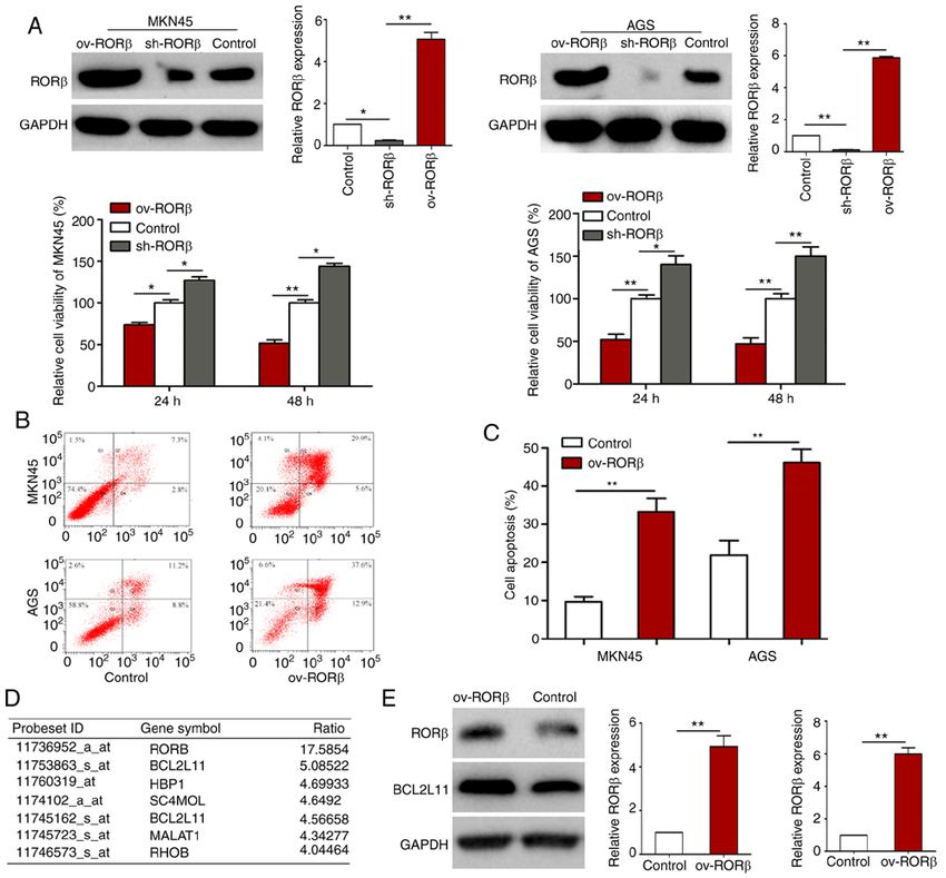
4 WEN et al: RORβ SUPPRESSES THE STEMNESS OF GASTRIC CANCER CELLS
AGCA A‑3', reverse, 5'‑TCGC TCTGGAGGACCTGGTA‑3' Los Angeles, USA. GSEA (23) was used to analyze the asso‑
and probe, 5'Fam‑ATTCAGACCC TCA AGC TGG CGG CC ciation between RORβ expression levels and GCSC markers.
A‑3'Tamra; vimentin forward, 5'‑CAATGAGTCCCTG GA
ACGCC‑3', reverse, 5'‑GGTGACGAGCCAT TTCCTCC‑3' Top/Fop luciferase reporter assay. RORβ‑overexpression GC
and probe, 5'Fam‑ACCA AGACAC TAT TGG CCG CCTGC cells were seeded into 6‑well plates at a density of 1x10⁶ at 37˚C
AGG‑3'Tamra; GAPDH forward, 5'‑ATCATCCCTGCCT CT with 5% CO2 for 24 h. A total of 8 µl Fugene 6 (Roche Diagnostics)
ACTG G‑3', reverse, 5'‑GTCAGGTCCACCACTGACAC‑3' was incubated with 100 µl OPTI‑MEM (Invitrogen; Thermo
and probe, 5'Fam‑ACCTTGCCCACAGCCTTGGC‑3'Tamra; Fisher Scientific, Inc.) at room temperature for 5 min. Then, a
The relative mRNA expression levels were calculated using total of 2 µg plasmid mixture including TOP (MilliporeSigma),
2‑ΔΔCq method (21). FOP (MilliporeSigma) and PRL (Promega Corporation) (4:4:1)
were added to the aforementioned mixture and incubated at
Western blotting. Cells were lysed using RIPA Lysis Buffer room temperature for 15 min. Then the mixture of Fugene 6 and
(Beyotime Institute of Biotechnology). The protein concentra‑ plasmids were added to the 6‑well plate and mixed, and incu‑
tions were determined using the bicinchoninic acid (BCA) bated at 37˚C in a 5% CO2 incubator for 24 h. The cell lysates
assay. Proteins were separated via 12% SDS‑PAGE and were transferred to the centrifuge tube after cell lysis. GloMax
blocked with 5% bovine serum albumin at 4˚C overnight. A 20/20 Luminometer was used to detect the fluorescence activity.
total of 20 ml of 12% separation gel was prepared, and the 5% A total of 20 µl of the aforementioned cell lysates were added
stacking gel was added on top of the separation gel. In total, with 100 µl of LAR II. After mixing, firefly luciferase activity
20‑40 µl protein samples with 5 µl protein marker were added was detected. Then 100 µl Stop&Glo Reagent was added to
to the corresponding lanes. Then the protein samples were detect Renilla luciferase activity. The Dual‑Luciferase® Reporter
transferred to the cellulose nitrate membranes by electropho‑ (DLR™) Assay System (Promega Corporation) was used.
resis. The cellulose nitrate membranes were incubated with
the following primary antibodies at 4˚C overnight: Anti‑c‑Myc Statistical analysis. Statistical analysis was performed using
(product code ab32072; 1:2,000), anti‑cyclin D1 (product code SPSS 19.0 software (IBM Corp.). Statistical differences
ab16663; 1:2,000), anti‑Slug (product code ab51772; 1:2,000), between groups were performed using one way ANOVA (with
anti‑Twist (product code ab50887; 1:2,000), anti‑E‑cadherin least significant difference test or Tamhane's T2 post hoc test)
(product code ab40772; 1:2,000), anti‑N‑cadherin (product or unpaired Student's t‑test. P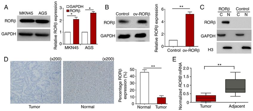
ONCOLOGY REPORTS 46: 180, 2021                                                                  5

Figure 1. Expression levels of RORβ in GC cells. (A) RORβ expression levels in GC cells were analyzed using western blotting. (B) Overexpression of RORβ
in AGS cells was examined using western blotting. (C) AGS cells were subjected to nuclear/cytosolic fractionation, and RORβ expression levels were analyzed
using western blotting. (D) RORβ expression levels in GC tissues were analyzed using immunohistochemical staining. (E) mRNA expression levels of RORβ
were analyzed in GC tissues using reverse transcription‑quantitative PCR. Data were analyzed using a Student's t‑test. *P
6                                  WEN et al: RORβ SUPPRESSES THE STEMNESS OF GASTRIC CANCER CELLS

Figure 3. RORβ exerts anti‑proliferative and pro‑apoptotic effects on GC cells. (A) MKN45 and AGS cells were transfected with control, RORβ overexpression
vector or RORβ shRNA and confirmed by western blotting. The viability of GC cells was evaluated using an CCK‑8 assay. Data were analyzed using one‑way
ANOVA. (B and C) Apoptosis of GC cells was detected using flow cytometry. GC cells were transfected with control or ROR β‑overexpression vector for 24 h.
Cell apoptosis was evaluated using the values in quadrants 2 and 4. Data were analyzed using a unpaired Student's t‑test. (D) GeneChip was used to evaluate
the gene expression profiles. The top differentially expressed genes are listed. (E) Protein expression levels of BCL2L11 were analyzed using western blotting.
*
 P
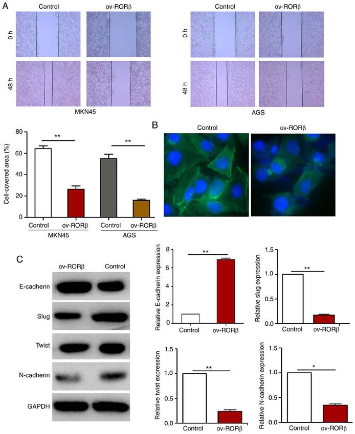
ONCOLOGY REPORTS 46: 180, 2021                                                              7

Figure 4. RORβ inhibits EMT in GC cells. GC cells were transfected with control or RORβ overexpression vector. (A) Cell migration was measured using
a wound healing assay. **P
8                                WEN et al: RORβ SUPPRESSES THE STEMNESS OF GASTRIC CANCER CELLS

Figure 5. RORβ inhibits stemness properties of GCSCs. GC cells were transfected with control, RORβ overexpression vector or RORβ shRNA vector.
(A) Images of colospheres of GC cells. Self‑renewal capacity of GCSCs was determined using a sphere formation assay. (B and C) mRNA expression levels
of CSC markers in RORβ‑overexpressing or RORβ‑silenced AGS cells and MKN45 cells were analyzed using RT‑qPCR and confirmed by western blotting.
(D and E) Tumorigenicity of RORβ‑overexpressing cells. Nude mice were inoculated with 1x10⁵, 5x10⁵ and 1x10⁶ RORβ‑overexpressing AGS cells (n=5). The
numbers and volumes of the tumors were observed within 4 weeks. (F) RNA and proteins from the tumors were extracted. Expression levels of EMT markers
in the aforementioned tumors were analyzed using RT‑qPCR and western blotting. (G) RNA and proteins from the tumors were extracted. Expression levels
of GCSC markers in the aforementioned tumors were analyzed using RT‑qPCR and western blotting. Data were analyzed using a unpaired Student's t‑test.
*
 P
ONCOLOGY REPORTS 46: 180, 2021                                                                    9

Figure 6. RORβ inhibits the Wnt/β‑catenin signaling pathway in GC stem cells. MKN45 and AGS cells were transfected with control or RORβ overexpression
vector. (A) Expression levels of c‑Myc, cyclin D1 and p‑β‑catenin were analyzed using western blotting. **P
10                           WEN et al: RORβ SUPPRESSES THE STEMNESS OF GASTRIC CANCER CELLS

    Previous studies have reported that the Wnt signaling          Competing interests
pathway is an important regulatory pathway in GCSCs (31,32).
Tan et al (33) revealed that CSCs in the AQP5+ tissues promoted    The authors declare that they have no competing interests.
GC in vivo by activating the Wnt signaling pathway in newly
generated AQP5‑creERT2 model mice. In the present study,           References
RORβ downregulated the expression levels of downstream mole‑
cules of the Wnt signaling pathway in GCSCs, which suggested        1. Torre LA, Bray F, Siegel RL, Ferlay J, Lortet‑Tieulent J and Jemal A:
that RORβ regulated the stemness of GCSCs by downregulating            Global cancer statistics. CA Cancer J Clin 65: 87‑108, 2015.
                                                                    2. Mariette C, Renaud F, Piessen G, Gele P, Copin MC, Leteurtre E,
the activity of the Wnt signaling pathway. Therefore, RORβ, as         Delaeter C, Dib M, Clisant S, Harter V, et al: The FREGAT
a Wnt inhibitor, may represent a novel therapy to reduce CSC           biobank: A Clinico‑biological database dedicated to esophageal
activity in GC cases with upregulated expression levels of RORβ.       and gastric cancers. BMC Cancer 18: 139, 2018.
                                                                    3. Pan Y, Ma S, Cao K, Zhou S, Zhao A, Li M, Qian F and Zhu C:
    In conclusion, the findings of the present study revealed          Therapeutic approaches targeting cancer stem cells. J Cancer Res
that RORβ significantly inhibited the stemness properties              Ther 14: 1469‑1475, 2018.
of GC cells by downregulating the Wnt signaling pathway.            4. Taniguchi H, Moriya C, Igarashi H, Saitoh A, Yamamoto H,
                                                                       Adachi Y and Imai K: Cancer stem cells in human gastrointes‑
Therefore, RORβ may represent a potential novel antitumor              tinal cancer. Cancer Sci 107: 1556‑1562, 2016.
agent for the treatment of GC. However, the downstream target       5. Guo Y, Feng K, Wang Y and Han W: Targeting cancer stem cells
molecule of RORβ as an antitumor agent for the treatment of            by using chimeric antigen receptor‑modified T cells: A poten‑
                                                                       tial and curable approach for cancer treatment. Protein Cell 9:
GC needs to be further clarified in the future.                        516‑526, 2018.
                                                                    6. Yang Y, Wu KE, Zhao E, Li W, Shi L, Xie G, Jiang B, Wang Y,
Acknowledgements                                                       Li R, Zhang P, et al: B7‑H1 enhances proliferation ability
                                                                       of gastric cancer stem‑like cells as a receptor. Oncol Lett 9:
                                                                       1833‑1838, 2015.
Not applicable.                                                     7. Yang YC, Wang SW, Hung HY, Chang CC, Wu IC, Huang YL,
                                                                       Lin TM, Tsai JL, Chen A, Kuo FC, et al: Isolation and charac‑
                                                                       terization of human gastric cell lines with stem cell phenotypes.
Funding                                                                J Gastroenterol Hepatol 22: 1460‑1468, 2007.
                                                                    8. Bekaii‑Saab T and El‑Rayes B: Identifying and targeting
The present study was financially supported by the Zhejiang            cancer stem cells in the treatment of gastric cancer. Cancer 123:
                                                                       1303‑1312, 2017.
Provincial Medical and Health science Foundation (grant             9. Nguyen PH, Giraud J, Chambonnier L, Dubus P, Wittkop L,
nos. 2021441200, 2015KYB020 and 2020380312), the                       Belleannée G, Collet D, Soubeyran I, Evrard S, Rousseau B, et al:
National Science of Foundation Committee of China (grant               Characterization of biomarkers of tumorigenic and chemoresis‑
                                                                       tant cancer stem cells in human gastric carcinoma. Clin Cancer
no. 81602586) and the Zhejiang Provincial Traditional                  Res 23: 1586‑1597, 2017.
Chinese Medicine Science Research Foundation (grant no.            10. Gong X, Azhdarinia A, Ghosh SC, Xiong W, An ZQ, Liu QY and
2015ZA010).                                                            Carmon KS: LGR5‑targeted antibody‑drug conjugate eradicates
                                                                       gastrointestinal tumors and prevents recurrence. Mol Cancer
                                                                       Ther 15: 1580‑1590, 2016.
Availability of data and materials                                 11. Nguyen PH, Giraud J, Staedel C, Chambonnier L, Dubus P,
                                                                       Chevret E, Bœuf H, Gauthereau X, Rousseau B, Fevre M, et al:
                                                                       All‑trans retinoic acid targets gastric cancer stem cells and
All datasets generated and analyzed during the present study           inhibits patient‑derived gastric carcinoma tumor growth.
are available from the corresponding author on reasonable              Oncogene 35: 5619‑5628, 2016.
                                                                   12. Fu Y, Du PZ, Zhao J, Hu CE, Qin YY and Huang GJ: Gastric
request.                                                               Cancer Stem cells: Mechanisms and therapeutic approaches.
                                                                       Yonsei Med J 59: 1150‑1158, 2018.
Authors' contributions                                             13. Zhan T, Rindtorff N and Boutros M: Wnt signaling in cancer.
                                                                       Oncogene 36: 1461‑1473, 2017.
                                                                   14. Carlberg C, Hooft van Huijsduijnen R, Staple JK, DeLamarter JF
ZW, MC and WG conceived and designed the study. PD,                    and Becker‑André M: RZRs, a new family of retinoid‑related
KG and YF performed the statistical analysis. MG and QW                orphan receptors that function as both monomers and homodi‑
                                                                       mers. Mol Endocrinol 8: 757‑770, 1994.
performed the experiments. All authors read and approved the       15. Feng SJ, Xu S, Wen ZZ and Zhu YL: Retinoic acid‑related orphan
final manuscript.                                                      receptor RORβ, circadian rhythm abnormalities and tumorigen‑
                                                                       esis (Review). Int J Mol Med 35: 1493‑1500, 2015.
                                                                   16. Mühlbauer E, Bazwinsky‑Wutschke I, Wolgast S, Labucay K and
Ethics approval and consent to participate                             Peschke E: Differential and day‑time dependent expression of
                                                                       nuclear receptors RORα, RORβ, RORγ and RXRα in the rodent
All animal experiments were approved by the Institutional              pancreas and islet. Mol Cell Endocrinol 365: 129‑138, 2013.
                                                                   17. Wen ZZ, Pan TH, Yang SS, Liu JW, Tao HY, Zhao YM, Xu DT,
Animal Care and Use Committee of Zhejiang University                   Shao W, Wu J, Liu XY, et al: Up‑regulated NRIP2 in colorectal
(Hangzhou, China) and complied with the Animal Welfare Act             cancer initiating cells modulates the Wnt pathway by targeting
to euthanize all animals in the experiment. GC tissues were            RORβ. Mol Cancer 16: 20, 2017.
                                                                   18. Risinger JI, Allard J, Chandran U, Day R, Chandramouli GVR,
collected at the Sir Run Run Shaw Hospital of the Zhejiang             Miller C, Zahn C, Oliver J, Litzi T, Marcus C, et al: Gene expres‑
University following ethical approval from the Institutional           sion analysis of early stage endometrial cancers reveals unique
Review Board from Sir Run Run Shaw Hospital (Hangzhou,                 transcripts associated with grade and histology but not depth of
                                                                       invasion. Front Oncol 3: 139, 2013.
China). All patients provided written informed consent.            19. Edge SB and Compton CC: The American Joint Committee on
                                                                       Cancer: The 7th edition of the AJCC cancer staging manual and
Patient consent for publication                                        the future of TNM. Ann Surg Oncol 17: 1471‑1474, 2010.
                                                                   20. Mulcahy DM: The animal welfare act and the conduct and
                                                                       publishing of Wildlife research in the United States. ILAR J 58:
Not applicable.                                                        371‑378, 2017.
ONCOLOGY REPORTS 46: 180, 2021 11
21. Livak KJ and Schmittgen TD: Analysis of relative gene expres‑ 28. Slusarski DC, Corces VG and Moon RT: Interaction of Wnt and a
sion data using real‑time quantitative PCR and the 2(‑Delta Delta Frizzled homologue triggers G‑protein‑linked phosphatidylino‑
C(T)) method. Methods 25: 402‑408, 2001. sitol signalling. Nature 390: 410‑413, 1997.
22. Huang da W, Sherman BT and Lempicki RA: Systematic and 29. Nguyen LV, Vanner R, Dirks P and Eaves CJ: Cancer Stem cells:
integrative analysis of large gene lists using DAVID bioinfor‑ An evolving concept. Nat Rev Cancer 12: 133‑143, 2012.
matics resources. Nat Protoc 4: 44‑57, 2009. 30. Schulenburg A, Blatt K, Cerny‑Reiterer S, Sadovnik I,
23. Subramanian A, Tamayo P, Mootha VK, Mukherjee S, Ebert BL, Herrmann H, Marian B, Grunt TW, Zielinski CC and Valent P:
Gillette MA, Paulovich A, Pomeroy SL, Golub TR, Lander ES and Cancer stem cells in basic science and in translational oncology:
Mesirov JP: Gene set enrichment analysis: A knowledge‑based Can we translate into clinical application? J Hematol Oncol 8:
approach for interpreting genome‑wide expression profiles. Proc 16, 2015.
Natl Acad Sci USA 102: 15545‑15550, 2005.
31. Luo SQ and Rubinsztein DC: BCL2L11/BIM: A novel molecular
24. André E, Gawlas K and Becker‑André M: A novel isoform of
link between autophagy and apoptosis. Autophagy 9: 104‑105,
the orphan nuclear receptor RORbeta is specifically expressed in
2013.
pineal gland and retina. Gene 216: 277‑283, 1998. 32. Mao J, Fan S, Ma W, Fan P, Wang B, Zhang J, Wang H, Tang B,
25. Tiffon C, Giraud J, Molina‑Castro SE, Peru S, Seeneevassen L, Zhang Q, Yu X, et al: Roles of Wnt/β ‑catenin signaling in the
Sifré E, Staedel C, Bessède E, Dubus P, Mégraud F, et al: TAZ gastric cancer stem cells proliferation and salinomycin treat‑
Controls Helicobacter pylori‑induced epithelial‑mesenchymal ment. Cell Death Dis 5: e1039, 2014.
transition and cancer stem cell‑like invasive and tumorigenic 33. Tan SH, Swathi Y, Shawna Tan S, Goh J, Seishima R,
properties. Cells 9: 1462, 2020. Murakami K, Oshima M, Tsuji T, Phuah P, Tan LT, et al: AQP5
26. Yang M, Jin M, Li K, Liu H, Yang X, Zhang X, Zhang B, Gong A enriches for stem cells and cancer origins in the distal stomach.
and Bie Q: TRAF6 promotes gastric cancer cell self‑renewal, Nature 578: 437‑443, 2020.
proliferation, and migration. Stem Cells Int 2020: 3296192, 2020.
27. Azimi M, Totonchi M, Rahimi M, Firouzi J, Sahranavard P,
Emami Razavi A, Memari F, Kamali F and Ebrahimi M: An This work is licensed under a Creative Commons
integrated analysis to predict micro‑RNAs targeting both stem‑ Attribution-NonCommercial-NoDerivatives 4.0
ness and metastasis in human gastric cancer. J Gastroenterol International (CC BY-NC-ND 4.0) License.
Hepatol 36: 436‑445, 2021.You can also read