CD52 is a novel target for the treatment of FLT3-ITD-mutated myeloid leukemia - Nature
←
→
Page content transcription
If your browser does not render page correctly, please read the page content below
Karnan et al. Cell Death Discovery (2021)7:121
                                    https://doi.org/10.1038/s41420-021-00446-8                                                                                                           Cell Death Discovery

                                     ARTICLE                                                                                                                                                    Open Access

                                    CD52 is a novel target for the treatment of
                                    FLT3-ITD-mutated myeloid leukemia
                                    Sivasundaram Karnan1, Ichiro Hanamura2, Akinobu Ota 1, Souichi Takasugi2, Ayano Nakamura2, Miyuki Takahashi2,
                                    Kaori Uchino 2, Satsuki Murakami2, Md Wahiduzzaman1, Lam Quang Vu2, Md Lutfur Rahman1,
                                    Muhammad Nazmul Hasan 1, Toshinori Hyodo1, Hiroyuki Konishi 1, Shinobu Tsuzuki1, Kazuhiro Yoshikawa3,
                                    Susumu Suzuki3,4, Ryuzo Ueda4, Masayuki Ejiri5, Yoshitaka Hosokawa1 and Akiyoshi Takami 2

                                      Abstract
                                      Internal tandem duplication (ITD) of FMS-like tyrosine kinase 3 (FLT3) confers poor prognosis and is found in
                                      approximately 25% of cases of acute myeloid leukemia (AML). Although FLT3 inhibitors have shown clinical benefit
                                      in patients with AML harboring FLT3-ITD, the therapeutic effect is limited. Here, to explore alternative therapeutics,
                                      we established a cellular model of monoallelic FLT3ITD/WT cells using the CRISPR-Cas9 system in a human myeloid
                                      leukemia cell line, K562. cDNA microarray analysis revealed elevated CD52 expression in K562–FLT3ITD/WT cells
                                      compared to K562–FLT3WT/WT cells, an observation that was further confirmed by quantitative real-time-PCR and
                                      flow cytometric analyses. The elevated expression of CD52 in K562–FLT3ITD/WT cells was decreased in wild-type FLT3
                                      (FLT3-WT) knock-in K562–FLT3ITD/WT cells. In K562–FLT3ITD/WT cells, a STAT5 inhibitor, pimozide, downregulated
                                      CD52 protein expression while an AKT inhibitor, afuresertib, did not affect CD52 expression. Notably, an anti-CD52
                  1234567890():,;
1234567890():,;
                  1234567890():,;
1234567890():,;

                                      antibody, alemtuzumab, induced significant antibody-dependent cell-mediated cytotoxicity (ADCC) in K562-
                                      FLT3ITD/WT cells compared to K562–FLT3WT/WT cells. Furthermore, alemtuzumab significantly suppressed the
                                      xenograft tumor growth of K562–FLT3ITD/WT cells in severe combined immunodeficiency (SCID) mice. Taken
                                      together, our data suggested that genetically modified FLT3-ITD knock-in human myeloid leukemia K562 cells
                                      upregulated CD52 expression via activation of STAT5, and alemtuzumab showed an antitumor effect via induction
                                      of ADCC in K562–FLT3ITD/WT cells. Our findings may allow establishment of a new therapeutic option, alemtuzumab,
                                      to treat leukemia with the FLT3-ITD mutation.

                                    Introduction                                                                                        (TKD) and regulates cell survival, proliferation, and
                                       The human FMS-like tyrosine kinase 3 (FLT3) gene,                                                differentiation1,3,4. An in-frame FLT3 internal tandem
                                    which encodes a class III receptor tyrosine kinase, is                                              duplication mutation (FLT3-ITD) often occurs in or near
                                    located on chromosome arm 13q121,2. The gene is                                                     the gene sequence encoding the protein’s juxtamem-
                                    expressed in (and displayed on) normal hematopoietic                                                brane (JM) domain5,6. FLT3-ITD results in ligand-
                                    stem cells, and in the acute myeloid leukemia (AML)                                                 independent dimerization, autophosphorylation, and
                                    cells of most patients. The binding of FLT3 to FLT3                                                 constitutive activation of downstream signaling path-
                                    ligands activates the intracellular tyrosine kinase domain                                          ways, including mitogen-activated protein kinase/
                                                                                                                                        extracellular signal-regulated kinase (MAPK/ERK),
                                                                                                                                        phosphatidylinositol 3-kinase/AKT, and signal transdu-
                                    Correspondence: Ichiro Hanamura (hanamura@aichi-med-u.ac.jp)
                                    1
                                     Department of Biochemistry, Aichi Medical University, Nagakute, Aichi, Japan                       cer and activator of transcription 5 (STAT5)7,8.
                                    2
                                     Division of Hematology, Department of Internal Medicine, Aichi Medical                               FLT3-ITD is found in approximately 25% of cases of
                                    University, Nagakute, Aichi, Japan
                                                                                                                                        AML, and 2–4% of cases of chronic myeloid leukemia
                                    Full list of author information is available at the end of the article
                                    These authors contributed equally: Sivasundaram Karnan, Ichiro Hanamura                             (CML)9,10. Patients with AML having FLT3-ITD are
                                    Edited by Alessandro Rufini

                                    © The Author(s) 2021
                                                        Open Access This article is licensed under a Creative Commons Attribution 4.0 International License, which permits use, sharing, adaptation, distribution and reproduction
                                                        in any medium or format, as long as you give appropriate credit to the original author(s) and the source, provide a link to the Creative Commons license, and indicate if
                                    changes were made. The images or other third party material in this article are included in the article’s Creative Commons license, unless indicated otherwise in a credit line to the material. If
                                    material is not included in the article’s Creative Commons license and your intended use is not permitted by statutory regulation or exceeds the permitted use, you will need to obtain
                                    permission directly from the copyright holder. To view a copy of this license, visit http://creativecommons.org/licenses/by/4.0/.

                                    Official journal of the Cell Death Differentiation Association
Karnan et al. Cell Death Discovery (2021)7:121                                                                  Page 2 of 14

highly refractory to conventional chemotherapy11–13.             was significantly increased compared with that in the
Recently developed FLT3 kinase inhibitors are clini-             parent K562–FLT3WT/WT (p < 0.01) (Fig. 1c).
cally active14; however, the treatment outcome of
patients with FLT3-ITD remains unsatisfactory due to             Gene expression changes induced by the FLT3-ITD
inhibitor insensitivity and/or acquired drug resistance.         mutation in K562
Therefore, new therapeutic strategies with molecular-              To identify candidate genes related to the FLT3-ITD
level targets, particularly those that differ in their mode      mutation, we performed comprehensive cDNA micro-
of action from classical kinase inhibition, might                array analysis with parent K562–FLT3WT/WT and
improve the clinical outcomes of patients with FLT3-             K562–FLT3ITD/WT cells. The analysis identified seven
ITD leukemia.                                                    genes that were upregulated (>2.0-fold) and 65 genes
  To this end, detailed investigation and understanding of       that were downregulated (
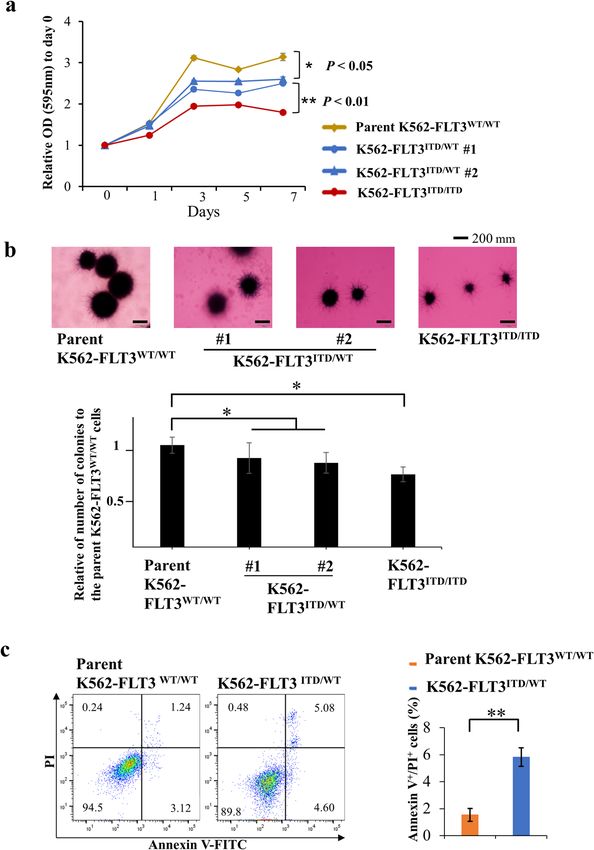
Karnan et al. Cell Death Discovery (2021)7:121                                                                                                    Page 3 of 14

  Fig. 1 Cell growth and colony formation assay of FLT3-ITD knock-in K562 cells. a MTT assay for the growth rate of K562 cell clones (yellow, parent
  K562–FLT3WT/WT; blue, K562–FLT3ITD/WT; red, K562–FLT3ITD/ITD). The relative optical density (OD) at 595 nm on each indicated day was normalized to
  (divided by) that on Day 0. b Representative images of soft agar colony formation assay for the parent K562–FLT3WT/WT, K562–FLT3ITD/WT, and
  K562–FLT3ITD/ITD cell clones (upper panel). Number of colonies of indicated K562 cell clones (lower panel). Two hundred cells of each clone were seeded in
  a six-well plate. After 14 days, the cells were stained with MTT, imaged, and counted. c Proportion of apoptotic cells was increased in K562–FLT3ITD/WT cells
  compared with K562–FLT3WT/WT cells. Data are expressed as mean ± SE (n = 3). Asterisks indicate statistically significant differences between indicated K562
  cell clones using two-tailed non-paired one-way analysis of variance (ANOVA), followed by post hoc Student’s t-test analysis. *p < 0.05, **p < 0.01.

Elevated CD52 expression in patients with AML harboring                           available. We found that the median CD52 expression in
FLT3-ITD                                                                          patients with FLT3-ITD was nominally (though not sig-
  To investigate the relationship between CD52 expres-                            nificantly) higher than that in patients lacking FLT3-ITD
sion levels and FLT3-ITD in patient samples, we analyzed                          (Supplemental Fig. S5). These results suggested that
public domain data (GSE34860) for which CD52 mRNA                                 FLT3-ITD may increase CD52 expression in patients
expression in patients with AML harboring FLT3-ITD is                             with AML.

Official journal of the Cell Death Differentiation Association
Karnan et al. Cell Death Discovery (2021)7:121                                                                                          Page 4 of 14

  Fig. 2 Comparative gene expression profiling and quantitative real-time PCR (qRT-PCR) analysis. a A heat map of upregulated or
  downregulated genes in the parent K562–FLT3WT/WT, K562–FLT3ITD/WT, and K562–FLT3ITD/ITD cell clones, as determined by microarray analysis. cDNA
  microarray analysis was performed using the Agilent Whole Human Genome cDNA Microarray Kit (4 × 44 K; Design ID, 026652). A fold change of >2.0
  was considered an upregulated gene, and a fold change of
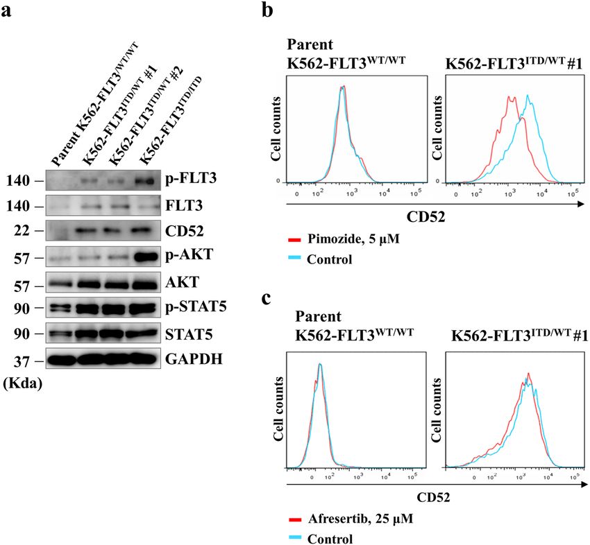
Karnan et al. Cell Death Discovery (2021)7:121                                                                                            Page 5 of 14

  Fig. 3 Conversion of FLT3-ITD to the wild-type FLT3 sequence in K562–FLT3ITD/WT cells by the CRISPR-Cas9 system. The FLT3-ITD sequence in
  the K562–FLT3ITD/WT cells was replaced by the wild-type FLT3 (FLT3-WT) sequence. a Agarose gel electrophoresis of genomic PCR products
  containing the FLT3-ITD domain in the indicated K562 cells. b qRT-PCR analysis of CD52, BTG2, and ID2 genes in the indicated K562 cell clones.
  Relative gene expression levels are shown after normalization to GAPDH mRNA level. Data are expressed as mean ± SE (n = 3). Asterisks indicate
  statistically significant differences between K562–FLT3ITD/WT and rescued K562–FLT3WT/WT using two-tailed non-paired one-way analysis of variance
  (ANOVA), followed by post hoc Student’s t-test analysis. ***p < 0.01. c Flow cytometric analysis for CD52 expression in the rescued K562-FLT3WT/WT #1
  (red, left panel), the parent K562–FLT3WT/WT (red, right panel), and K562–FLT3ITD/WT #1 (blue, both panels) cells. CD52 expression was decreased in
  both rescued K562–FLT3WT/WT #1 and parent K562–FLT3WT/WT cells compared with that in K562–FLT3ITD/WT #1 cells.

FLT3-ITD enhances CD52 expression via accumulation of                         cells, the level of phospho-STAT5 was elevated in
phosphorylated STAT5                                                          K562–FLT3ITD/WT cells, and the level of phospho-AKT was
  We next investigated the mechanism of CD52 upregulation                     further elevated in K562–FLT3ITD/ITD cells (Fig. 4a). Expo-
in K562–FLT3ITD/WT cells. First, we confirmed the expres-                      sure of the cells to pimozide, a STAT5 inhibitor, resulted in
sion of CD52 and the expression and phosphorylation levels                    decreases in CD52 protein levels in K562–FLT3ITD/WT cells,
of FLT3, AKT, and STAT5 in parent K562–FLT3WT/WT,                             an effect not seen with afuresertib, an AKT inhibitor
K562–FLT3ITD/WT (K562–FLT3ITD/WT #1 and #2),                                  (Fig. 4b, c). These results suggested that the accumulation of
and K562–FLT3ITD/ITD cells by immunoblotting (Fig. 4a).                       CD52 protein in cells harboring FLT3-ITD occurs via acti-
We observed that, compared to parent K562-FLT3WT/WT                           vation of STAT5 transcriptional activity.

Official journal of the Cell Death Differentiation Association
Karnan et al. Cell Death Discovery (2021)7:121 Page 6 of 14
Fig. 4 FLT3-ITD enhances CD52 expression via upregulation of phosphorylated STAT5. a Western blot analysis showing the expression of
CD52 and phosphorylation levels of FLT3, AKT, and STAT5 in the indicated clones of K562. b Flow cytometric analysis showing the effect of a STAT5
inhibitor, pimozide, on CD52 expression in parent K562–FLT3WT/WT cells (left panel) and K562–FLT3ITD/WT cells (right panel). Cells were treated
with 5 μM pimozide for 5 days. c Flow cytometric analysis showing the effect of afuresertib, an AKT inhibitor, on CD52 expression in parent
K562–FLT3WT/WT cells (left panel) and K562–FLT3ITD/WT cells (right panel). Cells were treated with 25 μM afuresertib for 5 days. Phosphate-buffered
saline (PBS) was used as the control.
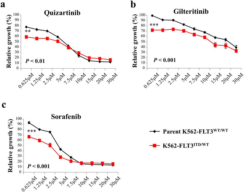
Effects of FLT3 inhibitors on cell proliferation in Alemtuzumab inhibits xenograft tumor growth of
K562–FLT3ITD/WT cells K562–FLT3ITD/WT cells in SCID mice
To clarify the efficacy of FLT3 inhibitors in We examined the effect of alemtuzumab on in vivo
K562–FLT3ITD/WT cells, we performed MTT assays tumor growth of K562–FLT3ITD/WT cells using a xeno-
assessing the growth of K562–FLT3ITD/WT cells in the graft tumor model in SCID mice. As expected, the growth
absence and presence of the indicated FLT3 inhibitors of K562–FLT3ITD/WT tumors was significantly attenuated
(quizartinib, gilteritinib, and sorafenib). We found that in mice treated with alemtuzumab compared to tumor
K562–FLT3ITD/WT cells were more sensitive to each of growth in tumor-bearing mice treated with vehicle alone
the three FLT3 inhibitors than were parent (p < 0.01) (Fig. 6c, d).
K562–FLT3WT/WT cells (Fig. 5a–c). These results sug-
gested that FLT3 cellular signaling is functionally active in Cytotoxic effects of alemtuzumab on AML cells with FLT3-
K562–FLT3ITD/WT cells. ITD
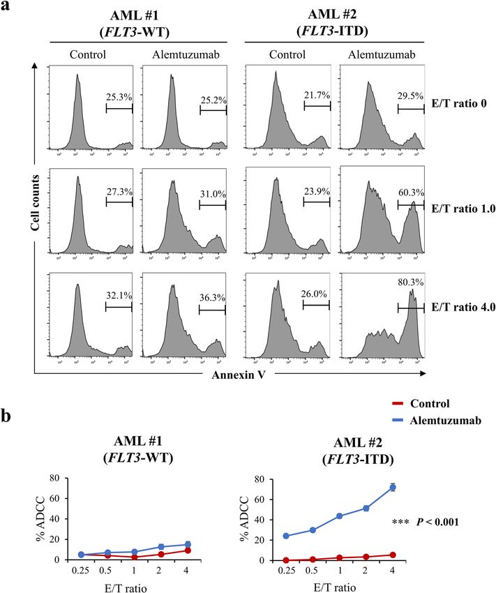
Finally, we performed the ADCC assay with alemtuzu-
Comparison of ADCC with alemtuzumab in parent mab on newly diagnosed AML patient samples that har-
K562–FLT3WT/WT and K562–FLT3ITD/WT cells bor the FLT3-ITD mutation. We found that alemtuzumab
Alemtuzumab-induced NK cell-mediated ADCC was showed ADCC in cells from an AML patient with FLT3-
examined in parent K562–FLT3WT/WT and K562– ITD, but did not show ADCC in cells from an AML
FLT3ITD/WT cells. ADCC with alemtuzumab was con- patient with FLT3-WT (Fig. 7a, b). In addition, alemtu-
sistently elevated in K562–FLT3ITD/WT cells compared to zumab showed the ADCC in MOLM-13 cells, a human
that in parent K562–FLT3WT/WT cells, regardless of the AML cell line harboring FLT3-ITD, which was slightly
E/T (effector cell/target cell) ratio (p < 0.01) (Fig. 6a, b). resistant to quizartinib compared with MOLM-14 cells
Official journal of the Cell Death Differentiation Association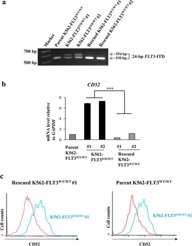
Karnan et al. Cell Death Discovery (2021)7:121                                                                                            Page 7 of 14

  Fig. 5 Effect of FLT3 inhibitors on cell proliferation of parent K562–FLT3WT/WT and K562–FLT3ITD/WT cells. a–c Effect of quizartinib (a),
  gilteritinib (b), and sorafenib (c) on cell proliferation in parent K562–FLT3WT/WT and K562–FLT3ITD/WT cells. Cells were treated with the indicated
  concentration of inhibitors for 48 h. Cell growth was measured by the MTT assay. Black and red lines indicate parent K562–FLT3WT/WT cells and
  K562–FLT3-ITD/WT cells, respectively. Data are expressed as mean ± SE (n = 3). Asterisks indicate statistically significant differences between parent
  K562–FLT3WT/WT cells and K562–FLT3ITD/WT using two-tailed non-paired one-way analysis of variance (ANOVA), followed by post hoc Student’s t-test
  analysis. **p < 0.01, ***p < 0.001.

which were also a human AML cell line with FLT3-ITD                           (Fig. 6c, d). Additionally, we demonstrated that alemtu-
(Supplementary Fig. S7a–c).                                                   zumab showed ADCC in cells from AML patients with
                                                                              FLT3-ITD and MOLM-13 cells that had FLT3-ITD
Discussion                                                                    (Fig. 7a, b and Supplementary Fig. S7a–c). To our
  In this study, we generated a cellular model of the FLT3-                   knowledge, this work is the first to describe the generation
ITD mutation using the CRISPR-Cas9 system and a                               of FLT3-ITD mutants using human myeloid leukemia
human myeloid leukemia cell line, K562 (Supplemental                          cells and the CRISPR-Cas9 system; these cells lines
Figs. S1a, b and S2a–d). We found that the expression of                      allowed us to elucidate the relationship between the
CD52 (at both the mRNA and protein level) was increased                       FLT3-ITD mutation and CD52 overexpression. Our
in FLT3-ITD knock-in K562 cells (K562–FLT3ITD/WT                              findings suggest the possibility of a new therapeutic
cells) compared to the parent and to rescued                                  option, the anti-CD52 antibody alemtuzumab, to treat
K562–FLT3WT/WT cells (Figs. 2a, b and 3a–c). Further-                         leukemia carrying the FLT3-ITD mutation.
more, our analysis with a database deposited in the public                      We demonstrated that FLT3-ITD was associated with
domain showed that CD52 mRNA expression in samples                            increased expression of CD52 in genetically modified
from patients with FLT3-ITD-positive AML tended to be                         FLT3-ITD knock-in K562 cells and in patients with AML
higher than that in samples from patients with FLT3-ITD-                      (Fig. 2a, b and Supplemental Fig. S5). Multiple studies have
negative AML (Supplemental Fig. S5). Together, these                          investigated the molecular mechanisms and cell phenotype
results indicated the possibility that FLT3-ITD cellular                      underlying FLT3-ITD; however, we are not aware of any
signaling is closely associated with CD52 expression.                         reports examining the relationship between FLT3-ITD and
Moreover, we found that alemtuzumab, an anti-CD52                             CD52. This issue may reflect differences in the methods
antibody, induced stronger ADCC in K562–FLT3ITD/WT                            used to establish transfectants harboring the FLT3-ITD
cells compared with that in K562–FLT3WT/WT cells                              mutation. In cells into which FLT3-ITD is introduced by a
(Fig. 6a, b) and dramatically suppressed tumor growth by                      conventional method (e.g., using a viral vector), the ectopic
K562–FLT3ITD/WT cells in mouse xenograft experiments                          FLT3-ITD gene would be overexpressed, potentially

Official journal of the Cell Death Differentiation Association
Karnan et al. Cell Death Discovery (2021)7:121 Page 8 of 14 Fig. 6 Anti-tumor effects of alemtuzumab in K562–FLT3ITD/WT cells. a Comparison of NK cell-mediated antibody-dependent cell-mediated cytotoxicity (ADCC) with alemtuzumab in parent K562–FLT3WT/WT (left panel) and K562–FLT3ITD/WT (right panel) cells at the indicated E/T (effector cell/target cell) ratios. Phosphate-buffered saline (PBS) was used as control for alemtuzumab. b Percent ADCC by E/T ratio with control (left panel) and alemtuzumab (right panel). Red and blue lines indicate parent K562–FLT3WT/WT and K562–FLT3ITD/WT cells, respectively. Alemtuzumab showed higher percent ADCC in K562–FLT3ITD/WT cells than that in parent K562–FLT3WT/WT cells, whereas control did not. Data are expressed as mean ± SE (n = 3). c, d Effect of alemtuzumab on the tumor growth of xenografted K562–FLT3ITD/WT cells. c Mice implanted with tumors of xenografted K562–FLT3ITD/WT cells and treated with control (PBS) or alemtuzumab are pictured at the time of euthanasia on day 14. d Tumor volume of xenografted K562–FLT3ITD/WT cells treated with control or alemtuzumab on the indicated day. Data are expressed as mean ± SE (n = 6). Asterisks indicate statistically significant differences between control and alemtuzumab and K562–FLT3ITD/WT using two-tailed non-paired one-way analysis of variance (ANOVA), followed by post hoc Student’s t-test analysis. **p < 0.01. Official journal of the Cell Death Differentiation Association
Karnan et al. Cell Death Discovery (2021)7:121                                                                                                Page 9 of 14

  Fig. 7 Effects of alemtuzumab in human primary AML cells. a Comparison of NK cell-mediated cytotoxicity (antibody-dependent cellular
  cytotoxicity, ADCC) with alemtuzumab in primary cells from an AML patient with FLT3-WT (AML #1, left panel) or from an AML patient with FLT3-ITD
  (AML #2, right panel) cells at the indicated E/T (effector cell/target cell) ratios. Phosphate-buffered saline (PBS) was used as control for alemtuzumab.
  b Percent ADCC by E/T ratio with control (red line) and alemtuzumab (blue line) in cells from AML patients. Alemtuzumab showed higher percent
  ADCC in the cells from the AML patient with FLT3-ITD (right panel) than that in those from the AML patient with FLT3-WT (left panel). Data are
  expressed as mean ± SE (n = 3). Asterisks indicate statistically significant differences between control and alemtuzumab in indicated cells using two-
  tailed non-paired one-way analysis of variance (ANOVA), followed by post hoc Student’s t-test analysis. ***p < 0.001.

resulting in a phenotype distinct from that of actual patient                     FLT3-ITD has been reported to activate PI3K/AKT
leukemia cells, which typically may not overexpress the                         signaling via STAT5-mediated activation in AML cells15–17.
mutant gene. In contrast, we employed genome editing,                           In the present work, we confirmed that phospho-STAT5
permitting the construction of a FLT3-ITD-containing                            levels were increased in K562–FLT3ITD/WT cells com-
leukemia cell model that more closely resembles the actual                      pared to parental cells; additionally, the levels of phospho-
condition of leukemia cells in patients. Thus, genome                           AKT appeared to show progressive increases when
editing may have revealed alterations of cell phenotype                         comparing K562–FLT3WT/WT, K562–FLT3ITD/WT, and
specific to FLT3-ITD that previously have been overlooked.                       K562–FLT3ITD/ITD cells (Fig. 4a). Regarding the rela-
We also found that ID2 and BTG2 were upregulated                                tionship between cellular signaling and CD52 expression,
in K562–FLT3ITD/WT cells compared to parent                                     we observed that CD52 expression in K562–FLT3ITD/WT
K562–FLT3WT/WT cells (Fig. 2c, d). The molecular func-                          cells was decreased upon exposure to pimozide, a STAT5
tion and significance of the elevated expression of these                        inhibitor, whereas exposure to afuresertib, an AKT inhi-
genes will need to be examined in future studies.                               bitor, had no significant effect of on CD52 expression in

Official journal of the Cell Death Differentiation Association
Karnan et al. Cell Death Discovery (2021)7:121                                                                 Page 10 of 14

K562-FLT3ITD/WT cells (Fig. 4b, c). This observation              We found that FLT3 inhibitors (quizartinib, gilteritinib
suggested that signaling by STAT5, but not that by AKT,         and sorafenib) suppress the proliferation of
is involved in FLT3-ITD-induced upregulation of CD52.           K562–FLT3ITD/WT cells compared with that of parent
  Using MMT and colony formation assays, we found that          K562–FLT3WT/WT cells (Fig. 5a–c), suggesting that
cell growth was decreased in the K562–FLT3ITD/WT and            FLT3 signaling is functionally active in K562–FLT3ITD/WT
K562–FLT3ITD/ITD cells compared with that in the parent         cells. Although FLT3 inhibitors can prolong the survival
K562–FLT3WT/WT cells (Fig. 1a, b). These findings differ         of patients harboring cancers with FLT3-ITD24, the out-
from those of the previous literature, which reported that      come of treatment in these patients remains unsatisfac-
FLT3-ITD enhanced cell proliferation18. We speculate            tory due to insensitivity to these compounds and/or
that the impaired growth seen in the present work reflects       acquired drug resistance25,26. Secondary mutations in the
oncogenic death19 in our FLT3-ITD cells caused by               sequences encoding the FLT3 TKD have been observed in
excessive strong proliferation signaling resulting from the     a subset of those patients27,28. Therefore, an anti-tumor
additional FLT3 cellular signaling in K562 cells, that          antibody targeting CD52, such as alemtuzumab, is a
already are subject to BCR-ABL tyrosine kinase activity.        potential therapeutic alternative for AML with FLT3-ITD,
  CD52, which also is known as CAMPATH-1, is a gly-             given that this antibody targets and kills leukemic cells by
coprotein that is expressed on the cell surface of lym-         a mechanism that is different from that of the FLT3
phocytes, monocytes, and dendritic cells20. Alemtuzumab,        kinase inhibitors.
a humanized anti-CD52 antibody, has been used for                 A major limitation of our study is that we did not
depleting lymphocytic leukemia cells in patients with           analyze many primary samples. Further studies investi-
CLL. Alemtuzumab induces the killing of CD52-positive           gating ADCC with alemtuzumab, and the anti-tumor
lymphocytes via ADCC and complement-dependent                   effects of alemtuzumab in a xenograft model using more
cytotoxicity20,21. In the present study, alemtuzumab            patient samples will be needed to validate our proposed
exposure provided increased ADCC in K562–FLT3ITD/WT             treatment strategy for FLT3-ITD leukemia.
cells compared with that in parent K562–FLT3WT/WT                 Taken together, the findings of this study revealed
cells (Fig. 6a, b), and dosing with alemtuzumab sup-            CD52 as a molecular target for the antibody treatment of
pressed the growth of K562–FLT3ITD/WT tumors in a               FLT3-ITD leukemia. Since alemtuzumab is an approved
mouse xenograft model (Fig. 6c, d). These results sug-          drug, further clinical studies using alemtuzumab are
gested that alemtuzumab suppresses, via ADCC, the               warranted to evaluate our proposed treatment strategy
in vivo growth of K562–FLT3ITD/WT cells, which have             for FLT3-ITD leukemia. The present study offers valu-
elevated expression of CD52. Interestingly, a previous          able clues for further improvement of the outcomes in
study reported that the administration of alemtuzumab in        this challenging disease.
NOD SCID gamma (NSG) mice significantly suppressed
the engraftment of AML patient-derived CD52-positive            Materials and methods
leukemia cells with the FLT3-ITD mutation, while not            Cell culture and reagents
suppressing engraftment of leukemia cells lacking the             The human CML cell line K562 was obtained from the
FLT3-ITD mutation22. These data suggest that alemtu-            Japanese Collection of Research Bioresource Cell Bank.
zumab may be effective for the treatment of myeloid             The human AML cell line MOLM-13 and MOLM-14
leukemia with FLT3-ITD. Additionally, alemtuzumab               were purchased from DSMZ (German Collection of
has been employed as a therapeutic option for reducing          Microorganisms and Cell Cultures). Primary human AML
the risk of graft-versus-host disease (GVHD) by elim-           samples from newly diagnosed patients were obtained
inating lymphocytes23. Therefore, it may be possible to         from the sample archive at the Aichi Medical University
improve the outcome of cases with FLT3-ITD myeloid              Hospital. All samples were obtained after written
leukemia using alemtuzumab as a GVHD prophylaxis for            informed consent, and the use of biological samples for
patients undergoing allogeneic hematopoietic stem cell          research was approved by the ethics committee of the
transplantation.                                                Aichi Medical University Hospital (Approval Number
  In addition to our modification of K562 cells, we              2020-156), in accordance with the Declaration of Helsinki.
established LCL-FLT3-ITDITD/WT cells. We demon-                 The cells were cultured in RPMI-1640 supplemented with
strated that LCL-FLT3-ITDITD/WT cells showed increased          10% fetal bovine serum (FBS) at 37 °C in a 5% CO2
expression of CD52 mRNA, decreased cell proliferation,          humidified atmosphere. Afuresertib, gilteritinib, and qui-
and increased levels of apoptosis compared to the parent        zartinib were purchased from Selleck Chemicals (Hous-
LCL-FLT3-ITDWT/WT cells (Supplemental Fig. S6a–d).              ton, TX, USA). Sorafenib was obtained from ChemScene
These results suggest that FLT3-ITD effects on CD52             (Monmouth Junction, NJ, USA). Pimozide was obtained
expression and cell proliferation are not unique to             from Sigma-Aldrich (St. Louis, MO, USA). Alemtuzumab,
K562 cells.                                                     a recombinant humanized monoclonal antibody against

Official journal of the Cell Death Differentiation Association
Karnan et al. Cell Death Discovery (2021)7:121                                                                 Page 11 of 14

human CD52, was purchased from Sanofi K.K. (Tokyo,               using the CRISPR-Cas9 system as above (Supplemental
Japan).                                                         Fig. S2a). The resulting clone was designated LCL-
                                                                FLT3ITD/WT.
Establishment of genetically FLT3-ITD knock-in cell clones
generated using the CRISPR-Cas9 system                          cDNA microarray analysis
  The CRISPR-Cas9 system was used to convert the wild-            cDNA microarray analysis was performed according to
type sequence of FLT3 (FLT3-WT) to FLT3-ITD, as                 the manufacturer’s protocol (Agilent Technologies, Santa
described elsewhere29. pSpCas9 (BB)-2A-GFP (PX458)              Clara, CA, USA). Briefly, cDNA synthesis and cRNA
was a gift from Dr Feng Zhang (Addgene plasmid # 48138;         labeling with cyanine 3 (Cy3) dye were performed using
Addgene)30. A single guide RNA (sgRNA) sequence for             the Agilent Low Input Quick Amp Labeling Kit (Agilent
FLT3 was selected using Optimized CRISPR Design                 Technologies). Cy3-labeled cRNA then was purified,
(http://crispr.mit.edu/). The sgRNA sequence for FLT3           fragmented, and hybridized to a Human Gene Expression
exon 14 was 5′-GTAGAAGTACTCATTATCTG-3′                          4x44K v2 Microarray Chip containing 27,958 Entrez Gene
(Supplemental Fig. S1a). A 1034-bp DNA fragment con-            RNAs using a Gene Expression Hybridization Kit
taining the 24-bp FLT3-ITD sequence was obtained by             (Agilent Technologies). To compare gene expression
PCR amplification of genomic DNA from a human                    profiles between the parent K562–FLT3WT/WT cells and
myeloid leukemia cell line, AMU-AML2 cells, previously          K562–FLT3ITD/WT cells, raw fluorescence values were
established in our laboratory. Amplification of the FLT3-        normalized and clustered according to the differential
ITD gene was performed using primers as follows: for-           gene expression. The raw and normalized microarray data
ward (Fw), 5′-ACTCAAGTGATCCTCCCATC-3′, and                      have been submitted to the GEO database at NCBI as
reverse (Rev), 5′-TGACTGGGTTGACACCCCA-3′. The                   Accession Number GSE116727.
DNA fragment containing the 24-bp FLT3-ITD sequence
was inserted into a plasmid vector, pcDNA 3.1 (+), using        Quantitative reverse transcription real-time PCR (qRT-PCR)
TA cloning, yielding a plasmid that was designated                qRT-PCR analyses for FLT3, CD52 (cluster of differ-
pcDNA/FLT3-ITD. The sgRNA sequence for FLT3 was                 entiation 52), ID2 (inhibitor of DNA binding 2), and BTG2
cloned by ligating corresponding oligonucleotides into the      (B-cell translocation gene 2) were performed using SYBR
BbsI site of PX458, yielding a plasmid designated FLT3/         Green I, as described in a previous study31. The GAPDH
PX458. To convert FLT3-WT to FLT3-ITD in K562 cells,            transcript (encoding glyceraldehyde phosphate dehy-
FLT3/PX458 and pcDNA/FLT3-ITD were co-transfected               drogenase, a housekeeping protein) was used as an
into K562 cells using a 4D-Nucleofector™ instrument             internal control. The sequences of the primers for CD52,
(Lonza Japan, Tokyo, Japan). A single clone was selected        ID2, BTG2, and GAPDH used in this study are listed in
from a 96-well plate, expanded in a 12-well plate, and then     Supplemental Table S1.
used for biological assays. For sequence analysis and agarose
gel electrophoresis analysis, the FLT3 gene was amplified by     Cell growth assay
PCR using the following primers: Fw, 5′-AGAAGTG                   Cell growth rate was determined by an MTT (3-(4,5-
GAAGAAGAGGTGG-3′, and Rev, 5′-TCCAAGACAA                        dimethylthiazol-2-yl)-2,5-diphenyltetrazolium bromide)
CATCTCATTC-3′. The FLT3 gene amplified from geno-                assay. Briefly, parent K562-FLT3WT/WT and K562-
mic DNA was subjected to sequence analysis to confirm            FLT3ITD/WT cells (1 × 10³ cells/well) were seeded into
the monoallelic presence of the 24-bp duplication (FLT3-        96-well plates, and the plates were incubated for the
ITD) in two independent K562–FLT3ITD/WT clones                  indicated intervals at 37 °C in a 5% CO2 environment.
(designated #1 and #2), and the biallelic presence of           Subsequently, 10 μL of MTT solution (5 mg/mL) was
the 24-bp duplication in one K562–FLT3ITD/ITD clone             added to each well and the plates were incubated for
(Supplemental Fig. S1a–d). To convert the FLT3-ITD allele       another 4 h. Next, cell lysis buffer was added to the wells
in K562–FLT3ITD/WT cells to the FLT3-WT sequence, 1 μg          to lyse the cells and dissolve the colored formazan crystals
each of FLT3/PX458 and pcDNA/FLT3-WT were co-                   produced by the reduction of MTT. The optical density
transfected into K562–FLT3ITD/WT cells (1 × 10⁶ cells/mL)       (595 nm) of the colored product was measured at each of
using a 4D-Nucleofector™ as above.                              the time points (0, 1, 3, 5, and 7 days) using a Spec-
                                                                traMAX M5 spectrophotometer (Molecular Devices,
Construction of model cell line using lymphoblastoid cell       Sunnyvale, CA, USA).
line (LCL) cell line
  A human B-cell-derived LCL was kindly provided by             Annexin V assay
Dr. Sonta Shin-ichi (Fetal Life Science Center, Aichi,            The cells were cultured in 6-well plates (5 × 10⁵ cells/
Japan). We established a LCL cell clone containing the          well) for 24 h, followed by incubation with fluorescein
monoallelic 24-bp duplication of the FLT3-ITD mutation          isothiocyanate (FITC)-conjugated annexin V (Biolegend,

Official journal of the Cell Death Differentiation Association
Karnan et al. Cell Death Discovery (2021)7:121 Page 12 of 14
San Diego, CA, USA) and propidium iodide (PI) at Cytotoxicity was calculated according to the following
approximately 25 °C for 15 min. Fluorescence intensities formula: % ADCC = 100 × (E − S) / (100 − S), where E is
of FITC and PI were determined by flow cytometric the concentration of annexin V-positive cells in the
analysis using a FACSCanto II instrument (BD, Franklin experimental well and S is the concentration of annexin
Lakes, NJ, USA). V-positive cells in the absence of alemtuzumab (i.e., when
target cells were incubated with NK cells alone).
Soft agar colony formation assay
An aliquot (2 mL) of RPMI-1640 culture medium con- Xenograft experiment
taining 0.3% agar was used as the bottom gel in each well Animal experiments were approved by the ethical
of a 6-well plate. For each well, 5000 parent committee of Aichi Medical University and performed
K562–FLT3WT/WT or K562–FLT3ITD/WT cells were sus- according to their guidelines. Female Fox Chase severe
pended in 2 mL RPMI-1640 culture medium containing combined immunodeficiency (SCID) mice (CB17/Icr-
0.15% agar, and the cell suspension was poured onto the Prkdcscid/IcrIcoCrl) were purchased from CLEA Japan,
bottom gel. After two weeks, colonies were stained with Inc. (Tokyo, Japan) and bred at the Institute of Animal
MTT solution. The wells were photographed under Experiments, Aichi Medical University, in specified-
bright-field microscopy (IX-73, Olympus, Tokyo, Japan). pathogen-free animal facilities. All mice used in this
The number of colonies was counted using Colony study were 6–8 weeks old and weighed 17–18 g each at
Counter software (BZ-X800, Keyence, Tokyo, Japan). the time of implantation. SCID mice were injected sub-
cutaneously in the left flank with K562–FLT3ITD/WT cells
Western blot analysis (1 × 10⁷ cells/mouse). Tumor growth was monitored by
Western blot analysis was performed as described in a measuring the tumors along the perpendicular long and
previous study32. The antibodies used in this study are short axes (length and width, respectively). Tumor
listed in Supplemental Table S2. Immune complexes were volumes were calculated using the formula for the volume
detected using ImmunoStar LD (Wako Pure Chemical of a modified ellipsoid (volume = 1/2 × length × width2).
Industries, Ltd., Osaka, Japan) in conjunction with a LAS- When the implanted tumors reached a mean size of
4000 image analyzer (GE Healthcare, Tokyo, Japan). 100 mm3, mice were randomly divided into two groups.
Animals of the alemtuzumab group were administered
ADCC assay intraperitoneally with the antibody (1 mg/kg, 2 doses/
We analyzed ADCC induced by anti-CD52 antibody, week). Animals of the control groups were administered
alemtuzumab, in the parent K562–FLT3WT/WT, equivalent volumes of phosphate-buffered saline (PBS;
K562–FLT3ITD/WT, MOLT-13, and AML patient cells vehicle) according to the same regimen. Following the
using flow cytometry with staining for annexin V. Natural start of treatment, tumor dimensions were measured
killer (NK) cells were prepared by isolating CD56-positive every 3 or 4 days through Day 14, at which point the
cells from the peripheral blood mononuclear cells animals were euthanized.
(PBMCs) of a healthy donor using anti-CD56 antibody
conjugated with magnetic microbeads in combination Statistical analysis
with the autoMACS system (Miltenyi Biotec, Bergisch Experimental results are expressed as mean ± standard
Gladbach, Germany). The prepared NK cells were error (SE). The statistical significance of the comparisons
co-cultured with the parent K562–FLT3WT/WT, among groups was determined using two-tailed non-
K562–FLT3ITD/WT, MOLT-13, and AML patient cells in paired one-way analysis of variance (ANOVA) with post
RPMI-1640 with 10% FBS in the presence of alemtuzu- hoc Student’s t-test where indicated. Values of *p < 0.05,
mab; the mixtures were incubated at 37 °C for 15 h. The **p < 0.01, and ***p < 0.001 (indicated by asterisks) were
cells then were stained with allophycoerithrin (APC)- considered statistically significant. Statistical analyses
conjugated anti-glycophorin A antibody and phycoery- were performed using the SPSS program (version 23.0;
thrin (PE)-conjugated anti-CD56 antibody (Biolegend) for SPSS, Inc., Chicago, IL, USA).
20 min at 4 °C. After washing twice with 500 µL annexin
buffer (10 mM HEPES, 150 mM NaCl, and 2 mM CaCl2,
[pH 7.4]), the cells were incubated with FITC-conjugated Acknowledgements
annexin V (Biolegend) for 10 min at approximately 20 °C. The authors thank Ms. Yuka Oohigashi and Ms. Taeko Nakamura for their
valuable secretarial assistance and FORTE Science Communications (Tokyo,
Then, flow cytometry was performed using a Fortessa Japan) for their editorial assistance. This study was supported in part by grants
instrument (BD Biosciences, Franklin Lakes, NJ, USA); from the Ministry of Education, Culture, Sports and Technology of Japan
annexin V-positive cells gated on the glycophorin A+ (MEXT; 19K08825 to I.H., 19K09292 to S.K., 18K07277 to S.S., and 18K08342 to
A.O.), by a research grant from the Hirose International Scholarship Foundation,
CD3− population were analyzed by FlowJo software and by grants from the Japan Agency for Medical Research and Development
(version 10; Tree Star, Inc., Ashland, OR, USA). (AMED; 19ae0101074s040 to R.U.).
Official journal of the Cell Death Differentiation AssociationKarnan et al. Cell Death Discovery (2021)7:121 Page 13 of 14
Author details 3. Small, D. et al. STK-1, the human homolog of Flk-2/Flt-3, is selectively
1
Department of Biochemistry, Aichi Medical University, Nagakute, Aichi, Japan. expressed in CD34+human bone marrow cells and is involved in the pro-
2
Division of Hematology, Department of Internal Medicine, Aichi Medical liferation of early progenitor/stem cells. Proc. Natl Acad. Sci. USA 91, 459–463
University, Nagakute, Aichi, Japan. 3Research Creation Support Center, Aichi (1994).
Medical University, Nagakute, Aichi, Japan. 4Department of Tumor 4. Lyman, S. D. et al. Molecular cloning of a ligand for the flt3/flk-2 tyrosine
Immunology, Aichi Medical University, Nagakute, Aichi, Japan. 5Department of kinase receptor: a proliferative factor for primitive hematopoietic cells. Cell 75,
Pharmacy, Aichi Medical University, Nagakute, Aichi, Japan 1157–1167 (1993).
5. Nakao, M. et al. Internal tandem duplication of the flt3 gene found in acute
Author contributions myeloid leukemia. Leukemia 10, 1911–1918 (1996).
(1) Contributions of all named authors to the manuscript: S.K.; conception and 6. Yokota, S. et al. Internal tandem duplication of the FLT3 gene is preferentially
design of the project, analysis and interpretation of data, and writing the seen in acute myeloid leukemia and myelodysplastic syndrome among var-
manuscript. I.H.; conception and design of the project, analysis and ious hematological malignancies. A study on a large series of patients and cell
interpretation of data, and writing the manuscript. A.O.; development of lines. Leukemia 11, 1605–1609 (1997).
methodology. S.Ta.; acquisition of data. A.N.; acquisition of data. M.T.; 7. Takahashi, S. Downstream molecular pathways of FLT3 in the pathogenesis of
acquisition of data. K.U.; acquisition of data. S.M.; acquisition of data. M.W.; acute myeloid leukemia: biology and therapeutic implications. J. Hematol.
acquisition of data. L.Q.V.; acquisition of data. M.L.R.; acquisition of data. M.N.H.; Oncol. 4, 13 (2011).
acquisition of data. T.H.; acquisition of data. H.K.; acquisition of data. S.Ts.; 8. Quentmeier, H., Reinhardt, J., Zaborski, M. & Drexler, H. G. FLT3 mutations in
acquisition of data. K.Y.; acquisition of data. S.S.; acquisition of data. R.U.; acute myeloid leukemia cell lines. Leukemia 17, 120–124 (2003).
acquisition of data. M.E.; acquisition of data. Y.H.; acquisition of data. A.T.; 9. Annamaneni, S. et al. Incidence of internal tandem duplications and D835
acquisition of data. (2) Contributions of all named authors to the figures: Figure mutations of FLT3 gene in chronic myeloid leukemia patients from Southern
1: S.K. and T.H. generated the data and prepared figures. Figure 2: S.K. and I.H. India. Hematology 19, 129–1235 (2014).
generated the data and the prepared the figures. S.Ta., H.K., R.U., and K.Y. 10. Xu, B., Tian, H. & Zhou, S. Y. Detection of FLT3 gene and FLT3/ITD gene
analyzed the microarray data and prepared the panel B. Figure 3: S.K. and M.L.R. mutation in chronic myeloid leukemia and its significance. Ai. Zheng 23,
generated the data and prepared the figures. Figure 4: S.K. generated the data 1218–1221 (2004).
and prepared the figures. A.O., M.N.H., and M.W. analyzed the data. Figure 5: 11. Kiyoi, H. et al. Prognostic implication of FLT3 and N-RAS gene mutations in
S.K., M.T. and L.Q.V. generated the data and prepared the figures. Figure 6: S.K., acute myeloid leukemia. Blood 93, 3074–3080 (1999).
S.Ta., A.N., and S.S. generated the data and prepared the figures. A.T. and Y.H. 12. Meshinchi, S. et al. Prevalence and prognostic significance of Flt3 internal
analyzed the data. Figure 7: S.K., S.Ta., A.N., and S.S. generated the data and tandem duplication in pediatric acute myeloid leukemia. Blood 97, 89–94
prepared the figures. (2001).
13. Stirewalt, D. L. et al. Size of FLT3 internal tandem duplication has prognostic
Funding significance in patients with acute myeloid leukemia. Blood 107, 3724–3726
This study was supported in part by grants from the Ministry of Education, (2006).
Culture, Sports and Technology of Japan (19K08825 to IH, 18K07277 to SS, 14. Perl, A. E. et al. Selective inhibition of FLT3 by gilteritinib in relapsed or
18K08342 to AO), the Japan Agency for Medical Research and Development refractory acute myeloid leukaemia: a multicentre, first-in-human, open-label,
(AMED; 19ae0101074s040 to RU), and by a research grant from Kyowa Kirin. phase 1-2 study. Lancet Oncol. 18, 1061–1075 (2017).
15. Choudhary, C. et al. Activation mechanisms of STAT5 by oncogenic Flt3-ITD.
Ethics statement Blood 110, 370–374 (2007).
This research complies with the ethical guidelines of the Japanese Ministry of 16. Nogami, A. et al. FLT3-ITD confers resistance to the PI3K/Akt pathway inhi-
Health, Labour and Welfare. bitors by protecting the mTOR/4EBP1/Mcl-1 pathway through STAT5 activa-
tion in acute myeloid leukemia. Oncotarget 6, 9189–9205 (2015).
Conflict of interest 17. Brandts, C. H. et al. Constitutive activation of Akt by Flt3 internal tandem
I.H. received honoraria and/or membership on an entity’s board of directors, duplications is necessary for increased survival, proliferation, and myelold
speakers’ bureau, or advisory committees from Celgene, Janssen, Takeda, Ono, transformation. Cancer Res. 65, 9643–9650 (2005).
Bristol-Myers Squibb (BMS), Novartis, Daiichi Sankyo, Kyowa Kirin, Eisai, Nihon- 18. Kiyoi, H., Kawashima, N. & Ishikawa, Y. FLT3 mutations in acute myeloid leu-
Shinyaku, Pfizer, AbbVie, Otsuka, Shionogi, Mundi, CSL Behring, and Merck kemia: therapeutic paradigm beyond inhibitor development. Cancer Sci. 111,
Sharp & Dohme (MSD). I.H. and A.T. received research funding from BMS, MSD, 312–322 (2020).
Astellas, Otsuka, Ono, Kyowa Kirin, Sanofi, Shionogi, Zenyaku, Daiichi Sankyo, 19. Chi, S. et al. Oncogenic Ras triggers cell suicide through the activation of a
Taiho, Takeda, Chugai, Eli Lilly, Nihon Shinyaku, Novartis, Pfizer, Celgene, caspase-independent cell death program in human cancer cells. Oncogene 18,
Fukuyu Hospital, and Yamada Yohojo. RU received research funding from 2281–2290 (1999).
Kyowa Kirin, Chugai Pharmaceutical, and Ono Pharmaceutical. The remaining 20. Hu, Y. et al. Investigation of the mechanism of action of alemtuzumab in a
authors declare no competing financial interests. human CD52 transgenic mouse model. Immunology 128, 260–270 (2009).
21. Zent, C. S. et al. Direct and complement dependent cytotoxicity in CLL cells
from patients with high-risk early-intermediate stage chronic lymphocytic
Publisher’s note leukemia (CLL) treated with alemtuzumab and rituximab. Leuk. Res. 32,
Springer Nature remains neutral with regard to jurisdictional claims in 1849–1856 (2008).
published maps and institutional affiliations. 22. Blatt, K. et al. Identification of campath-1 (CD52) as novel drug target in
neoplastic stem cells in 5q-patients with MDS and AML. Clin. Cancer Res. 20,
Supplementary information The online version contains supplementary 3589–3602 (2014).
material available at https://doi.org/10.1038/s41420-021-00446-8. 23. Potter, V. T. et al. Long-term outcomes of alemtuzumab-based reduced-
intensity conditioned hematopoietic stem cell transplantation for myelodys-
Received: 2 October 2020 Revised: 22 January 2021 Accepted: 9 March plastic syndrome and acute myelogenous leukemia secondary to myelodys-
2021 plastic syndrome. Biol. Blood Marrow Transpl. 20, 111–117 (2014).
24. Fathi, A. T. & Chabner, B. A. FLT3 inhibition as therapy in acute myeloid
leukemia: a record of trials and tribulations. Oncologist 16, 1162–1174
(2011).
25. Piloto, O. et al. Prolonged exposure to FLT3 inhibitors leads to resistance
References via activation of parallel signaling pathways. Blood 109, 1643–1652
1. Rosnet, O. et al. Human FLT3/FLK2 gene: cDNA cloning and expression in (2007).
hematopoietic cells. Blood 82, 1110–1119 (1993). 26. Smith, C. C., Lin, K., Stecula, A., Sali, A. & Shah, N. P. FLT3 D835 mutations confer
2. Carow, C. E. et al. Localization of the human stem cell tyrosine kinase-1 gene differential resistance to type II FLT3 inhibitors. Leukemia 29, 2390–2392
(FLT3) to 13q12−>q13. Cytogenet. Cell Genet. 70, 255–257 (1995). (2015).
Official journal of the Cell Death Differentiation AssociationKarnan et al. Cell Death Discovery (2021)7:121 Page 14 of 14
27. Alvarado, Y. et al. Treatment with FLT3 inhibitor in patients with FLT3-mutated 30. Ran, F. A. et al. Genome engineering using the CRISPR-Cas9 system. Nat.
acute myeloid leukemia is associated with development of secondary FLT3- Protoc. 8, 2281–2308 (2013).
tyrosine kinase domain mutations. Cancer 120, 2142–2149 (2014). 31. Asai, A. et al. High-resolution 400 K oligonucleotide array comparative geno-
28. Larrosa-Garcia, M. & Baer, M. R. FLT3 inhibitors in acute myeloid leukemia: mic hybridization analysis of neurofibromatosis type 1-associated cutaneous
current status and future directions. Mol. Cancer Ther. 16, 991–1001 (2017). neurofibromas. Gene 558, 220–226 (2015).
29. Ota, A. et al. Delta40p53alpha suppresses tumor cell proliferation and induces 32. Wahiduzzaman, M. et al. Novel combined Ato-C treatment synergistically
cellular senescence in hepatocellular carcinoma cells. J. Cell Sci. 130, 614–625 suppresses proliferation of Bcr-Abl-positive leukemic cells in vitro and in vivo.
(2017). Cancer Lett. 433, 117–130 (2018).
Official journal of the Cell Death Differentiation AssociationYou can also read