Molecular Targeting of H/MDM-2 Oncoprotein in Human Colon Cancer Cells and Stem-like Colonic Epithelial-derived Progenitor Cells
←
→
Page content transcription
If your browser does not render page correctly, please read the page content below
ANTICANCER RESEARCH 41: 27-42 (2021)
doi:10.21873/anticanres.14749

                   Molecular Targeting of H/MDM-2 Oncoprotein
                    in Human Colon Cancer Cells and Stem-like
                     Colonic Epithelial-derived Progenitor Cells
                ANUSHA THADI1, WILLIAM F. MORANO1, MARIAN KHALILI1, BLAKE D. BABCOCK1,
                        MOHAMMAD F. SHAIKH1, DESHKA S. FOSTER1, YELENA PIAZZA2,
                        ELIZABETH M. GLEESON1, EVE GOLDSTEIN1, LINDSAY STEELE1,
                  PAUL M. CAMPBELL3, BO LIN4, MATTHEW R. PINCUS4 and WILBUR B. BOWNE1,5

                                      1Division
                                        of Surgical Oncology, Department of Surgery,
                         Drexel University College of Medicine, Philadelphia, PA, U.S.A.;
                              2Department of Pathology and Laboratory Medicine,

                         Drexel University College of Medicine, Philadelphia, PA, U.S.A.;
           3Cancer Signaling and Epigenetics Program, The Marvin and Concetta Greenberg Pancreatic

                       Cancer Institute Fox Chase Cancer Center, Philadelphia, PA, U.S.A.;
    4Department of Pathology and Laboratory Medicine, SUNY Downstate Medical Center, Brooklyn, NY, U.S.A.;
                  5Department of Surgery, Thomas Jefferson University, Philadelphia, PA, U.S.A.

Abstract. Background/Aim: We have tested whether the anti-          caspase 3). In vivo, PNC-27 caused necrosis of tumor nodules
cancer peptide, PNC-27, that kills cancer cells but not normal      but not of normal tissue. Conclusion: PNC-27 selectively kills
cells by binding to cancer cell membrane HDM-2 forming pores,       colon cancer stem cells by binding of this peptide to membrane
kills CD44+ colon cancer stem cells. Materials and Methods:         H/MDM-2.
Flow cytometry determined the CD44 and HDM-2 expression
on six-colon cancer cell lines and one normal cell line (CCD-       Colorectal cancer is the third most common cause of cancer and
18Co). MTT, LDH release, annexin V binding and caspase 3            cancer-related deaths worldwide. In patients with metastatic,
assays were used to assess PNC-27-induced cell death.               unresectable disease, systemic therapy is the treatment of choice
Bioluminescence imaging measured PNC-27 effects on in vivo          (1, 2). However, the majority of patients with advanced disease
tumor growth. Results: High percentages of cells in all six tumor   will become refractory to treatment and develop disease
lines expressed CD44. PNC-27 co-localized with membrane             progression despite first-line therapy, becoming potential
HDM-2 only in the cancer cells and caused total cell death          candidates for second-line systemic therapy (3). Mutations in
(tumor cell necrosis, high LDH release, negative annexin V and      cancer stem cells (CSCs) and their self-renewal properties make
                                                                    them a root cause of systemic disease through metastasis-
                                                                    initiating cells (MICs) and disease relapse (4). Therefore,
                                                                    identification of therapeutic agents against CSC targets has
This article is freely accessible online.                           become critical in developing new cancer treatment strategies.
                                                                       One candidate agent that has been found to be effective
Correspondence to: Wilbur B. Bowne, MD, Thomas Jefferson
University Hospital, 1100 Walnut Street, Philadelphia, PA 19107,
                                                                    against CSCs is the anti-cancer peptide, PNC-27 and its
U.S.A. Tel: +1 2155036855, Fax: +1 215503850, e-mail:               shorter homologue, PNC-28. PNC-27 contains an HDM-2
Wilbur.Bowne@jefferson.edu; Bo Lin, MD, PhD, State University       binding segment from human p53 (corresponding to residues
of New York, Downstate Medical Center, 450 Clarkson Avenue,         12-26) attached on its carboxyl terminal end to a leader
Brooklyn, NY 11203, U.S.A. Tel: +1 7182701643, Fax: +1              peptide, called the membrane residency peptide (MRP), that
7182703303, e-mail: Bo.Lin@downstate.edu; Matthew R. Pincus,        inserts into cell membranes (5). Both peptides have been
MD, Ph.D., State University of New York, Downstate Medical
                                                                    found to induce tumor cell necrosis in a wide variety of solid
Center, NY 11203, U.S.A. Tel: +1 7182701643, Fax: +1
7182703303, e-mail: mrpincus2010@gmail.com
                                                                    tissue and hematopoietic cancer cells both in vivo and in vitro
                                                                    but have no effect on the viability or growth of normal cells
Key Words: Cancer stem cells, PNC-27, H/MDM-2, tumor cell           including, importantly, human hematopoietic stem cells (5, 6).
necrosis, peritoneal carcinomatosis.                                A number of studies have suggested that specificity of these

                                                                                                                                  27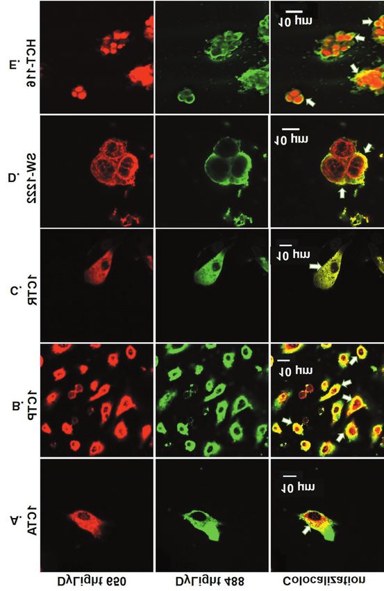
ANTICANCER RESEARCH 41: 27-42 (2021)

peptides is caused by their binding to the double minute-          Materials and Methods
binding protein of human or mouse origin (H/MDM-2) that is
uniquely expressed in the cell membranes of cancer cells at        Peptides and chemicals. The test peptide, PNC-27, whose sequence
significantly higher levels than that expressed on normal or       is H-Pro-Pro-Leu-Ser-Gln-Glu-Thr-Phe-Ser-Asp-Leu-Trp-Lys-
untransformed cells (7). Formation of this peptide-protein         Leu-Leu-Lys-Lys-Trp-Lys-Met-Arg-Arg-Asn-Gln-Phe-Trp-Val-Lys-
                                                                   Val-Gln-Arg-Gly-OH, contains the MDM-2 binding domain from
complex induces transmembrane pore formation resulting in
                                                                   human p53 and is comprised of p53 aa 12-26 (bold) linked to the
rapid tumor cell necrosis (6-8). Subsequent studies have           membrane penetrating sequence or membrane residency peptide
further confirmed that PNC-27 selectively induces tumor cell       (MRP) (italics). It was synthesized using a solid phase method with
necrosis by co-localizing with H/MDM-2 resulting in rapid          HPLC purity >95% (Biopeptides Corp, USA). The negative control
tumor cell necrosis (9-11).                                        peptide, PNC-29, whose sequence is H-Met-Pro-Phe-Ser-Thr-Gly-
   In a major in vivo and in vitro study of the effects of         Lys-Arg-Ile-Met-Leu-Gly-Glu-Lys-Lys-Trp-Lys-Met-Arg-Arg-Asn-
PNC-27 on stem cell-enriched human acute myelogenous               Gln-Phe-Trp-Val-Lys-Val-Gln-Arg-Gly-OH (aa sequence from
                                                                   human cytochrome p450 in bold, MRP sequence in italics) was also
leukemia (AML) cells from multiple patients with this
                                                                   synthesized by solid phase method with >95% HPLC purity
disease, identified by their expression with the stem cell         (Biopeptides Corp, USA). TritonX-100 and Bovine serum albumin
marker protein, CD-34, PNC-27 was found to be strongly             (BSA) were purchased from Sigma-Aldrich (St. Louis, MO, USA).
cytotoxic to each AML tumor, with IC50 values that                 Trypsin and Pen Strep were obtained from Life Technologies
correlated with the extent of expression of HDM-2 on the           (Grand Island, NY, USA). All other chemicals were purchased from
tumor cell surfaces (12). In addition, AML-cancer stem cell        Sigma Aldrich.
(CD-34-expressing)-enriched tumor cells were implanted in
                                                                   Cell culture. Modified human colonic epithelial cells (HCEC)
the bone marrows of nude mice that were treated with PNC-
                                                                   1CTA, 1CTP and 1CTR were a generous gift from Dr. Jerry W.
27 or the negative control peptide, PNC-29. After treatment,       Shay, University of Texas Southwestern, Dallas, TX, USA. Stem
bone marrow explants from these mice were transplanted             cells were cultured in 4:1 DMEM: Media 199 + 2% Cosmic calf
into the bone marrows of nude mice. White cell counts in           serum + 10 μg/ ml insulin + 20 μg/ ml human recombinant
the PNC-27-treated groups were found to be reduced to              epidermal growth factor + 1 μg/ml hydrocortisone + 2 μg/ml apo-
normal levels and their survival times were significantly          transferin + 0.05pM sodium selenite. The human stem-like colonic
longer than those for the negative control peptide-treated         progenitor cells, 1CTA and 1CTP, were derived from HCECs by
                                                                   shRNA knockdown of the APC (adenomatous polyposis coli) gene
group whose white cell counts remained elevated and
                                                                   and p53 respectively whereas, 1CTR are HCECs expressing
exhibited no off-target effects (12). These results suggested      KRASV12. HCECs have been reported to express significant levels
that PNC-27 is a potent agent that kills hematopoietic             of stem cell markers, CD29, CD44 and CD166 (17-19). The human
cancer stem cells, but does not affect normal cells, including     male colon cancer cell line, SW-1222, was a generous gift from
normal (non-malignant) hematopoietic stem cells as found           Meenhard Herlyn, Wistar Institute, Philadelphia, PA, USA, and
in our prior studies (5).                                          these cells were cultured in DMEM + 10% FBS + 1% pen/strep.
   In this article, we have extended our studies on the effects       The human male colon cancer cell line, HCT-116, the murine colon
                                                                   cancer cell line, CT-26, and the normal CCD-18Co human female
of PNC-27 on cancer stem cells to human solid tissue
                                                                   colonic fibroblast cell line were acquired from the American type
tumors, in particular human colon cancers. Like                    culture collection, Manassas, VA, USA. HCT-116 cells [containing
hematopoietic cancers, colon cancers have been found to            CD24+CD44+ CSC subpopulations, that cannot differentiate but result
contain tumor stem cells (13-16) as indicated by expression        in more aggressive and metastatic tumors (14)] were cultured in
of surface markers identifying these cell types, most notably      McCoy’s 5A + 10% FBS + 1% pen/strep, and CCD-18Co cells were
the glycoprotein, CD-44. This protein is expressed on colon        cultured in EMEM + 10% FBS + 1% pen/strep. All cells were
cancer stem cell surfaces in different splice variant forms, all   maintained in a humidified incubator with 5% CO2 at 37˚C.
                                                                      CT-26 cells were cultured in RPMI-1640 media supplemented
of which bind to hyaluronan to activate mitogenic signal
                                                                   with 10% fetal bovine serum (FBS) and 1% penicillin/streptomycin
transduction pathways. Solid tissue tumor cells expressing         (pen/strep). DMEM, RPMI, Media 199, FBS and cosmic calf serum
this protein, but not those that do not express this surface       were acquired from Thermo Fischer Scientific, Waltham, MA, USA.
protein, have been found to regenerate themselves and to
divide into a variety of more differentiated forms of tumor        Flow cytometry. Cells were harvested and washed in chilled
cells when transplanted into nude mice (13).                       Dulbecco’s phosphate buffered saline without Ca2+ and Mg2+
   We, herein, show that PNC-27 is effective in killing a          (DPBS) and diluted to have 100,000 cells/ 100 μl/ assay in chilled
variety of colon cancer cells, each of which contains              FACS buffer. Ten percent fetal bovine serum (FBS) + 2 mM
                                                                   ethylenediaminetetraacetic acid (EDTA) was added to DPBS to
significant CD-44 surface protein expression and colocalizes
                                                                   prepare FACS buffer. Cells were incubated with polyclonal rabbit
with surface membrane expression of H/MDM-2 on each                anti-HDM-2 (1:40) or mouse anti-CD44 (1:100) and isotype
cancer cell line but does not affect normal colonic fibroblast     controls for 1 h at 4˚C after blocking them in FACS buffer for 30
negative control cells, which do not express significant levels    min on ice. At the end of incubation, cells were washed 3X with
of membrane HDM-2.                                                 chilled FACS buffer and incubated with DyLight® 650 labeled goat

28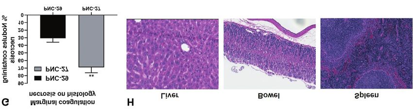
Thadi et al: Molecular Targeting of H/MDM-2 in Colon Cancer Cell Lines

anti-rabbit (1:250) and Alexa Fluor 488 labeled anti-mouse (1:500)     h, 50 μM, 0.5 μM; CT-26, 6 h, 50 μM, 5 μM. Following treatment,
antibodies at 4˚C, 1 h. Cells were then washed 3X with chilled         cells were harvested and washed in chilled DPBS. After
FACS buffer. Fluorescent signals from anti-CD44 and anti-MDM2          centrifugation, cells were diluted to 1×10⁶ cells/ml in chilled annexin
stained cells were measured using a BD Accuri™ C6 cytometer            V buffer to have 100,000 cells/100 μl/assay. Subsequently, cells were
system. Polyclonal rabbit anti-HDM-2 (catalog# AF1244) and rabbit      incubated with 5 μl FITC annexin V and 5 μl of 10 μg/ml propidium
isotype control (catalog# AB-105-C) were obtained from Novus           iodide (PI) for 15 min at room temperature. Stained cells were then
Biologicals, Centennial, CO, USA. Mouse isotype control (catalog#      analyzed using BD Accuri™ C6 cytometer system. Annexin V
sc3878) were obtained from Santa Cruz Biotechnology, CA, USA.          buffer, FITC annexin V reagent and propidium iodide were provided
Monoclonal mouse anti-CD44 (catalog# 156-3C11) was purchased           in the apoptosis detection kit from BD Biosciences (BD Biosciences,
from Thermo Fischer Scientific, MA, USA. Alexa Fluor 488 labeled       CA, USA).
anti-mouse antibody (catalog# ab96879) and DyLight® 650 labeled
goat anti-rabbit (catalog# ab96902) was obtained from Abcam,           Caspase activity assay. All cells were cultured at a concentration of
Cambridge, MA, USA.                                                    3-5×10⁶ cells in a T150 flask and grown to confluence. For each cell
                                                                       line, untreated cells (negative control), cells treated with staurosporine
Confocal microscopy. Colocalization experiments using confocal         that is known to induce apoptosis (positive control) and cells treated
microscopy were performed on all cell lines in a manner identical      with PNC-27 were assayed for caspase- 3 activity using Caspase-3
to the procedure used in a prior publication (10). Briefly, cells      Colorimetric Assay Kit (BioVision, Inc., Milpitas, CA, USA) as per
grown in confocal dishes overnight to 50-60% density were treated      the manufacturer’s instructions. Staurosporine was present at 1 μM
for 1h and maintained in 5% CO2 humidified air at 37˚C. 1CTA,          for CTA, CTP and SW-1222 cells, 2 μM for CTR cells, 0.5 μM for
1CTP, 1CTR and SW-1222 were treated with 60 μM of PNC-27               HCT cells, and 5 μM for CT-26 cells. PNC-27 was present at a
while HCT-116 and CT-26 were treated with 25 μM of PNC-27.             concentration of 80 μM and an incubation time of 24 h for 1CTA,
After 3X washes with 1X DPBS (containing Ca2+ and Mg2+), cells         1CTP, 1CTR and SW-1222 cells; a concentration of 50 μM and
were immediately fixed in 4% formaldehyde. Fixation was                incubation time of 24h for HCT116 cells and at a concentration of 50
performed for 10 min at 37˚C. Fixation was followed by blocking        μM and an incubation time of 10h for CT-26 cells.
in 2% BSA in 1X DPBS (containing Ca2+ and Mg2+) for 1 h at
25˚C. Cells were then incubated in polyclonal rabbit anti-MDM-2        In vivo studies. To test whether PNC-27 induces tumor cell necrosis
(catalog# AF1244) at a dilution of 1:10 and polyclonal mouse anti-     in vivo without affecting normal cells in tissues, we treated a
p53 (DO-1) at a dilution of 1:40 with shaking overnight at 4˚C.        murine colon cancer cell line (CT-26, see above in this section) that
Cells were washed 3X with 1XDPBS (containing Ca2+ and Mg2+)            was transplanted intraperitoneally into nude mice. Our animal
and incubated with polyclonal anti-rabbit red fluorophore (DyLight®    protocol, approved by the Drexel University Institutional Animal
650) and polyclonal anti-mouse green fluorophore (DyLight® 488)        Care and Use Committee (IACUC), was adhered to throughout.
at a dilution of 1:200 in the dark for 1 h at 25˚C. After 3X washes    Five to six-week-old female Nu/Nu nude mice were acclimatized
with 1X DPBS (containing Ca2+ and Mg2+) and nuclei staining with       for three to five days in the barrier housing where they were
DAPI, cell were visualized in confocal dishes under the Olympus        provided with food, bedding and water for the duration of the
Fluoview FV3000 confocal microscope (Olympus America INC,              experiment. Peritoneal carcinomatosis was induced by
Center Valley, PA, USA) at 60X resolution. Image analysis was          intraperitoneal (IP) injection of 2.5×10⁶ luciferase-transfected CT-
performed using cellSens software. Polyclonal mouse anti-p53 (DO-      26 (CT-26-Luc) cells in 500 μl of Dulbecco’s phosphate-buffered
1) was obtained from Santa Cruz Biotechnology, Santa Cruz, CA,         saline (DPBS). Lipofectamine 2000 Transfection Reagent (Thermo
USA and polyclonal anti-mouse green fluorophore (DyLight® 488)         Fisher Scientific, Rockford, IL, USA) was used to transfect CT-26
was obtained from Abcam Inc., Cambridge, MA, USA.                      cells with a firefly luciferase gene (luc2) containing plasmid
                                                                       (manufacturer protocol was followed). Stable transfection was
MTT cell proliferation assay, LDH cytotoxicity assay. Cells were       confirmed in vitro via bioluminescent imaging. In vivo, established
seeded in 24 well culture plate and grown to 70-80% confluence.        tumor in our murine colon cancer peritoneal carcinomatosis model
Cells were then treated with increasing concentrations of PNC-27       was monitored using the In Vivo Imaging System (IVIS) Lumina
and PNC-29 for 4 h. At the end of the treatment, MTT (3-[4,5-          XR series III [Perkin Elmer (Caliper), Waltham, MA]. The mice
dimethylthiazol-2yl]-2,5-diphenyl tetrazolium bromide) assay           were monitored via IVIS on days 1, 3, 5, 7 and 9. After
(Promega, Madison, WI) was carried out to determine the effect of      establishment of tumor on day 9 as evidenced by IVIS, mice were
PNC27/29 on cell proliferation. LDH (lactate dehydrogenase)            divided into two groups and treated as follows: Intraperitoneal (IP)
release from cells was determined using CytoTox96 assay                injection, once per 24 h, with 1. PNC-29 (100 μg/10 μl sterile
(Promega) with lysis buffer as a positive control. Manufacturer’s      water/ g mouse, negative control) and 2. PNC-27 (100 μg/10 μl
instructions were followed for both assays.                            sterile water/g mouse, treatment cohort) for 14 days. On day 24,
                                                                       mice were sacrificed via CO2 inhalation and tumors were excised.
Annexin V/PI apoptosis assay. Cells were seeded at a density of        Tumor size and extent of hemorrhage were quantified on
100,000 cells/well in 6-well plates and grown to 60-70% confluence.    necroscopy. Marginal coagulation necrosis was assessed using
Cells were then treated either with PNC-27 or an apoptosis inducer,    histological sections prepared from fixed tumor nodules from each
staurosporine. Incubation times and agent concentrations were          of the treatment groups. A treatment-blinded rater was used to
optimized to obtain the maximal level of apoptosis with the positive   examine necropsy specimens. An average of twenty-two tumor
control agent, staurosporine, for each cell line as follows,           nodules were examined per animal. Necrosis was quantified
respectively: CTA, 16 h, 80 μM, 1 μM; CTP, 6 h, 80 μM, 1 μM;           histologically using Aperio eSlide Manager (Leica Biosystems,
CTR, 6 h, 80 μM, 2 μM; SW122, 6h, 80 μM , 1 μM; HCT116, 16             Buffalo Grove, IL, USA).

                                                                                                                                              29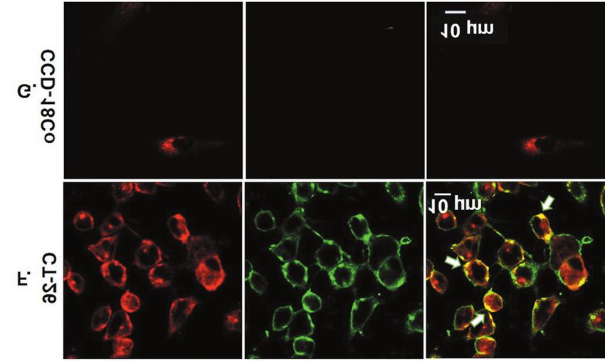
ANTICANCER RESEARCH 41: 27-42 (2021)

                                                                                                              Figure 1. Continued

Computational and statistical methods. GraphPad Prism 8.0              Results and Discussion
(GraphPad Software, La Jolla, CA, USA) was used to analyze all
quantitative assay results from flow cytometry, confocal microscopy,
MTT assays and LDH assays. Results are shown as means±1 SEM.
                                                                       Expression of CD44 and HDM-2 in colon cancer cell
Statistical significance between assay values was determined using     membranes. Human and murine colon cancer cells were
the two-tailed Student’s t-test. p-Values of 0.05 were considered to   validated for their heterogeneity and presence of colon
indicate statistically significant difference.                         cancer stem cell marker, CD44. CD44 has been widely

30
Thadi et al: Molecular Targeting of H/MDM-2 in Colon Cancer Cell Lines
Figure 1. Membrane expression of H/MDM-2 in CD44+ and CD44-subpopulations of colon cancer cells. Flow cytometry was utilized to identify
the expression of stem cell-associated marker, CD44 in colon cancer cells. In (A) I. 1CTA, (B) I. 1CTP, (C) I. 1CTR, (D) I. SW-1222, (E) I. HCT-
116 and (F) I. CT-26, cells were gated based on isotype controls and sorted into CD44+ and CD44– subpopulations. To evaluate levels of H/MDM-
2 in CD44+ and CD44– subpopulations of each cell, all cells were co-stained with H/MDM-2 as shown in panels II and III. CD44+ and CD44–
cells stained positively for membrane H/MDM-2 are shown in quadrant labeled as H/MDM-2+. Membrane H/MDM-2 was detected in all 6 colon
cancer cell lines and a significantly high percentage of CD44+ cells expressed membrane H/MDM-2. (G) I shows percentage of non-malignant/
normal CCD-18Co cells stained with membrane H/MDM-2 were negligible compared to colon cancer cells. Very few (about 1.4%) of these cells
were positive for HDM-2 which indicates that H/MDM-2 is specifically upregulated in malignant cells. Dot plots shown for each cell line are
representative plots; data are from 3 independent experiments.
studied as a CSC surface marker and utilized to segregate 2 was negligible in normal CCD-18Co cells (which
CSCs in many cancers (20). In our study, we found a contained no CD44 positive cells) (Figure 1G). This finding
significantly high fraction of CD44+ CSCs in various human suggests that membrane H/MDM-2 can be a potential
and murine colon cancer cells (~35-76%) as shown in Figure therapeutic target in CD44+ CSCs, which can ameliorate
1. Furthermore, high percentages of cells in all six-colon aggressive tumors that develop resistance to the conventional
cancer cell lines expressed H/MDM-2 in their cell chemotherapies without affecting normal cells.
membranes (lowest, 47% in HCT 116 cells, highest 92% in
CT 26 cells). Moreover, a high percentage of the CD44+ PNC-27 interacts with HDM-2 on the membrane of colon
CSCs were positively stained for the oncoprotein, membrane cancer cells. PNC-27 was designed to target overexpressed
H/MDM-2 (Figure 1A-F). In contrast, membrane H/MDM- membrane HDM-2 and promote cell death. Our aim was to
31ANTICANCER RESEARCH 41: 27-42 (2021)
Figure 2. Continued
32Thadi et al: Molecular Targeting of H/MDM-2 in Colon Cancer Cell Lines
Figure 2. Colocalization of PNC-27 with HDM-2 on the colon cancer cell plasma membrane. (A) 1CTA, (B) 1CTP, (C) 1CTR, (D) SW-1222, (G)
CCD-18Co were treated with 60 μM whereas (E) HCT-116 and (F) CT-26 cells were treated with 25 μM of PNC-27 for 1 h followed by imaging
under Olympus Fluoview FV3000 confocal microscope at 60× magnification. Green and red fluorescence correspond to DyLight® 488 anti-hu-
p53/PNC-27 and DyLight® 650 anti-H/MDM2, respectively. Yellow fluorescence was produced by the overlap of green and red emission signals at
the membrane of colon cancer cells. (G) Normal CCD-18Co cells did not emit any yellow fluorescence.
determine if membrane HDM-2 expressed on colon cancer In order to understand the mechanism of cell death, we
cells containing significant populations of cancer stem cells can measured the levels of necrotic, early apoptotic, and late
be a targeted by PNC-27. We performed co-localization studies apoptotic markers in these cells after treatment with PNC-
using confocal microscopy to determine if PNC-27 can interact 27. As shown in Figure 4A-F, 1CTA, 1CTP, 1CTR, SW-
with HDM-2 on the plasma membrane of cancer stem cells. As 1222, HCT-116 and CT-26 cells treated with increasing doses
shown in Figure 2, PNC-27, indicated by green fluorescence of PNC-27 for a 4 h incubation time resulted in LDH
and HDM-2, indicated by red fluorescence are both localized leakage, indicative of loss of membrane integrity, in a dose-
to the cell surface. Prominent yellow fluorescence on the cell dependent manner. Treatment with increasing doses of our
surface, resulting from direct overlap of green and red control peptide, PNC-29, did not yield an increase in LDH.
emission, clearly demonstrates co-localization of PNC-27 and CCD-18Co cells did not undergo necrosis when treated with
HDM-2 on the membrane of colon cancer cells. PNC-27 or PNC-29.
Importantly, for all six cancer cell lines, at specific
Binding of PNC-27 to HDM-2 results in cell death by concentrations of PNC-27, the level of LDH release was
necrosis and shows no sign of apoptotic activity. Further found to be the same as that for the lysis controls, i.e., the
studies were done to determine if the PNC-27-HDM-2 positive controls in which each cell line was lysed with lysis
complex on the cell surface of cancer cells will result in their buffer to determine the level of LDH representing total cell
cell death. The MTT cell viability assay was performed on lysis or total cell death. Thus, PNC-27 killed all tumor cells
these cells at different concentrations of PNC-27 and in each cell line, a finding that indicates that this peptide
negative control peptide PNC-29. As summarized in Figure killed all tumor (CD44-expressing) stem cells.
3A-F (black bar graphs), we observed a dose-dependent In contrast, results from the annexin V assay for apoptosis
decrease in cell viability of colon cancer cells treated with demonstrated no significant difference in detection of
PNC-27 for an incubation time of 4 h. This effect was not annexin V-positive cells in the PNC-27 treated group of
seen with our control peptide, PNC-29 (Figure 3A-F, gray cancer cells compared to stained untreated cells (Figure 5).
bar graphs). Additionally, we found that PNC-27 is not toxic Likewise, similar absence of caspase-3 activity in PNC-27-
to normal CCD-18Co cells (Figure 3G). treated cells compared to untreated cells further confirmed
33ANTICANCER RESEARCH 41: 27-42 (2021)
Figure 3. Effect of PNC-27 on cell viability of colon cancer cells. PNC-
27 decreased cell viability (MTT assay) in (A) 1CTA, (B) 1CTP, (C)
1CTR, (D) SW-1222, (E) HCT-116 and (F) CT-26 with increasing
concentrations after 4 h. PNC29 does not induce any cell killing. (G)
PNC-27 did not induce cell death in normal CCD-18Co cells. Cell
viability is presented as mean absorbance at 570 nm ±SEM as a
function of increasing concentrations of peptide. Cells were treated in
triplicates in each experiment. Data were generated from 3 independent
experiments.
34Thadi et al: Molecular Targeting of H/MDM-2 in Colon Cancer Cell Lines
Figure 4. PNC-27 induces cell death and results in LDH release in a
dose-dependent manner. Cells were incubated with increasing
concentrations of PNC-27 and negative control, PNC-29 for 4 h. PNC-
27 treatment resulted in cell death by necrosis as assessed by amount of
LDH released in media of (A) 1CTA, (B) 1CTP, (C) 1CTR, (D) SW-1222,
(E) HCT-116 and (F) CT-26. No significant amount of LDH was released
in media of CCD-18Co cells when treated with PNC-27 or PNC-29. Cell
death by necrosis is presented as mean absorbance at 490 nm ±SEM.
Data was generated from 3 independent experiments and each
experiment was carried out in triplicates.
35ANTICANCER RESEARCH 41: 27-42 (2021)
Figure 5. Continued
the cell death mechanism to be independent of apoptosis. On PNC-27 induces significant tumor-specific hemorrhagic
the other hand, our positive control, staurosporine, a known necrosis in vivo. To test whether PNC-27 induces tumor cell
activator of apoptosis, resulted in significant annexin V- necrosis without affecting normal cells in vivo, we
positive cell and caspase-3 activity (Figure 5). Therefore, established a murine colon cancer peritoneal carcinomatosis
PNC-27 induces its cytotoxic effects via necrosis model by IP injection of CT-26-Luc in athymic Nu/Nu mice.
independent of apoptosis, as we found for all of the other Tumor development was monitored via IVIS
cell lines that we have studied previously (5-11, 21). bioluminescence imaging and relative luminescence units
36Thadi et al: Molecular Targeting of H/MDM-2 in Colon Cancer Cell Lines
Figure 5. Continued
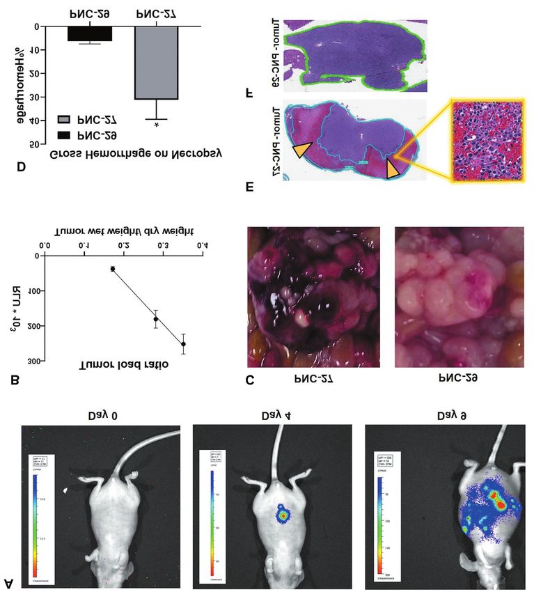
(RLU) were recorded as shown in Figure 6A and B. Once (Figure 6C, left) while tumors of mice treated with the
tumors were established, PNC-27 was tested for its efficacy negative control PNC-29 peptide showed significantly fewer
for inducing tumor-specific necrosis without damaging regions of hemorrhage that were also significantly smaller
normal cells. Treatment was initiated when the presence of than found in mice treated with PNC-27 (Figure 6C, right).
tumor was confirmed via IVIS. Upon necropsy on day 15 These results are quantitated in Figure 6D showing that
post treatment, tumors of mice treated with PNC-27 hemorrhagic necrosis was dramatically higher in the PNC-
demonstrated multiple peritoneal areas of gross hemorrhage 27-treated tumors.
37ANTICANCER RESEARCH 41: 27-42 (2021) Figure 5. Anti-tumor effect of PNC-27 does not induce apoptotic markers in the six colon cancer cell lines. Percent apoptosis induced in response to PNC- 27 treatment was determined by staining cells with FITC-annexin V to detect early apoptotic marker, phosphatidylserine using flow cytometry. The X-axis is the staining intensity for annexin V, and the Y-axis is the intensity of staining for propidium iodide (PI) that stains the cell nuclei. (A-D): For each of the six cell lines, four flow cytometry patterns are shown, labeled sequentially from left to right as (A-D). (A) is the pattern for untreated cells, not stained with either agent (annexin V or PI); (B) is the pattern for untreated cells that have been stained with both agents; (C) is the pattern for cells treated with staurosporine; and (D) is the pattern for cells treated with PNC-27. Percentages of staining patterns of cells are given for each cell line for each quadrant in the flow cytometric pattern. Percent cells positive for apoptosis, i.e., positive for annexin V and negative for PI, were detected in the lower right quadrant (Q-LR) for each cell line. (E) is a bar graph of % apoptotic cells (annexin V+ PI– cells) in the three groups: no treatment [corresponding to graph (B)], treated with staurosporine [corresponding to graph (C)] and treated with PNC-27 [corresponding to graph (D)]. Results are presented as mean of % annexin V+PI– cells ±SEM and were generated from 3 independent experiments. (F) is a bar graph summarizing caspase levels that were measured after treatment with PNC-27 in the same three groups, respectively, as described in (E) above. Caspase levels are represented as mean of caspase-3 activity at absorbance 405 nm±SEM. Data was generated from 3 independent experiments and ****p≤0.0001, ***p≤0.001 and **p≤0.01 in comparison to untreated cells. 38
Thadi et al: Molecular Targeting of H/MDM-2 in Colon Cancer Cell Lines

   Further demonstration of PNC-27-induced hemorrhagic               very likely due to pore formation given that the rise in
necrosis of the peritoneal tumor is shown in Figure 6E               cytosolic LDH was rapid and, at higher concentrations of
showing the histopathology of hemotoxylin-eosin stains of            peptide, reached maximal levels within four hours as observed
sections taken from the tumor. In this section, the eosin-           in prior studies (9-11). Our findings that PNC-27 induced
staining sections (red-colored areas enclosed by the blue            rapid cancer cell death without expression of early (annexin
boundary curve) show hemorrhagic necrosis while the                  V staining of exposed phospholipid) or late (caspase 3)
hemoxylin-staining (blue-colored) sections are tumor cells.          markers for apoptosis in any of the cell lines tested in this
Section 6E inset shows the same area at a higher                     study (Figures 5) strongly support tumor cell necrosis and not
magnification. Figure 6F shows that in tumors treated with the       apoptosis as the mechanism of cell death. Further evidence
negative control peptide, PNC-29, the histopathology on              that apoptosis was not the cause of PNC-27-induced cancer
similar sections through the tumor shows absence of any              cell death is that staurosporine, that is known to be a strong
hemorrhagic necrosis and only tumor cells. The results of            inducer of apoptosis, required from 10-24 h to induce total
histopathologic quantitation of the percentage of tumor              cancer cell death in each of the cell lines that we tested while
nodules showing hemorrhagic necrosis in mice treated with            PNC-27 induced total cancer cell death in 4 h for each cell
PNC-27 or with PNC-29 (negative control peptide) are shown           line tested and induced maximal release of LDH over this time
in Figure 6G in which it is clear that hemorrhagic necrosis is       course. In prior studies, we have found that PNC-27 actually
significantly more prominent in the PNC-27-treated mice.             induces immediate release of LDH into the medium
   Since PNC-27 was administered intraperitoneally, it was           suggesting early cancer cell membrane damage (5-11, 21).
possible that it exerted a general cytotoxic effect that affected    Electron microscopic analysis of these cancer cells treated
tumor and non-tumor cells i.e., off target effects. As shown in      with PNC-27 after several minutes of incubation shows
Figure 6H, histopathological sections taken from other major         transmembrane pore formation in these cells (6, 8). Although
tissues, i.e., liver, bowel and spleen, revealed no cytotoxic        the initial events in apoptosis leading to cell death can occur
effects on these tissues. Thus, the cell-killing effects of PNC-     early after an initiating event, cell death with membrane
27 in this in vivo model are specific for the tumor cells and        damage resulting in release of LDH requires hours to days
not normal cells showing no gross toxicity or apparent cell          (22).
death in healthy GI organs of PNC-27 treated mice.                      An important finding in this study was that a significant
                                                                     fraction of each of the cell lines expressed the CD44 protein
Conclusion                                                           marker for colonic stem cells. Since, as suggested by the cell
                                                                     viability and LDH release data shown in Figures 3 and 4,
PNC-27 is cytotoxic to all six colon cancer cell lines used in       respectively, at higher concentrations of PNC-27 for each cell
this study. This effect is specific to colon cancer cells since      line, there was 100 percent cell death, we conclude that PNC-
this peptide had no effect on the viability of control normal        27 is cytotoxic to colon cancer stem cells. Since it is also toxic
colonic fibroblasts (CCD-18Co cells). Specificity of PNC-27          to leukemia stem cells (12), it may be an effective agent
for killing tumor cells exclusively was further supported by         against tumor stem cells in general.
our findings in the in vivo peritoneal carcinomatosis model in          It is also of interest that PNC-27 was cytotoxic to six
nude mice in which this peptide induced widespread                   different cell lines that differed significantly phenotypically
hemorrhagic tumor cell necrosis of an intraperitoneally              and genotypically from one another. For example, as noted in
implanted syngeneic CT-26 colon cancer with no evidence of           the Materials and Methods section, HCT 116 cells express, in
cell damage to normal cells in a variety of tissues in these         addition to CD 44, CD 24 and, unlike the other cell lines,
mice.                                                                cannot differentiate but result in more aggressive and
   Furthermore, in our in vitro studies, all six-colon cancer cell   metastatic tumors. Human stem-like colonic progenitor cells,
lines expressed significant levels of H/MDM-2 in their               1CTA and 1CTP, were derived from HCECs by shRNA
membranes (Figure 1) while the normal control cells                  knockdown of APC and p53 respectively whereas, 1CTR are
expressed barely detectable membrane levels of this protein          HCECs expressing KRASV12. Thus, the cytotoxic effects of
suggesting that cancer cell membrane expression of this              PNC-27 on these cells is independent of the pathways
protein is important for the cytotoxic effects of PNC-27 on          involved in cell transformation and very likely result from the
these cancer cells. As found in a number of previous studies,        observation that all cell lines express H/MDM-2 in their
PNC-27 colocalized with H/MDM-2 in the membranes of the              membranes enabling PNC-27 binding to HDM-2 with
colon cancer cells (Figure 2). As shown in these previous            subsequent transmembrane pore formation.
studies, binding of PNC-27 to HDM-2 in cancer cell
membranes induces the formation of transmembrane pores               Conflicts of Interest
resulting in cell death (6, 8, 12). This mechanism of PNC-27-
induced cancer cell death for the cells tested in this study was     The Authors have no conflicts of interest to declare.

                                                                                                                                    39
ANTICANCER RESEARCH 41: 27-42 (2021)
Figure 6. Continued
Authors’ Contributions EMG conducted experiments, acquired, and analyzed the data. DSF,
LS and PMC analyzed the data and reviewed the manuscript. WBB,
AT designed and conducted to experiments, acquired and analyzed BL and MRP designed experiments, analyzed the data, wrote the
the data, wrote the article. WFM, MK, BDB, EG, MFS, YP, and article and provided administrative support.
40Thadi et al: Molecular Targeting of H/MDM-2 in Colon Cancer Cell Lines Figure 6. Effect of PNC-27 in vivo on CT-26 peritoneal carcinomatosis (CT-26 inoculated mice). Nu/Nu mice were inoculated with CT-26-Luc cells and IVIS was used to detect the bioluminescent signal from tumors (See Materials and Methods Section). (A) Shows tumor development in nude mice monitored using IVIS on day 0, 4 and 9. (B) Shows tumor development in Nu/Nu mice represented by RLU measured by IVIS as a function of tumor wet weight/ dry weight. (C) Left panel shows necrotic hemorrhage of tumors from mice treated with PNC-27; Right panel: there was no hemorrhage seen in tumors from PNC-29 treated mice. (D) The bar graph shows the histomorphology analysis of tumors with significantly higher percent gross hemorrhage of tumors treated with PNC-27 as compared with tumors treated with PNC-29. Percent gross hemorrhage was calculated as % area of necrosis relative to area of full specimen. (E) Histopathology of a peritoneal tumor nodule in PNC-27-treated mouse showing marginal coagulation necrosis (eosinophilic area) surrounding area of tumor (hematoxylin or blue stain). E-inset is a higher power view of (E) showing hemorrhagic necrosis. (F) section of a typical tumor nodule taken from the peritoneum of a PNC-29 treated mouse showing only tumor cells and no necrosis. (G) Bar graph represents relatively higher percentage of nodules containing necrosis from PNC-27-treated mice as compared with PNC-29-treated mice. Results are represented as mean±SEM with **p≤0.01 and *p
ANTICANCER RESEARCH 41: 27-42 (2021)

     Oncol 15(12): 3588-3600, 2008. PMID: 18931881. DOI:                 16 Botchkina IL, Rowehl RA, Rivadeneira DE, Karpeh MS Jr.,
     10.1245/s10434-008-0147-0                                              Crawford H, Dufour A, Ju J, Wang Y, Leyfman Y and Botchkina
9    Thadi A, Gleeson EM, Khalili M, Shaikh MF, Goldstein E,                GI: Phenotypic subpopulations of metastatic colon cancer stem
     Morano WF, Daniels LM, Grandhi N, Glatthorn H, Richard SD,             cells: Genomic analysis. Cancer Genomics Proteomics 6(1): 19-
     Campbell PM, Sarafraz-Yazdi E, Pincus MR and Bowne WB:                 29, 2009. PMID: 19451087.
     Anti-cancer tumor cell necrosis of epithelial ovarian cancer cell   17 Graillot V, Dormoy I, Dupuy J, Shay JW, Huc L, Mirey G and
     lines depends on high expression of hdm-2 protein in their             Vignard J: Genotoxicity of cytolethal distending toxin (cdt) on
     membranes. Ann Clin Lab Sci 50(5): 611-624, 2020. PMID:                isogenic human colorectal cell lines: Potential promoting effects
     33067207.                                                              for colorectal carcinogenesis. Front Cell Infect Microbiol 6: 34-
10   Thadi A, Lewis L, Goldstein E, Aggarwal A, Khalili M, Steele           34, 2016. PMID: 27047802. DOI: 10.3389/fcimb.2016.00034
     L, Polyak B, Seydafkan S, Bluth MH, Ward KA, Styler M,              18 Roig AI, Eskiocak U, Hight SK, Kim SB, Delgado O, Souza RF,
     Campbell PM, Pincus MR and Bowne WB: Targeting membrane                Spechler SJ, Wright WE and Shay JW: Immortalized epithelial
     hdm-2 by pnc-27 induces necrosis in leukemia cells but not in          cells derived from human colon biopsies express stem cell
     normal hematopoietic cells. Anticancer Res 40(9): 4857-4867,           markers and differentiate in vitro. Gastroenterology 138(3):
     2020. PMID: 32878773. DOI: 10.21873/anticanres.14488                   1012-1021.e1011-1015, 2010. PMID: 19962984. DOI:
11   Davitt K, Babcock BD, Fenelus M, Poon CK, Sarkar A, Trivigno           10.1053/j.gastro.2009.11.052
     V, Zolkind PA, Matthew SM, Grin’kina N, Orynbayeva Z,               19 Eskiocak U, Kim SB, Ly P, Roig AI, Biglione S, Komurov K,
     Shaikh MF, Adler V, Michl J, Sarafraz-Yazdi E, Pincus MR and           Cornelius C, Wright WE, White MA and Shay JW: Functional
     Bowne WB: The anti-cancer peptide, pnc-27, induces tumor cell          parsing of driver mutations in the colorectal cancer genome
     necrosis of a poorly differentiated non-solid tissue human             reveals numerous suppressors of anchorage-independent growth.
     leukemia cell line that depends on expression of hdm-2 in the          Cancer Res 71(13): 4359-4365, 2011. PMID: 21527559. DOI:
     plasma membrane of these cells. Ann Clin Lab Sci 44(3): 241-           10.1158/0008-5472.Can-11-0794
     248, 2014. PMID: 25117093.                                          20 Biddle A, Gammon L, Fazil B and Mackenzie IC: Cd44 staining
12   Wang H, Zhao D, Nguyen LX, Wu H, Li L, Dong D, Troadec E,              of cancer stem-like cells is influenced by down-regulation of
     Zhu Y, Hoang DH, Stein AS, Al Malki M, Aldoss I, Lin A,                cd44 variant isoforms and up-regulation of the standard cd44
     Ghoda LY, McDonald T, Pichiorri F, Carlesso N, Kuo YH,                 isoform in the population of cells that have undergone epithelial-
     Zhang B, Jin J and Marcucci G: Targeting cell membrane hdm2:           to-mesenchymal transition. PLoS One 8(2): e57314, 2013.
     A novel therapeutic approach for acute myeloid leukemia.               PMID: 23437366. DOI: 10.1371/journal.pone.0057314
     Leukemia 34(1): 75-86, 2020. PMID: 31337857. DOI: 10.1038/          21 Pincus MR, Fenelus M, Sarafraz-Yazdi E, Adler V, Bowne W
     s41375-019-0522-9                                                      and Michl J: Anti-cancer peptides from ras-p21 and p53
13   Thapa R and Wilson GD: The importance of cd44 as a stem cell           proteins. Curr Pharm Des 17(25): 2677-2698, 2011. PMID:
     biomarker and therapeutic target in cancer. Stem Cells Int 2016:       21728981. DOI: 10.2174/138161211797416075
     2087204, 2016. PMID: 27200096. DOI: 10.1155/2016/2087204            22 Green DR: Apoptotic pathways: Ten minutes to dead. Cell
14   Yeung TM, Gandhi SC, Wilding JL, Muschel R and Bodmer                  121(5): 671-674, 2005. PMID: 15935754. DOI: 10.1016/j.cell.
     WF: Cancer stem cells from colorectal cancer-derived cell lines.       2005.05.019
     Proc Natl Acad Sci USA 107(8): 3722-3727, 2010. PMID:
     20133591. DOI: 10.1073/pnas.0915135107
15   Liu Y and Bodmer WF: Analysis of p53 mutations and their
     expression in 56 colorectal cancer cell lines. Proc Natl Acad Sci                                       Received November 1, 2020
     USA 103(4): 976-981, 2006. PMID: 16418264. DOI: 10.1073/                                                Revised November 16, 2020
     pnas.0510146103                                                                                        Accepted November 22, 2020

42
You can also read