Enhanced Hybridization Selectivity Using Structured GammaPNA Probes - MDPI
←
→
Page content transcription
If your browser does not render page correctly, please read the page content below
molecules
Article
Enhanced Hybridization Selectivity Using Structured
GammaPNA Probes
Taylor D. Canady 1,† , April S. Berlyoung 1,† , Joe A. Martinez 1 , Cole Emanuelson 1 ,
Cheryl A. Telmer 2 , Marcel P. Bruchez 1,2 and Bruce A. Armitage 1, *
 1   Department of Chemistry and Center for Nucleic Acids Science and Technology, Carnegie Mellon University,
     4400 Fifth Avenue, Pittsburgh, PA 15213-3890, USA; canmatics@gmail.com (T.D.C.);
     april.berlyoung@gmail.com (A.S.B.); joe.martinez.jam@gmail.com (J.A.M.);
     emanuelsonc@gmail.com (C.E.); bruchez@cmu.edu (M.P.B.)
 2   Department of Biological Sciences, Carnegie Mellon University, 4400 Fifth Avenue,
     Pittsburgh, PA 15213-3890, USA; ctelmer@cmu.edu
 *   Correspondence: army@cmu.edu
 †   These authors contributed equally to this work.

 Academic Editor: Katherine Seley-Radtke

 Received: 30 January 2020; Accepted: 18 February 2020; Published: 21 February 2020

 Abstract: High affinity nucleic acid analogues such as gammaPNA (γPNA) are capable of invading
 stable secondary and tertiary structures in DNA and RNA targets but are susceptible to off-target
 binding to mismatch-containing sequences. We introduced a hairpin secondary structure into
 a γPNA oligomer to enhance hybridization selectivity compared with a hairpin-free analogue.
 The hairpin structure features a five base PNA mask that covers the proximal five bases of the
 γPNA probe, leaving an additional five γPNA bases available as a toehold for target hybridization.
 Surface plasmon resonance experiments demonstrated that the hairpin probe exhibited slower
 on-rates and faster off-rates (i.e., lower affinity) compared with the linear probe but improved single
 mismatch discrimination by up to a factor of five, due primarily to slower on-rates for mismatch vs.
 perfect match targets. The ability to discriminate against single mismatches was also determined in
 a cell-free mRNA translation assay using a luciferase reporter gene, where the hairpin probe was
 two-fold more selective than the linear probe. These results validate the hairpin design and present a
 generalizable approach to improving hybridization selectivity.

 Keywords: γPNA; antisense; hybridization; selectivity

1. Introduction
     High affinity recognition and accurate discrimination of nucleic acids is the foundation for
many biotechnology tools and biomedical applications ranging from PCR-based diagnostics and
fluorescence in situ hybridization (FISH) to antisense blocking of translation and genome editing.
Nevertheless, the enduring challenge of technologies that rely on nucleic acid hybridization is the
trade-off between affinity and selectivity [1]. The affinity of an oligonucleotide probe for its DNA or
RNA target can be fine-tuned by adjusting the length and/or the nucleobase composition. On the other
hand, optimizing the selectivity of a probe for its target is much more difficult due to the demand for
discriminating single mismatches. Making the probe short so that a mismatch prevents hybridization
at the experimental temperature raises the likelihood that other perfectly matched sequences will
be present elsewhere in the genome/transcriptome, leading to off-target hybridization. Making the
probe long in order to find a unique sequence also enables off-target hybridization due to the modest
destabilizing effect of a single mismatch in a high-affinity probe.

Molecules 2020, 25, 970; doi:10.3390/molecules25040970                        www.mdpi.com/journal/molecules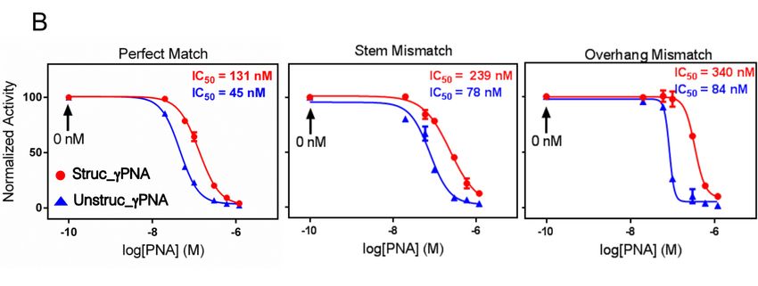
secondary structure into the probe [2–4]. Thus, intramolecular base pairing within the probe
competes with intermolecular base pairing to the target. This naturally decreases the affinity of the
structured probe for its target, relative to a corresponding unstructured probe, but it increases the
sensitivity to a single mismatch.
The simplest type of nucleic acid secondary structure is the hairpin, in which two remote
Molecules 2020, 25, 970 2 of 12
complementary sequences form a stem through Watson–Crick base pairing, extruding the
intervening nucleotides into a loop. One side of the stem, i.e., the probe, is complementary to the
The
target, quest
while thetoother
blockside
off-target hybridization
of the stem serves ashas
the led to many
“mask”, whichinnovative
providesapproaches
the competitionthat vary with
(Scheme
the application. Of relevance to the work we report here is the strategy of
1). The ability of a hairpin secondary structure to improve hybridization selectivity was first reported intentionally building a
secondary structure
in the context into the probe
of fluorescent [2–4]. beacons
molecular Thus, intramolecular
[2,3] and hasbase sincepairing within thetoprobe
been extended othercompetes
types of
with intermolecular
hybridization probesbase [4]. pairing to the
In addition, target. This naturally
intermolecular maskingdecreases
of a probethe affinityhybridization
through of the structuredto a
probe for its target, relative to a corresponding unstructured probe, but it
competing oligonucleotide in order to improve selectivity has also been reported (Scheme 1) [5,6]. increases the sensitivity to a
singleAsmismatch.
chemists have developed new generations of synthetic oligonucleotides, significant affinity
The simplest
improvements havetypebeenofrealized.
nucleic Peptide
acid secondary
nucleic acidstructure
(PNA)isisthe onehairpin,
such example in which two
[7,8], remote
where the
complementary
high affinity and sequences form a stem
the biochemical through
stability Watson–Crick
have base pairing,
led to numerous extrudingincluding
applications, the intervening
PCR-
nucleotides
clamping [9]into andabacterial
loop. One side of the[10],
genotyping stem, i.e.,even
with the probe,
higheris complementary
affinity reported for to athe target, while the
second-generation
other
PNA side
known of theas stem serves as (γPNA)
gammaPNA the “mask”, which
[11,12], provides
which the competition
features a helix-inducing(Scheme 1). The ability
substituent at theof
a hairpincarbon
gamma secondary of thestructure to improve
PNA backbone. hybridization
A 10mer γPNA binds selectivity
to its was first reported
complementary DNAin the context
and RNA
of fluorescent
targets with mid-fMmolecular beacons
Kd values [2,3]
[12]. Theand hasaffinity
high since been
of γPNAextended
allowstoitother typesgenomic
to invade of hybridization
DNA in
probes
order to[4].
bind Inits
addition, intermolecular
target site, masking ofinainprobe
leading to applications through
vivo gene hybridization
editing [13,14] andtotelomere
a competing
FISH
oligonucleotide
[15,16]. in order to improve selectivity has also been reported (Scheme 1) [5,6].
Use of
Scheme 1. Use of aa masking
masking oligonucleotide to improve selectivity of hybridization probes.
As chemists
While have developed
high affinity is essentialnew generationsrequiring
for applications of synthetic oligonucleotides,
invasion significant
of genomic DNA, use ofaffinity
γPNA
improvements have been realized. Peptide nucleic acid (PNA) is one such example [7,8], where
in other contexts where the target site is more accessible (e.g., antisense) is susceptible to off-targetthe high
affinity and to
effects due thethe
biochemical stability
tolerance for singlehave led to numerous
mismatches. applications,
This report describesincluding PCR-clamping
our efforts [9]
to use a hairpin
and bacterial genotyping [10], with even higher affinity
secondary structure to improve the selectivity of a γPNA probe. reported for a second-generation PNA known
as gammaPNA (γPNA) [11,12], which features a helix-inducing substituent at the gamma carbon of
the PNA backbone. A 10mer γPNA binds to its complementary DNA and RNA targets with mid-fM
2. Results
Kd values [12]. The high affinity of γPNA allows it to invade genomic DNA in order to bind its target
2.1. Hairpin
site, leadingDesign and Characterization
to applications in in vivo gene editing [13,14] and telomere FISH [15,16].
While high affinity is essential for applications requiring invasion of genomic DNA, use of γPNA
To test the impact of secondary structure on γPNA hybridization selectivity, we designed
in other contexts where the target site is more accessible (e.g., antisense) is susceptible to off-target
struc_γPNA, which consists of a 10 nucleotide (nt) target-recognition domain composed of γPNA
effects due to the tolerance for single mismatches. This report describes our efforts to use a hairpin
monomers, an octaethylene glycol (EG8) linker, and a 5 nt PNA mask complementary to the last 5 nt
secondary structure to improve the selectivity of a γPNA probe.
of the target-recognition domain (Table 1). The probe should fold into a hairpin structure [17] with a
five
2. base pair (bp) γPNA-PNA stem, an EG8 loop, and a 5 nt γPNA overhang, or “toehold”, where
Results
hybridization to a target can be nucleated (Scheme 2) [18]. Using PNA rather than γPNA as the mask
should
2.1. decrease
Hairpin Designtheand
stability of the stem [19], thereby increasing the likelihood that binding to a target
Characterization
nucleic acid will result in opening of the stem to form a stable duplex. Designing the probe to have
To test the impact of secondary structure on γPNA hybridization selectivity, we designed
struc_γPNA, which consists of a 10 nucleotide (nt) target-recognition domain composed of γPNA
monomers, an octaethylene glycol (EG8 ) linker, and a 5 nt PNA mask complementary to the last
5 nt of the target-recognition domain (Table 1). The probe should fold into a hairpin structure [17]
with a five base pair (bp) γPNA-PNA stem, an EG8 loop, and a 5 nt γPNA overhang, or “toehold”,
where hybridization to a target can be nucleated (Scheme 2) [18]. Using PNA rather than γPNA as the
mask should decrease the stability of the stem [19], thereby increasing the likelihood that binding to a
Molecules 2020, 25, 970                                                                                   3 of 12

target nucleic acid will result in opening of the stem to form a stable duplex. Designing the probe to
have an overhang should minimize any penalty in hybridization kinetics compared with a traditional
molecular beacon design, where the stem is fully base-paired. The unstructured probe (unstruc_γPNA)
consisting solely of the 10 nt γPNA target-recognition domain was also synthesized for comparison.

                            Table 1. GammaPNA (γPNA) probes and DNA targets.

                    Sequence Name                         Sequence a,b                    Mismatch
                       struc_γPNA           H2 N-K-TCTGGGTTCG-EG8 -cgaac-H          N/A
                      unstruc_γPNA               H2 N-K-TCTGGGTTCG-H                N/A
                   Perfect Match (PM)              50 -AGACCCAAGC-30                N/A
               Stem Mismatch (S-MM-7T)             50 -AGACCCTAGC-30                (T-T)
Molecules 2020,Stem
                25, x Mismatch (S-MM-6A)
                      FOR PEER REVIEW             50 -AGACCAAAGC-30                (A-G)        3 of 13
              Stem Mismatch (S-MM-7G)              50 -AGACCCGAGC-30               (G-T)
           Overhang Mismatch (O-MM-4A)            50 -AGAACCAAGC-30                (A-G)
an overhang should minimize any penalty in hybridization         kinetics compared with a traditional
           Overhang Mismatch (O-MM-3T)             50 -AGTCCCAAGC-30                (T-T)
molecular       beaconMismatch
           Overhang       design,(O-MM-4T)
                                   where the stem 5is   fully base-paired.
                                                    0 -AGATCCAAGC-3   0     The unstructured
                                                                                   (T-G)       probe
(unstruc_γPNA)
      a γPNA upperconsisting    solely of the 10 nt γPNA target-recognition domain was also synthesized
                     case, PNA lower case, sequences written C-to-N. (The C-terminus is an amide, hence “H2 N”).
for comparison.
      b Mismatches in DNA targets underlined.

     Scheme 2. Chemical structures of PNA and γPNA and design of structured and unstructured
     Scheme 2. Chemical structures of PNA and γPNA and design of structured and unstructured γPNA
     γPNA probes.
     probes.
     Intramolecular folding of struc_γPNA was confirmed by UV melting studies, in which a melting
transition was observed atTable
                             Tm =1.40
                                    GammaPNA
                                      ◦ C at both (γPNA)
                                                  0.8 and probes
                                                           8.0 µMand   DNA
                                                                   strand     targets.
                                                                            concentrations (Figure 1). The lack
of a concentration Sequence
                     dependence
                              Name for the transition is consistent
                                                             Sequencewith formation Mismatch
                                                                        a,b              of an intramolecular
hairpin as opposed to    an intermolecular duplex.
                      struc_γPNA                        Although γPNAs 8generally
                                                H2N-K-TCTGGGTTCG-EG           -cgaac-H exhibit
                                                                                            N/A hyperchromic
transitions near 40 ◦unstruc_γPNA                     H2N-K-TCTGGGTTCG-H
                     C due to melting of their intrinsic    helical structure (even when N/Atheir sequences do
                  Perfect Match  (PM)                   5′-AGACCCAAGC-3′                    N/A
not allow hairpin formation), kinetic data presented below are consistent with hairpin formation by
              Stem
struc_γPNA. For     Mismatch (S-MM-7T)
                  comparison,                            a similar hairpin consisting of a(T-T)
                                we recently reported 5′-AGACCCTAGC-3′                       4 bp γPNA-γPNA
              Stem Mismatch (S-MM-6A)                   5′-AGACCAAAGC-3′                   (A-G)
stem and a peptide loop, which melted at 76 ◦ C [20].
                Stem Mismatch (S-MM-7G)                 5′-AGACCCGAGC-3′                   (G-T)
              Overhang Mismatch (O-MM-4A)               5′-AGAACCAAGC-3′                   (A-G)
              Overhang Mismatch (O-MM-3T)               5′-AGTCCCAAGC-3′                   (T-T)
              Overhang Mismatch (O-MM-4T)               5′-AGATCCAAGC-3′                   (T-G)
     aγPNA upper case, PNA lower case, sequences written C-to-N. (The C-terminus is an amide, hence
     “H2N”). b Mismatches in DNA targets underlined.

     Intramolecular folding of struc_γPNA was confirmed by UV melting studies, in which a melting
transition was observed at Tm = 40 °C at both 0.8 and 8.0 μM strand concentrations (Figure 1). The
lack of a concentration dependence for the transition is consistent with formation of an intramolecular
hairpin as opposed to an intermolecular duplex. Although γPNAs generally exhibit hyperchromic
transitions near 40 °C due to melting of their intrinsic helical structure (even when their sequences
do not allow hairpin formation), kinetic data presented below are consistent with hairpin formation
by struc_γPNA. For comparison, we recently reported a similar hairpin consisting of a 4 bp γPNA-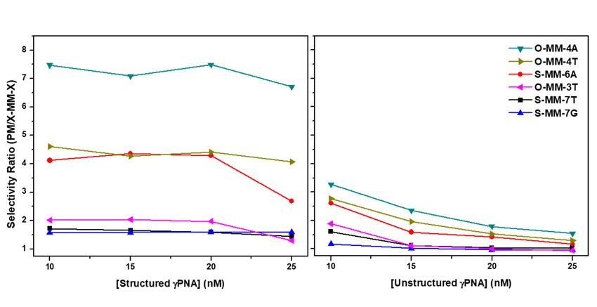
Molecules 2020, 25, 970 4 of 12
Molecules 2020, 25, x FOR PEER REVIEW 4 of 13
Figure 1. UV melting transition observed at 275 nm for struc_γPNA is independent of strand concentration
Figure 1. UV melting transition observed at 275 nm for struc_γPNA is independent of strand
over a 10-fold range. Buffer contained 10 mM Tris-HCl (pH = 7.4), 100 mM NaCl, 0.1 mM Na2 EDTA.
concentration over a 10-fold range. Buffer contained 10 mM Tris-HCl (pH = 7.4), 100 mM NaCl, 0.1
mM Na2EDTA.Selectivity Analyzed by SPR
2.2. Hybridization
We used surface
2.2. Hybridization plasmon
Selectivity Analyzedresonance
by SPR (SPR) experiments to characterize hybridization of the
structured and the unstructured γPNA probes to DNA targets that were either perfectly matched
to the We10mer
used recognition
surface plasmon domain resonance (SPR)(PM)
of the probes experiments
or wouldtoform characterize
a single hybridization
mismatch to various of the
structured and the unstructured γPNA probes to DNA targets that
positions within either the stem or the overhang region of the structured probe. For each experiment, were either perfectly matched to
the 10mer recognition domain of the probes (PM) or would
a biotinylated version of the DNA target was immobilized on a streptavidin-coated chip, and a form a single mismatch to various
positions
solution of within
eithereither the stemor
struc_γPNA orunstruc_γPNA
the overhang region of theconcentrations
at various structured probe. wasFor eachover
flowed experiment,
the chip.
aThe
biotinylated version of the DNA target was immobilized
sequences of the DNA targets are given in Table 1 and are named so as to indicate the on a streptavidin-coated chip, and of
location a
solution
the mismatchof either struc_γPNA
in either the stem or (S)
unstruc_γPNA
or the overhang at various
(O), and concentrations was flowed
the specific position andover the chip.
the mutation
The sequences of the DNA targets are
(e.g., MM-7T indicates the base at position seven is a T). given in Table 1 and are named so as to indicate the location
of theFigure
mismatch in either the stem (S) or the overhang (O), and the
2 shows SPR sensorgrams for hybridization of 10 nM struc_γPNA (A) and unstruc_γPNA specific position and the mutation
(e.g., MM-7T
(B) to the various indicates
DNAthe base at
targets. position
It is seven is
immediately a T). that the on-rate for the structured probe is
apparent
Figure 2 shows SPR sensorgrams
ca three-fold lower than for the unstructured probe. Two for hybridization of 10factors
nM struc_γPNA (A) andtounstruc_γPNA
likely contribute this difference.
(B)
First, the unstructured probe can nucleate via any subset of its 10 nucleotides, the
to the various DNA targets. It is immediately apparent that the on-rate for whichstructured probe is
are all accessible
ca
to three-fold
the target, lowerwhereas thanthefor PNAthe mask
unstructured probe. Two
on the structured factors
probe blockslikely contribute
access to five ofto the
thisnucleotides.
difference.
First, the unstructured probe can nucleate via any subset of its
Second, even if nucleation between struc_γPNA and a DNA target does occur via the toehold, 10 nucleotides, which are all accessible
the time
to the target,
required whereas
to open the PNAand
the hairpin mask form on additional
the structured baseprobe
pairs blocks accessthe
to stabilize to duplex
five of the
couldnucleotides.
allow the
Second,
probe toeven if nucleation
dissociate from thebetweenimmobilized struc_γPNA
target. and a DNA target does occur via the toehold, the
time required to open the hairpin and
There is also a smaller but measurable difference form additionalin base pairsfor
off-rate to the
stabilize the duplex
two probes with could allow
their perfect
the
matchprobe to dissociate
targets. from thephase
The dissociation immobilized target.
of the experiment, where buffer rather than probe is flowing over
There is also a smaller but measurable
the surface of the chip, begins at ca. 420 s. The unstructured difference in off-rate
probe forshows
the two probes
very littlewith their perfect
dissociation from
match targets. The dissociation phase of the experiment, where
the PM target, consistent with prior reports of γPNA kinetics [12,21]. In contrast, the structured buffer rather than probe is flowing
probe
over
clearlythebegins
surface toof the chip,from
dissociate begins theatchip
ca. 420 s. The
surface as unstructured
soon as the buffer probe showsflowing
begins very little
overdissociation
the surface;
from
Figure S1 (Supplementary Materials) shows an overlay of the two dissociation curves. the
the PM target, consistent with prior reports of γPNA kinetics [12,21]. In contrast, Thestructured
difference
probe
likely clearly begins to
arises because thedissociate
struc_γPNA fromcan thereform
chip surface as soon
the hairpin as the buffer
structure begins flowing
as it dissociates from the overDNAthe
surface; Figure S1 (Supplementary Materials) shows
target, slowing its re-association and leading to net loss from the chip surface.an overlay of the two dissociation curves. The
difference
Comparedlikely toarises becausematch
the perfect the struc_γPNA can reform
target, all mismatch the hairpin
targets showedstructure as it dissociates
slower hybridization forfrom
both
the DNA target, slowing its re-association and leading to net loss
the structured and the unstructured probes. In order to compare the selectivities of the two probes,from the chip surface.
we divided the maximum response units (determined at t = 420 s) for the perfect match targets by those
for each mismatch target. The resulting selectivity ratios (SR) are plotted in Figure 3 for each of the
γPNA concentrations tested. The improved selectivity for the structured probe is evident, with higher
SR values for each target. Moreover, the SR is invariant in the range of 10–20 nM for struc_γPNAMolecules 2020, 25, 970 5 of 12
but decreases nearly two-fold for unstruc_γPNA. Overall, these results demonstrate that the hairpin
Molecules 2020, 25, x FOR PEER REVIEW 5 of 13
secondary structure of struc_γPNA leads to significant improvement in mismatch discrimination.
Figure 2. Surface plasmon resonance (SPR) sensorgrams for 10 nM struc_γPNA (A) and unstruc_γPNA
(B). DNA target sequences are given in Table 1. All data are plotted as n = 3 average ± SD.
Compared to the perfect match target, all mismatch targets showed slower hybridization for
both the structured and the unstructured probes. In order to compare the selectivities of the two
probes, we divided the maximum response units (determined at t = 420 s) for the perfect match targets
by those for each mismatch target. The resulting selectivity ratios (SR) are plotted in Figure 3 for each
of the γPNA concentrations tested. The improved selectivity for the structured probe is evident, with
higher SR values for each target. Moreover, the SR is invariant in the range of 10–20 nM for
struc_γPNA but decreases nearly two-fold for unstruc_γPNA. Overall, these results demonstrate that
the hairpin secondary structure of struc_γPNA leads to significant improvement in mismatch
discrimination.
Figure 2. Surface plasmon resonance (SPR) sensorgrams for 10 nM struc_γPNA (A) and unstruc_γPNA
Figure
(B). DNA2.target
Surface plasmonare
sequences resonance
given in(SPR)
Tablesensorgrams forplotted
1. All data are 10 nM as n = 3 average
struc_γPNA (A) ±
and
SD.unstruc_γPNA
(B). DNA target sequences are given in Table 1. All data are plotted as n = 3 average ± SD.
A B
Compared to the perfect match target, all mismatch targets showed slower hybridization for
both the structured and the unstructured probes. In order to compare the selectivities of the two
probes, we divided the maximum response units (determined at t = 420 s) for the perfect match targets
by those for each mismatch target. The resulting selectivity ratios (SR) are plotted in Figure 3 for each
of the γPNA concentrations tested. The improved selectivity for the structured probe is evident, with
higher SR values for each target. Moreover, the SR is invariant in the range of 10–20 nM for
struc_γPNA but decreases nearly two-fold for unstruc_γPNA. Overall, these results demonstrate that
the hairpin secondary structure of struc_γPNA leads to significant improvement in mismatch
discrimination.
A B
Figure 3. Selectivity ratio of (A) struc_γPNA and (B) unstruc_γPNA as determined by dividing the
Figure 3.
association Selectivity
max ratio of (A)
(max response struc_γPNA
units and (B) 1unstruc_γPNA
(RU) in Figures and 2) of the as determined
perfect matchby to dividing the
each respective
association max (max response
target mutant association max. units (RU) in Figures 1 and 2) of the perfect match to each respective
target mutant association max.
It is clear from the SPR data that the position of the mismatch has a significant effect on the
It is clear from the SPR data that the position of the mismatch has a significant effect on the
hybridization selectivity, even though the order of selectivity is not dependent on whether the probe is
hybridization selectivity, even though the order of selectivity is not dependent on whether the probe
structured. Since we only studied a subset of all possible mismatches and locations, it is interesting
is structured. Since we only studied a subset of all possible mismatches and locations, it is interesting
to see that the most strongly discriminated mismatches are those that are closer to the middle of the
probe (O-MM_4A, O-MM-4T, and S-MM-6A). This is consistent with prior literature demonstrating the
greater destabilizing effects of internal mismatches as opposed to terminal mismatches, where fraying
of the base pairs weakens discrimination of noncomplementary nucleotides [22].
For the structured probe, the nature of the mismatch at position four has a significant effect on
selectivity; the SR for O-MM-4A (A-G mismatch) is 70% higher than for O-MM-4T (T-G mismatch), likely
due to the ability of the latter target to form a more stable G-T wobble pair with the γPNA probe [22].
In contrast, the unstructured probe only discriminates the G-A mismatch by 17% over the G-T mismatch.
Figure 3. Selectivity ratio of (A) struc_γPNA and (B) unstruc_γPNA as determined by dividing the
The nature of the mismatch at position 7 (T-T vs. G-T) does not significantly impact selectivity, although
association max (max response units (RU) in Figures 1 and 2) of the perfect match to each respective
the SR < 2 for these targets leaves little opportunity for selectivity differences to emerge.
target mutant association max.
It is clear from the SPR data that the position of the mismatch has a significant effect on the
hybridization selectivity, even though the order of selectivity is not dependent on whether the probe
is structured. Since we only studied a subset of all possible mismatches and locations, it is interestingMolecules 2020, 25, 970 6 of 12
2.3. UV Melting Analysis of Select γPNA-DNA Duplexes
The improved selectivity of the structured γPNA probe was evident in the kinetics experiments
presented above. We next characterized the thermal stability of the duplexes formed between the γPNA
probes and three DNA targets, namely the perfect match and the O-MM-4X targets, where kinetic
discrimination was highest. Cooperative, reversible melting transitions were observed in all cases; melting
curves can be found in the Supporting Information. The melting temperatures (Tm ) are collected in Table 2.
Table 2. UV melting temperature (◦ C) for select γPNA-DNA duplexes.
Target unstruc_γPNA struc_γPNA
PM 77.6 ± 0.1 80.0 ± 1.4
O-MM-4A 54.3 ± 1.3 58.7 ± 0.1
O-MM-4T 61.9 ± 0.1 66.0 ± 1.4
The data indicate that the hairpin conformation of struc_γPNA does not have a detrimental effect on
DNA hybridization. In fact, the Tm values are 2–4 ◦ C higher for the structured versus the unstructured
probe. This implies that the overhanging nucleotides present after hybridization of struc_γPNA provide
added stability. While this phenomenon was reported previously for PNA [23], the EG8 linker separating
the PNA mask from the γPNA target-recognition domain in the present case would be expected to
minimize interactions between the two domains after hybridization. Of greater interest is the fact that
the effect of mismatches on melting (∆Tm ) is about 2 ◦ C lower for the structured probe. The lack of any
improvement in selectivity in these experiments can be attributed to the fact that the hairpin secondary
structure is significantly destabilized at the temperatures where the mismatched duplexes melt. Since the
hairpin cannot re-fold after melting, no effect on mismatch discrimination is observed, in contrast to the
kinetic discrimination observed at 25 ◦ C in the SPR experiments described above.
2.4. Antisense Effects in a Luciferase Reporter Assay
Both γPNA probes are complementary to a 10 nucleotide site at the 50 -UTR terminus of a
firefly luciferase (Fluc) mRNA, which was shown previously to be a potent antisense target site for
γPNA [24]. We also constructed mutant Fluc mRNAs bearing mismatches within either the stem
(50 -AGACCUAGC-30 ) or the overhang (50 -AGAACCAAGC-30 ) regions of the target-recognition site
(Figure 4A). These correspond to DNA probes S-MM-7T and O-MM-4A from the earlier experiments,
where strong kinetic discrimination was observed for the overhang mismatch but not the stem mismatch
(Figure 3).
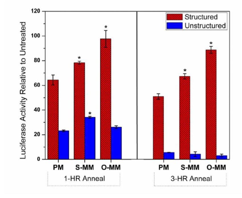
To compare the antisense activity of the γPNAs, we annealed various concentrations of either
probe with freshly transcribed mRNA for one hour at 37 ◦ C before adding to rabbit reticulocyte lysate
to start translation. Dose-response curves shown in Figure 4B reveal nearly three-fold higher potency
for the unstructured probe against the perfect match mRNA (IC50 = 45 nM vs. 131 nM). Since these
experiments were done at a temperature below the Tm for unfolding of the hairpin, struc_γPNA would
be expected to be less potent. When a U-T mismatch was present in the stem domain, the IC50 values for
the two probes increased by roughly the same amount, i.e., the unstructured probe remained three-fold
more potent. However, when an A-G mismatch was present in the overhang domain, the IC50 value
for the structured probe increased by 42%, while that for the unstructured probe increased only 7%,
leading to a four-fold difference in potency. Thus, the mismatch discrimination effects observed for
O-MM-4A and S-MM-7T by SPR and luciferase reporter assay are qualitatively consistent.
We repeated these experiments at a single γPNA concentration (100 nM) but with an extended
annealing time of 3 h to determine whether the effects shown in Figure 4 were due to kinetic differences
in probe hybridization. The results are shown in Figure 5, where the relative luciferase activity
(i.e., the ratio of luciferase signal in the presence and the absence of the γPNA) is plotted for both
probes and all three targets after 1 or 3 h annealing. Thus, a higher value corresponds to a weakerMolecules 2020, 25, 970 7 of 12
Molecules 2020, 25, x FOR PEER REVIEW 7 of 13
would be expected to be less potent. When a U-T mismatch was present in the stem domain, the IC50
antisense effect. For struc_γPNA, extended annealing did not alter the selectivity of the probe.
values for the two probes increased by roughly the same amount, i.e., the unstructured probe
Slightly greaterthree-fold
remained knockdown moreofpotent.
luciferase was observed.
However, when an A-GInterestingly,
mismatch unstruc_γPNA
was present in theexhibited
overhang much
larger effectsthe
domain, dueICto prolonged annealing. Specifically, greater knockdown was observed, but the
50 value for the structured probe increased by 42%, while that for the unstructured
selectivity
probe was lost. Itonly
increased is unclear why thetounstructured
7%, leading probe appeared
a four-fold difference to loseThus,
in potency. selectivity at the longer
the mismatch
annealing time. However, the important finding of this experiment is that the ability
discrimination effects observed for O-MM-4A and S-MM-7T by SPR and luciferase reporter assay of the structured
are
probequalitatively consistent.
to discriminate between perfect and mismatched targets is due to thermodynamic rather than
kinetic differences.
Figure 4. (A) Design of firefly luciferase (Fluc) mRNA targets for antisense experiments with
unstructured
Molecules Figure
2020, 25, 4. and structured
(A) PEER
x FOR Design of γPNA
REVIEW probes performed
firefly luciferase in rabbit
(Fluc) mRNA reticulocyte
targets lysate.
for antisense Mismatchwith
experiments sites8 are
of 13
unstructured
indicated with (*).and
(B)structured γPNA probes
Dose-response curvesperformed in rabbit
determined after 1reticulocyte
h annealinglysate. ◦ C.
at 37Mismatch sites are
indicated with (*). (B) Dose-response curves determined after 1 h annealing at 37 °C.
We repeated these experiments at a single γPNA concentration (100 nM) but with an extended
annealing time of 3 h to determine whether the effects shown in Figure 4 were due to kinetic
differences in probe hybridization. The results are shown in Figure 5, where the relative luciferase
activity (i.e., the ratio of luciferase signal in the presence and the absence of the γPNA) is plotted for
both probes and all three targets after 1 or 3 h annealing. Thus, a higher value corresponds to a weaker
antisense effect. For struc_γPNA, extended annealing did not alter the selectivity of the probe.
Slightly greater knockdown of luciferase was observed. Interestingly, unstruc_γPNA exhibited much
larger effects due to prolonged annealing. Specifically, greater knockdown was observed, but the
selectivity was lost. It is unclear why the unstructured probe appeared to lose selectivity at the longer
annealing time. However, the important finding of this experiment is that the ability of the structured
probe to discriminate between perfect and mismatched targets is due to thermodynamic rather than
kinetic differences.
Figure 5. Effect of annealing time on antisense target discrimination. Samples contained 10 nM mRNA
and 100 5.
Figure nM γPNA.
Effect Fluc activity
of annealing time was compared
on antisense to control
target samplesSamples
discrimination. lacking either γPNA
contained probe
10 nM mRNAand are
expressed as a percentage of activity on the y-axis. At a 1 h annealing time (left
and 100 nM γPNA. Fluc activity was compared to control samples lacking either γPNA probe and panel), the struc_γPNA
demonstrated
are expresseda as statistically significant
a percentage (* = p on
of activity < 0.05) difference
the y-axis. At in
a 1luciferase
h annealinginhibition between
time (left the the
panel), perfect
match (PM) and
struc_γPNA both mismatch
demonstrated targets, whereas
a statistically the unstruc_γPNA
significant only showed
(* = p < 0.05) difference a significant
in luciferase difference
inhibition
betweenthe
between thePMperfect
andmatch (PM) At
S-MM-7U. anda both mismatchtime
3 h annealing targets, whereas
(right panel),the unstruc_γPNA
there only showed
was no inhibition difference
(pa >significant difference
0.05) amongst between
the three thewhen
targets PM and S-MM-7U.
using At a 3 h annealing
the unstruc_γPNA, whereas time
the(right panel), there
struc_γPNA retained
was no inhibition
discrimination of thedifference
mismatched (p >targets.
0.05) amongst
All data the
shownthree
aretargets when
presented as using of n = 3 ± SD.
the unstruc_γPNA,
an average
whereas the struc_γPNA retained discrimination of the mismatched targets. All data shown are
presented as an average of n = 3 ± SD.
3. Discussion
High affinity oligonucleotides such as γPNA possess the ability to overcome target secondaryMolecules 2020, 25, 970 8 of 12
3. Discussion
High affinity oligonucleotides such as γPNA possess the ability to overcome target secondary and
tertiary structures that impose kinetic and thermodynamic obstacles to hybridization. Even genomic
DNA can be successfully hybridized by γPNA, as evidenced by recent reports of in vivo [13] and
in utero [14] gene editing induced by strand-invading γPNAs. However, the capacity to target
highly structured DNA or RNA comes with a substantial risk, namely tolerance of single- or even
multiple-mismatch sites, potentially leading to off-target hybridization. In the best case, this would
diminish the potency of on-target hybridization while, in the worst case, it would lead to deleterious
side effects. The results described above illustrate a generalizable approach to improving the selectivity
of γPNAs.
The SPR results demonstrate the impact of the hairpin design on hybridization selectivity.
Compared to the unstructured analogue, struc_γPNA showed ca. three-fold slower hybridization
kinetics, as expected due to needing to unfold the hairpin structure in order to fully hybridize to the
DNA target. There was also a slight increase in off-rate, presumably due to the ability of struc_γPNA
to refold into a hairpin upon dissociation. Slower binding and faster release translate into decreased
overall affinity. This was not reflected in the melting data due to the fact that the hairpin structure
melts at a lower temperature than even the mismatched duplexes. Although the overall affinity was
lower, the selectivity of struc_γPNA was markedly improved over that of unstruc_γPNA for each of
the single mismatches tested (Figure 3). Moreover, we observed that the selectivity of the hairpin probe
was relatively insensitive to γPNA concentration, whereas the linear probe’s selectivity decreased
progressively as its concentration increased. For example, struc_γPNA was 2.2-fold more selective at
10 nM concentration, but this rose to 4.5-fold at 25 nM probe.
The hairpin probe also exhibited higher selectivity in cell-free antisense experiments after both
1 and 3 h annealing times. The selectivity of struc_γPNA was similar for both annealing times, whereas
unstruc_γPNA’s selectivity was lost at the longer annealing time. Evidently, longer time allows the
unstructured probe to hybridize to the mismatch sites, leading to inhibition of translation. On the other
hand, the kinetic barrier imposed by the hairpin structure presented a sufficiently strong barrier to
hybridization to the off-target sites to allow it to retain its selectivity even after extended annealing.
Both probes exhibited greater antisense effects after longer incubation, but the improved
potency was much more apparent for the unstructured γPNA, suggesting a greater kinetic barrier
to hybridization by this probe compared with struc_γPNA. This is surprising, given the faster
hybridization and the slower dissociation of this probe in the SPR experiments. One possible explanation
for this result is initial spurious hybridization of unstruc_γPNA to off-target sites elsewhere in the
luciferase mRNA before binding to the higher affinity perfect match site in the 50 -UTR. Such sites
might be more accessible and/or abundant than the target site, allowing faster hybridization, yet their
lower affinity would minimize any potential antisense effects. In contrast, the structured probe would
have slower association at the off-target sites, leading to an antisense effect that was nearly maximized
even after only 1 h of incubation.
The improved mismatch discrimination of struc_γPNA validates the hairpin design, but much
more needs to be done to optimize the approach. In particular, the length and the affinity of the
mask domain need to be studied in detail. One would expect the selectivity to improve with the
stability of the hairpin, but the cost in potency could be substantial. Moreover, moving the mask to
different positions along the probe could lead to significant differences in both selectivity and potency.
Finally, analyzing probe selectivity through both biophysical and biochemical methods is important
since real-world performance in an application can be affected by factors beyond hybridization kinetics
and affinity.Molecules 2020, 25, 970 9 of 12
4. Materials and Methods
4.1. γPNA/DNA Oligomers
All γPNA oligomers reported here were purchased from PNA Innovations Inc and gave satisfactory
HPLC and MS data. This vendor is no longer in business. Concentrations of struc_γPNA (ε260
= 146,700 M−1 cm−1 ) and unstruc_γPNA (ε260 = 94,400 M−1 cm−1 ) were determined by UV-vis
absorption on a Cary 300 spectrophotometer (Agilent, Santa Clara, CA, USA). Sequences of all
biotinylated target DNA oligonucleotides used in SPR direct binding experiments are also given below.
DNA oligonucleotides were ordered from Integrated DNA Technologies (idtdna.com, Coralville,
IA, USA).
4.2. Surface Plasmon Resonance (SPR)
SPR experiments were performed using a Biacore T100 instrument (GE Healthcare, Marlborough,
MA, USA) and four-channel carboxymethyl dextran matrix sensor chips. These commercially available
CM5 chips (GE Healthcare) were further functionalized with streptavidin (approximately 5000 RUs) via
NHS-EDC coupling. The 50 biotinylated DNA targets (all oligonucleotide DNA targets used are shown
in the Table 3) were individually immobilized (approximately 120 RUs) to the streptavidin labeled
surface, and chips were finalized for PNA injections by priming five times with buffer. The buffer used
for chip preparation and all subsequent SPR experiments was 10 mM HEPES pH 7.4, 100 mM NaCl,
3 mM Na2 EDTA, and 0.005% v/v polysorbate 20 (HBS-EP Buffer).
Table 3. Biotinylated DNA oligonucleotides used for SPR experiments.
DNA Sequence Sequence a
Perfect Match (PM) 50 -Bt-TTTTTAGACCCAAGC-30
Stem Mismatch (S-MM-7T) 50 -Bt-TTTTTAGACCCTAGC-30
Stem Mismatch (S-MM-6A) 50 -Bt-TTTTTAGACCAAAGC-30
Stem Mismatch (S-MM-7G) 50 -Bt-TTTTTAGACCCGAGC-30
Overhang Mismatch (O-MM-4A) 50 -Bt-TTTTTAGAACCAAGC-30
Overhang Mismatch (O-MM-3T) 50 -Bt-TTTTTAGTCCCAAGC-30
Overhang Mismatch (O-MM-4T) 50 -Bt-TTTTTAGATCCAAGC-30
a Bt = Biotinylated, mismatches underlined.
Direct binding experiments were conducted in triplicate (sensorgrams provided are an average
of all three experiments with a standard deviation shown at 418 s) as well as background subtracted
for streptavidin and buffer backgrounds. Various concentrations (10, 15, 20, or 25 nM) of structured
or unstructured γPNA were injected over the prepared sensor chip for 400 s (flow rate = 30 µL/min).
This was followed by a dissociation cycle via buffer injection for 600 s (flow rate = 30 µL/min).
Finally, a regeneration cycle was conducted to wash any residual γPNA from the sensor chip (30 s
injection of 1 M NaCl, 10 mM NaOH flow rate = 50 µL/min), followed by a buffer injection to re-establish
a baseline for subsequent injections (150 s injection of buffer flow rate = 30 µL/min).
Hybridization on rates were calculated using the slopes of the raw sensorgrams between 80–100 s
(Equation (1)).
RU80 s − RU100 s
On Rate = (1)
20 s
On rate ratios were calculated by dividing the PM on rate by each mismatch (Equation (2)).
PMOn Rate
On Rate Ratio = (2)
MMOn RateMolecules 2020, 25, 970 10 of 12
Selectivity ratios were calculated for all mismatch DNAs using the maximum at 418 s. Max RUs of
the PM were divided between that of the mismatches in order to compare the penalty each mismatch
imposed on γPNA binding (Equation (3)).
PMMAX
Selectivity Ratio = (3)
MMMAX
4.3. UV Melting
Samples containing 2 µM each of DNA target and γPNA probe were mixed in buffer containing
10 mM Tris-HCl (pH = 7.4), 100 mM NaCl, and 0.1 mM Na2 EDTA and placed in 1 mL, 1 cm pathlength
quartz cuvettes. Samples were heated to 90 ◦ C for 5 min before cooling to 15 ◦ C at a rate of 1 ◦ C/min,
collecting absorbance values at 260 nm at 1.0 ◦ C intervals. Samples were then heated back to 90 ◦ C at
the same rate and data collection interval. No hysteresis was observed between heating and cooling
curves; heating curves are shown in the text.
4.4. Luciferase Assays
Mutated Fluc template production and in vitro transcription were performed. A previously
cloned firefly luciferase template (T7 promoter) was used as the “perfect match” template (Fluc-PM)
and used to create the additional O-MM-4 (Fluc-O-MM) and S-MM-7 (Fluc-S-MM) firefly templates.
To create the mutant subclones, Fluc-PM was digested at a PvuII (upstream of T7 promoter) and
HindIII (downstream of T7 promoter) cut site. The digested Fluc vector was then purified using
agarose gel electrophoresis (0.4%). The O-MM-4 and the S-MM-7 forward and reverse sequences
were ordered from Integrated DNA Technologies (idtdna.com) and contained the PvuII and HindIII
overhangs. The O-MM-4 and the S-MM-7 oligonucleotides were pre-annealed into duplex formation
prior to T4-ligation.
O-MM-4A insert sequences:
50 -CTG GCT TAT CGA AAT TAA TAC GAC TCA CTA TAG GGA GAA CCA
50 -AGC TTG GTT CTC CCT ATA GTG AGT CGT ATT AAT TTC GAT AAG CCA G
S-MM-7U insert sequences:
50 -CTG GCT TAT CGA AAT TAA TAC GAC TCA CTA TAG GGA GAC CCT
50 -AGC TAG GGT CTC CCT ATA GTG AGT CGT ATT AAT TTC GAT AAG CCA G
After ligation, the plasmid was transfected (Mach1/T1 Escherichia coli) and plated (plasmid
confers ampicillin resistance) overnight. The resultant colonies were selected and sent for sequencing
for verification.
PCR amplification of the firefly luciferase plasmid was then performed. The firefly plasmids
were PCR amplified using the NEB PCR Protocol for Phusion High-Fidelity DNA Polymerase (cycled
35 times, PCR program 98 ◦ C, 2 min; 98 ◦ C, 10 s; 45 ◦ C, 15 s; 72 ◦ C, 2 min; 72 ◦ C, 1 min; hold at 4 ◦ C).
Primer design: T7 transcription site 50 -TACGACTCACTATAGGG
poly A tail site: 50 -TTTTTTTTTTTTTTTTTTTTTTTTTTTTTT
The products were purified using the Thermo Scientific Gel Extraction Kit protocol (Thermo
Scientific, Waltham, MA, USA) and verified using agarose gel electrophoresis (1.8 kB).
4.5. Transcription Reaction and Purification
The transcription reaction followed the Thermo Scientific conventional transcription protocol
(50 µL final volume) and consistently gave high RNA product yield (~2.5 µM, determined via NanoDrop
spectrophotometer, Thermo Fisher, Waltham, MA, USA). The transcription reaction was conducted
at 37 ◦ C for 2 h. The transcription products were purified using the GeneJET RNA Cleanup and
Concentration Micro Kits (Thermo Scientific, Waltham, MA, USA), and concentration was measured
using a NanoDrop spectrophotometer.Molecules 2020, 25, 970 11 of 12
The γPNA (struc_γPNA or unstruc_γPNA) and the mRNA were annealed together in the presence
of 79 mM potassium chloride (designed to match the K+ concentration in the rabbit reticulocyte lysate,
RRL Promega) and DEPC-treated water. The RNA concentration for all translation experiments was
set at 10 nM in the final translation reaction at a final volume of 15 µL. The probe concentration varied
depending on the desired dose. The probe/mRNA was annealed at 37 ◦ C for 1 h.
The translation reaction was conducted using the Luciferase Assay System (E1500, Promega,
Madison, WI, USA) (rabbit reticulocyte lysate). The entire 15 µL of annealing solution (above) was
mixed into 20 µL lysate. The final γPNA concentration was determined based on the 35 µL final
translation reaction volume. The translation reaction was conducted at 30 ◦ C for 1.5 h. Then, 15 µL of
lysate solution was mixed into 15 µL of Promega Luciferase Assay Reagent (E1483) and added to a
Nunc 96 well plate (flat white, Thermo-Fisher, Waltham, MA, USA). The bioluminescence reading was
collected on a TECAN Infinite M1000 plate reader (TECAN, Morrisville, NC, USA).
Supplementary Materials: The following are available online. Figure S1: Overlay of SPR sensorgrams for
struc_γPNA and unstruc_γPNA with perfect match DNA target showing greater dissociation of struc_γPNA;
Figure S2: UV melting curves of unstruc_γPNA with perfect match and single mismatch DNA targets; Figure S3:
UV melting curves of struc_γPNA with perfect match and single mismatch DNA targets.
Author Contributions: Conceptualization, T.D.C., A.S.B., M.P.B. and B.A.A.; methodology, T.D.C., A.S.B., C.A.T.,
M.P.B. and B.A.A.; formal analysis, T.D.C., A.S.B., M.P.B. and B.A.A.; investigation, T.D.C., A.S.B., J.A.M., C.E.;
writing-original draft preparation, T.D.C. and B.A.A., writing-review and editing, A.S.B., J.A.M., C.A.T., M.P.B.,
supervision, T.D.C., M.P.B., B.A.A.; project administration, B.A.A.; funding acquisition, B.A.A. All authors have
read and agreed to the published version of the manuscript.
Funding: This research was funded by the David Scaife Family Charitable Foundation, Award 141RA01 to B.A.A.
Conflicts of Interest: The funders had no role in the design of the study; in the collection, analyses, or interpretation
of data; in the writing of the manuscript, or in the decision to publish the results. B.A. owns equity in Trucode
Gene Repair, Inc., which is commercializing γPNA probes for gene-editing applications.
References
1. Demidov, V.V.; Frank-Kamenetskii, M.D. Two Sides of the Coin: Affinity and Specificity of Nucleic Acid
Interactions. Trends Biochem. Sci 2004, 29, 62–71. [CrossRef] [PubMed]
2. Bonnet, G.; Tyagi, S.; Libchaber, A.; Kramer, F.R. Thermodynamic Basis of the Enhanced Specificity of
Structured DNA Probes. PNAS 1999, 96, 6171–6176. [CrossRef] [PubMed]
3. Tsourkas, A.; Behlke, M.A.; Rose, S.D.; Bao, G. Hybridization Kinetics and Thermodynamics of Molecular
Beacons. Nucleic Acids Res. 2003, 31, 1319–1330. [CrossRef] [PubMed]
4. Xiao, Y.; Plakos, K.J.I.; Lou, X.; White, R.J.; Qian, J.; Plaxco, K.W.; Soh, H.T. Fluorescence Detection of
Single-Nucleotide Polymorphisms with a Single, Self-Complementary, Triple-Stem DNA Probe. Angew. Chem.
Int. Ed. 2009, 48, 4354–4358. [CrossRef] [PubMed]
5. Zhang, D.Y.; Chen, S.X.; Yin, P. Optimizing the Specificity of Nucleic Acid Hybridization. Nat. Chem. 2012, 4,
208–214. [CrossRef] [PubMed]
6. Wu, L.R.; Wang, J.S.; Fang, J.Z.; Evans, E.R.; Pinto, A.; Pekker, I.; Boykin, R.; Ngouenet, C.; Webster, P.J.;
Beechem, J.; et al. Continuously Tunable Nucleic Acid Hybridization Probes. Nat. Methods 2015, 12,
1191–1196. [CrossRef]
7. Egholm, M.; Buchardt, O.; Christensen, L.; Behrens, C.; Freier, S.M.; Driver, D.A.; Berg, R.H.; Kim, S.K.;
Nordén, B.; Nielsen, P.E. PNA Hybridizes to Complementary Oligonucleotides Obeying the Watson-Crick
Hydrogen-Bonding Rules. Nature 1993, 365, 566–568. [CrossRef]
8. Nielsen, P.E.; Egholm, M.; Berg, R.H.; Buchardt, O. Sequence-Selective Recognition of DNA by Strand
Displacement with a Thymine-Substituted Polyamide. Science 1991, 254, 1498–1500. [CrossRef]
9. Ørum, H.; Nielsen, P.E.; Egholm, M.; Berg, R.H.; Buchardt, O.; Stanley, C. Single Base Pair Mutation Analysis
by PNA Directed PCR Clamping. Nucleic Acids Res. 1993, 21, 5332–5336. [CrossRef]
10. Stender, H.; Fiandaca, M.; Hyldig-Nielsen, J.J.; Coull, J. PNA for Rapid Microbiology. J. Microbiol. Methods
2002, 48, 1–17. [CrossRef]Molecules 2020, 25, 970 12 of 12
11. Dragulescu-Andrasi, A.; Rapireddy, S.; Frezza, B.M.; Gayathri, C.; Gil, R.R.; Ly, D.H. A Simple γ-Backbone
Modification Preorganizes Peptide Nucleic Acid into a Helical Structure. J. Am. Chem. Soc. 2006, 128,
10258–10267. [CrossRef] [PubMed]
12. Sahu, B.; Sacui, I.; Rapireddy, S.; Zanotti, K.J.; Bahal, R.; Armitage, B.A.; Ly, D.H. Synthesis and
Characterization of Conformationally Preorganized, (R)-Diethylene Glycol-Containing γ-Peptide Nucleic
Acids with Superior Hybridization Properties and Water Solubility. J. Org. Chem. 2011, 76, 5614–5627.
[CrossRef] [PubMed]
13. Bahal, R.; McNeer, N.A.; Quijano, E.; Liu, Y.; Sulkowski, P.; Turchick, A.; Lu, Y.-C.; Bhunia, D.C.; Manna, A.;
Greiner, D.L.; et al. In vivo correction of anaemia in b-thalassemic mice by gPNA-mediated gene editing
with nanoparticle delivery. Nat. Commun. 2016, 7, 13304. [CrossRef] [PubMed]
14. Ricciardi, A.S.; Bahal, R.; Farrelly, J.S.; Quijano, E.; Bianchi, A.H.; Luks, V.L.; Putman, R.; López-Giráldez, F.;
Coşkun, S.; Song, E.; et al. In Utero Nanoparticle Delivery for Site-Specific Genome Editing. Nat. Commun.
2018, 9, 2481. [CrossRef]
15. Pham, H.H.; Murphy, C.T.; Sureshkumar, G.; Ly, D.H.; Opresko, P.L.; Armitage, B.A.
Cooperative Hybridization of γPNA Miniprobes to a Repeating Sequence Motif and Application to Telomere
Analysis. Org. Biomol. Chem. 2014, 12, 7345–7354. [CrossRef]
16. Orenstein, A.; Berlyoung, A.S.; Rastede, E.E.; Pham, H.H.; Fouquerel, E.; Murphy, C.T.; Leibowitz, B.J.;
Yu, J.; Srivastava, T.; Armitage, B.A.; et al. γPNA FRET Pair Miniprobes for Quantitative Fluorescent In Situ
Hybridization to Telomeric DNA in Cells and Tissue. Molecules 2017, 22, 2117. [CrossRef]
17. Armitage, B.; Ly, D.; Koch, T.; Frydenlund, H.; Ørum, H.; Schuster, G.B. Hairpin-Forming Peptide Nucleic
Acid Oligomers. Biochemistry 1998, 37, 9417–9425. [CrossRef]
18. Zhang, D.Y.; Winfree, E. Control of DNA Strand Displacement Kinetics Using Toehold Exchange. J. Am.
Chem. Soc. 2009, 131, 17303–17314. [CrossRef]
19. Sacui, I.; Hsieh, W.-C.; Manna, A.; Sahu, B.; Ly, D.H. Gamma Peptide Nucleic Acids: As Orthogonal Nucleic
Acid Recognition Codes for Organizing Molecular Self-Assembly. J. Am. Chem. Soc. 2015, 137, 8603–8610.
[CrossRef]
20. Tan, X.; Bruchez, M.P.; Armitage, B.A. Closing the Loop: Constraining TAT Peptide by gammaPNA Hairpin
for Enhanced Cellular Delivery of Biomolecules. Bioconjug. Chem. 2018, 29, 2892–2898. [CrossRef]
21. Lusvarghi, S.; Murphy, C.T.; Roy, S.; Tanious, F.A.; Sacui, I.; Wilson, W.D.; Ly, D.H.; Armitage, B.A. Loop and
Backbone Modifications of PNA Improve G Quadruplex Binding Selectivity. J. Am. Chem. Soc. 2009, 131,
18415–18424. [CrossRef] [PubMed]
22. SantaLucia, J.J.; Hicks, D. The Thermodynamics of DNA Structural Motifs. Annu. Rev. Biophys. Biomol. Struct.
2004, 33, 415–440. [CrossRef] [PubMed]
23. Datta, B.; Armitage, B.A. Hybridization of PNA to Structured DNA Targets: Quadruplex Invasion and the
Overhang Effect. J. Am. Chem. Soc. 2001, 123, 9612–9619. [CrossRef]
24. Canady, T.D.; Telmer, C.A.; Oyaghire, S.N.; Armitage, B.A.; Bruchez, M.P. In Vitro Reversible Translation
Control Using γPNA Probes. J. Am. Chem. Soc. 2015, 137, 10268–10275. [CrossRef] [PubMed]
Sample Availability: Samples of γPNAs and plasmids will be made available upon request from the authors.
© 2020 by the authors. Licensee MDPI, Basel, Switzerland. This article is an open access
article distributed under the terms and conditions of the Creative Commons Attribution
(CC BY) license (http://creativecommons.org/licenses/by/4.0/).You can also read