Histomorphological Characteristics and Pathological Types of Hyperproliferation of Gastric Surface Epithelial Cells - Hindawi.com
←
→
Page content transcription
If your browser does not render page correctly, please read the page content below
Hindawi
Gastroenterology Research and Practice
Volume 2021, Article ID 8828326, 11 pages
https://doi.org/10.1155/2021/8828326

Research Article
Histomorphological Characteristics and Pathological Types of
Hyperproliferation of Gastric Surface Epithelial Cells

          Yangkun Wang ,1 Lan Shen,1 Guang Zhao,2 Baohui Li,2 Jianxue Bu,2 Chaoya Zhu,3
          Bo Jiang,4 and Sunan Wang 5
          1
            Department of Pathology, Shenzhen Hospital of Southern Medical University, Shenzhen, Guangdong Province 518100, China
          2
            Department of Pathology, The 989th Hospital of the Joint Logistics Support Force of the Chinese People’s Liberation Army, Luoyang,
            Henan Province 471031, China
          3
            Department of Pathology, The Third Affiliated Hospital of Zhengzhou University, Zhengzhou, Henan Province 450052, China
          4
            The 990th Hospital of Joint Logistics Support Force of the Chinese People’s Liberation Army, Zhumadian,
            Henan Province 463000, China
          5
            Shen Zhen Polytechnic, Shenzhen, Guangdong Province 518055, China

          Correspondence should be addressed to Sunan Wang; drwangsunan@126.com

          Received 14 September 2020; Revised 8 February 2021; Accepted 20 February 2021; Published 10 March 2021

          Academic Editor: Oronzo Brunetti

          Copyright © 2021 Yangkun Wang et al. This is an open access article distributed under the Creative Commons Attribution License,
          which permits unrestricted use, distribution, and reproduction in any medium, provided the original work is properly cited.

          Objective. To investigate the histomorphological characteristics and pathological types of hyperproliferation of gastric surface
          epithelial cells. Methods. Hematoxylin and Eosin, Periodic acid–Schiff, and immunohistochemical staining were performed on
          biopsy specimens obtained from 723 patients with hyperproliferation of gastric surface epithelial cells and/or hyperplasia of
          gastric pits. Follow-up gastroscopic reexaminations were performed on 475 patients included. Improvement probability was
          analyzed using Kaplan-Meyer as well as Cox proportional hazards models. Results. Seven different histomorphologies and
          clinicopathologies of hyperproliferation of gastric surface epithelial cells were identified: (1) common hyperplasia of gastric
          epithelial cells, which was characterized by focal glandular epithelial hyperplasia of gastric pits with chronic inflammation; (2)
          drug-induced hyperplasia of gastric epithelial cells, which was characterized by increased hyperplasia of gastric pits and cells
          arranged in a monolayer; (3) Helicobacter pylori (Hp) infection-induced hyperplasia of gastric epithelial cells, which was
          characterized by the disappearance of oval, spherical, and bounded membrane-enclosed mucus-containing granules in the
          cytoplasm and on the nucleus together with cytoplasmic swelling and vacuolation; (4) metaplastic hyperplasia of gastric
          epithelial cells, which was characterized by the coexistence of intestinal metaplastic cells with hyperplastic gastric epithelial cells;
          (5) atrophic hyperplasia of gastric epithelial cells, which was characterized by the mucosal atrophy accompanied with
          hyperplasia of gastric pits; (6) low-grade neoplasia of epithelial cells, which was characterized by the mild to moderate dysplasia
          of gastric epithelial cells; and (7) high-grade neoplasia of epithelial cells, which was characterized by the evident dysplasia of
          hyperplastic epithelial cells and losses of cell polarity. The different pathological types are associated with different improvement
          probabilities. Conclusions. This study demonstrated the histomorphological characteristics and pathological types, which might
          guide clinicians to track malignant cell transformation, perform precise treatment, predict the clinical prognosis, and control the
          development of gastric cancer.

1. Introduction                                                            tion of gastric cancer is increasing with the application of
                                                                           screening endoscopy [3]. Because gastroscopy provides the
Gastric cancer remains the third leading cause of cancer-                  character and extent of the lesions, which provides a refer-
related deaths in the world [1]. The mortality from gastric                ence for clinicians to make accurate diagnoses and perform-
cancer can be reduced because of the early-stage tumor                     ing precise treatment [4–7]. In addition, histopathological
detection and clinic pathological diagnosis [2]. Early detec-              examination is considered necessary for the clinic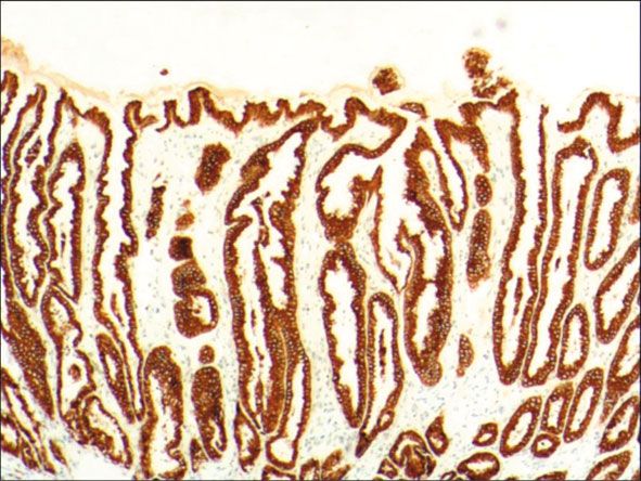
2                                                                                          Gastroenterology Research and Practice

pathological diagnosis due to the vague and nonspecific clin-          were obtained from Shenzhen Dameng Bio-Medical Tech-
ical symptoms [8, 9].                                                 nology Co., Ltd. All primary antibodies were obtained from
     The hyperproliferation of surface epithelial cells of the gas-   Shenzhen Dameng Bio-Medical Technology Co., Ltd. Sec-
tric mucosa, which secrete protective and lubricant insoluble         ondary antibody for immunohistochemistry and DAB (3,3 ′
mucus containing high concentrations of bicarbonate, is also          -diaminobenzidine) chromogen kit were bought from Shenz-
known as hyperplasia of gastric pits and is of diagnostic value       hen Dameng Bio-Medical Technology Co., Ltd.
in pathohistological practice [10]. Because this epithelial cell
proliferation, which breaks the balance between cell prolifera-       2.3. Hematoxylin and Eosin (H&E) Staining. Biopsies were
tion and apoptosis, is accepted as one of the risk factors for gas-   fixed in neutral buffered 10% formalin (to preserve their his-
tric carcinogenesis [11]. Malignancies can develop through            tological structure), embedded in paraffin, and sectioned at
benign hyperplasia of gastric surface epithelial cells and are pre-   four micrometer. Next, sections were stained with H&E
ceded by atypical hyperplasia, which can progress into intrae-        according to the following protocol. The sections were depar-
pithelial neoplasia and malignant forms [11, 12]. The                 affinized in xylene for 15 min twice, gradually hydrated in
proliferation of surface epithelial cells of the gastric mucosa       100%, 80%, and 70% alcohol for 5 min twice. After being
reportedly results from infected gastric mucosa, drug stimula-        washed in distilled water for 5 min, the sections were
tion, immune factors and genetic factors, etc. In addition to         immersed in Hematoxylin solution for 5 min and then
many diverse etiologies, there are various histomorphologies          dipped in distilled water for 2-3 s. Next, sections were differ-
caused by the proliferation of gastric epithelial cells, which lead   entiated in 1% HCl ethanol for 1-3 s and rinsed with distilled
to great differences in treatment and prognosis [13]. Therefore,       water for 30 s. After being washed in distilled water for 30 s,
detecting the histomorphologies caused by hyperplasia of gas-         the sections were immersed in 80%, 90%, and 100% alcohol
tric surface epithelial cells is significantly important for clini-    for 10 min. The sections were subsequently incubated in
cians to understand the development process of lesions, thus          0.5% eosin aqueous solution for 3 min. The gastric tissue
making accurate diagnosis and performing precise treatment.           structure and cell morphology were observed under the opti-
     In the present study, 723 cases of hyperplasia of gastric        cal microscope.
pits and/or hyperplasia of gastric surface epithelial cells in
gastroscopic biopsy specimens were collected, of which 475            2.4. Periodic Acid–Schiff (PAS) Staining. Biopsies were fixed
cases were reexamined by gastroscopic biopsy, to investigate          in neutral buffered 10% formalin, embedded in paraffin,
the morphological and pathological characteristics caused by          and cut into 4 μm thick sections. Next, gastric tissue sections
hyperplasia of gastric surface epithelial cells. This study is        were stained with PAS according to the following protocol.
conducive for clinicians to perform accurate treatment of             The sections were deparaffinized in xylene and graded alco-
gastric surface epithelial cell hyperplasia/gastric pits hyper-       hols. After being washed in distilled water, the sections were
plasia and the follow-up of malignant transformation. In              immersed into periodic acid solution for 15-20 min, followed
addition, our findings provide insight into the nature and             by being washed with distilled water. The sections were then
development process of lesions.                                       treated with Schiff’s reagent for 30-60 min and rinsed with
                                                                      sulphurous acid solution. Sections were subsequently washed
2. Materials and Methods                                              with distilled water for 2-3 min and counterstained with
                                                                      methylgreen for 10-20 min. After staining, tissue sections
2.1. Study Subjects. A total of 723 patients diagnosed with           were differentiated with hydrochloric acid alcohol, clarified
hyperplasia of gastric pits and/or hyperplasia of gastric sur-        in xylol, and mounted. Positive cells (red; nuclei, blue) were
face epithelial cells were included in this study. These 723          monitored using the optical microscope.
patients included 448 men and 275 women were hospitalized
in the department of gastroenterology of Shenzhen Hospital            2.5. EnVision Two-Step Immunohistochemical Staining.
of Southern Medical University, the 990th Hospital of Joint           Immunohistochemical staining was performed with an Envi-
Logistics Support force of the Chinese People’s Liberation            sion Kit. The operations are performed according to the
Army, and the Third Affiliated Hospital of Zhengzhou Uni-               manufacturer’s instructions. Briefly, paraffin-embedded gas-
versity, and underwent endoscopic examination between                 tric tissue sections were deparaffinized, hydrated, and rinsed
December 2018 and December 2019. The age of patients                  with distilled water. Then, the sections were placed in Tris-
ranged from 24 to 78 years, with an average age of 45.2 years.        buffered saline (TBS) for 10 min. Next, endogenous peroxi-
Two or three tissues were harvested from each site of a               dase activity was blocked by incubating sections for 5 min
patient and were used as gastroscopic biopsy specimens. This          in peroxidase blocking reagent containing H2O2 and NaN3.
study was approved by the ethics committee of Shenzhen                Sections were subsequently treated with TBS for 10 min. Each
Hospital of Southern Medical University, the 990th Hospital           of the primary antibodies (CEA, CK7, CK20, Hp, MUC1,
of Joint Logistics Support force of the Chinese People’s Liber-       MUC2, MUC5AC, MUC6, p53, and Ki-67) was incubated
ation Army, and the Third Affiliated Hospital of Zhengzhou              with the sections for 30 min at room temperature. After a
University. Written informed consent was obtained from all            10-min wash in TBS, sections were incubated in EnVi-
study subjects prior to the procedure.                                sionTM. After a 10-min wash in TBS, a secondary antibody
                                                                      was applied for 10 min. The chromogenic substrate solution
2.2. Materials. The Hematoxylin and Eosin (H&E) staining              was incubated for 10 min followed by distilled water rinsing.
Kit, Periodic Acid Schiff (PAS) Stain Kit, and Envision Kit            Color was developed with DAB and sections were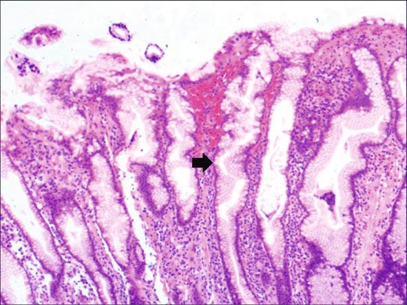
Gastroenterology Research and Practice                                                                                            3

counterstained with hematoxylin. Known gastric mucosa                 Figure 4(a) shows mild to moderate dysplasia of gastric
sections were used as positive controls, and PBS buffer            epithelial cells which were located at the base of the glandu-
instead of the primary antibody was used as the negative con-     lar epithelium, with increased nuclear length, retained
trol. The positive expression of MUC2, MUC5AC, and                polarity, and visible mitoses. Figure 5(a) demonstrates evi-
MUC6 was determined when yellow-brown granules were               dent dysplasia of hyperplastic epithelial cells and cell polar-
found in the cytoplasm. The positive expression of P53 and        ity disorder. Besides, cells were morphologically columnar
Ki-67 was determined when yellow-brown granules were              to cuboidal, with large nuclei, increased nuclear cytoplasmic
observed within the cell nucleus.                                 ratio, prominent nucleoli, and increased mitotic figures
                                                                  (Figure 5(a)). These varying degrees of cellular and struc-
2.6. Follow-Up Examination. The follow-up was performed           tural atypias suggested hyperplasia and dysplasia of gastric
on 475 patients who were pathologically diagnosed as hyper-       epithelial cells. These observations also indicated the neo-
proliferation of gastric surface epithelial cells and/or hyper-   plastic hyperplasia caused by hyperplasia and dysplasia of
plasia of gastric pits. At least two tissues were harvested       gastric epithelial cells. In this study, the hyperplasia and
from each site of the patient between December 2018 and           dysplasia of gastric epithelial cells, which are characterized
December 2019. The follow-up included endoscopic, histo-          by varying degrees of cellular and structural atypias and
morphological, and immunohistochemical examinations               are able to result in neoplastic hyperplasia, are referred to
and was performed from December 2018 to June 2020. The            as the “epithelial cell lesion on gastric mucosal surface.”
time range for reexamination was divided into 3 stages:           The positive PAS staining (indicating neutral mucin)
1st~3rd months, 4th~6th months, and 7 th~12th months.             showed in Figure 1 suggested the presence of gastric epithe-
                                                                  lial cells accompanied with hyperplasia of gastric pits.
                                                                  Therefore, “epithelial cell lesion on gastric mucosal surface”
2.7. Statistics. Cox proportional hazards models were used to     is also referred to as the “gastric pits lesions or columnar
calculate the hazard ratios (HRs) and 95% confidence inter-        mucous cell lesions”.
vals (CI). p values of 0.05 were considered significant.
                                                                  3.2. Histomorphologic Characteristics of Epithelial Cell Lesion
3. Results                                                        on Gastric Mucosal Surface. As shown in Figure 1(a), the
                                                                  common hyperplasia of gastric epithelial cells is morpholog-
3.1. The Formation of Epithelial Cell Lesion on Gastric           ically characterized by focal glandular epithelial hyperplasia,
Mucosal Surface. To investigate the histomorphologic              accompanied by chronic inflammation of gastric mucosa.
changes caused by hyperplasia of gastric epithelial cells, we     The height of hyperplastic gastric pits is between 0.5 and
performed H&E staining and immunohistochemical staining           1.0 mm. (Figure 1(a)). While the drug-induced hyperplasia
on gastroscopic biopsies. Results from H&E staining showed        of gastric epithelial cells is characterized by no or very little
that commonly the hyperplasia of gastric mucosal epithelium       inflammatory cell infiltration in the stroma, with the cells
is morphologically focal glandular epithelial hyperplasia, and    arranged in a monolayer (Figure 2(a)). Infection-induced
the height of hyperplastic gastric pits is between 0.5 and        hyperplasia of gastric epithelial cells is characterized by cyto-
1.0 mm when there is lymphocyte infiltration (Figure 1(a)).        plasmic swelling and vacuolation. As shown in Figure 3(a),
The hyperplasia of gastric pits indicated the histomorpholo-      oval, spherical, and bounded membrane-enclosed mucus-
gical hyperplasia of the surface epithelium and gastric foveo-    containing granules in the cytoplasm and on the nucleus
lar epithelium.                                                   are disappeared, with cytoplasmic swelling and vacuolation.
    While the drug-induced hyperplasia of gastric epithelial      Figure 3(a) also demonstrates that hyperplastic gastric pits
cells is characterized by no or very little inflammatory cell      are histologically thick and wide, with wide interstitium
infiltration in the stroma, with the height of hyperplastic sur-   caused by inflammation, edema, and vascular congestion.
face epithelial cells between 1 and 1.5 mm and the cells being    Figure 6(a) shows that intestinal metaplastic cells coexisted
arranged in a monolayer (Figure 2(a)). Figure 3(a) shows that     with hyperplastic gastric epithelial cells, and cells were orga-
oval, spherical, and bounded membrane-enclosed mucus-             nized in a monolayer or stratified epithelium arrangement,
containing granules in the cytoplasm and on the nucleus           with a nuclear length 1–2 times of that in normal epithelial
are disappeared, with cytoplasmic swelling and vacuolation.       cells. In this study, such a lesion is referred to as “metaplastic
Figure 3(a) also demonstrates that hyperplastic gastric pits      hyperplasia of gastric epithelial cells.” Figure 7(a) shows
are histologically thick and wide, with wide interstitium         mucosal atrophy (loss of glands) accompanied with hyper-
caused by inflammation, edema, and vascular congestion.            plasia of gastric pits, with varying degrees of decrease and
Figure 3(b) shows a positive expression of Hp, confirming          even disappearance of gastric fundus glands, cardiac glands,
the Hp infection. These findings confirmed that the damage          and pyloric glands. However, such hyperplasia is compensa-
to surface epithelial cells caused by Hp, drug stimulation,       tory and regional and cannot be determined morphologically
and autoimmune factors can lead to the hyperplasia of gastric     as intraepithelial neoplasia. In this study, such a lesion is
epithelial cells. It was also found that hyperplasia beyond the   called “atrophic epithelial hyperplasia”. Figure 4(a) shows
normal range caused hyperplasia of gastric pits, with the         histological morphology characterized by mild to moderate
height of hyperplastic gastric pits exceeding twice of the        dysplasia of gastric epithelial cells, which were located at
height of normal gastric pits, and more than three consecu-       the base of the glandular epithelium, with increased nuclear
tive pits proliferated.                                           length and retained polarity. Such hyperplasia is referred to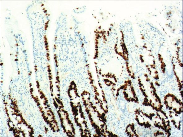
4 Gastroenterology Research and Practice
(a) (b)
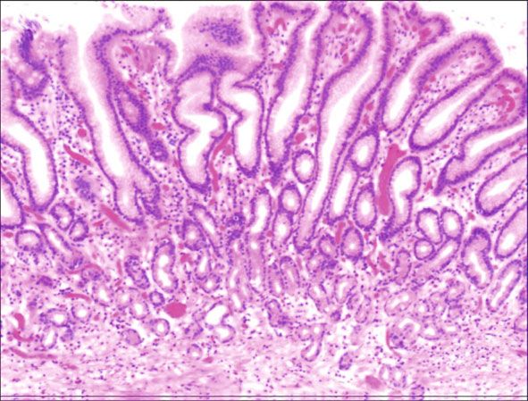
Figure 1: The histomorphologic characteristics of common hyperplasia of gastric epithelial cells. (a) H&E staining results showing
morphologically focal glandular epithelial hyperplasia, with chronic inflammation of gastric mucosa, with the height of hyperplastic
gastric pits between 0.5 and 1.0 mm. Characteristic image at 200x objective magnification was shown. (b) PAS staining results showing
gastric epithelial cells with strong expression of neutral mucin. Characteristic image at 100x objective magnification was shown. The
arrows refer to hyperplastic gastric pits.
(a) (b)
Figure 2: The histomorphologic characteristics of drug-induced hyperplasia of gastric epithelial cells. (a) H&E results showing no or very
little inflammatory cell infiltration in the stroma, with the height of hyperplastic gastric epithelial cells between 1 and 1.5 mm and cells
arranged in a monolayer. (b) Immunohistochemical staining results showing positive expression of MUC5AC. Characteristic images of
each group at 200x objective magnification were shown.
(a) (b)
Figure 3: The histomorphologic characteristics of Hp infection-induced hyperplasia of gastric epithelial cells. (a) H&E staining results
showing the disappearance of oval, spherical, and bounded membrane-enclosed mucus-containing granules in the cytoplasm and on the
nucleus are disappeared, with cytoplasmic swelling and vacuolation. Characteristic image at 100x objective magnification was shown. (b)
Immunohistochemical staining results showing positive expression of Hp. Characteristic image at 400x objective magnification was shown.
as “low-grade neoplasia of epithelial cells.” Figure 5(a) dem- sia of epithelial cells.” The pathological types and histological
onstrates evident dysplasia of hyperplastic epithelial cells. diagnostic criteria of epithelial cell lesions on gastric mucosal
Besides, cells were morphologically columnar to cuboidal, surface are explicated in Table 1.
with large nuclei, increased nuclear cytoplasmic ratio, prom-
inent nucleoli, and increased mitotic figures (Figure 5(a)). In 3.3. Results from PAS Staining and Immunohistochemical
this study, such a lesion is referred to as “high-grade neopla- Staining. To identify the hyperplasia of gastric epithelial cells,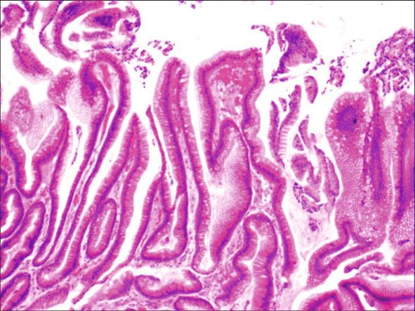
Gastroenterology Research and Practice                                                                                                            5

                                                  (a)                                          (b)

Figure 4: The histomorphologic characteristics of low-grade neoplasia of epithelial cells. (a) H&E staining results showing mild to moderate
dysplasia of gastric epithelial cells, which were located at the base of the glandular epithelium, with increased nuclear length, retained polarity,
and visible mitoses. Characteristic image at 100x objective magnification was shown. (b) Immunohistochemical staining results showing the
expression of ki-67. Characteristic image at 200x objective magnification was shown.

                                                  (a)                                          (b)

Figure 5: The histomorphologic characteristics of high-grade neoplasia of epithelial cells. (a) H&E staining results showing evident dysplasia
of hyperplastic epithelial cells, with cells morphologically columnar to cuboidal, large nuclei, increased nuclear cytoplasmic ratio, prominent
nucleoli, and increased mitotic figures. Characteristic image at 100x objective magnification was shown. (b) Immunohistochemical staining
results showing the expression of p53. Characteristic image at 200x objective magnification was shown.

                                                  (a)                                          (b)

Figure 6: The histomorphologic characteristics of metaplastic hyperplasia of gastric epithelial cells. (a) H&E staining results showing
coexistence of intestinal metaplastic cells with hyperplastic gastric epithelial cells, with cells organized in a monolayer or stratified
epithelium arrangement and a nuclear length 1–2 times of that in normal epithelial cells. Characteristic image at 100x objective
magnification was shown. (b) Immunohistochemical staining results showing positive expression of MUC2. Characteristic image at 200x
objective magnification was shown.

PAS staining was performed. In addition, to investigate the                 infection-induced hyperplasia of gastric epithelial cells,
expression of MUC1, MUC2 [14], MUC5AC (a gastric-type                       which was also confirmed by PAS staining (Figure 3(b)).
secreted mucin), MUC6, CK7, CK20, CEA, p53, and Ki-67                       CK7 and CK20 were positively expressed (+) in cases of
in the development of epithelial cell lesions on gastric muco-              low-grade neoplasia of epithelial cells and high-grade neopla-
sal surface, immunohistochemical staining was performed.                    sia of epithelial cells, while negatively expressed in other cases
The PAS staining and immunohistochemical staining results                   (Table 2). Negative expression of CEA was detected in cases
are explicated in Table 2. According to Table 2, positive                   of common and drug-induced hyperplasia of gastric epithe-
expression of Hp was only identified in the case of Hp                       lial cells, while positive expression (+) of CEA was identified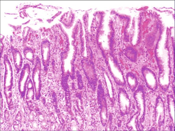
6                                                                                                 Gastroenterology Research and Practice

                                                (a)                                         (b)

Figure 7: The histomorphologic characteristics of atrophic hyperplasia of gastric epithelial cells. (a) H&E staining results showing mucosal
atrophy (loss of glands) accompanied with hyperplasia of gastric pits, with very few atrophic pyloric glands. (b) Immunohistochemical
staining results showing positive expression of MUC6 in pyloric glands. Characteristic images at 100x objective magnification were shown.

                            Table 1: Histomorphologic changes caused by hyperplasia of gastric epithelial cells.

Hyperplasia types                                                           Histomorphologic characteristics
Common hyperplasia of gastric                Morphologically focal glandular epithelial hyperplasia, with chronic inflammation of gastric
epithelial cells                                         mucosa; the height of hyperplastic gastric pits between 0.5 and 1.0 mm.
Drug-induced hyperplasia of gastric        No or very little inflammatory cell infiltration in the stroma; the height of hyperplastic gastric pits
epithelial cells                                            is between 1 and 1.5 mm, with the cells arranged in a monolayer.
                                           Oval, spherical, and bounded membrane-enclosed mucus-containing granules in the cytoplasm
Hp infection-induced hyperplasia of
                                             and on the nucleus are disappeared, with cytoplasmic swelling and vacuolation and positive
gastric epithelial cells
                                                                                     expression of Hp
                                             Coexistence of intestinal metaplastic cells with hyperplastic gastric epithelial cells; cells were
Metaplastic hyperplasia of gastric
                                           organized in a monolayer or stratified epithelium arrangement, with a nuclear length 1–2 times of
epithelial cells
                                                                              that in normal epithelial cells
                                                 Compensatory and regional hyperplasia; it cannot be determined morphologically as
Atrophic hyperplasia of gastric epithelial   intraepithelial neoplasia; mucosal atrophy (loss of glands) accompanied with hyperplasia of
cells                                       gastric pits, with varying degrees of decrease and even disappearance of gastric fundus glands,
                                                                           cardiac glands, and pyloric glands.
                                              Mild to moderate dysplasia of gastric epithelial cells, which were located at the base of the
Low-grade neoplasia of epithelial cells
                                              glandular epithelium, with increased nuclear length, retained polarity, and visible mitoses.
                                           Evident dysplasia of hyperplastic epithelial cells; cells were morphologically columnar to cuboidal,
High-grade neoplasia of epithelial cells with large nuclei, increased nuclear cytoplasmic ratio, prominent nucleoli, and increased mitotic
                                                                                          figures.

                                     Table 2: PAS staining and immunohistochemical staining results.

                                                                                                                          Percentage of Ki67-
Hyperplasia types                          Hp CK7 CK20 CEA MUC1 MUC2 MUC5AC MUC6 p53 PAS
                                                                                                                             positive cells
Common hyperplasia of gastric
                                           —     —      —      —       —        —         +++        —       —      +            1~7%
epithelial cells
Drug-induced hyperplasia of gastric
                                           —     —      —      —       —        —         +++        —              +           11~19%
epithelial cells
Hp infection-induced hyperplasia of
                                            +    —      —       +      —        —          +         —              +           14~21%
gastric epithelial cells
Metaplastic hyperplasia of gastric
                                           —     —      —       +       +       +          +        +++             +           9~32%
epithelial cells
Atrophic hyperplasia of gastric
                                           —     —              +       +       —          +          +             +           18~34%
epithelial cells
Low-grade neoplasia of epithelial cells    —     +       +    +++       +       —          —          +      +     —            26~35%
High-grade neoplasia of epithelial cells   —     +       +    +++       +       —          —          +     +++    —            32~40%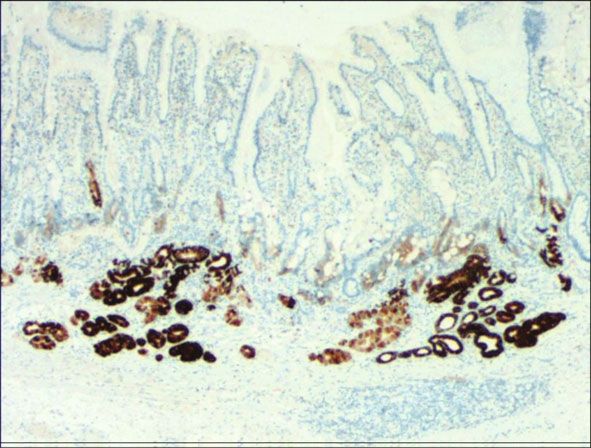
Gastroenterology Research and Practice                                                                                                 7

in cases of Hp infection-induced, metaplastic and atrophic            showed high rate of malignant transformation despite mor-
hyperplasia of gastric epithelial cells. The strongly positive        phologically benign lesions.
expression (+ + +) of CEA was found in cases of low-grade
and high-grade neoplasia of epithelial cells. Negative expres-        3.5. Results from Univariate and Multivariate Models.
sion of MUC1 was found in cases of the common, drug-                  Kaplan-Meier survival curves (Figure 8) demonstrates that
induced, and Hp infection-induced hyperplasia of gastric              the proportionality assumption was always satisfied. Next,
epithelial cells, while positive expression (+) of MUC1 was           Cox proportional hazards model (or Cox regression) was
identified in cases of metaplastic hyperplasia of gastric epi-         used to evaluate the probability of cure and improvement.
thelial cells, atrophic hyperplasia of gastric epithelial cells,      The Cox model shows that with drug-induced hyperplasia
low-grade neoplasia of epithelial cells, and high-grade neo-          of gastric epithelial cells used as a reference, common hyper-
plasia of epithelial cells. The positive expression of MUC2           plasia of gastric epithelial cells, and Hp infection-induced
was only identified in the case of metaplastic hyperplasia of          hyperplasia of gastric epithelial cells were associated with
gastric epithelial cells, which was confirmed by PAS staining          higher improvement probability [HR: 4.95 (95% CI: 2.95–
(Figure 6(b)). Strongly positive expression of MUC5AC was             8.30), p < 0:001 and HR: 4.81 (95% CI: 2.81–8.21), p < 0:001
identified in cases of common and drug-induced hyperplasia             , respectively] (Table 4). The Cox model also demonstrates
of gastric epithelial cells, which was confirmed by PAS stain-         reduced improvement probability related with atrophic
ing (Figure 2(b)). Additionally, MUC5AC was found to pos-             hyperplasia of gastric epithelial cells [HR: 0.43 (95% CI:
itively be expressed in the cases of Hp-induced, metaplastic,         0.23–0.82), p = 0:01] and neoplasia of epithelial cells [HR:
and atrophic hyperplasia of gastric epithelial cells while neg-       0.16 (95% CI: 0.04–0.67), p = 0:01] (Table 4). These results
atively expressed in cases of low-grade and high-grade neo-           are consistent with the results from follow-up examination,
plasia of epithelial cells. Negative expression of MUC6 was           thus indicating that the histomorphological characteristics
found in cases of the common, drug-induced, and Hp                    and pathological types are helpful to predict the clinical prog-
infection-induced hyperplasia of gastric epithelial cells, while      nosis of the hyperplasia and dysplasia of gastric epithelial
positive expression (+) of MUC6 was identified in cases of             cells.
atrophic hyperplasia of gastric epithelial cells, low-grade neo-
plasia of epithelial cells, and high-grade neoplasia of epithe-       4. Discussion
lial cells. The strongly positive expression (+) of MUC6 was
identified in cases of metaplastic hyperplasia of gastric epi-         Normally, the gastric mucosa is histologically composed of
thelial cells. It was also found that P53 showed positive             epithelium, lamina propria, and mucosa. Gastric epithelial
expression in the cases of low-grade neoplasia of epithelial          cells are also known as surface epithelial cells or gastric foveo-
cells and strongly positive in the cases of high-grade neopla-        lar epithelial cells. Most gastric mucosal surface epithelial
sia of epithelial cells, which was also confirmed by PAS stain-        cells are surface mucous cells, and cells are arranged in a
ing (Figure 5(b)). The percentage of Ki67-positive cells in           monolayer, with an inconspicuous nucleolus. There are oval,
cases of the common, drug-induced, Hp infection-induced,              spherical, and bounded membrane-enclosed mucus-
metaplastic, atrophic hyperplasia, and low-grade and high-            containing granules in the cytoplasm and on the nucleus with
grade neoplasia of epithelial cells was 1~7%, 11~19%,                 strongly positive PAS staining [15]. Studies have shown that
14~21%, 9~32%, 18~34%, 26~35%, and 32~40%, respec-                    the drug stimulation, autoimmune diseases, and infection
tively (Table 2).                                                     (especially the Hp infection) are able to destroy the structure
                                                                      of gastric epithelial cells, thus leading to hyperplasia of gastric
3.4. Results from Follow-Up Examination. Follow-up exami-             mucosal surface epithelial cells [16, 17]. In this study, the
nation was performed on cases with at least two harvested tis-        hyperplasia and dysplasia of gastric epithelial cells are
sues. Therefore, a total of 475 patients were included in this        referred to as the “epithelial cell lesion on gastric mucosal
study. Follow-up findings were shown in Table 3. Of the                surface.” Many etiologies can result in the hyperplasia of gas-
475 patients, 214 patients (45.1%) were cases of common               tric epithelial cells, which also show different histomorpholo-
hyperplasia of gastric epithelial cells, with 74.3% improved          gical characteristics with the development of lesions.
and cured cases while 6.5% worsened cases. The 475 patients           However, gastric foveolar hyperplasia described in the clini-
included 22 cases of drug-induced hyperplasia of gastric epi-         copathological case report reflects few histomorphological
thelial cells, with the improved and cured cases of 90.0%. The        features, thus providing clinicians little valuable information
475 patients also included 115 cases of Hp-induced hyperpla-          to assess the relationship between gastric foveolar hyperpla-
sia of gastric epithelial cells, with the improved and cured          sia and carcinogenesis. The present study provides the histo-
cases of 73.9% and worsened cases of 10.9%. There were 37             morphological features and pathological types of “epithelial
cases of metaplastic hyperplasia of gastric epithelial cells,         cell lesion on gastric mucosal surface,” which is of impor-
with the improved and cured cases of 51.4% and worsened               tance for clinicians to track malignant cell transformation,
cases of 13.5%. 29 cases of low-grade neoplasia of epithelial         perform precise treatment, predict the clinical prognosis,
cells were followed up, and only 17.2% were found to be               and control the occurrence and development of gastric
improved and cured cases while 10.3% were found to be                 cancer.
worsened. These findings indicated that metaplastic hyper-                  Gastric mucosal atrophy is accepted as an important pre-
plasia of gastric epithelial cells, atrophic hyperplasia of gastric   cancerous lesion [18]. Worsened cases (6.5%) of common
epithelial cells, and low-grade neoplasia of epithelial cells         hyperplasia of gastric epithelial cells suggest that common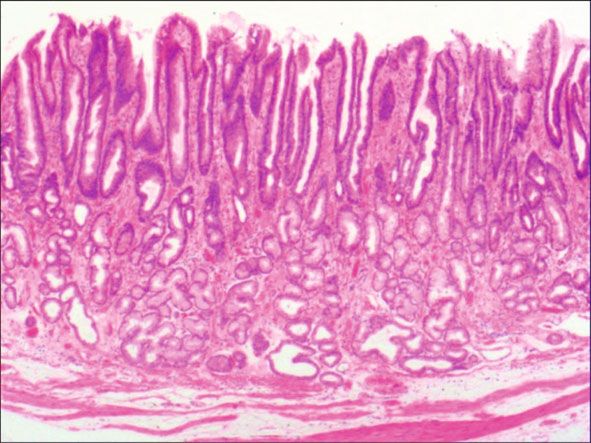
8 Gastroenterology Research and Practice
Table 3: Follow-up results.
Cases with little change Cured/improved cases Worsened cases
Hyperplasia types Cases
(%) (%) (%)
Common hyperplasia of gastric epithelial cells 214 41 (19.2) 159 (74.3) 14 (6.5)
Drug-induced hyperplasia of gastric epithelial cells 22 2 (9.1) 20 (90.9) 0 (0)
Hp infection-induced hyperplasia of gastric epithelial
115 20 (17.4) 87 (75.7) 8 (6.9)
cells
Metaplastic hyperplasia of gastric epithelial cells 46 14 (30.4) 27 (58.7) 5 (10.9)
Atrophic hyperplasia of gastric epithelial cells 37 9 (24.3) 23 (62.2) 5 (13.5)
Low-grade neoplasia of epithelial cells 29 21 (72.4) 5 (17.2) 3 (10.3)
High-grade neoplasia of epithelial cells 12 0 12 (100%)∗ 0
∗
: Lesions were all removed through ESD.
The present study demonstrated that 7.0% cases of drug-
LML function for patterns 1-6
induced hyperplasia of gastric epithelial cells were exacer-
bated, while 73.9% cases of drug-induced hyperplasia of gas-
2.5 tric epithelial cells were improved and cured, which indicates
the vast majority of cured cases after aggressive anti-Hp ther-
apy. Semiannual gastroscopic biopsy reexaminations are rec-
0.0
ommended to closely monitor the proliferation of epithelial
Log of a negative log
cells because Hp infection has been recognized as the main
–2.5 cause of gastric adenocarcinoma [17, 19]. 10.9% cases of
metaplastic hyperplasia of gastric epithelial cells were found
–5.0 to be exacerbated in our work, which indicates a high ten-
dency to malignant transformation. Intestinal metaplasia is
reported as a defensive, reparative, and reactive response to
–7.5 inflammatory stimuli and injury, and hyperplasia no longer
continues once the cause of hyperplasia is eliminated [20,
–10.0 21]. Therefore, semiannual gastroscopic biopsy reexamina-
tion is also recommended for patients with metaplastic
0.00 5.00 10.00 15.00 20.00
hyperplasia of gastric epithelial cells. In the present study,
Time (month) 10.3% cases of low-grade epithelial neoplasia were aggra-
vated. Consistent with previous studies, closely followed up
Helicobacter pylori (Hp)
is recommended for patients with low-grade epithelial neo-
infection-induced
hyperplasia of gastric plasia [22]. We recommend semiannual gastroscopic biopsy
epithelial cells reexaminations for patients with low-grade epithelial neopla-
Metaplastic hyperplasia of sia and suggest endoscopic submucosal dissection (ESD) for
gastric epithelial cells lesion resection once aggravated morphological changes are
Common hyperplasia of observed. Current treatment strategies for high-grade intrae-
gastric epithelial cells pithelial neoplasia include endoscopic therapy, surgical treat-
Neoplasia of epithelial cells ment, and follow-up [23]. In the present study, 12 patients
Atrophic hyperplasia of were with high-grade neoplasia of epithelial cells. These 12
gastric epithelial cells cases were followed up with endoscopic reexamination
Drug-induced hyperplasia within 3 months after treatment. Patients with negative
of gastric epithelial cells
results were reexamined endoscopically within 6 months
Figure 8: The Kaplan-Meyer survival plots of the study population. after treatment. Patients with negative results from reexami-
nation were reexamined endoscopically 1 year later. Patients
with negative results for two consecutive times were reexa-
hyperplasia of gastric epithelial cells is morphologically mined endoscopically every 1 year. Our findings suggest that
benign lesions with potential to be advanced. 90.9% of cases the classification of epithelial cell lesions on gastric mucosal
of drug-induced hyperplasia of gastric epithelial cells were surface according to the morphological features, etiologies,
found to be improved and cured, suggesting histologically and development rules is conducive for clinicians to achieve
benign lesions with a low probability of malignant transfor- precise treatment. Consistently, Cox model showed increased
mation, which indicates that clinicians can make the patients improvement probability in cases of common hyperplasia of
recover by adjusting the drug dose. Gastroscopic biopsy reex- gastric epithelial cells and Hp infection-induced hyperplasia
amination is recommended within half a year for patients of gastric epithelial cells [HR: 4.95 (95% CI: 2.95–8.30), p <
with drug-induced hyperplasia of gastric epithelial cells. 0:001 and HR: 4.81 (95% CI: 2.81–8.21), p < 0:001,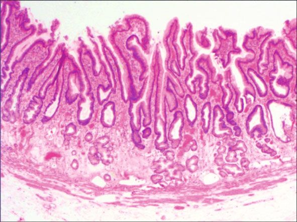
Gastroenterology Research and Practice                                                                                                9

                                        Table 4: Cox-modelled hazard ratio (HR) for all outcomes.

Hyperplasia types                                                            HR                      95% CI                       p
Drug-induced hyperplasia of gastric epithelial cells                         1.00                       —                        —
Common hyperplasia of gastric epithelial cells                               4.95                   2.95–8.30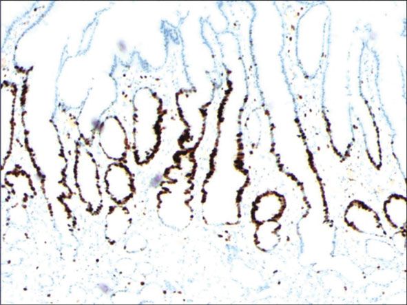
10                                                                                               Gastroenterology Research and Practice

thus not conducive to guide clinicians to achieve accurate                [6] L. Fustar-Preradovic, B. Coha, and I. Pajic-Penavic, “A correl-
treatment. The present study demonstrated the histomor-                       ative study of histology and imprint cytology of gastric mucosa
phological characteristics and pathological types of epithelial               biopsy in the diagnosis gastric cancer,” Collegium Antropologi-
cell lesions on gastric mucosal surface, as well as the changes               cum, vol. 34, no. 2, pp. 355–358, 2010.
in expression of Hp, MUC1, MUC2, MUC5AC, MUC6, CK7,                       [7] N. Pokhrel, B. Khanal, K. Rai, M. Subedi, and N. R. Bhattarai,
CK20, CEA, p53, and Ki-67 during the development of the                       “Application of PCR and microscopy to detect helicobacter
lesions. Our findings provide in-depth understanding of the                    pylori in gastric biopsy specimen among acid peptic disorders
nature and the development of epithelial cell lesions on gas-                 at tertiary care centre in Eastern Nepal,” Canadian Journal of
                                                                              Infectious Diseases and Medical Microbiology, vol. 2019, Arti-
tric mucosal surface, which is conducive to clinicians to per-
                                                                              cle ID 3695307, 6 pages, 2019.
form follow-up of malignant cell transformation, predict the
clinical prognosis, and achieve accurate treatment to control             [8] M. Gundogan, N. C. Demirkan, K. Tekin, and H. Aybek, “Gas-
                                                                              tric histopathological findings and ghrelin expression in mor-
the occurrence and development of gastric cancer.
                                                                              bid obesity,” Turkish Journal of Pathology, vol. 29, no. 1,
                                                                              pp. 19–26, 2013.
Data Availability                                                         [9] M. el Khadir, S. Alaoui, D. A. Benajah et al., “VacA genotypes
Corresponding authors and first authors are in charge of the                   and cagA-EPIYA-C motifs of helicobacter pylori and gastric
                                                                              histopathological lesions,” International Journal of Cancer,
underlying data supporting the results of this study.
                                                                              vol. 147, no. 11, pp. 3206–3214, 2020.
Conflicts of Interest                                                    [10] J. Fischer, P. J. Klein, M. Vierbuchen, B. Skutta, G. Uhlenbruck,
                                                                              and R. Fischer, “Characterization of glycoconjugates of
The authors declare that there is no conflict of interest                      human gastrointestinal mucosa by lectins. I. Histochemical
regarding the publication of this article.                                    distribution of lectin binding sites in normal alimentary
                                                                              tract as well as in benign and malignant gastric neoplasms,”
                                                                              Journal of Histochemistry & Cytochemistry, vol. 32, no. 7,
Authors’ Contributions                                                        pp. 681–689, 1984.
Yangkun Wang and Sunan Wang performed the conceptual-                    [11] T. Yamaguchi, N. Nakajima, H. Kuwayama, Y. Ito, A. Iwasaki,
ization. Yangkun Wang did the methodology, investigation,                     and Y. Arakawa, “Gastric epithelial cell proliferation and apo-
                                                                              ptosis in Helicobacter pylori-infected mice,” Alimentary Phar-
software, and funding acquisition and wrote the original
                                                                              macology & Therapeutics, vol. 14, pp. 68–73, 2000.
draft. Lan Shen, Guang Zhao, Baohui Li, and Jianxue Bu per-
formed the data curation, investigation, and software.                   [12] N. Shin, H. J. Jo, W. K. Kim et al., “Gastric pit dysplasia in adja-
                                                                              cent gastric mucosa in 414 gastric cancers: prevalence and
Chaoya Zhu did the validation and visualization. Bo Jiang
                                                                              characteristics,” The American Journal of Surgical Pathology,
and Sunan Wang wrote, reviewed, and edited the manuscript                     vol. 35, no. 7, pp. 1021–1029, 2011.
and performed the supervision and project administration.
                                                                         [13] F. Boccellato, S. Woelffling, A. Imai-Matsushima et al.,
                                                                              “Polarised epithelial monolayers of the gastric mucosa reveal
Acknowledgments                                                               insights into mucosal homeostasis and defence against infec-
                                                                              tion,” Gut, vol. 68, no. 3, pp. 400–413, 2019.
This work was supported by the key scientific and technolog-
ical project of Henan Province (grant number                             [14] P. Leone, A. Buonavoglia, R. Fasano et al., “Insights into the
                                                                              regulation of tumor angiogenesis by micro-RNAs,” Journal of
132102310008).
                                                                              Clinical Medicine, vol. 8, no. 12, p. 2030, 2019.
                                                                         [15] A. M. Costa, R. M. Ferreira, I. Pinto-Ribeiro et al., “Helicobac-
References                                                                    ter pylori activates matrix metalloproteinase 10 in gastric epi-
 [1] A. P. Thrift and H. B. El-Serag, “Burden of gastric cancer,”             thelial cells via EGFR and ERK-mediated pathways,” The
     Clinical Gastroenterology and Hepatology, vol. 18, no. 3,                Journal of Infectious Diseases, vol. 213, no. 11, pp. 1767–
     pp. 534–542, 2020.                                                       1776, 2016.
 [2] M. Rugge, R. M. Genta, F. Di Mario et al., “Gastric cancer as       [16] S. G. Barreto and J. A. Windsor, “Redefining early gastric can-
     preventable disease,” Clinical Gastroenterology and Hepatol-             cer,” Surgical Endoscopy, vol. 30, no. 1, pp. 24–37, 2016.
     ogy, vol. 15, no. 12, pp. 1833–1843, 2017.                          [17] S. Ouyang, G. Zhu, L. Ouyang et al., “Bapx1 mediates trans-
 [3] D. Y. Graham, M. Kato, and M. Asaka, “Gastric endoscopy in               forming growth factor-β\-induced epithelial-mesenchymal
     the 21st century: appropriate use of an invasive procedure in            transition and promotes a malignancy phenotype of gastric
     the era of non-invasive testing,” Digestive and Liver Disease,           cancer cells,” Biochemical and Biophysical Research Communi-
     vol. 40, no. 7, pp. 497–503, 2008.                                       cations, vol. 486, no. 2, pp. 285–292, 2017.
 [4] H. J. Kim, N. Kim, C. Y. Yun, and H. S. Lee, “The clinical          [18] I. Oda, S. Hoteya, and M. Fujishiro, “Status of Helicobacter
     meaning of the “indefinite for atrophy” lesions within gastric            pylori infection and gastric mucosal atrophy in patients with
     mucosa biopsy specimens in a region with a high prevalence               gastric cancer: analysis based on the Japan Endoscopy Data-
     of gastric cancer,” Helicobacter, vol. 24, no. 5, article e12605,        base,” Digestive Endoscopy, vol. 31, no. 1, p. 103, 2019.
     2019.                                                               [19] Q. Zhang, M. Wang, F. Huang et al., “H. pylori infection-
 [5] S. P. Romero, F. Alberca de las Parras, A. S. del Río, J. López-         induced MSC differentiation into CAFs promotes epithelial-
     Picazo, J. J. Gutiérrez, and J. L. Molina, “Quality indicators in        mesenchymal transition in gastric epithelial cells,” Interna-
     gastroscopy. Gastroscopy procedure,” Revista Española de                 tional Journal of Molecular Medicine, vol. 32, no. 6,
     Enfermedades Digestivas, vol. 111, pp. 699–709, 2019.                    pp. 1465–1473, 2013.
Gastroenterology Research and Practice 11
[20] J. A. Trieu, M. Bilal, H. Saraireh, and A. Y. Wang, “Update on
the diagnosis and management of gastric intestinal metaplasia
in the USA,” Digestive Diseases and Sciences, vol. 64, no. 5,
pp. 1079–1088, 2019.
[21] S. Ono, Y. Ono, and N. Sakamoto, “Spraying l-menthol
enhances gastric intestinal metaplasia in linked color imag-
ing,” Digestive Endoscopy, vol. 31, no. 3, pp. e70–e71, 2019.
[22] J. W. Hwang, Y. S. Bae, M. S. Kang et al., “Predicting pre- and
post-resectional histologic discrepancies in gastric low-grade
dysplasia: a comparison of white-light and magnifying endos-
copy,” Journal of Gastroenterology and Hepatology, vol. 31,
no. 2, pp. 394–402, 2016.
[23] Y. W. Son, A. Kim, and H. H. Jeon, “Efficacy and safety of
endoscopic submucosal dissection for gastric epithelial neo-
plasia in elderly patients aged 80 years and older,” Aging Clin-
ical and Experimental Research, vol. 31, no. 12, pp. 1833–1838,
2019.
[24] X. He, X. Xu, G. Zhu, and H. Ye, “Circulating uPA as a poten-
tial prognostic biomarker for resectable esophageal squamous
cell carcinoma,” Medicine (Baltimore), vol. 98, no. 9, article
e14717, 2019.
[25] J. Folkman, “Tumor angiogenesis: therapeutic implications,”
The New England Journal of Medicine, vol. 285, no. 21,
pp. 1182–1186, 1971.
[26] A. Forma, M. Tyczyńska, P. Kędzierawski, K. Gietka, and
M. Sitarz, “Gastric Carcinogenesis: A Comprehensive Review
of the Angiogenic Pathways,” Clinical Journal of Gastroenter-
ology, vol. 14, no. 1, pp. 14–25, 2021.
[27] M. Fassan, M. Simbolo, E. Bria et al., “High-throughput muta-
tion profiling identifies novel molecular dysregulation in high-
grade intraepithelial neoplasia and early gastric cancers,” Gas-
tric Cancer, vol. 17, no. 3, pp. 442–449, 2014.
[28] S. Tanaka, M. Mizuno, T. Maga et al., “H. pylori decreases gas-
tric mucin synthesis via inhibition of galactosyltransferase,”
Hepato-Gastroenterology, vol. 50, no. 53, pp. 1739–1742, 2003.
[29] J. C. Byrd, P. Yan, L. Sternberg, C. K. Yunker, J. M. Scheiman,
and R. S. Bresalier, “Aberrant expression of gland-type gastric
mucin in the surface epithelium of Helicobacter pylori-
infected patients,” Gastroenterology, vol. 113, no. 2, pp. 455–
464, 1997.
[30] A. Teixeira, L. David, C. A. Reis, J. Costa, and M. Sobrinho-
Simoes, “Expression of mucins (MUC1, MUC2, MUC5AC,
and MUC6) and type 1 Lewis antigens in cases with and with-
out helicobacter pylori colonization in metaplastic glands of
the human stomach,” The Journal of Pathology, vol. 197,
no. 1, pp. 37–43, 2002.
[31] C. A. Reis, L. David, P. Correa et al., “Intestinal metaplasia of
human stomach displays distinct patterns of mucin (MUC1,
MUC2, MUC5AC, and MUC6) expression,” Cancer Research,
vol. 59, no. 5, pp. 1003–1007, 1999.
[32] D. R. Hess, “Retrospective studies and chart reviews,” Respira-
tory Care, vol. 49, no. 10, pp. 1171–1174, 2004.
[33] E. M. Schaeffer, “Re: defining a clinically significant struvite
stone: a non-randomized retrospective study,” The Journal of
Urology, vol. 204, no. 3, pp. 597-598, 2020.You can also read