An Efficient and Footprint-Free Protocol for the Transdifferentiation of Hepatocytes Into Insulin-Producing Cells With IVT mRNAs
←
→
Page content transcription
If your browser does not render page correctly, please read the page content below
ORIGINAL RESEARCH
                                                                                                                                                published: 05 June 2020
                                                                                                                                         doi: 10.3389/fgene.2020.00575

                                              An Efficient and Footprint-Free
                                              Protocol for the Transdifferentiation
                                              of Hepatocytes Into
                                              Insulin-Producing Cells With IVT
                                              mRNAs
                                              Shinan Ma 1 , Mengjie Yang 1,2 , Wenhui Zhou 1 , Longjun Dai 3,4 , Yan Ding 1 , Xingrong Guo 1 ,
                                              Yahong Yuan 1 , Junming Tang 1 , Dongsheng Li 1* and Xiaoli Wang 1*
                                              1
                                               Hubei Key Laboratory of Embryonic Stem Cell Research, Taihe Hospital, Hubei University of Medicine, Shiyan, China,
                                              2
                                               Department of Medical, Southeast University, Nanjing, China, 3 Department of Neurosurgery, Taihe Hospital, Hubei
                                              University of Medicine, Shiyan, China, 4 Department of Surgery, University of British Columbia, Vancouver, BC, Canada

                         Edited by:
                        William Cho,          Background: Direct transdifferentiation of adult somatic cells into insulin-producing
      Queen Elizabeth Hospital (QEH),
                         Hong Kong
                                              cells (IPCs) is a promising approach for cell-based therapies for type 1 diabetes mellitus.
                       Reviewed by:
                                              Liver cells are an ideal source for generating IPCs because they have regenerative
                         Najah Nassif,        ability and a developmental process similar to that of the pancreas. Pancreas versus
     University of Technology Sydney,         liver fate is regulated by TALE homeoprotein (TGIF2) during development. Here, we
                             Australia
              Ann Margaret Simpson,           wanted to investigate whether TGIF2 could enhance the efficiency of transdifferentiation
     University of Technology Sydney,         of hepatocytes into IPCs induced by three pancreatic transcription factors (pTFs), i.e.,
                             Australia
                                              Pdx1, NeuroD, and Mafa, which are crucial for pancreatic development in the embryo.
                  *Correspondence:
                          Dongsheng Li        Methods: The in vitro transcribed (IVT) mRNAs of TGIF2 and the three pTFs were
                   dsli1698@126.com
                                              synthesized in vitro and sequentially supplemented in hepatocytes. On day 6, the
                            Xiaoli Wang
                   xiaolitina@126.com         expression of transcription factors was assessed by quantitative real-time polymerase
                                              chain reaction (qRT-PCR), and insulin expression was detected by immunofluorescence.
                   Specialty section:
                                              Glucose-stimulated insulin secretion was assessed by enzyme-linked immunosorbent
         This article was submitted to
                                  RNA,        assay (ELISA). The key genes controlling cell polarity and the Wnt/PCP signaling
               a section of the journal       pathway were assayed by qRT-PCR, and the level of JNK protein phosphorylation,
                  Frontiers in Genetics
                                              which regulates the Wnt/PCP signaling pathway, was detected by western blotting.
       Received: 09 December 2019
           Accepted: 11 May 2020              Results: IVT mRNAs could be efficiently transfected into hepatocytes. Quantitative real-
          Published: 05 June 2020
                                              time polymerase chain reaction results revealed that compared with ectopic expression
                              Citation:
       Ma S, Yang M, Zhou W, Dai L,
                                              of the three pTFs alone, ectopic expression of the three pTFs plus TGIF2 could strongly
   Ding Y, Guo X, Yuan Y, Tang J, Li D        reduce hepatic gene expression and subsequently improve the induction of a set of
  and Wang X (2020) An Efficient and
                                              pancreatic genes. Immunofluorescence analysis showed that TGIF2 expression could
               Footprint-Free Protocol
            for the Transdifferentiation      double the transdifferentiation yield; 30% of the cells were insulin positive if induced by
of Hepatocytes Into Insulin-Producing         TGIF2 plus the 3 pTFs, while only 15% of the cells were insulin positive if induced by the
                Cells With IVT mRNAs.
                 Front. Genet. 11:575.
                                              three pTFs alone. ELISA analysis confirmed that glucose-stimulated insulin secretion
      doi: 10.3389/fgene.2020.00575           was less efficient after transfection with the three pTFs alone. The differentiated cells

Frontiers in Genetics | www.frontiersin.org                                         1                                                June 2020 | Volume 11 | Article 575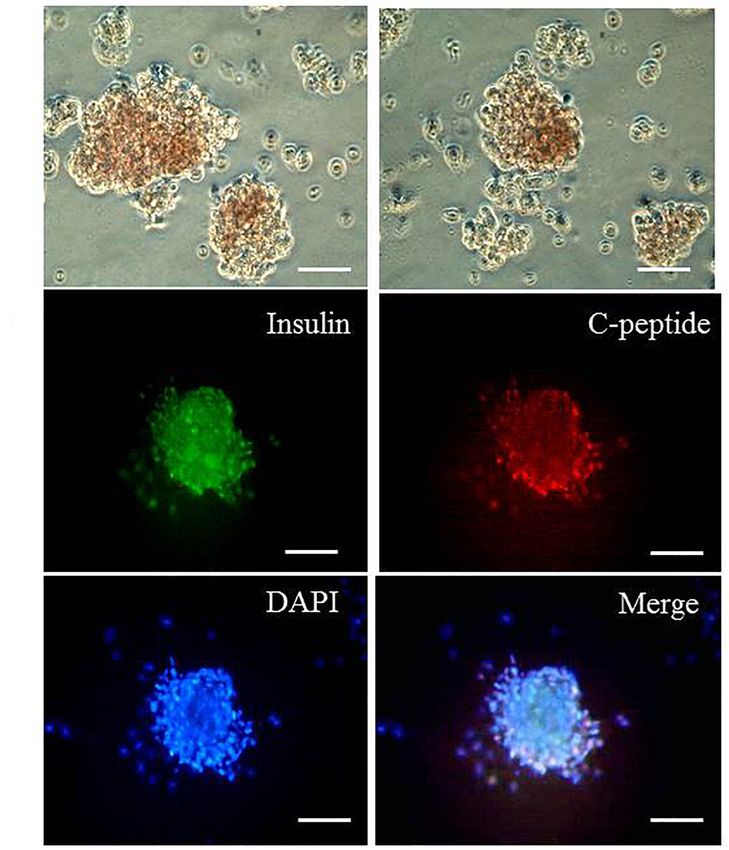
Ma et al.                                                                                                                 Hepatocytes Into IPCs With IVT mRNAs

                                              derived from the addition of TGIF2 mRNA could form islet-like clusters. By contrast, the
                                              cells differentiated with the three pTFs did not form clusters under the same conditions.
                                              Tgif2 induced transdifferentiation more efficiently by remodeling the expression of genes
                                              in the Wnt/PCP pathway. Overexpression of TGIF2 in hepatocytes could activate the
                                              expression of key genes controlling cell polarity and genes in the Wnt/PCP signaling
                                              pathway, increasing the level of JNK protein phosphorylation.
                                              Conclusions: Our study established a novel footprint-free protocol for efficient
                                              transdifferentiation of hepatocytes into IPCs using IVT mRNAs of TGIF2 and 3 pTFs,
                                              which paved the way toward a clinical application.
                                              Keywords: transdifferentiation, IVT mRNA, TGIF2, hepatocytes, insulin-producing cells, transcription factors

BACKGROUND                                                                           crucial role in the middle stage of pancreatic development,
                                                                                     controlling the transformation of cells into pancreatic endocrine
Type 1 diabetes mellitus (T1D) is an autoimmune-mediated                             precursor cells. Mafa participates in late development of the
disease that is characterized by pancreatic beta cells being                         pancreas, inducing further maturation of pancreatic endocrine
attacked by the immune system. The primary therapy is the                            precursor cells into islet beta cells (Kaneto et al., 2009).
injection of exogenous insulin to normalize blood glucose levels,                    Pancreatic transcription factors are usually delivered by vectors
but this is often accompanied by diabetic complications.                             such as plasmids or viruses. These delivery methods cannot
A preferable treatment is transplantation of pancreatic                              accurately modulate the expression of transcription factors,
islets. Unfortunately, the limited supply of donor islets and                        and the obtained cells have some potential biosafety hazards
the requirement for life-long immune suppression make                                that mean they cannot be used for follow-up clinical research.
this approach impractical for large-scale application. This                          The use of in vitro transcribed (IVT) mRNA has several
hurdle has led researchers to seek new alternative therapies                         advantages in the regulation of transdifferentiation. It does
(Omami et al., 2017; Nevo-Shenker et al., 2019). Cell-based                          not need to reach the nucleus to be functional and does not
therapy has opened a new corridor for T1D therapy, as                                integrate into the genome, which means it has no risk of
beta-like cells or insulin-producing cells (IPCs) have been                          insertion mutagenesis. Protein expression can also be controlled
successfully induced in laboratories (Kopan et al., 2018;                            accurately by adding IVT mRNAs at different times and
Espona-Noguera and Ciriza, 2019).                                                    dosages (Ida et al., 2018). These characteristics make IVT
    Direct transdifferentiation of some somatic cells into other                     mRNAs very convenient and safe for future clinical use. In our
functioning cell types is a novel approach for cell-based therapies                  previous work, we successfully differentiated human umbilical
(Heathman et al., 2015; Allickson, 2017). Recently, transcription                    cord mesenchymal stem cells into IPCs with PDX1 mRNA
factor-mediated transdifferentiation has successfully regenerated                    (Wang et al., 2014).
beta-like cells or IPCs from other types of cell sources (Vieira                         Although the sequential introduction of three pTFs could
et al., 2017; Amirruddin et al., 2019; Cierpka-Kmiec et al.,                         improve the efficiency of hepatocyte transdifferentiation
2019). Hepatocytes are first on the list of cell sources with                        into IPCs, this efficiency was consistently limited to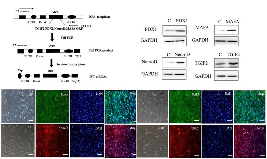
Ma et al.                                                                                                                      Hepatocytes Into IPCs With IVT mRNAs

MATERIALS AND METHODS                                                                  In vitro Transcribed mRNA Synthesis and
                                                                                       Transfection
Isolation and Culture of Mouse                                                         ORFs encoding TGIF2, PDX1, NeuroD1, and Mafa were cloned
Hepatocytes                                                                            from mouse islet cDNA by polymerase chain reaction (PCR).
This experiment was performed in compliance with the relevant                          In vitro transcribed template construction and RNA synthesis
Chinese regulations and approved by the Hubei University of                            are schematized in Figure 1A. mRNAs were synthesized with
Medicine Animal Ethics Committee. A two-step collagenase                               the use of the MEGAscript T7 kit (Ambion) according to the
perfusion method was used to isolate hepatocytes from C57/BL6                          manufacturer’s instructions. The reaction used ARCA cap
mice aged 8–10 weeks (Nagasaki et al., 2014). Inhalation of                            analog (New England Biolabs), 5-methylcytidine triphosphate
2% isoflurane was performed to anesthetize the mice, and                               instead of cytidine and pseudouridine triphosphate instead of
then the abdominal cavity was opened to reveal the portal                              uridine (TriLink Biotechnologies). Reactions were incubated
vein. Calcium- and magnesium-free phosphate-buffered saline                            for 5 h at 37◦ C followed by Antarctic Phosphatase (New
(PBS) was perfused through the portal vein at 5 mL/min for                             England Biolabs) treatment for 2 h at 37◦ C to remove
5 min and then changed to Dulbecco’s modified Eagle’s medium                           residual triphosphates. The synthesized RNA was purified
(DMEM) with 1 mg/mL collagenase II solution at 8 mL/min for                            with Ambion MEGAclear spin columns (Ambion) and
approximately 10 min. The entire liver was removed to a petri                          quantified using a Nanodrop spectrophotometer (Thermo
dish containing DMEM medium at room temperature. The crude                             Fisher Scientific). mRNA transfection was carried out
hepatocyte suspension was filtered through a gauze mesh filter                         with TransIT-mRNA (Mirus). In vitro transcribed mRNAs
(100 µm) and centrifuged. The cells were plated at a density                           were diluted in Opti-MEM basal media (Gibco), followed
of 0.4 × 10⁶ cells/mL and cultured in DMEM supplemented                                by the addition of boost reagent and TransIT-mRNA
with 10% fetal calf serum, 100 units/mL penicillin, 100 ng/mL                          sequentially. After 2 min incubation at room temperature
streptomycin, 250 ng/mL amphotericin B (Biological Industries),                        (RT), the RNA–lipid complexes were delivered to the culture
20 ng/mL EGF and 10 mM nicotinamide (Sigma) at 37◦ C in a                              medium. Four hours later, the medium was replaced with
humidified atmosphere with 5% CO2 and 95% air.                                         normal culture medium.

  FIGURE 1 | In vitro synthesis and identification of mRNAs. (A) Schematic diagram of different kinds of RNA synthesis in vitro. (B) The four kinds of IVT mRNAs were
  transfected into hepatocytes, and 12 h later, the cells were evaluated for protein expression by western blotting. (C) The four kinds of IVT mRNAs were transfected
  into hepatocytes, and 12 h later, protein expression was measured by immunocytochemistry (scale bar: 100 µm). BF, bright field.

Frontiers in Genetics | www.frontiersin.org                                        3                                                June 2020 | Volume 11 | Article 575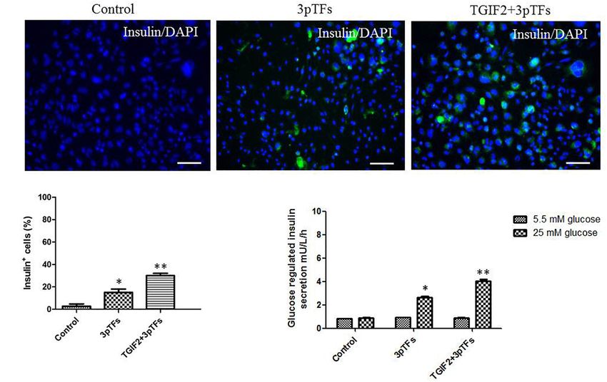
Ma et al.                                                                                                         Hepatocytes Into IPCs With IVT mRNAs

Differentiation of Hepatocytes Into IPCs                                     TABLE 1 | The primers for qRT-PCR.

The hepatocytes were infected with TGIF2, PDX1, NeuroD1, and                 Gene name             Sequence                                   Size (bp)
Mafa mRNAs sequentially from day 1 to day 4, with each day
assigned to one transcription factor. At day 6, the cells were               Afp F                 CGAGGAGTGTTGCCAAGGAAA                         141

harvested to assess transcription factor expression and insulin              Afp R                 CAGAAGCCTAGTTGGATCATG
                                                                             Alb F                 TGCTGCTGATTTTGTTGAGG                          169
expression. For islet-like cluster formation, the cells differentiated
                                                                             Alb R                 GCAGCACTTTTCCAGAGTGG
for 6 days were dissociated and seeded in the well with gelatin
                                                                             Hnf4a F               AACCACGCTACTTGCCTTTGCT                        104
(0.1%; Sigma) for another 3 days.
                                                                             Hnf4a R               TCTGATGGGACACAGCCTACTTCT
                                                                             Hex F                 GAGGTTCTCCAACGACCAGA                          202
Immunocytochemistry                                                          Hex R                 GTCCAACGCATCCTTTTTGT
The cells were fixed with 4% paraformaldehyde (Sigma) for                    Lgr5 F                CAGTGTTGTGCATTTGGGGG                          136
10 min and washed three times with PBS. The cells were                       Lgr5 R                CAAGGTCCCGCTCATCTTGA
permeabilized with 0.1% Triton-X 100 (Sigma) for 20 min                      Cdx2 F                AAACCTGTGCGAGTGGATG                           221
and blocked with 2% bovine serum albumin at RT for                           Cdx2 R                CTGCGGTTCTGAAACCAAAT
60 min. Then, the cells were incubated with rabbit anti-                     Foxa2 F               CATCCGACTGGAGCAGCTA                           178

mouse insulin (1:100) and rabbit anti-mouse C-peptide (1:100)                Foxa2 R               GCGCCCACATAGGATGAC
                                                                             Neurod F              AAGGCAAGGTGTCCCGAGGC                          109
(Santa Cruz Biotechnology) antibodies for 60 min at 37◦ C.
                                                                             Neurod R              CATCAGCCCGCTCTCGCTGT
After rinsing with PBS three times, the cells were incubated
                                                                             Pdx1 F                CCACCAAAGCTCACGCGTGGA                         156
with FITC-conjugated rabbit anti-mouse IgG 1:200 (Santa Cruz
                                                                             Pdx1 R                GGCGGGGCCGGGAGATGTATT
Biotechnology) secondary antibodies for 60 min, followed by
                                                                             Tgif2 F               CTATCTGCACCGCTACAACG                          107
rinsing three times with PBS. The cell nuclei were counterstained
                                                                             Tgif2 R               GGGCATTGATGAACCAGTTAC
with DAPI. The cells were examined under a fluorescence
                                                                             Isl1 F                GCGGCAATCAGATTCACGT                           181
microscope (Leica DMIRE2).
                                                                             Isl1 R                GCGCATTTGATCCCGTACAA
                                                                             Mafa F                CTTCAGCAAGGAGGAGGTCA                          195
Western Blot Analysis                                                        Mafa R                TTGTACAGGTCCCGCTCTTT

Cells transfected with IVT mRNAs were washed with PBS                        NKX6.1 F              ACCTTTGGGCTCACATAACCC                         120
                                                                             NKX6.1 R              AGGATGTCGTTGATGCCGTG
three times and collected with cell lysis buffer. Cell lysates
                                                                             NKX2.2 F              GAGGGCCTCCAATACTCCCT                          105
were incubated on ice for 30 min. Proteins in the cell lysates
                                                                             NKX2.2 R              GTCATTGTCCGGTGACTCGT
were separated by 12% sodium dodecyl sulfate-polyacrylamide
                                                                             Vangl F               CCCCGAGTCTTCGTGTTACG                           89
gel electrophoresis and electrotransferred to nitrocellulose
                                                                             Vangl R               AAGATGCGCACACCGTAGAA
membranes. The blot was placed in blocking buffer for
                                                                             Celsr F               GAGGCCATCACCAACTTCCC                          157
1 h at RT, followed by incubation with a 1:500 dilution of
                                                                             Celsr R               TTACCAGCTCTACCCAAACGG
rabbit anti-human TGIF2, PDX1, NeuroD1, Mafa, c-Jun N-
                                                                             Tcf7 F                GGAGCTGCAGCCATATGATAGA                        205
terminal kinase (JNK), and p-JNK antibodies (Abcam) overnight                Tcf7 R                AGATAGTGCATGCCACCTGC
at 4◦ C. The blots were rinsed with PBS with Tween-20                        Tle3 F                GAAGTCAAGCTCACTTGGCG                           92
three times and incubated with mouse anti-rabbit horseradish                 Tle3 R                TGACACGGAATTGTTCGTGC
peroxide-conjugated secondary antibody (1:1000) for 60 min,                  Camk2b F              GTTTGGATTTGCGGGAACGC                          102
and bands were detected by means of chemiluminescence                        Camk2b R              TACAGGATCACCCCACATGC
with ECL Hyper film.

Quantitative RT-PCR                                                          Detection of Insulin Secretion
Total RNA was extracted using TRIzol (Ambion) following                      After differentiation, the cells were washed five times with
the manufacturer’s recommendations and quantified by UV                      PBS and incubated for 1 h in Krebs-Ringer bicarbonate buffer
spectroscopy. To prepare RNA for quantitative real-time                      (120 mmol/L NaCl, 2.5 mmol/L CaCl2 , 1.1 mmol/L MgCl2 ,
PCR analysis, 2 µg of total RNA was converted to cDNA                        25 mmol/L NaHCO3 , 0.1% bovine serum albumin) containing
with the use of the Fast Quant reverse transcription kit                     5.5 mmol/L glucose. The cells were washed again and incubated
with gDNase (TIANGEN). Quantitative real-time RT-PCR was                     in Krebs-Ringer bicarbonate buffer containing 25 mmol/L
performed using a standard SYBR Green PCR kit (Invitrogen)                   glucose for an additional 1 h. Supernatants from cells stimulated
protocol on an ABI Step One Plus sequence Detection                          with 5.5 or 25 mmol/L glucose were collected and analyzed with a
System (Applied Biosystems). Each sample was detected in                     human insulin enzyme-linked immunosorbent assay (ELISA) kit
triplicate. All the values were normalized to the reference                  (Millipore) according to the manufacturer’s instructions.
genes and calculated using the software REST. Statistical
significance in qPCR experiments was calculated using the REST               Dithizone Staining
randomization test. The primer sets used in this study are listed            The differentiated cell clusters on the gelatin coated plate were
in Table 1.                                                                  washed with PBS three times, and 10 µL of the dithizone stock

Frontiers in Genetics | www.frontiersin.org                              4                                           June 2020 | Volume 11 | Article 575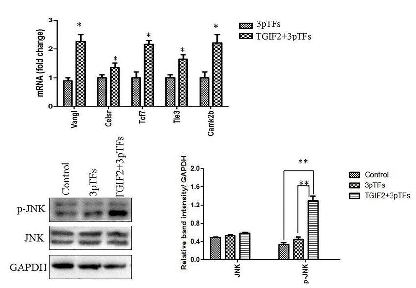
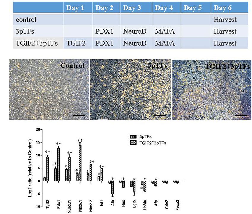
Ma et al.                                                                                                                    Hepatocytes Into IPCs With IVT mRNAs

(10 mg/mL) was then added to 1 mL of the culture medium.                             Statistical Analysis
Cells were incubated at 37◦ C for 15 min and rinsed three                            The results are presented as the mean ± SD. The statistical
times with PBS. Culture dishes were refilled with differentiation                    significance of differences was tested using Student’s t-test in
medium, and clusters were observed with the use of an inverted                       Microsoft Office Excel. Values of p < 0.05 were considered
light microscope.                                                                    statistically significant.

Cellular Insulin Content
Insulin was extracted by the acid/ethanol method (Hamid et al.,                      RESULTS
2002). The approximately 2.5 × 10⁵ cells per well were extracted
by incubation overnight at 4◦ C with 500 µL of acid/ethanol                          Efficient Transfection of Hepatocytes
solution (1.5% HCl, 75% ethanol, 23.5% H2 O), homogenized and                        With IVT mRNAs
incubated overnight with rotation. Then homogenized samples                          In vitro transcribed mRNA is a useful tool in gene delivery.
were centrifuged at 2000 rpm (15min, 4◦ C) and supernatant was                       It has higher safety than other approaches due to the
nertralized with 1 mol/L Tris-Cl buffer (PH7.4). The insulin levels                  avoidance of genomic insertion, and it can be translated
were measured using the ELISA kit and the results were presented                     efficiently in cells. The synthesis of mRNAs in our study
as means ± SD.                                                                       is illustrated in a flow chart (Figure 1A). According to the

  FIGURE 2 | Morphological changes and gene expression during transdifferentiation of hepatocytes into IPCs. (A) The hepatocyte transdifferentiation protocol for
  obtaining IPCs with IVT mRNAs. (B) Changes in the morphology of hepatocytes on day 6 of differentiation (scale bar: 250 µm). (C) RT-qPCR analysis of pancreatic
  gene expression and hepatic gene expression on day 6 after transdifferentiation. The data were normalized and represented as the log2 expression ratio between
  transduced and control cells. Values shown are the mean ± SEM (n = 3) *p < 0.05.

Frontiers in Genetics | www.frontiersin.org                                      5                                               June 2020 | Volume 11 | Article 575
Ma et al.                                                                                                                        Hepatocytes Into IPCs With IVT mRNAs

flow chart, we synthesized four kinds of mRNAs: TGIF2,                                  with a modified protocol reported by Dana et al. (2014). Their
Pdx1, NeuroD1, and Mafa. To confirm that the IVT mRNAs                                  experimental results suggested that transcription factor-induced
could enter the hepatocytes and subsequently be translated                              transdifferentiation from liver to pancreas is a progressive and
into protein, the four IVT mRNAs were transfected into                                  hierarchical process. To analyze the effect of TGIF2 during
the liver cells. After 12 h, the cells were harvested to                                transdifferentiation, we compared the effects of three major
detect protein expression through immunostaining and                                    pTFs, PDX1, NeuroD1, and Mafa, to those of TGIF2 plus
western blotting. The results showed that all four kinds                                these three pTFs on hepatocyte to IPC transdifferentiation.
of IVT mRNAs entered the hepatocytes efficiently by a                                   Cultured hepatocytes were infected with TGIF2, Pdx1, NeruoD1
cationic vehicle and were translated into proteins properly                             and Mafa mRNA in a stepwise manner, as illustrated in
(Figures 1B,C). The use of IVT mRNAs provides the                                       Figure 2A. The cell morphology changed rapidly, and the
possibility of controlled and temporary delivery of the desired                         cells began to coagulate 6 days after differentiation. The cells
transcripts to induce protein expression. This method is safe                           that were first transduced with TGIF2 changed more obviously
for use in gene therapy and will likely receive better clinical                         than the cells that were transduced with only the mRNAs
acceptance in the future.                                                               of the three pTFs (Figure 2B). To determine the changes
                                                                                        in transcription factor expression during the differentiation
                                                                                        period, the expression of liver and pTFs was assayed by
Morphological Characteristics and Gene                                                  qRT-PCR at different times after stepwise transfection with
Expression During Transdifferentiation of                                               IVT mRNAs. Ectopically transduced Tgif2 mRNA strongly
Hepatocytes Into IPCs                                                                   inhibited hepatic gene expression and initiated a set of
Many studies have suggested that the simultaneous expression                            pancreatic genes. Importantly and as expected, the levels of
of several pTFs increases the efficiency of transdifferentiation                        pancreatic transcripts were higher in cells transfected with all
compared with that of transdifferentiation induced by individual                        four mRNAs than in those transfected with the three pTFs
pTFs. We first started the transdifferentiation of hepatocytes                          alone (Figure 2C).

  FIGURE 3 | Detection of insulin expression in differentiated cells and their response to glucose. (A) Immunofluorescence staining of treated hepatocytes for insulin
  (scale bar: 100 µm). (B) The percentage of insulin-positive cells was calculated by counting at least 1000 positive cells from at least two independent experiments.
  (C) Detection of insulin release in response to glucose by ELISA. Data are presented as the mean ± SD of three independent experiments (*p < 0.05 and
  **p < 0.001).

Frontiers in Genetics | www.frontiersin.org                                         6                                                June 2020 | Volume 11 | Article 575
Ma et al. Hepatocytes Into IPCs With IVT mRNAs
Sequential Transduction With IVT
mRNAs of TGIF2 and Three pTFs
Increases the Efficiency of Hepatocyte
Transdifferentiation Into IPCs
Immunocytochemistry was performed to test for insulin-positive
cells after differentiation (Figure 3A). The group induced with
TGIF2 plus the three pTFs showed an increase in the number of
IPCs by 30%, while the group induced with the three pTFs alone
showed an increase of only 15% (Figure 3B). Secretion of insulin
was measured after the cells were stimulated by glucose. Insulin
secretion by IPCs from the groups induced with TGIF2 plus the
three pTFs or the three pTFs alone was measured by ELISA. As
shown in Figure 3C, the IPCs derived via these methods secreted
less insulin in the medium with a lower glucose concentration
(5.5 mM) and more insulin in the medium with a higher glucose
concentration (25 mM). Additionally, the IPCs derived from
group treated with TGIF2 plus the three pTFs had higher insulin
secretion levels than the IPCs derived from the group treated with
the three pTFs alone.
Islet-Like Cluster Formation
We then tested whether the differentiated cells could form
islet-like clusters. The cells that were differentiated for 6 days
were plated in the well coated with gelatin for another 3 days.
The differentiated cells derived from the addition of TGIF2 FIGURE 4 | Islet-like cluster formation. (A) Dithizone staining of differentiated
mRNA could form islet-like clusters. By contrast, control non- cells that were induced with TGIF2 together with three pTFs and grown on
gelatin for another 3 days (100 µm). The hepatocytes differentiated by the
transduced hepatocytes and cells differentiated with the three three pTFs failed to grow under the same conditions. (B) Detection of
pTFs did not form clusters under the same conditions. Dithizone coexpression of insulin and C-peptide for islet-like clusters by immunostaining
staining was used to detect the expression of insulin in the (scale bar: 100 µm).
islet-like cell clusters (Figure 4A). Immunocytochemistry was
performed to test the expression of insulin and C-peptide
proteins in the islet-like cell clusters and determined that the showed that TGIF2 could increase the level of JNK protein
clusters were insulin and C-peptide positive (Figure 4B). This phosphorylation, which played an important role in controlling
indicates that TGIF2 is important for control islet-like cluster the Wnt/PCP signaling pathway (Figures 5B,C). Our results
formation. The cellular insulin content of the islet-like clusters showed that TGIF2 may promote efficient transdifferentiation of
was 6.43 ± 0.09 IU/L. hepatocytes by changing cell polarity.
Tgif2 Induces Transdifferentiation More
DISCUSSION
Efficiently by Remodeling the Expression
of Genes in the Wnt/PCP Pathway Developmentally, the liver and pancreas are closely related. They
WNT signaling can endow cells with plasticity, which allows share a number of characteristics, including the ability to respond
them to change their transcriptional program and developmental to glucose levels, and a large group of specific transcription
fate when transfected with ectopic transcription factors. It has factors. Thus, transconversion from liver to pancreas might be
been shown that the Wnt/PCP signaling pathway is crucial for much easier than conversion to any other organ. To increase
directing endodermal progenitors toward the liver or islets during the efficiency of transdifferentiation, several research groups used
the process of embryonic development. To study the effect of simultaneous ectopic expression of three transcription factors,
TGIF2 during differentiation, the cells were collected to analyze PDX1, NeuroD1, and Mafa, to mimic progressive pancreatic
the expression of genes in the Wnt/PCP signaling pathway one development. When ectopic transcription factors are added
day after transfection of TGIF2 mRNA. Our results showed that in a stepwise manner, the efficiency of transdifferentiation
TGIF2 overexpression in hepatocytes can activate the expression increases (Tang et al., 2013). However, delivery of transcriptional
of key genes controlling cell polarity and genes in the Wnt/PCP factors into hepatocytes using virus-mediated strategies results
signaling pathway (Figure 5A). Many core Wnt/PCP cell polarity in immune-based hepatotoxicity, which is not suitable for
genes, such as Vangl and Celsr, as well as downstream Wnt clinical applications.
transducers, such as Tcf7, Camk2b, and Tle3, were found to In vitro transcribed mRNAs can produce proteins without
be upregulated in transfected cells. The western blot results any risk of randomly integrating echogenic genes into the
Frontiers in Genetics | www.frontiersin.org 7 June 2020 | Volume 11 | Article 575Ma et al. Hepatocytes Into IPCs With IVT mRNAs FIGURE 5 | Changes in the Wnt/PCP signaling pathway during the transdifferentiation of hepatocytes to IPCs. (A) Detection of expression of genes in the Wnt/PCP pathway compared to that in the control. Data are presented as the mean ± SD of three independent experiments (*p < 0.05). (B) Detection of JNK or phosphorylated JNK protein by western blotting. (C) The relative densities of JNK and phosphorylated JNK protein bands were normalized to actin in the same samples. Data are presented as the mean ± SD of three independent experiments (**p < 0.001). genome, and this is a very useful approach for regenerative progenitor-like phenotype is induced (Spagnoli and Brivanlou, medicine (Johler et al., 2015; Nevo-Shenker et al., 2019). 2008; Wang et al., 2017; Espona-Noguera and Ciriza, 2019). In vitro transcribed mRNAs have been frequently used to Cells differentiated with three pTFs could not form islet-like produce induced pluripotent stem cells or transdifferentiating clusters. Therefore, we used TGIF2 together with three pTFs cells in a precisely controlled pattern. Previously, we reported to induce differentiation. The cells changed rapidly to round a method for inducing human embryonic stem cells (hESC) or oval shapes and began to gather together to form islet- to differentiate into IPCs by transfecting IVT mRNAs of like clusters after transfection of the four kinds of mRNAs transcription factors specific for pancreatic development (Wang in differentiation medium, and the clusters were insulin- and et al., 2015). The limitations of transdifferentiation from C-peptide-positive. It seems that before the cell identity switch, hepatocytes to IPCs could be the lack of appropriate interaction the reprogramming activity of TGIF2 supports a “lineage- partners or the presence of antagonistic factors in hepatocytes restricted” dedifferentiation step. that lock the cell identity, hampering cell plasticity and In this study, we demonstrate that freshly isolated adult conversion. Considering these lineage restrictions, we assumed mouse hepatocytes can be cultured in vitro and induce that the developmental regulator TGIF2, a decision-making transdifferentiation into IPCs. We focused on the combined factor for pancreas versus liver fate, might be an effective introduction of IVT mRNAs of TGIF2 and three pTFs to reprogramming determinant to achieve transdifferentiation of assess their application for the transdifferentiation of hepatocytes hepatocytes into pancreatic islet cells. It has been found into IPCs, thereby establishing an efficient, footprint-free, and that overexpressing TGIF2 in hepatocytes could cause them simplified protocol to transdifferentiate hepatocytes into IPCs. to undergo extensive transcriptional remodeling, in which The administration of TGIF2 mRNA combined with sequential the original hepatic identity is suppressed and a pancreatic addition of three pTF mRNAs could induce transdifferentiation Frontiers in Genetics | www.frontiersin.org 8 June 2020 | Volume 11 | Article 575
Ma et al.                                                                                                                             Hepatocytes Into IPCs With IVT mRNAs

more efficiently than sequential administration of the three                               CONCLUSION
ectopic pTFs alone. The transdifferentiated cells exhibited
insulin production in vitro, especially when challenged                                    We established an efficient method to genetically manipulate
with glucose.                                                                              hepatocytes by IVT mRNAs encoding three pTFs, PDX1,
   The mechanism of TGIF2 function during transdifferentiation                             NeuroD1, and Mafa together with TGIF2. We were able to
from hepatocytes to IPCs is the basis of cell-based therapy                                successfully establish a rapid and footprint-free protocol for
for T1D in clinical applications. Our results showed that                                  trans-differentiation of hepatocytes into hepatocytes into IPCs.
TGIF2 could increase the level of JNK protein phosphorylation,                             Since it is footprint-free and integration-free to the host genome.
and JNK protein played an important role in the signaling                                  Thus, our method may be well suited for future regenerative
pathway. This provides a foundation for further studies                                    therapies for diabetes.
on the role of JNK protein phosphorylation in the trans-
differentiation of hepatocytes into insulin-secreting cells
induced by TGIF2.                                                                          DATA AVAILABILITY STATEMENT
   In summary, our work demonstrates that the protocol that
used TGIF2 combined with the three pTFs could promote                                      All datasets generated for this study are included in the
cell fate alteration and induce hepatocyte transdifferentiation                            article/supplementary material.
toward IPCs more efficiently than the protocol that used
the three pTFs alone. Through activating the expression
of Wnt/PCP pathway-related genes, TGIF2 remodels                                           AUTHOR CONTRIBUTIONS
cell fate. To bring transdifferentiation-based therapies
closer to clinical application, it is necessary to further                                 SM, MY, and WZ contributed to the data curation, investigation,
increase their efficacy and further reduce the number                                      and methodology. LD contributed to the writing—review and
of transdifferentiated cells required. mRNA-based gene                                     editing. YD, XG, YY, and JT contributed to verifying all the
delivery can be precisely controlled via the expression                                    experimental results. DL and XW contributed to the writing—
level and timing. In vitro transcribed mRNAs are useful                                    original draft and funding acquisition.
for highly efficient gene manipulation in hepatocytes, and
differentiated cells have great potential for therapeutic
applications. However, further investigation is required to                                FUNDING
transplant differentiated cells into the renal capsule of
diabetic mice. The technology of mRNA-based gene delivery                                  This study was supported by the Natural Science Foundation
can also be utilized as a useful tool to deliver genes                                     of Hubei Provincial (2019CFB115); Department of Education
in vivo, but the mRNA should be wrapped by biodegradable                                   Cultivating Project for Young Scholar at Hubei University of
nanomaterials to further increase its stability. It is very safe                           Medicine (2018QDJZR02); Science and Technology Department
for gene therapy use and will likely receive better clinical                               Foundation of Hubei Province (2018ACA162); The Key Science
acceptance in the future.                                                                  and Technology Project of Hubei Province (2016ACA157).

REFERENCES                                                                                 Espona-Noguera, A., and Ciriza, J. (2019). Review of advanced hydrogel-
                                                                                              based cell encapsulation systems for insulin delivery in type 1 diabetes
Allickson, J. G. (2017). Emerging translation of regenerative therapies. Clin.                mellitus.     Pharmaceutics       11:597.   doi:     10.3390/pharmaceutics1111
   Pharmacol. Ther. 101, 28–30. doi: 10.1002/cpt.549                                          0597
Amirruddin, N. S., Low, B. S. J., Lee, K. O., Tai, E. S., and Teo, A. K. K. (2019).        Hamid, M., McCluskey, J. T., McClenaghan, N. H., and Flatt, P. R. (2002).
   New insights into human beta cell biology using human pluripotent stem cells.              Comparison of the secretory properties of four insulin-secreting cell lines.
   Semin. Cell Dev Biol. 1084-9521, 30308–30312. doi: 10.1016/j.semcdb.2019.                  J. Endoc. Res. 28, 35–47. doi: 10.1081/erc-120004536
   11.004                                                                                  Heathman, T. R., Nienow, A. W., McCall, M. J., Coopman, K., Kara, B., and
Berneman-Zeitouni, D., Molakandov, K., Elgart, M., Mor, E., Fornoni, A.,                      Hewitt, C. J. (2015). The translation of cell-based therapies: clinical landscape
   Dominguez, M. R., et al. (2014). The temporal and hierarchical control of                  and manufacturing challenges. Regenerat. Med. 10, 49–64. doi: 10.2217/rme.
   transcription factors-induced liver to pancreas transdifferentiation. PLoS One             14.73
   9:e87812. doi: 10.1371/journal.pone.0087812                                             Ida, H., Akiyama, T., Ishiguro, K., Goparaju, S. K., Nakatake, Y.,
Cerda-Esteban, N., Naumann, H., Ruzittu, S., Mah, N., Pongrac, I. M., Cozzitorto,             Chikazawa-Nohtomi, N., et al. (2018). Establishment of a rapid and
   C., et al. (2017). Stepwise reprogramming of liver cells to a pancreas progenitor          footprint-free protocol for differentiation of human embryonic stem
   state by the transcriptional regulator Tgif2. Nat. Commun. 8:14127. doi: 10.               cells into pancreatic endocrine cells with synthetic mRNAs encoding
   1038/ncomms14127                                                                           transcription factors. Stem Cell Res. Ther. 9:277. doi: 10.1186/s13287-018-
Cierpka-Kmiec, K., Wronska, A., and Kmiec, Z. (2019). In vitro generation of                  1038-3
   pancreatic beta-cells for diabetes treatment. I. beta-like cells derived from           Johler, S. M., Rejman, J., Guan, S., and Rosenecker, J. (2015). Nebulisation of IVT
   human pluripotent stem cells. Folia Histochem. Cytobiol. 57, 1–14. doi: 10.5603/           mRNA complexes for intrapulmonary administration. PLoS One 10:e0137504.
   FHC.a2019.0001                                                                             doi: 10.1371/journal.pone.0137504.
Dana, B., Kfir, M., Marina, E., Eytan, M., Alessia, F., Miriam, R., et al. (2014).         Kaneto, H., Matsuoka, T. A., Katakami, N., and Matsuhisa, M. (2009).
   The temporal and hierarchical control of transcription factors-induced liver               Combination of MafA, PDX-1 and NeuroD is a useful tool to efficiently induce
   to pancreas transdifferentiation. PLoS One 9:e87812. doi: 10.1371/journal.pone.            insulin-producing surrogate beta-cells. Curr. Med. Chem. 16, 3144–3151. doi:
   0087812                                                                                    10.2174/092986709788802980

Frontiers in Genetics | www.frontiersin.org                                            9                                                  June 2020 | Volume 11 | Article 575
Ma et al. Hepatocytes Into IPCs With IVT mRNAs Kopan, C., Tucker, T., Alexander, M., Mohammadi, M. R., Pone, E. J., and Lakey, Vieira, A., Druelle, N., Avolio, F., Napolitano, T., Navarro-Sanz, S., Silvano, S., et al. J. R. T. (2018). Approaches in immunotherapy, regenerative medicine, and (2017). beta-Cell replacement strategies: the increasing need for a “beta-Cell bioengineering for Type 1 diabetes. Front. Immunol. 9:1354. doi: 10.3389/ Dogma”. Front. Genet. 8:75. doi: 10.3389/fgene.2017.00075 fimmu.2018.01354 Wang, H., Ren, Y., Hu, X., Ma, M., Wang, X., Liang, H., et al. (2017). Effect of Meivar-Levy, I., and Ferber, S. (2015). Reprogramming of liver cells into insulin- wnt signaling on the differentiation of islet beta-cells from adipose-derived stem producing cells. Best Pract. Res. Clin. Endocrinol. Metab. 29, 873–882. doi: cells. Biomed. Res. Int. 2017:2501578. doi: 10.1155/2017/2501578 10.1016/j.beem.2015.10.006 Wang, X. L., Hu, P., Guo, X. R., Yan, D., Yuan, Y., Yan, S. R., et al. (2014). Meivar-Levy, I., and Ferber, S. (2019). Liver to pancreas transdifferentiation. Curr. Reprogramming human umbilical cord mesenchymal stromal cells to islet- Diabetes Rep. 19:76. doi: 10.1007/s11892-019-1198-2 like cells with the use of in vitro-synthesized pancreatic-duodenal homebox 1 Nagasaki, H., Katsumata, T., Oishi, H., Tai, P. H., Sekiguchi, Y., Koshida, R., messenger RNA. Cytotherapy 16, 1519–1527. doi: 10.1016/j.jcyt.2014.05.017 et al. (2014). Generation of insulin-producing cells from the mouse liver using Wang, X. L., Yu, L., Ding, Y., Guo, X. R., Yuan, Y. H., and Li, D. S. (2015). beta cell-related gene transfer including Mafa and Mafb. PLoS One 9:e113022. Gene manipulation of human embryonic stem cells by in vitro-synthesized doi: 10.1371/journal.pone.0113022 mRNA for gene therapy. Curr. Gen. Ther. 15, 428–435. doi: 10.2174/ Nevo-Shenker, M., Phillip, M., Nimri, R., and Shalitin, S. (2019). Type 1566523215666150515144533 1 diabetes mellitus management in young children: implementation of Zhu, Y., Liu, Q., Zhou, Z., and Ikeda, Y. (2017). PDX1, Neurogenin-3, and MAFA: current technologies. Pediatr. Res. 87, 624–629. doi: 10.1038/s41390-019- critical transcription regulators for beta cell development and regeneration. 0665-4 Stem Cell Res. Ther. 8:240. doi: 10.1186/s13287-017-0694-z Omami, M., McGarrigle, J. J., Reedy, M., Isa, D., Ghani, S., Marchese, E., et al. (2017). Islet microencapsulation: strategies and clinical status in diabetes. Curr. Conflict of Interest: The authors declare that the research was conducted in the Diabetes Rep. 17:47. doi: 10.1007/s11892-017-0877-0 absence of any commercial or financial relationships that could be construed as a Spagnoli, F. M., and Brivanlou, A. H. (2008). The Gata5 target, TGIF2, potential conflict of interest. defines the pancreatic region by modulating BMP signals within the endoderm. Development) 135, 451–461. doi: 10.1242/dev.00 Copyright © 2020 Ma, Yang, Zhou, Dai, Ding, Guo, Yuan, Tang, Li and Wang. 8458 This is an open-access article distributed under the terms of the Creative Commons Tang, D. Q., Shun, L., Koya, V., Sun, Y., Wang, Q., Wang, H., et al. Attribution License (CC BY). The use, distribution or reproduction in other forums (2013). Genetically reprogrammed, liver-derived insulin-producing cells is permitted, provided the original author(s) and the copyright owner(s) are credited are glucose-responsive, but susceptible to autoimmune destruction in and that the original publication in this journal is cited, in accordance with accepted settings of murine model of type 1 diabetes. Am. J. Transl. Res. 5, academic practice. No use, distribution or reproduction is permitted which does not 184–199. comply with these terms. Frontiers in Genetics | www.frontiersin.org 10 June 2020 | Volume 11 | Article 575
You can also read