CRISPR/CAS12A MULTIPLEX GENOME EDITING OF SACCHAROMYCES CEREVISIAE AND THE CREATION OF YEAST PIXEL ART - JOVE
←
→
Page content transcription
If your browser does not render page correctly, please read the page content below
Journal of Visualized Experiments                                                                                            www.jove.com

Video Article
CRISPR/Cas12a Multiplex Genome Editing of Saccharomyces cerevisiae and
the Creation of Yeast Pixel Art
                 1,2                1                               3,4                1               1
Klaudia Ciurkot , Brenda Vonk , Thomas E. Gorochowski , Johannes A. Roubos , René Verwaal
1
DSM Biotechnology Center
2
Biochemistry and Molecular Biology, Department of Chemistry, University of Hamburg
3
BrisSynBio, University of Bristol, Life Sciences Building
4
School of Biological Sciences, University of Bristol, Life Sciences Building

Correspondence to: René Verwaal at Rene.Verwaal@dsm.com

URL: https://www.jove.com/video/59350
DOI: doi:10.3791/59350
Keywords: Bioengineering, Issue 147, CRISPR/Cas12a, CRISPR/Cpf1, CRISPR/Cas9, multiplex genome editing, Saccharomyces cerevisiae, yeast
pixel art
Date Published: 5/28/2019
Citation: Ciurkot, K., Vonk, B., Gorochowski, T.E., Roubos, J.A., Verwaal, R. CRISPR/Cas12a Multiplex Genome Editing of Saccharomyces
cerevisiae and the Creation of Yeast Pixel Art. J. Vis. Exp. (147), e59350, doi:10.3791/59350 (2019).

Abstract
    High efficiency, ease of use and versatility of the clustered regularly interspaced short palindromic repeats/CRISPR-associated protein 9
    (CRISPR/Cas9) system has facilitated advanced genetic modification of Saccharomyces cerevisiae, a model organism and workhorse in
    industrial biotechnology. CRISPR-associated protein 12a (Cas12a), an RNA-guided endonuclease with features distinguishable from Cas9 is
    applied in this work, further extending the molecular toolbox for genome editing purposes. A benefit of the CRISPR/Cas12a system is that it
    can be used in multiplex genome editing with multiple guide RNAs expressed from a single transcriptional unit (single CRISPR RNA (crRNA)
    array). We present a protocol for multiplex integration of multiple heterologous genes into independent loci of the S. cerevisiae genome using
    the CRISPR/Cas12a system with multiple crRNAs expressed from a single crRNA array construct. The proposed method exploits the ability
    of S. cerevisiae to perform in vivo recombination of DNA fragments to assemble the single crRNA array into a plasmid that can be used for
    transformant selection, as well as the assembly of donor DNA sequences that integrate into the genome at intended positions. Cas12a is pre-
    expressed constitutively, facilitating cleavage of the S. cerevisiae genome at the intended positions upon expression of the single crRNA array.
    The protocol includes the design and construction of a single crRNA array and donor DNA expression cassettes, and exploits an integration
    approach making use of unique 50-bp DNA connectors sequences and separate integration flank DNA sequences, which simplifies experimental
    design through standardization and modularization and extends the range of applications. Finally, we demonstrate a straightforward technique for
    creating yeast pixel art with an acoustic liquid handler using differently colored carotenoid producing yeast strains that were constructed.

Video Link
    The video component of this article can be found at https://www.jove.com/video/59350/

Introduction
    CRISPR/Cas enzymes have unquestionably revolutionized molecular biology and been widely adopted as tools for engineering genomes at
                                            1
    a speed that was previously unfeasible . The first modification of a Saccharomyces cerevisiae genome by the CRISPR/Cas9 genome editing
                                          2
    system was reported by DiCarlo et al. , demonstrating successful gene knock-out and making point mutations using externally introduced
    oligonucleotides. Further yeast CRISPR toolbox developments included: transcriptional regulation by fusion of catalytically inactive dead
                                                                                                          3
    Cas9 (dCas9) with transcriptional effector domains to enable activation and silencing of transcription , application for both genome editing and
                                                                                                                  4
    regulatory functions for metabolic pathway engineering by simultaneous activation, repression and deletion , deletion of large fragments from the
                           5                                   6
    S. cerevisiae genome , and multiple-chromosome fusions .

    CRISPR/Cas genome editing systems find their origin in adaptive immune systems of bacteria and archaea and these systems have been
    adapted by molecular biologists for genome editing. Their functionality is based on the Clustered Regularly Interspaced Short Palindromic
    Repeats (CRISPR) DNA regions encoding RNA responsible for the recognition of the foreign DNA or RNA and the CRISPR associated genes
                                                        1,7,8,9
    (Cas) which encodes RNA-guided endonucleases               . Based on the recent genome analysis of CRISPR/Cas systems it was proposed to divide
                                                                                 10
    the CRISPR/Cas systems into two classes, five types and 16 subtypes . The two classes are distinguished based on the organization of effector
    complexes involved in target cleavage. Typically, CRISPR/Cas systems with a multi-subunit organisation are categorized as class 1, whereas
                                                         10,11                                                                             10,12
    single subunit effector complexes belong to class 2         . In this paper, we explore the class 2 type V Cas12a, formerly called Cpf1     , which is
                                                                                                                                                        12
    an alternative to the class 2 type II Cas9. Although Cas9 is well-characterized and widely used in research, Cas12a offers additional features .
    Firstly, Cas12a forms a complex with CRISPR RNA (crRNA) of 42 to 44 nucleotides without requiring an additional trans-activating CRISPR
    RNA (tracrRNA). Therefore, a shorter guide RNA can be utilized in genome editing with CRISPR/Cas12a systems compared to CRISPR/Cas9.
                                                                                                                           13
    Secondly, the unique endonuclease and endoribonuclease activity of Cas12a enables maturation of its pre-crRNA . This RNase activity allows
    for the encoding of multiple crRNAs on a single CRISPR crRNA array, whereas Cas9 requires the separate expression of each so-called single-

Copyright © 2019 Creative Commons Attribution 3.0 License                                                      May 2019 | 147 | e59350 | Page 1 of 15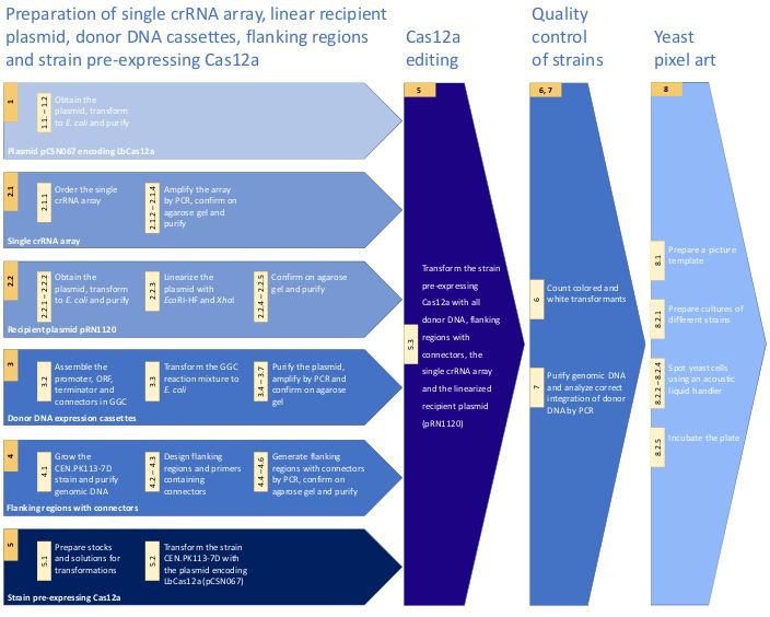
Journal of Visualized Experiments                                                                                            www.jove.com

  guide RNA (sgRNA) or alternatively for example expression of an additional endonuclease (e.g., Csy4) in combination with recognition motifs for
                                  14,15
  Csy4 surrounding each sgRNA          . Thirdly, Cas12a target site recognition requires a protospacer adjacent motif (PAM) at the 5’ end from the
  target and cleaves after the +18/+23 position from its PAM resulting in cleaved DNA with sticky ends, whereas Cas9 requires a PAM located on
                                                                                                   12
  the 3’ end from the target and cleaves after the -3 position creating blunt end cuts in the DNA . Fourthly, the consensus nucleotide sequence
  of the PAM differs between Cas12a ((T)TTV) and Cas9 (NGG), which makes Cas12a a promising candidate for targeting T-rich promoter and
                         16                                                                                               17
  terminator sequences . Finally, a recent study reported greater target specificity for Cas12a than for the native Cas9 .

  We present a protocol for using the CRISPR/Cas12a system for genome editing of S. cerevisiae with a particular focus on the introduction
  of multiple DNA expression cassettes into independent genomic loci simultaneously (multiplex genome editing) using a single crRNA array.
  The key steps of the protocol are depicted in Figure 1. As a proof of concept, the CRISPR/Cas12a system was applied for introduction of
                                                                                                                18
  three expression cassettes into the genome of S. cerevisiae which enable the production of β-carotene as schematically shown in Figure 2.
  Production of β-carotene affects the phenotype of S. cerevisiae: i.e., upon successful introduction of all three heterologous genes required for
  carotenoids biosynthesis, the white S. cerevisiae cells turn yellow or orange, depending on the expression strength of each gene’s promoter.
  Due to the simple visual read-out of this pathway, it has been introduced to develop advanced CRISPR-based systems and methods for genome
          19,20
  editing      . In this work, expression cassettes encoding the carotenoid genes crtE, crtYB and crtI have been constructed using a Golden Gate
                               21
  cloning (GGC) approach with heterologous promoters and homologous terminators used to drive expression of the genes. The expression
  cassettes are surrounded by unique 50-base pairs (bp) sequences, called connectors, that allow for in vivo assembly with integration flank
  DNA sequences (flanking regions) with the same 50-bp sequences, and subsequent integration into the genomic DNA of yeast at the position
  determined by the flanking regions. By using different promoter strengths, strains with different levels of carotenoids production were obtained
                                                                                                   22
  resulting in variation in color of the cells. These strains - inspired by the “Yeast Art Project” - were used in a spotting setup with an acoustic
  liquid handler to create a 4-color high-resolution “yeast photograph” of Rosalind Franklin. Franklin (1920-1958) was an English chemist and X-ray
                                                                                                         23,24,25
  crystallographer well known for her contribution to the discovery of the DNA structure by Photo 51             .

Protocol

1. Preparation of the Cas12a plasmids
  NOTE: The plasmid containing the Lachnospiraceae bacterium ND2006 Cas12a (LbCpf1, pCSN067) codon optimized for expression in S.
                                          19
  cerevisiae, was previously constructed , deposited at a plasmid repository (see the Table of Materials). This is a single-copy episomal S.
  cerevisiae/E. coli shuttle plasmid containing a KanMX resistance marker gene to allow for selection of S. cerevisiae transformants on geneticin
  (G418).

  1. Obtain the pCSN067 plasmid (see the Table of Materials).
  2. Amplify the pCSN067 plasmid to obtain a high amount.
      1. Transform 25 µL of purchased chemically competent E. coli cells with the plasmid pCSN067 according to the manufacturer’s protocol.
           Dilute the transformation mix 10 and 50 times in 2x peptone-yeast (PY). Plate out 10x and 50x dilutions on 2x PY agar plates
           containing ampicillin (0.1 g/L) and incubate overnight at 37 °C.
      2. Pick 2 to 3 colonies and inoculate each colony in 3 mL of 2x PY and grow overnight at 37 °C in a shaking incubator at 180 rpm.
      3. Purify the plasmid using a plasmid purification kit according to manufacturer's instructions.

2. Preparation of the single crRNA array expression cassette
  1. Prepare the single crRNA array.
                                                                                                                    2
     NOTE: The single crRNA array comprises an SNR52 RNA polymerase III promoter from S. cerevisiae , a direct repeat specific for LbCas12a
                                                                                       19                                                    2
     and a spacer (genomic target sequence), together repeated for each target and ends with a SUP4 terminator from S. cerevisiae . The
     single crRNA array is assembled by in vivo recombination into the linearized plasmid pRN1120 to generate a circular plasmid, thus regions
     homologous to plasmid pRN1120 must be present at the start and end of the single crRNA array (see Figure 2A). It is recommended to
                                                                                             19
     in advance evaluate the functionality of a number of designed crRNAs separately . This information is subsequently used to select most
     functional crRNAs to combine these into the direct repeat and spacer sequences to create a single crRNA array for the multiplexing purpose.
        1. Order the single crRNA array for multiplex genome editing experiments as synthetic DNA (see the DNA sequence of the single crRNA
            array in Supplementary Table 1).
        2. Amplify the ordered single crRNA array (e.g., using primers KC-101 and KC-102 (Supplemental Table 2)). Prepare the PCR
            amplification mix containing: 0.5 µL of DNA polymerase, 10 µL of 5x buffer required for the DNA polymerase, 1 µL of 10 mM dNTPs,
            2.5 µL of 10 µM forward primer, 2.5 µL of 10 µM reverse primer, 2 µL of DNA template at a concentration of 5 ng/µL and ultrapure H2O
            up to a total volume of 50 µL.
              1. Perform the reaction in a thermocycler using the following program: (i) 98 °C for 3 min, (ii) 98 °C for 10 s, (iii) 60 °C for 20 s, (iv)
                   72 °C for 15 s – repeat steps (ii) to (iv) 30 times, (v) 72 °C for 5 min (vi) hold at 12 °C until further analysis.

        3. Analyze the PCR products by electrophoresis by running the samples on a 0.8% agarose gel at 5 V/cm for 40 min using a DNA loading
           dye and DNA ladder with DNA fragments in a range of 100 to 10,000 bp.
        4. Purify the PCR products using a PCR purification kit according to the instructions of the manufacturer.

  2. Prepare the single crRNA array recipient plasmid.
                                                                                                        19
     NOTE: The single crRNA array is expressed from the S. cerevisiae/E. coli shuttle plasmid pRN1120 (see the Table of Materials). This
     multi-copy plasmid contains a NatMX resistance marker gene to allow selection of S. cerevisiae transformants on nourseothricin (NTC).
       1. Obtain the pRN1120 plasmid.
       2. Amplify the pRN1120 plasmid to obtain a high amount.

Copyright © 2019 Creative Commons Attribution 3.0 License                                                      May 2019 | 147 | e59350 | Page 2 of 15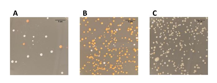
Journal of Visualized Experiments                                                                                          www.jove.com

              1. Transform 25 µL of purchased chemically competent E. coli cells with plasmid pRN1120 according to the manufacturer’s
                 protocol. Dilute the transformation mix 10 and 50 times in 2x PY. Plate out 10x and 50x dilutions on 2x PY agar plates containing
                 ampicillin (0.1 g/L) and incubate overnight at 37 °C.
              2. Pick 2 to 3 colonies and inoculate each colony in 3 mL of 2x PY and grow overnight at 37 °C in a shaking incubator at 180 rpm.
              3. Purify the plasmid using a plasmid purification kit according to the manufacturer's instructions.

        3. Linearize plasmid pRN1120 with EcoRI-HF and XhoI. For this, prepare a digestion mix composed of 1 µg of pRN1120, 5 µL of 10x
           buffer (1x buffer contains 50 mM potassium acetate, 20 mM Tris-acetate, 10 mM magnesium acetate, 100 µg/mL bovine serum
           albumin [BSA]; pH 7.9), 1 µL of EcoRI-HF (20 U), 1 µL of XhoI (20 U) and ultrapure H2O up to a total volume of 50 µL. Incubate the
           digestion mix at 37 °C for 2 h and inactivate at 65 °C for 20 min.
        4. Analyze the linearized plasmid by electrophoresis on an agarose gel (0.8%, 40 min, 5 V/cm) using a DNA loading dye and DNA ladder
           with DNA fragments in a range of 100 to 10,000 bp. As a control include a circular plasmid in the analysis.
        5. Purify the linearized plasmid using a PCR purification kit according to the instructions of the manufacturer.

3. Preparation of Promoter-ORF-Terminator (POT) donor DNA constructs
  1. Order a set of promoter (P) of different strength, open reading frame (O) and terminator (T) sequences as synthetic DNA such that each
                                                                                                                                                 26
     element contains standardized 4-bp recognition sequences that are flanked by BsaI sites to enable Golden Gate Cloning (GGC) assembly
     (see the detailed designs in Supplementary Table 3 and sequences in Supplementary Table 4).
  2. Assemble POT expression cassettes composed of a promoter, open reading frame, terminator and connectors sequences via a 4-
                                             21
     part assembly using a GGC reaction , into a destination vector that already contains pre-specified 50-bp connectors sequences (see
                                                  26,27
     Supplementary Table 4 and references              ).
       1. Measure the concentration of DNA parts using a spectrophotometer. Dilute each DNA part in ultrapure H2O to a final concentration of
           15 fmol/µL.
       2. Prepare a reaction mix composed of DNA fragments: 2 µL of promoter, 2 µL of open reading frame, 2 µL of terminator and 2 µL
                                                                       26
           backbone (Level 1 destination vectors as described in ), 4 µL of 5x T4 DNA ligase buffer, 2.5 µL of 1 U/µL T4 DNA Ligase, 1.5 µL of
           20 U/µL BsaI-HF and ultrapure H2O up to a total volume of 20 µL.
       3. Perform the GGC reaction in a thermocycler using the following program: (i) 37 °C for 2 min, (ii) 16 °C for 5 min – repeat steps (i) and
           (ii) 50 times, (iii) 50 °C for 60 min, (iv) 80 °C for 45 min, (v) hold at 12 °C until further analysis.
                                                                  28
  3. Transform 25 µL of purchased chemically competent E. coli cells with 3 µL of the GGC reaction mix according to manufacturer’s protocol.
     Dilute the transformation mix 10 and 50 times in 2x PY. Plate out 10x and 50x dilutions on 2x PY agar plates containing ampicillin (0.1 g/L)
     and incubate overnight at 37 °C.
  4. Pick 2 to 3 colonies and inoculate each colony in 3 mL of 2x PY and grow overnight at 37 °C in a shaking incubator at 180 rpm.
  5. Purify the plasmids using a plasmid purification kit according to manufacturer's instructions.
  6. Check if POT expression cassettes were assembled correctly in the GGC reaction by PCR.
       1. Design primers complementary to the connector sequence present at the start and the end of each expression cassette (see Figure
            2B). For connectors chosen in this protocol use primers KC-103 to KC-108 (see Supplementary Table 2).
       2. Prepare PCR amplification mixes for each plasmid containing: 0.5 µL of proofreading DNA polymerase, 10 µL of 5x buffer required for
            the DNA polymerase, 1 µL of 10 mM dNTPs, 2.5 µL of 10 µM forward primer, 2.5 µL of 10 µM reverse primer, 2 µL of DNA template
            with a concentration of 5 ng/µL, and ultrapure H2O up to a total volume of 50 µL.
       3. Perform the PCR reaction in a thermocycler using the following program: (i) 98 °C 3 min, (ii) 98 °C for 10 s, (iii) 60 °C for 20 s, (iv) 72
            °C for 2 min 30 s – repeat steps (ii) to (iv) 30 times, (v) 72 °C for 5 min, (vi) hold at 12 °C until further analysis.
            NOTE: Resulting PCR products consist of 50-bp of the 5’ connector, promoter, open reading frame, terminator and 50-bp of the 3’
            connector.

  7. Analyze the PCR products by electrophoresis by running samples on a 0.8% agarose gel at 5 V/cm for 40 min using a DNA loading dye and
     DNA ladder with DNA fragments in a range of 100 to 10,000 bp.

4. Preparation of integration flank DNA sequences containing connectors sequences
                                                                        29
  1. Purify genomic DNA from wild type S. cerevisiae CEN.PK113-7D .
       1. Grow the strain in a 500 mL shake flask filled with 100 mL of yeast extract peptone dextrose (YEPD, 2% glucose) medium at 30 °C and
            shaking at 250 rpm for 48 hours.
       2. Harvest the cells by centrifugation of 2 mL of broth at 16,000 x g for 1 min and discard the supernatant.
       3. Resuspend the cells in physiological salt (200 µL; 0.85% NaCl solution) with RNase (10 µL, 10 mg/mL) and yeast lytic enzyme (4 µL).
            Incubate the cell suspension at 37 °C for 15 min.
       4. Add 300 µL of cell lysis solution (see Table of Materials) and vortex shortly.
       5. Add 168 µL of protein precipitation solution (see Table of Materials) and vortex vigorously for 20 s.
       6. Separate the protein fraction by centrifugation at 16,000 x g and 4 °C for 10 min. Collect 600 µL of supernatant in a new tube and mix
            with 600 µL of isopropanol and vortex shortly.
       7. Recover DNA by spinning down at 16,000 x g at room temperature for 10 min. Discard the supernatant and keep the pellet.
       8. Wash the pellet with 200 µL of ethanol (70%). Centrifuge at 16,000 x g at room temperature for 10 min and remove the supernatant.
            Evaporate the ethanol by incubating the tube at room temperature for 10 min with the lid opened.
            NOTE: If liquid in the tube is still visible, repeat the step 4.1.8. Do not dry the pellet for longer than 10 min to prevent decreased
            solubility of the DNA.
       9. Dissolve DNA in 50 µL of TE buffer. Store purified DNA at 4 °C.

Copyright © 2019 Creative Commons Attribution 3.0 License                                                    May 2019 | 147 | e59350 | Page 3 of 15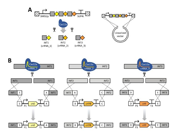
Journal of Visualized Experiments                                                                                                     www.jove.com

  2. For each integration site, design integration flank DNA sequences (approx. 500 bp) such that approximately 1000 bp of genomic DNA will be
     removed upon introduction of donor DNA (see the schematic design in Figure 2B and sequences in Supplementary Table 4).
  3. Design primers to generate the flanking regions by PCR.
       1. For the left flanking region, design forward and reverse primers to amplify approximately 500 bp of the genomic DNA region
           positioned 5’ (left) of the integration site of interest.
           NOTE: The forward primer includes 20 bp of homology with the intended flanking region. The reverse primer includes 20 bp with
           homology with the intended flanking region and contains the desired 50-bp connector sequence to enable in vivo assembly in the
           Cas12a editing on the genome later on.
       2. For the right flanking region, design forward and reverse primers to amplify approximately 500 bp of the genomic DNA region
           positioned 3’ (right) of the integration site of interest.
           NOTE: The forward primer includes 20 bp with homology with the intended flanking region and contains the desired 50-bp connector
           sequence to enable in vivo assembly in the Cas12a editing on the genome later on. The reverse primer includes 20 bp of homology
           with the intended flanking region.

  4. Amplify the flanking regions with the designed primers (e.g., primers KC-109 to KC-120 enclosed in Supplementary Table 2).
      1. Measure the concentration of purified genomic DNA that will serve as the template in the PCR. Adjust the DNA concentration to 50 ng/
           µL.
      2. Prepare PCR amplification mixes composed of genomic DNA (1 – 4 µL of 50 ng/µL genomic DNA dilution) purified in step 4.1,
           forward and reverse primer (10 µM each), 1 µL of 10 mM dNTPs, 10 µL of 5x buffer required for the DNA polymerase, 0.5 µL of DNA
           polymerase (1.0 U), and ultrapure H2O up to total volume of 50 µL.
      3. Perform PCRs in a thermocycler using the following program: (i) 98 °C for 3 min, (ii) 98 °C for 20 s, (iii) 60 °C for 20 s, (iv) 72 °C for 15
           s, repeat steps (ii) to (iv) 30 times, (v) 72 °C for 5 min, (vi) hold at 12 °C until further analysis.

  5. Analyze the PCR products by electrophoresis on a 0.8% agarose gel at 5 V/cm for 40 min using a DNA loading dye and DNA ladder with
     DNA fragments in a range of 100 to 10,000 bp.
  6. Purify the correct PCR products using a PCR purification kit according the instructions of the manufacturer.

5. Transformation to S. cerevisiae
                                                                                                              30                     31
  NOTE: Perform transformation using a protocol based on the methods developed by Gietz et al. (1995)              and Hill et al.        which can be used for
  various strains of S. cerevisiae. The protocol described below is sufficient for 1 transformation.

  1. Prepare solutions required for transformation.
       1. Prepare the following stock solutions and filter-sterilize: 10x TE buffer containing 100 mM Tris-HCl (pH 7.5), 10 mM EDTA, total volume
          of 50 mL; 1 M LiAc at pH 7.5, total volume of 50 mL; 50% PEG 4000, total volume of 100 mL.
          NOTE: Always check that PEG 4000 stock is at pH 5. This stock should not be stored longer than one month.
       2. Prepare the following solutions using stocks: Prepare LiAc-TE solution containing 0.1 M LiAc, 10 mM Tris-HCl, 1 mM EDTA, total
          volume of 0.5 mL. Prepare PEG-LiAc-TE solution containing 40% PEG 4000, 0.1 M LiAc, 10 mM Tris-HCl, 1 mM EDTA, total volume of
          1 mL.
          NOTE: It is crucial for successful transformation that PEG-LiAc-TE and LiAc-TE solutions are freshly prepared.

  2. First transformation round (prepare the strain pre-expressing Cas12a).
     NOTE: In all the transformation steps, use water with a pH higher than 5. It is recommended to use demineralized water in all the steps of the
     transformation.
        1. Prepare a pre-culture by growing strain CEN.PK113-7D in a 100 mL shake flask containing 20 mL of YEPD (2% glucose) medium and
             incubate overnight at 30 °C with shaking at 250 rpm.
        2. Measure the OD600 of the pre-culture (ODpc). Calculate the dilution factor (df) between the volume of pre-culture and the volume of
             fresh medium required for preparation of the cells pre-expressing Cas12a to be used in the transformation (transformation culture). In
             the calculations assume the optical density of the transformation culture (ODtc) to be 1.0 after the incubation step described in 5.2.3 (ti).

            where ti and τ are the incubation time and doubling time, respectively.
              1. Calculate the volume of the pre-culture (Vi) required for inoculation of the transformation culture (Vtc) based on the dilution factor.

        3. Prepare the transformation culture by inoculation of 20 mL of YEPD (2% glucose) (Vtc) with the volume of pre-culture determined in the
            previous step (Vi). Incubate at 30 °C with shaking at 250 rpm.
        4. Measure the OD600 of the transformation culture until an OD600 of 1.0 is reached.
        5. Harvest the cells by centrifugation of the 20 mL broth at 2,500 x g for 5 min. Discard the supernatant and wash the cells in 20 mL of
            room temperature demineralized water. Repeat the centrifugation step and keep the cell pellet.
        6. Resuspend the cells in 100 µL of LiAc-TE solution and transfer to a microcentrifuge tube.
        7. Add 5 µL of single-stranded carrier DNA (10 mg/mL salmon sperm DNA) and mix by pipetting.
        8. Pipette 1 µg of plasmid pCSN067 to the microcentrifuge tube.
            NOTE: The total volume of the DNA mixture should not exceed 100 µL to prevent a lower transformation efficiency.
        9. Add 600 µL of PEG-LiAc-TE solution and mix by pipetting. Incubate for 30 min at 30 °C while shaking at 450 rpm in a table top heat
            block.
        10. Add 70 µL of DMSO (100%) to the transformation mixture and mix by pipetting. Perform heat-shock by incubating the transformation
            mixture at 42 °C for 15 minutes in a water bath.

Copyright © 2019 Creative Commons Attribution 3.0 License                                                      May 2019 | 147 | e59350 | Page 4 of 15
Journal of Visualized Experiments www.jove.com
11. Recover the cells by transferring the mixture to a 15 mL round bottom tube and add 10 mL of YEPD (2% glucose) to the tube. Incubate
overnight at 30 °C with shaking at 250 rpm.
12. Centrifuge the transformation mix at 2,500 x g for 5 min. Discard the supernatant and resuspend the cell pellet in approximately 200 µL
of the remaining solution.
13. Plate out 150 µL of the transformation mix and a 20x dilution in YEPD (2% glucose) of transformation mix on YEPD (2% glucose) agar
plates supplemented with 0.2 g/L G418. Incubate the plates at 30 °C for 48 – 72 hours.
14. Pick a single transformant and re-streak on a YEPD (2% glucose) agar plate supplemented with 0.2 g/L G418 to obtain single colonies.
3. Second transformation round (perform multiplex genome editing with CRISPR/Cas12a).
1. Prepare a pre-culture by growing the strain pre-expressing Cas12a, created in the first transformation round (step 5.2), in a 100 mL
shake flask containing 20 mL of YEPD (2% glucose) medium supplemented with 0.2 g/L G418. Incubate overnight at 30 °C with
shaking 250 rpm.
NOTE: For multiple transformations, adapt the volume of the pre-culture.
2. Follow the steps 5.2.2 to 5.2.7 for the first transformation round.
NOTE: For multiple transformations, adapt the volumes of required solutions and culture of the strain pre-expressing Cas12a.
3. Pipette 1 µg of the single crRNA array, 1 µg of the linearized recipient plasmid for the crRNA array, 1 µg of donor DNA and 1 µg of each
flanking region (step 4.3) in the microcentrifuge tube.
NOTE: The total volume of the DNA mixture should not exceed 100 µL to prevent a lower transformation efficiency.
4. Prepare the following controls for the transformation: negative control (ultrapure H2O); positive control for determination of the
transformation efficiency (1 µg of circular pRN1120); a control verifying if introduction of donor DNA is conducted via CRISPR editing
(1 µg of circular pRN1120, 1 µg of all donor DNA expression cassettes and 1 µg of flanking regions but no single crRNA array); control
verifying if donor DNA can be integrated outside of target (1 µg of linearized pRN1120, 1 µg of donor DNA expression cassettes and 1
µg of the single crRNA array but no flanking regions); a control verifying full linearization of pRN1120 (1 µg of linearized pRN1120).
5. Follow the steps 5.2.9 to 5.2.12 for the first transformation round.
6. Plate out 150 µL of the transformation mix and 20x dilution in YEPD (2% glucose) of transformation mix on YEPD (2% glucose) agar
supplemented with 0.2 g/L G418 and 0.2 g/L NTC. Plate out controls on YEPD (2% glucose) agar supplemented with the appropriate
selection (G418 and/or NTC or no selection). Incubate the plates at 30 °C for 48 – 72 hours.
7. Pick a single colored transformant and re-streak on a YEPD (2% glucose) agar plate to obtain single colored colonies.
6. Evaluation of the genome editing efficiency
1. Count the number of colored colonies and white colonies on the transformation plates.
2. Calculate genome editing efficiency by dividing the number of colored colonies by the total number of colonies (both white and colored), as
shown in Table 1.
7. Confirmation of integration of donor DNA at the intended loci
1. Re-streak a colored single colony from a transformation plate on a YEPD (2% glucose) agar plate without G418 and NTC selection and
incubate for 48 hours at 30 °C.
2. Pick a single colony and inoculate a 500 mL shake flask filled with 100 mL of YEPD (2% glucose) medium. Incubate for 48 hours at 30 °C
and shaking at 250 rpm.
3. Isolate the genomic DNA as described in Section 4.1.
32
NOTE: Alternatively, use a protocol for preparation of yeast for colony PCR previously proposed by Looke et al. . In this case, growth in
liquid medium (Section 7.2) can be skipped.
4. Verify correct integration by amplification of two fragments per integrated expression cassette.
1. Design primers which anneal to genomic DNA outside of the transformed flanking regions and the gene of interest (see examples
in Supplementary Table 2, KC-121 to KC-132). When using primers KC-121 to KC-132, set the annealing temperature in the PCR
program to 62 °C.
2. Amplify region of interest as described in Section 4.4.2. Adapt the PCR program, specifically adjust the time of the extension step in
PCR according to the length of the template and manufacturer’s recommendations for the DNA polymerase.
5. Check the size of the PCR products by electrophoresis on an agarose gel (0.8%, 40 min, 5 V/cm) using a DNA loading dye and DNA ladder
with DNA fragments in a range of 100 to 10,000 bp.
8. Creation of yeast pixel art using an acoustic liquid handler
1. Prepare a picture template for the yeast pixel art.
1. Resize the original RGB picture (220 × 280 pixels, see the representative results), e.g. using ImageJ to create a final 64 × 96 pixels
(width × height) grey-scale image visualized in intended colors (Representative Results).
2. Convert the RGB picture into grey-scale using this formula:
where Igr, Ir, Ig, Ib are the grey, red, green and blue intensities, respectively.
3. In order to categorize the pixels, develop an ImageJ plugin applying the following rules: (a) If Igr is ≤ 64, use the dark orange yeast
(strain 1, Supplementary Table 3) for this pixel. (b) If 64 < Igr ≤ 128, use the orange yeast (strain 2, Supplementary Table 3) for this
Copyright © 2019 Creative Commons Attribution 3.0 License May 2019 | 147 | e59350 | Page 5 of 15Journal of Visualized Experiments www.jove.com
pixel. (c) If 128 < Igr ≤ 192, use the yellow yeast (strain 3, Supplementary Table 3) for this pixel. (d) If Igr > 192, use the white yeast
(CEN.PK113-7D) for this pixel.
2. Spot yeast cells to create the yeast pixel art.
1. Inoculate 500 mL shake flasks containing 100 mL of YEPD (2% glucose) medium with three differently colored carotenoid producing S.
cerevisiae strain and wild type CEN.PK113-7D. Incubate cultures overnight at 30 °C with shaking at 250 rpm.
2. Transfer 0.5 mL of the overnight culture to a tube filled with 0.5 mL of sterile non-ionic density gradient medium (see the Table of
Materials). Mix by vortexing briefly.
3. Transfer the cell suspension to a qualified reservoir, 2 x 3 well. Perform spotting using an acoustic liquid handler instrument from a
qualified reservoir source plate to a microplate (see the Table of Materials) containing 50 mL of YEPD (2% glucose) agar. To simplify
plating, define wells on plate, e.g. use a microplate as a 6144 well plate (64 × 96).
4. Spot 25 nL of each S. cerevisiae strain from the 2x 3 well reservoir source plate using a .csv file with the fluid calibration setting
6RES_AQ_GPSA2 onto the destination microplate. Define each of these 25 nL droplets as a pixel in the 64 x 96 grid which is
translated to the well positions (A01, B01, C01 etc.).
5. Incubate the microplate at 30 °C for 48 hours. To intensify the colors of the strains store the agar plate at 4 °C for at least 72 hours.
Representative Results
The protocol for multiplex genome editing using CRISRP/Cas12a was demonstrated by constructing three carotenoid producing S. cerevisiae
strains expressing the crtE, crtYB and crtI genes using heterologous promoters of high, medium and low strength: strain 1, 2 and, 3 respectively
(Supplementary Table 3). Construction of these strains required generation of three donor DNA expression cassettes and six flanking regions
per strain for targeting to three different loci in genomic DNA (shown in Figure 2B). As described herein, promoter, open reading frame,
terminator and two contiguous 50-bp connectors sequences were assembled into an expression cassette via a Golden Gate Cloning reaction
and the assembly was verified by PCR (Figure 3A). The single crRNA array was ordered as a synthetic DNA fragment and was amplified by
PCR (Figure 3B). The recipient plasmid for the single crRNA array (plasmid pRN1120) was linearized with EcoRI-HF and XhoI and linearization
was confirmed by electrophoresis (Figure 3C). The design and nucleotide sequences of the introduced donor DNA expression cassettes and
flanking regions are shown in Supplementary Table 3 and Supplementary Table 4. The sequence of single crRNA array expression cassettes
is provided in Supplementary Table 1. Functionality of the spacers included in the single crRNA array was tested beforehand by singleplex
19
genome editing with individual crRNAs .
The efficiency of genome editing using Cas12a was firstly evaluated based on the number of colored colonies obtained after transformation
(Table 1, Figure 4). The editing efficiency of the three constructed strains varied from 50% to 94%. Notably, introduction of expression cassettes
used to generate strain 1 displayed the lowest editing efficiency, possibly caused by the nature of the donor DNA (i.e., these expression
cassettes encode crtE, crtYB and crtI from three high strength promoters). Secondly, correct integration of the three donor DNA expression
cassettes at the intended loci on the genomic DNA was confirmed by PCR (Figure 5). Primers were designed in such a way that PCR products
were obtained when correct integration of donor DNA at the intended locus occurred. For each transformation experiment, eight colonies were
picked from the transformation plate and tested (note that only three are presented in Figure 5). In general, out of 8 colonies tested per donor
DNA, correct integration of the crtE donor DNA at the INT1 locus, crtYB at the INT2 locus and crtI at the INT3 locus was confirmed in >90% of
the transformants. These results demonstrate the CRISPR/Cas12a system in combination with a single crRNA array enables efficient multiplex
editing of the S. cerevisiae genome at multiple loci simultaneously.
Additionally, we demonstrate the creation of “yeast pixel art” using the three carotenoid producing strains that were constructed together with a
non-colored wild-type strain. Starting from a black and white picture of Rosalind Franklin (Figure 6A), a 4-color picture (Figure 6B) and spotting
list was created which was then used to spot the four different yeast strains on an agar microplate using an acoustic liquid handler, resulting in a
high-resolution “yeast painting” of Rosalind Franklin (Figure 6C,D,E).
Copyright © 2019 Creative Commons Attribution 3.0 License May 2019 | 147 | e59350 | Page 6 of 15Journal of Visualized Experiments www.jove.com Figure 1: Workflow of the protocol for CRISPR/Cas12a multiplex genome editing in S. cerevisiae. The workflow includes crucial steps of the presented method. For details see the Protocol. Please click here to view a larger version of this figure. Copyright © 2019 Creative Commons Attribution 3.0 License May 2019 | 147 | e59350 | Page 7 of 15
Journal of Visualized Experiments                                                                                      www.jove.com

  Figure 2: Scheme of CRISPR/Cas12a multiplex genome editing using a single crRNA array. (A) The single crRNA array is composed
  of three crRNAs units in their mature form, a 20-bp direct repeat specific for LbCas12a (grey squares) with a 23-bp guide sequence (colored
  diamonds). Expression of the crRNA array is enabled by the SNR52 promoter and SUP4 terminator. Transformation of S. cerevisiae with a
  linearized pRN1120 and the single crRNA array expression cassette containing homology with pRN1120 (diagonal stripes) allows for in vivo
  recombination into a circular plasmid in cells pre-expressing LbCas12a. The single crRNA array is subsequently processed by Cas12a. (B)
  Cas12a is directed to the intended INT1, INT2 and INT3 genomic target sites and creates double stranded breaks. In the transformation mixture,
  donor DNA consisting of flanking regions and the carotenoid gene expression cassette were included. Donor DNA assemblies were targeted to
  one stretch of DNA in genomic DNA around the INT1 (crtE), INT2 (crtYB) and INT3 (crtI) loci by in vivo recombination due to the presence of 50-
  bp homologous connectors sequences, indicated as 5, A, B, C, D or E. P1–P3, different promoters; T1–T3, different terminators. This figure has
                                            19                                                                                        40
  been modified from Verwaal et al. 2018 . Genetic constructs shown using Synthetic Biology Open Language (SBOL) Visual symbols . Please
  click here to view a larger version of this figure.

Copyright © 2019 Creative Commons Attribution 3.0 License                                                May 2019 | 147 | e59350 | Page 8 of 15
Journal of Visualized Experiments www.jove.com Figure 3: PCR verifying the genome editing experiments. (A) Verification of Golden Gate Cloning reactions of assembled donor DNA cassettes. Obtained results are in agreement with expected lengths. (B) PCR of the single crRNA array. (C) Linearization of plasmid pRN1120. Please click here to view a larger version of this figure. Figure 4: Plates of S. cerevisiae transformations using the multiplex genome editing approach. (A) Strain 1 expressing crtE, crtYB and crtI from three strong promoters (dark orange colonies). (B) Strain 2 expressing crtE, crtYB and crtI from three medium strength promoters (orange colonies). (C) Strain 3 expressing crtE, crtYB and crtI from three low strength promoters (yellow colonies). Please click here to view a larger version of this figure. Copyright © 2019 Creative Commons Attribution 3.0 License May 2019 | 147 | e59350 | Page 9 of 15
Journal of Visualized Experiments                                                                                                www.jove.com

  Figure 5: PCR verifying integration of the donor DNA expression cassettes at the intended loci within the genomic DNA. (A) Verification
  of three colonies of the strain 1. (B) Verification of three colonies of the strain 2. (C) Verification of three colonies of the strain 3. Please click here
  to view a larger version of this figure.

Copyright © 2019 Creative Commons Attribution 3.0 License                                                        May 2019 | 147 | e59350 | Page 10 of 15
Journal of Visualized Experiments                                                                                         www.jove.com

  Figure 6: Yeast pixel art of Rosalind Franklin. (A) Black and white RGB photo of 220 × 280 pixels of Rosalind Franklin that was used as a
  template. (B) Computer conversion of the black and white photo of Rosalind Franklin into a 4-color 64 × 96 pixel list. (C) Photo of yeast pixel art
  with 64 × 96 yeast colonies with a zoomed-in section. (D) Photo of an acoustic liquid handler with two full grown plates. (E) Photo of a full grown
  microplate with 64 × 96 yeast colonies. Please click here to view a larger version of this figure.

                                       Strain 1                            Strain 2                              Strain 3
Colored colonies                       16                                  279                                   220
White colonies                         16                                  18                                    18
Total colonies                         32                                  297                                   238
Efficiency                             50%                                 94%                                   92%

  Table 1: Editing efficiency of the multiplex genome editing approach.

                         a,b,c,d,e,f
crRNA array sequence
CATGTTTGACAGCTTATCATCGATAATCCGGAGCTAGCATGCGGCCGCTCTAGAACTAGTGGATCCCCCGGGCTGCAGTCTTTGAAAA
GATAATGTATGATTATGCTTTCACTCATATTTATACAGAAACTTGATGTTTTCTTTCGAGTATATACAAGG
TGATTACATGTACGTTTGAAGTACAACTCTAGATTTTGTAGTGCCCTCTTGGGCTAGCGGTAAAGGTGCGCA
TTTTTTCACACCCTACAATGTTCTGTTCAAAAGATTTTGGTCAAACGCTGTAGAAGTGAAAGTTGGTGCGC
ATGTTTCGGCGTTCGAAACTTCTCCGCAGTGAAAGATAAATGATCAATTTCTACTAAGTGTAGAT
CTGGTGGGAGAGAAAGCTTATGAAATTTCTACTAAGTGTAGATGTGCCGTAC
GCCGGAGCCGACGGAATTTCTACTAAGTGTAGATTGCCCCTCTTATACGATTATATTTT
TTTTTGTTTTTTATGTCTGGGGGGCCCGGTACCCAGCTTTTGTTCCCTTTAGTGAGG
GTTAATTCCGAGCTTGGCGTAATCATGGTCATAGCTGTTTCCTGTGTG
a. Homology to pRN1120 (bold).
b. SNR52 promoter (italics).
c. Genomic target sequences (underlined).
d. Guide direct repeats specific for LbCas12a (italics, bold).
e. SUP4 terminator (italics).
f. Homology to pRN1120 (bold).

  Supplementary Table 1: Single crRNA array for LbCas12a containing homology with plasmid pRN1120.

Copyright © 2019 Creative Commons Attribution 3.0 License                                                  May 2019 | 147 | e59350 | Page 11 of 15
Journal of Visualized Experiments www.jove.com
Name Sequence
a
Description
b Used in point
KC-101 CATGTTTGACAGCTTATCATC FW primer for amplification of 2.1.4
single crRNA array
KC-102 CACACAGGAAACAGCTATGAC RV primer for amplification of 2.1.4
single crRNA array
KC-103 AAGCGACTTCCAATCGCTTTGC FW primer for amplification of 3.6.1
donor DNA with connector 5
KC-104 AAAGCAAAGGAAGGAGAGAAC RV primer for amplification of donor 3.6.1
DNA with connector A
KC-105 CGGATCGATGTACACAACCG FW primer for amplification of 3.6.1
donor DNA with connector B
KC-106 CAACAGGAGGCGGATGGATATAC RV primer for amplification of donor 3.6.1
DNA with connector C
KC-107 AACGTTGTCCAGGTTTGTATCC FW primer for amplification of 3.6.1
donor DNA with connector D
KC-108 AGGTACAACAAGCACGACCG RV primer for amplification of donor 3.6.1
DNA with connector E
KC-109 CACTATAGCAATCTGGCTATATG FW primer for amplification of INT1 4.4
5' with connector 5
KC-110 AAACGCCTGTGGGTGTGGTAC RV primer for amplification of INT1 4.4
TGGATATGCAAAGCGATTGGAA 5' with connector 5
GTCGCTTGACTCCTCTGCCGTC
ATTCC
KC-111 TTGCCCATCGAACGTACAAG FW primer for amplification of INT1 4.4
TACTCCTCTGTTCTCTCCTTCCTT 3' with connector A
TGCTTTAAGCGTTGAAGTTTCCTC
TTTG
KC-112 TGTCAACTGGAGAGCTATCG RV primer for amplification of INT1 4.4
3' with connector A
KC-113 AGAAGATTTCTCTTCAATCTC FW primer for amplification of INT2 4.4
5' with connector B
KC-114 TGCTAAGATTTGTGTTCGTT RV primer for amplification of INT2 4.4
TGGGTGCAGTCGGTTGTGTACAT 5' with connector B
CGATCCGCCCTTATCAAGGATACC
TGGTTG
KC-115 ACGCTTTCCGGCATCTTCCA FW primer for amplification of INT2 4.4
GACCACAGTATATCCATCCGCCT 3' with connector C
CCTGTTGGGCGATTACACAAGCG
GTGG
KC-116 TCTCCTCTTCGATGACCGGG RV primer for amplification of INT2 4.4
3' with connector C
KC-117 GGTCGTTTTTGTGCAGCATATTG FW primer for amplification of INT3 4.4
5' with connector D
KC-118 GCGGAATATTGGCGGAACGG RV primer for amplification of INT3 4.4
ACACACGTGGATACAAACCTG 5' with connector D
GACAACGTTTTCCAAGGAGGTG
AAGAACG
KC-119 AAATAACCACAAACATCCTT FW primer for amplification of INT3 4.4
CCCATATGCTCGGTCGTGCTTGTT3' with connector E
GTACCTGATGGGACGTCAGCACT
GTAC
KC-120 GAGCTTACTCTATATATTCATTC RV primer for amplification of INT3 4.4
3' with connector E
KC-121 GTTACTAAACTGGAACTGTCCG FW primer for verification of 7.4.1
integration of con5-crtE-conA to
INT1 5'
Copyright © 2019 Creative Commons Attribution 3.0 License May 2019 | 147 | e59350 | Page 12 of 15Journal of Visualized Experiments www.jove.com
KC-122 CACTGCTAACTACGTTTACTTC FW primer for verification of 7.4.1
integration of con5-crtE-conA to
INT1 3'
KC-123 CACTGGAACTTGAGCTTGAG FW primer for verification of 7.4.1
integration of conB-crtYB-conC to
INT2 5'
KC-124 GTCTCCAGCTGAATTGGTCC FW primer for verification of 7.4.1
integration of conB-crtYB-conC to
INT2 3'
KC-125 CTCTCATGAAGCAGTCAAGTC FW primer for verification of 7.4.1
integration of conD-crtI-conE to
INT3 5'
KC-126 GATCGGTCAATTAGGTGAAG FW primer for verification of 7.4.1
integration of conD-crtI-conE to
INT3 3'
KC-127 CCTTGTCCAAGTAGGTGTCC RV primer for verification of 7.4.1
integration of con5-crtE-conA to
INT1 5'
KC-128 GCTGTCATGATCTGTGATAAC RV primer for verification of 7.4.1
integration of con5-crtE-conA to
INT1 3'
KC-129 CTGGCAATGTTGACCAATTGC RV primer for verification of 7.4.1
integration of conB-crtYB-conC to
INT2 5'
KC-130 CCAACGTGCCTTAAAGTCTG RV primer for verification of 7.4.1
integration of conB-crtYB-conC to
INT2 3'
KC-131 CCTTACCTTCTGGAGCAGCAG RV primer for verification of 7.4.1
integration of conD-crtI-conE to
INT3 5'
KC-132 CTGGTTACTTCCCTAAGACTG RV primer for verification of 7.4.1
integration of conD-crtI-conE to
INT3 3'
a. Bold sequences denote
connector sequences.
b. Forward and reverse primers
are designated as FW and RV,
respectively.
Supplementary Table 2: Primer sequences.
Supplementary Table 3: Design of constructed strains.
Copyright © 2019 Creative Commons Attribution 3.0 License May 2019 | 147 | e59350 | Page 13 of 15Journal of Visualized Experiments www.jove.com
Supplementary Table 4: Sequences of donor DNA expression cassettes and flaking regions. Please click here to download this file.
Discussion
The provided protocol describes multiplex genome editing of S. cerevisiae using Cas12a from Lachnospiraceae bacterium ND2006 in
combination with a single crRNA array and donor DNA. Design of the single crRNA array and donor DNA is explained in detail. In contrast to
the well-established CRISPR/Cas9 system, the CRISPR/Cas12a has the unique additional ability of processing multiple crRNAs expressed
13,33
from a single crRNA array . Due to this feature, simultaneous editing of multiple targets is easier to set up and can be achieved in a single
34
transformation. This single crRNA array approach was demonstrated before by Zetsche et al. who simultaneously edited up to four genes
35
in mammalian cells using AsCas12a, and by Swiat et al. who introduced four DNA fragments into a yeast genome using FnCas12a. To our
knowledge, a higher number of simultaneous genomic modifications using a Cas12a system has not been reported and the maximal limit of
targets per single array for Cas12a is yet to be determined. Further research utilizing single crRNA arrays in combination with Cas12a includes
33,36,37
multiplex transcriptional regulation in a wide range of organisms .
There are some critical steps in the presented protocol. Carefully design all DNA sequences that are involved in the Cas12a genome editing
experiment, especially in case when novel DNA sequences are introduced. Determine the functionality of new spacer sequences part of a
19
crRNA, for example by a singleplex genome editing experiment as described by Verwaal et al. before combining them into a single crRNA
array. Follow the recommendations for the preparation of transformation buffer solutions used in the Cas12a editing experiment to achieve a
good transformation efficiency of yeast.
There are some optional modifications of the technique. It is recommended to use 1 µg of each donor DNA, linearized pRN1120 or single
crRNA array expression cassette in the transformation, although the use of a lower DNA amount is also expected to result in a satisfactory
transformation efficiency. Perform a test transformation to determine whether lower DNA amounts can be used. The transformation of S.
cerevisiae might be performed using a different method than the one described in this protocol, for example the protocol described by Gietz et al.
38
(2007) . The guide RNA recipient plasmid pRN1120 is suitable for the expression of a single crRNA and single crRNA array of different Cas12a
19
variants (e.g., from Acidaminococcus spp. BV3L6 or Francisella novicida U112) as well as for expression of sgRNA in combination with Cas9 .
The donor DNA does not need to be limited to carotenoid gene expression cassettes and flanking regions that target donor DNA to the described
INT1, INT2 and INT3 sites in genomic DNA. Any DNA of interest can be introduced, in a multiplex manner, into genomic DNA of the host by the
design principles described in this protocol, or alternatively donor DNA can be used to delete DNA from a host genome. The modular structure
of single crRNA array facilitates easy adjustment of spacer and direct repeat sequences. Modification of spacer sequences allows for a change
of the intended integration locus which can be designed by one of the tools for identification of a genomic target site, e.g. GuideScan software
39
1.0 . Instead of using large flanking sequences that contain connectors sequences, 50-bp of the flanking region can be included in the donor
DNA sequences by incorporating these 50-bp flanking region sequences in the primers used in the PCR. In this case, in total just three instead of
nine donor DNA fragments are required for a successful multiplex genome editing experiment.
In summary, this protocol provides step-by-step directions to perform multiplex genome editing in S. cerevisiae using Cas12a in combination
with a single crRNA array approach. The protocol was demonstrated by multiplex genome editing using 9 donor DNA fragments and single
crRNA array coding for three gRNAs. We show high overall editing frequencies between 50% and 94% for the three strain designs reported here.
Concluding, the unique feature of Cas12a is the ability to process a single crRNA array into individual crRNAs in a cell, which makes Cas12a
an excellent tool to enable multiplex genome editing and develop transcriptional regulation modules targeting multiple expression cassettes in
one go. In the end, three strains were obtained producing carotenoids at a different level and colors in shades between yellow and orange. With
those strains and a wild-type strain, we showed how an acoustic liquid handler can be used straightforwardly to make yeast pixel art – this in
23
honor of Rosalind Franklin who contributed to the discovery of the DNA structure 65 years ago by her famous photo 51 .
Disclosures
The authors declare that there is a conflict of interest. The authors have filed IP related to presented methods.
Acknowledgments
This project received funding from the European Union’s Horizon 2020 research and innovation programme under grant agreement no.
686070 (DD-DeCaf) and 764591 (SynCrop), and from the research programme Building Blocks of Life with project number 737.016.005 by
the Netherlands Organisation for Scientific Research (NWO). T.E.G. was supported by the Royal Society (grant UF160357) and BrisSynBio,
a BBSRC/EPSRC Synthetic Biology Research Centre (grant BB/L01386X/1). We thank Zi Di and Jeffrey van Wijk for their contribution to the
yeast spotting experiments for creating the yeast pixel art.
References
1. Knott, G.J., Doudna J.A. CRISPR-Cas guides the future of genetic engineering. Science. 361 (6405), 866-869 (2018).
Copyright © 2019 Creative Commons Attribution 3.0 License May 2019 | 147 | e59350 | Page 14 of 15Journal of Visualized Experiments www.jove.com
2. DiCarlo, J.E. et al. Genome engineering in Saccharomyces cerevisiae. using CRISPR-Cas systems. Nucleic Acids Research. 41 (7),
4336-4343 (2013).
3. Gilbert, L.A. et al. CRISPR-mediated modular RNA-guided regulation of transcription in eukaryotes. Cell. 154 (2), 442-451 (2013).
4. Lian, J., HamediRad, M., Hu, S., Zhao, H. Combinatorial metabolic engineering using an orthogonal tri-functional CRISPR system. Nature
Communications. 8 (1), 1688 (2017).
5. Li, Z-H., Liu, M., Lyu, X-M., Wang, F-Q., Wei, D-Z. CRISPR/Cpf1 facilitated large fragment deletion in Saccharomyces cerevisiae. Journal of
Basic Microbiology. 58 (12), 1100-1104 (2018).
6. Shao, Y., Lu, N., Qin, Z., Xue, X. CRISPR-Cas9 facilitated multiple-chromosome fusion in Saccharomyces cerevisiae. ACS Synthetic Biology.
7 (11), 2706-2708 (2018).
7. Brouns, S. J. et al. Small CRISPR RNAs guide antiviral defense in prokaryotes. Science. 321 (5891), 960-964 (2008).
8. Jinek, M., et al. A programmable dual-RNA–guided DNA endonuclease in adaptive bacterial immunity. Science. 337 (6096), 816-821 (2012).
9. Abudayyeh, O.O. et al. C2c2 is a single-component programmable RNA-guided RNA-targeting CRISPR effector. Science. 353 (6299),
aaf5573 (2016).
10. Makarova, K.S. et al. An updated evolutionary classification of CRISPR–Cas systems. Nature Reviews Microbiology. 13 (11), 722-736 (2015).
11. Mohanraju, P. et al. Diverse evolutionary roots and mechanistic variations of the CRISPR-Cas systems. Science. 353 (6299), aad5147
(2016).
12. Zetsche, B. et al. Cpf1 is a single RNA-guided endonuclease of a class 2 CRISPR-Cas system. Cell. 163 (3), 759-771 (2015).
13. Fonfara, I., Richter, H., Bratovič, M., Le Rhun, A., Charpentier, E. The CRISPR-associated DNA-cleaving enzyme Cpf1 also processes
precursor CRISPR RNA. Nature. 532 (7600), 517-521 (2016).
14. Lian, J., HamediRad, M., & Zhao, H. Advancing metabolic engineering of Saccharomyces cerevisiae. using the CRISPR/Cas System.
Biotechnology Journal., 13 (9), 1700601 (2018).
15. Ferreira, R. et al. Multiplexed CRISPR/Cas9 genome editing and gene regulation using Csy4 in Saccharomyces cerevisiae. ACS Synthetic
Biology. 7 (1), 10-15 (2018).
16. Swarts, D.C., Martin J. Cas9 versus Cas12a/Cpf1: Structure–function comparisons and implications for genome editing. Wiley
Interdisciplinary Reviews: RNA. 9 (5), e1481 (2018).
17. Strohkendl, I., Saifuddin, F.A., Rybarski, J.R., Finkelstein, I.J., Russell, R. Kinetic Basis for DNA Target Specificity of CRISPR-Cas12a.
Molecular Cell. 71 (5), 816-824.e3 (2018).
18. Verwaal, R. et al. High-level production of beta-carotene in Saccharomyces cerevisiae. by successive transformation with carotenogenic
genes from Xanthophyllomyces dendrorhous. Applied and Environmental Microbiology. 73 (13), 4342-4350 (2007).
19. Verwaal, R., Buiting-Wiessenhaan, N., Dalhuijsen, S., Roubos, J. A. CRISPR/Cpf1 enables fast and simple genome editing of
Saccharomyces cerevisiae. Yeast. 35 (2), 201-211 (2018).
20. Jakociunas, T., Jensen, M.K., Keasling, J.D. CRISPR/Cas9 advances engineering of microbial cell factories. Metabolic Engineering. 34,
44-59 (2016).
21. Engler, C., Romy K., Marillonnet S. A one pot, one step, precision cloning method with high throughput capability. PloS One. 3 (11), e3647
(2008).
22. Yeast Art project. (2018, October 30). Retrieved from http://www.yeastart.org. (2018).
23. Franklin, R.E., Gosling, R.G. Molecular configuration in sodium thymonucleate. Nature. 171, 740-741 (1953).
24. Watson, J.D., Crick, F.H. A structure for deoxyribose nucleic acid. Nature. 171, 737-738 (1953).
25. Wilkins, M.H.F., Stokes, A.R., Wilson, H.R. Molecular structure of deoxypentose nucleic acids. Nature. 171, 738-740 (1953).
26. Young, E.M., et al. Iterative algorithm-guided design of massive strain libraries, applied to itaconic acid production in yeast. Metabolic
Engineering. 48, 33-43 (2018).
27. Roubos, J.A., Pel, H.J., Meijrink, B. Cloning Method. WO2013144257 (2013).
28. Mandel, M., Higa, A. Calcium-dependent bacteriophage DNA infection. Journal of Molecular Biology. 53 (1), 159-162 (1970).
29. Van Dijken, J.P. et al. An interlaboratory comparison of physiological and genetic properties of four Saccharomyces cerevisiae strains.
Enzyme and Microbial Technology. 26 (9-10), 706-714 (2000).
30. Gietz, R.D., Schiestl, R.H., Willems, A.R., Woods, R.A. Studies on the transformation of intact yeast cells by the LiAc/SS#DNA/PEG
procedure. Yeast. 11 (4), 355-360 (1995).
31. Hill, J., Donald, K.A., Griffiths, D.E., Donald, G. DMSO-enhanced whole cell yeast transformation. Nucleic Acids Research. 19 (20), 5791
(1991).
32. Looke, M., Kristjuhan, K., Kristjuhan, A. Extraction of genomic DNA from yeasts for PCR-based applications. Biotechniques. 50 (5), 325-328
(2011).
33. Tak, Y.E. et al. Inducible and multiplex gene regulation using CRISPR–Cpf1-based transcription factors. Nature Methods. 14 (12), 1163-1166
(2017).
34. Zetsche, B. et al. Multiplex gene editing by CRISPR–Cpf1 using a single crRNA array. Nature biotechnology. 35 (1), 31-34 (2017).
35. Swiat, M.A. et al. FnCpf1: a novel and efficient genome editing tool for Saccharomyces cerevisiae. Nucleic Acids Research. 45 (21),
12585-12598 (2017).
36. Li, L. et al. CRISPR-Cpf1-Assisted Multiplex Genome Editing and Transcriptional Repression in Streptomyces. Applied Environmental
Microbiology. 84 (18), e00827-18 (2018).
37. Zhang, X. et al. Multiplex gene regulation by CRISPR-ddCpf1. Cell Discovery. 3, 17018 (2017).
38. Gietz, R. D., Schiestl, R. H. Frozen competent yeast cells that can be transformed with high efficiency using the LiAc/SS carrier DNA/PEG
method. Nature Protocols. 2 (1), 1-4 (2007).
39. Perez, A.R. et al. GuideScan software for improved single and paired CRISPR guide RNA design. Nature Biotechnology. 35 (4), 347-349
(2017).
40. Cox, R. S. et al. Synthetic Biology Open Language Visual (SBOL Visual) Version 2.0. Journal of Integrative Bioinformatics. 15 (1) 1613-4516
(2018).
Copyright © 2019 Creative Commons Attribution 3.0 License May 2019 | 147 | e59350 | Page 15 of 15You can also read