Identification of a Novel Nucleobase-Ascorbate Transporter Family Member in Fish and Amphibians - MDPI
←
→
Page content transcription
If your browser does not render page correctly, please read the page content below
fishes
Article
Identification of a Novel Nucleobase-Ascorbate
Transporter Family Member in Fish and Amphibians
Diogo Oliveira 1,† , André M. Machado 1,† , Tiago Cardoso 1 , Mónica Lopes-Marques 1 ,
L. Filipe C. Castro 1,2, * and Raquel Ruivo 1, *
 1    CIIMAR—Interdisciplinary Centre of Marine and Environmental Research, U. Porto—University of Porto,
      Terminal de Cruzeiros do Porto de Leixões, Av. General Norton de Matos s/n, 4450-208 Matosinhos,
      Portugal; up201207827@fc.up.pt (D.O.); andre.machado@ciimar.up.pt (A.M.M.);
      tiago12525@gmail.com (T.C.); monicaslm@hotmail.com (M.L.-M.)
 2    Department of Biology, Faculty of Sciences, U. Porto—University of Porto, Rua do Campo Alegre 1021/1055,
      4169-007 Porto, Portugal
 *    Correspondence: filipe.castro@ciimar.up.pt (L.F.C.C.); rruivo@ciimar.up.pt (R.R.);
      Tel.: +351-223-401-800 (R.R.)
 †    These authors contributed equally to this work.

 Received: 24 October 2018; Accepted: 18 December 2018; Published: 1 January 2019

 Abstract: Nucleobase-Ascorbate Transporter (NAT) family includes ascorbic acid, nucleobases,
 and uric acid transporters: With broad evolutionary distribution. In vertebrates, four members have
 been previously recognized, the ascorbate transporters Slc23a1 and Slc3a2, the nucleobase transporter
 Slc23a4 and an orphan transporter Slc23a3. Using phylogenetic and synteny analysis, we identify
 a fifth member of the vertebrate slc23 complement (slc23a5), present in neopterygians (gars and
 teleosts) and amphibians, and clarify the evolutionary relationships between the novel gene and
 known slc23 genes. Further comparative analysis puts forward uric acid as the preferred substrate
 for Slc23a5. Gene expression quantification, using available transcriptomic data, suggests kidney and
 testis as major expression sites in Xenopus tropicalis (western clawed frog) and Danio rerio (zebrafish).
 Additional expression in brain was detected in D. rerio, while in the Neoteleostei Oryzias latipes
 (medaka) slc23a5 expression is restricted to the brain. The biological relevance of the retention of
 an extra transporter in fish and amphibians is discussed.

 Keywords: Nucleobase-Ascorbate Transporters (NAT); Slc23; Slc23a5; uric acid; antioxidant

1. Introduction
      Ascorbic acid (or its salt form ascorbate) is an essential enzyme cofactor, participating in collagen
and norepinephrine synthesis, as well as a potent antioxidant and free radical scavenger [1,2]. Despite
its importance, anthropoid primates, teleost fish, Guinea pigs, some bats, and Passeriformes bird
species, lack a functional L-gulono-Υ-lactone oxidase (GLO) gene and, thus, the ability to catalyze the
last step of ascorbic acid synthesis [3,4]. The scattered loss of the GLO gene was suggested to evolve
neutrally, with ascorbic acid requirements being met by dietary intake [3]. Unlike ascorbate, uric acid
is largely regarded as end-product of purine metabolism and metabolic waste [5]. Yet, human-biased
research has suggested alternative physiological functions for uric acid or urate (uric acid salts), such as
antioxidant and free radical scavenger, similarly to ascorbic acid, counterbalancing disease-related
oxidative stress [6–8].
      Ascorbic and uric acid serve as substrates to active transporters that belong to the
Nucleobase-Ascorbate Transporter (NAT) family; NAT family members, which include solute carrier
family 23 (Slc23) transporters, display a very broad evolutionary distribution, from bacteria to

Fishes 2019, 4, 1; doi:10.3390/fishes4010001                                      www.mdpi.com/journal/fishes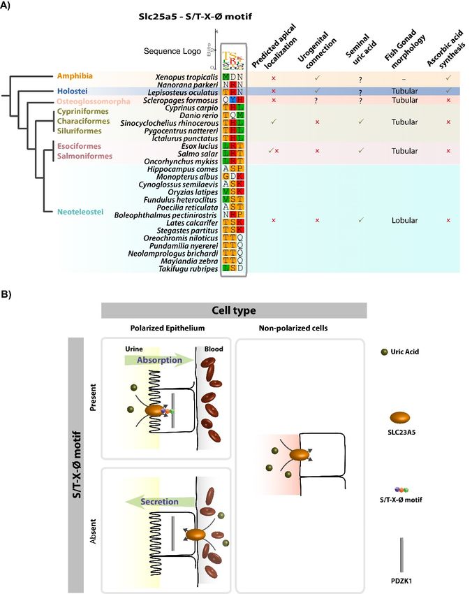
Fishes 2019, 4, 1 2 of 12
metazoans [1]. Sodium-dependent Slc23 transporters were suggested to maintain vertebrate ascorbic
acid levels: With Slc23a1 responsible for renal and intestine ascorbic acid absorption across brush
border epithelia and Slc23a2, which exhibits a widespread expression, responsible for ascorbic acid
supply to target tissues and cells, including neurons [1,9,10]. In vertebrates, two additional transporters
were identified, the orphan transporter Slc23a3 and the nucleobase transporter Slc23a4, described in
rats but found pseudogenized in humans [1,11]. Rat Slc23a4 is restricted to the small intestine and was
shown to predominantly mediate sodium-dependent uracil transport [11]. Slc23a3, on the other hand,
is expressed in proximal renal tubules but is unresponsive to both ascorbic acid and nucleobases [1].
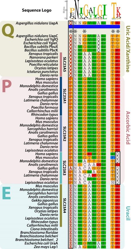
NAT family members display a highly conserved signature motif: [Q/E/P]-N-x-G-x-x-x-x-T-
[R/K/G] [1,11–13]. Functional characterization of a distinct member of the NAT family highlighted the
importance of the first amino acid position of the motif in the definition of substrate specificity [1,11–13].
Thus, these transporters can be divided into three functional groups. Slc23a1, Slc23a2, and Slc23a3
belong to the ascorbate group and harbor a conserved proline (P) in the first amino acid position
of the signature motif [1]. Glutamate (E) residue defines the uracil group, including bacterial anion
symporters and Slc23a4, while glutamine (Q) is present in the bacterial uric acid and/or xanthine anion
symporters [1,11–13]. Topology predictions, using proteins from the distinct groups, suggest a cytosolic
location of the motif [1,11,12]. Here we revise the portfolio of NAT family members in vertebrates.
2. Materials and Methods
2.1. Sequence Mining
Slc23 protein sequences were obtained from the GenBank database by BLASTp using human
Slc23a1 (NP_005838), Slc23a2 (NP_976072), and Slc23a3 (NP_001138362) sequences as query.
The sequences from Lethenteron japonica (Japanese lamprey) were obtained from the Japanese Lamprey
Genome Project (http://jlampreygenome.imcb.a-star.edu.sg/). The sequences from Leucoraja erinacea
(little skate) and Scyliorhinus canicula (small-spotted catshark) were obtained as nucleotide sequences
from SkateBase (http://skatebase.org) and translated. Accession numbers for all the retrieved
sequences are listed in Supplementary Table S1.
2.2. Synteny
Slc23 genes were mapped onto the respective species genomes, using the latest genome assemblies
available in the GenBank database (https://www.ncbi.nlm.nih.gov/). Genes flanking Slc23a1, Slc23a2,
Slc23a3, Slc23a4 and the novel Slc23a-like gene were identified and mapped using the human loci
as a reference. If the target gene was not found, the syntenic genomic region was retrieved using
neighboring genes as a reference.
2.3. Phylogenetic Analysis
The retrieved amino acid sequences were aligned using the MAFFT software web service (http:
//mafft.cbrc.jp/alignment/software/) in default automatic settings, with the L-INS-I refinement
method [14]. The alignment was stripped from columns with at least 20% of gaps resulting in alignment
with 75 sequences and 526 positions. The output alignment was used to construct a phylogenetic tree
using PhyML 3.0 with Smart Model Selection web service (http://www.atgc-montpellier.fr/phyml-
sms/) in default settings, which selected the LG +G+I+F model [15]. Bayesian-like transformation
of aLRT was selected to assess branch support. An additional phylogenetic tree, with automatic
bootstrapping (552 bootstraps) and using the default hill-climbing algorithm, was constructed with
RAxML, available in CIPRES Science Gateway.
2.4. Membrane Topology Prediction
Slc23a5 membrane topology predictions were performed for selected species, using distinct
prediction web servers: TMHMM Server v. 2.0 (http://www.cbs.dtu.dk/services/TMHMM/) and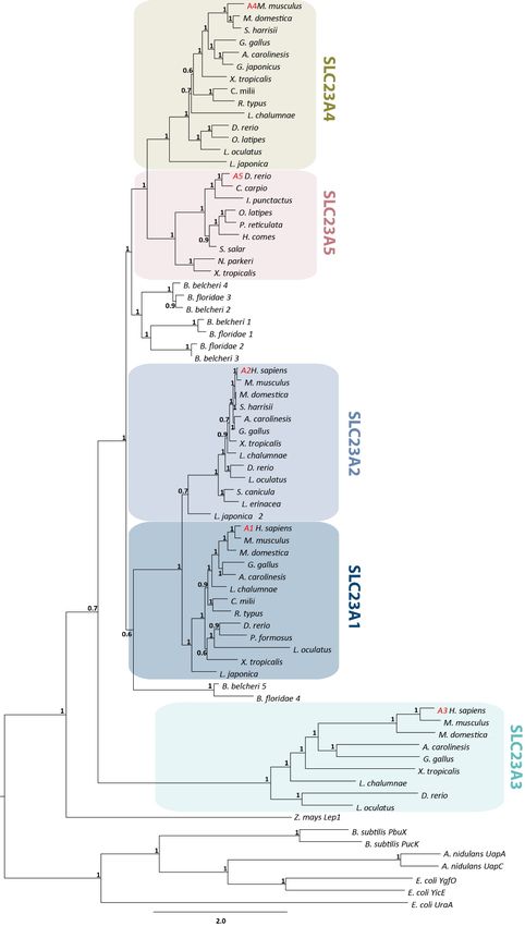
Fishes 2019, 4, 1                                                                                    3 of 12

TMPred (https://embnet.vital-it.ch/software/TMPRED_form.html). Human Slc23a1 was used as
a control.

2.5. Filtering and Quality Control of Sequence Reading Archive (SRA) Datasets
     For RNA-seq analysis, we included 39 SRA datasets from brain, intestine, kidney, liver, and testis
tissues of Homo sapiens (human), Mus musculus (mouse), Gallus gallus (chicken), Anolis carolinensis
(American green anole), Xenopus tropicalis (western clawed frog), Danio rerio (zebrafish), Lepisosteus
oculatus (spotted gar), and Oryzias latipes (Japanese medaka), which were obtained from the National
Center for Biotechnology (NCBI) Sequence Read Archive (SRA) (https://www.ncbi.nlm.nih.gov/sra/)
(Table S2). The read quality assessment of each SRA dataset was performed using FastQC software
(https://www.bioinformatics.Babraham.ac.uk/projects/fastqc/). Trimmomatic 0.36 [16] was used to
trim and drop reads with quality scores below 5, at leading and trailing ends, with an average quality
score below 20 in a 4 bp sliding window and with less than 36 bases length.

2.6. Relative Gene Expression Levels
      To assess the relative gene expression of slc23 genes from the selected species, reference sequences
and the corresponding annotations were collected from NCBI and Ensembl [17] databases. For human,
mouse, lizard, zebrafish and spotted gar, the GTF and DNA files (genome) were retrieved from Ensembl
(Release 89) [17]. For chicken and western clawed frog, transcript sequences (coding and non-coding)
and mapping (gene/transcript) from NCBI Refseq database were used after filtering [18] (for each
species the rna.fasta file from ftp://ftp.ncbi.nih.gov/genomes/refseq/ was used—See Tables S3 and
S4 for detail). The SRA reads of each specimen were aligned to the reference using Bowtie2 (default
parameters) [19], and gene expression was quantified as transcript per million (TPM) using RSEM
v.1.2.31 (RNA-seq by Expectation Maximization) software package [20]. Isoform expression levels for
each gene were summed to derive the TPM values. For genes with low relative expression values,
TPM values were log2-transformed after adding a value of one; TPM values
Fishes 2019, 4, 1                                                                                    4 of 12

sequence was retrieved from chondrichthyans, chondrosteans (basal actinopterygians), nor from
the sarcopterygian Latimeria chalumnae (coelacanth). Subsequent loss of slc23a5 in chondrichthyans
was further supported by the conservation of the genomic locus in Callorhynchus milii (Australian
ghostshark) (Figure S2). Regarding coelacanth, two scaffolds, denoting conservation of the flanking
genes, were found; while, no information was retrieved regarding the chondrostean loci (Figure S2).
The remaining genes of the slc23 family were retrieved and genomic loci were found generally
conserved across vertebrate species, with exception of the slc23a4 loci in fish (Figures S3–S6).
The branching pattern of our phylogenetic analysis suggests that after the expansion of the slc23a
gene family slc23a5 was subsequently lost in sharks, basal actinopterygians, coelacanth, and amniotes
(Figures 1 and S2). In agreement, we obtained three phylogenetic clusters possibly corresponding to
three ancestral lineages (Figures 1 and S1): One composed of typical slc23a1 and slc23a2 genes and
a second group including the previously reported slc23a4 and the novel slc23a5 genes, both out-grouped
by invertebrate sequences. A third cluster, which outgroups the remaining slc23 genes, included the
highly divergent slc23a3 genes, mirrored by the long branch lengths (Figure 1).

3.2. Slc23a5 Signature Motifs
     Further comparative protein sequence analysis highlighted the presence of a NAT family
signature motif with a conserved Q in the substrate determining position. According to previous
functional characterization of NAT family members, this residue is found in uric acid and/or
xanthine transporters, thus placing Slc23a5 in the uric acid and/or xanthine group (Figure 2) [1,11–13].
Besides the typical signature motif, a carboxyl terminus tripeptide motif was identified in some
SLC23 family members: An S/T-X-Ø tripeptide. S/T-X-Ø motifs were shown to interact with PDZ
domains of scaffolding proteins belonging to the sodium/hydrogen exchanger regulatory factor,
NHERF, family [21]. S/T-X-Ø are found highly conserved across species in Slc23a1 and Slc23a4,
in agreement with their renal and intestinal expression and role in ascorbic acid and nucleobase
absorption (Figure S7). Slc23a5, on the other hand, exhibits a highly conserved tripeptide motif in
Otocephala (Cypriniformes, Characiformes, and Siluriformes) but not Osteoglossomorpha (Figure 3A).
The Euteleostei Esociformes and Salmoniformes exhibit an inverted motif while in the Neoteleostei
no conservation was found. Finally, topology predictions yielded similar transmembrane profiles for
Slc23a5 sequences from D. rerio, O. latipes, Salmo salar (Atlantic salmon) and X. tropicalis, when compared
to human Slc23a1 (Figure S8). The NAT motif was predicted cytosolic in all analyzed sequences.
This suggests a conserved cellular import topology, as observed in other family members.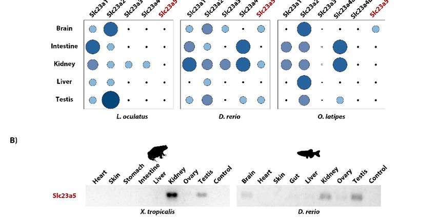
Fishes 2019, 4, 1                                                                                             5 of 12
Processes 2018, 6, x FOR PEER REVIEW                                                                          5 of 12

      Figure1.1.Maximum
      Figure      Maximum likelihood  phylogenetic
                              likelihood            tree describing
                                          phylogenetic              relationships
                                                          tree describing         among Nucleobase-Ascorbate
                                                                            relationships  among Nucleobase-
      Transporter (NAT) family(NAT)
      Ascorbate Transporter      members.  Node
                                       family     values represent
                                               members.             branchrepresent
                                                           Node values      support using thesupport
                                                                                     branch   aBayes algorithm.
                                                                                                     using the
      Accession numbersAccession
      aBayes algorithm.  for all sequences
                                     numbersarefor
                                                 provided   in Supplementary
                                                   all sequences  are providedMaterials.
                                                                                in Supplementary Materials.
Fishes 2019, 4, 1 6 of 12
Processes 2018, 6, x FOR PEER REVIEW 6 of 12
Figure 2.
Figure 2. Alignment of the signature motif of the Nucleobase-Ascorbate
Nucleobase-Ascorbate Transporter
Transporter (NAT)
(NAT) family.
family.
NAT family
NAT family members
members display
display aa highly
highly conserved
conserved signature
signature motif
motif defining
defining substrate specificity:
[Q/E/P]-N-x-G-x-x-x-x-T-[R/K/G]. Asterisks
[Q/E/P]-N-x-G-x-x-x-x-T-[R/K/G]. Asterisksindicate
indicateconserved
conservedpositions.
positions.The
The first
first amino acid
acid
position, with residues highlighted in red within a black box, defines substrate preference: Q—Uric
position, with residues highlighted in red within a black box, defines substrate preference: Q—Uric
acid/xanthine group;
acid/xanthine group; P—Ascorbate group and E—Uracil group. The mirror Q in the Slc23a3 group group is
is
also highlighted
also highlighted by
by aa black
black box.
box.Fishes 2019, 4, 1 7 of 12
Processes 2018, 6, x FOR PEER REVIEW 7 of 12
Figure3.3.The
Figure TheS/T-X-Ø
S/T-X-Ø tripeptide
tripeptide provides
provides insight
insightinto
intothethecellular
cellularfunction
functionofofSlc23a5
Slc23a5homologues
homologues
from fish and amphibians. (A) Alignment of the PDZ-binding tripeptide in
from fish and amphibians. (A) Alignment of the PDZ-binding tripeptide in Slc23a5 homologues Slc23a5 homologues from
fish fish
from and andamphibians.
amphibians. TheTheS/T-X-Ø
S/T-X-Ø tripeptide
tripeptideis isfound
found in Characiformes, and
in Cypriniformes, Characiformes, and
Siluriformeswhile
Siluriformes while
thethe Euteleostei
Euteleostei Esociformes
Esociformes and Salmoniformes
and Salmoniformes exhibit anexhibit
invertedan motif.
inverted motif.
Predicted
Predicted
cellular cellular localization
localization is indicatedisasindicated as well as morphological
well as morphological and physiological
and physiological adaptations adaptations
among the
among the different fish groups. (B) Schematic representation of tripeptide-dependent
different fish groups. (B) Schematic representation of tripeptide-dependent uric acid flows in polarized uric acid flows
in polarized
epithelia epithelia and non-polarized
and non-polarized cells.
cells. In polarized In polarized
epithelia epithelia
(i.e., kidney) the(i.e., kidney)
presence thetripeptide
of the presence motif
of the
tripeptide
suggests motif suggests
interaction with theinteraction
scaffolding with the scaffolding
protein PDZK1 which protein PDZK1
localizes which
apically, in localizes apically,
the absorptive side.in
the absorptive side.Fishes 2019, 4, 1 8 of 12
Processes 2018, 6, x FOR PEER REVIEW 8 of 12
3.3. Slc23a5 Expression
3.3. Expression Patterns
Patterns
Togain
To gainfurther
furtherinsight
insightintointothe
thefunctional
functional relevance
relevance of of this
this additional
additional gene,
gene, wewe collected
collected RNA-
RNA-seq
seq data
data and and determined
determined relative
relative expression
expression levelslevels of Slc23
of Slc23 genes.
genes. GeneGene expression
expression analysis
analysis suggested
suggested that
that in D. rerio and X. tropicalis, slc23a5 is mostly expressed in kidney and testis, further
in D. rerio and X. tropicalis, slc23a5 is mostly expressed in kidney and testis, further confirmed by PCR confirmed by
PCR (Figure
(Figure 4 and4Table
and Table S5). Expression
S5). Expression in the in the was
brain brainalso
wasdetected
also detected in D.and
in D. rerio rerio and
was was to
found found to
be the
be the exclusive
exclusive expression
expression site Neoteleostei
site in the in the Neoteleostei O. latipes.
O. latipes. The holostean
The holostean L. oculatus,
L. oculatus, on the on the hand,
other other
hand, yielded
yielded no significant
no significant expression
expression in the tissues.
in the tested tested tissues.
Figure
Figure 4.4. Expression
Expression patterns of Slc23
patterns of Slc23 isoforms
isoforms across
across vertebrates.
vertebrates. (A)
(A) RNAseq-based quantification.
RNAseq-based quantification.
Relative gene expression levels are provided as log -transformed transcript per million
Relative gene expression levels are provided as log22-transformed transcript per million (TPM) (TPM) values,
values,
from
from 0 to 8 TPM’s, NF – not found, ND – no sequence reading archive (SRA) data available for the
0 to 8 TPM’s, NF – not found, ND – no sequence reading archive (SRA) data available for the
specific tissue. (B) PCR-based amplification of Slc23a5 transcripts in D. rerio and X. tropicalis tissues.
specific tissue. (B) PCR-based amplification of Slc23a5 transcripts in D. rerio and X. tropicalis tissues.
4. Discussion
4. Discussion
Our findings indicate a restricted distribution of slc23a5 to taxa that share an aquatic nature of
Our findings indicate a restricted distribution of slc23a5 to taxa that share an aquatic nature of
their habitat. In fact, slc23a5 was found to be expressed in the kidney and testis of both X. tropicalis
their habitat. In fact, slc23a5 was found to be expressed in the kidney and testis of both X. tropicalis
and D. rerio. In the latter, as well as in the Neoteleostei O. latipes, brain expression was also detected.
and D. rerio. In the latter, as well as in the Neoteleostei O. latipes, brain expression was also detected.
Comparative analysis using previously characterized NAT family transporters placed this novel
Comparative analysis using previously characterized NAT family transporters placed this novel
transporter in the uric acid and/or xanthine transporter group. In fact, similarly to uric acid and/or
transporter in the uric acid and/or xanthine transporter group. In fact, similarly to uric acid and/or
xanthine transporters, Slc23a5 proteins harbor a Q in the first position of the NAT family signatureFishes 2019, 4, 1 9 of 12
xanthine transporters, Slc23a5 proteins harbor a Q in the first position of the NAT family signature
motif ([Q/E/P]-N-x-G-x-x-x-x-T-[R/K/G]). Yet, further functional studies are required to fully clarify
the molecular function of Slc23a5 proteins, notably its uric acid and/or xanthine binding abilities.
Given the nature of NAT family transporter ligands, including ascorbic and uric acids, xanthine
or nucleobases, renal Slc23a5 could participate in the homeostatic maintenance of excretion products,
in osmotic regulation, and/or in antioxidant processes. In fact, kidneys play a crucial role in
the maintenance of both ascorbic and uric acid levels through the excretion and reabsorptions
processes [6,21]. In kidney absorptive epithelium, transporters located on the filtrate-epithelial interface
(apical side) interact with the scaffolding protein PDZ-containing kidney protein 1 (PDZK1) [21].
PDZK1 belongs to the NHERF family of proteins which are mostly expressed in the apical side
of absorptive epithelia of the kidney, intestine, and liver, and establish protein-protein interaction
networks with receptors, transporters, and channels [22,23]. Besides its scaffolding role, NHERF family
members were also shown to modulate the activity of protein assemblages [23–25]. Within Slc23 family
members, S/T-X-Ø motifs are found in Slc23a1 and Slc23a4 proteins, which are present in renal and
intestinal polarized epithelia and participate in ascorbic acid and nucleobase absorption, and absent in
Slc23a2. Curiously, a highly conserved PDZK1 interacting motif (S/T-X-Ø) was also detected in Slc23a5
from Cypriniformes, Characiformes, and Siluriformes, but not in other fish groups or amphibians.
No relationship between salt and freshwater environments was found. Thus, the presence/absence
of this peptide signal might reflect distinct flows in the kidney: An apically expressed Slc23a5 likely
participates in absorptive flows, while a basolateral expression would be related to secretion (Figure 3B).
In the human proximal renal tubules, several transporters, localized to specific cellular
regions (apical or basolateral), seem to cooperate for efficient urate handling: Named the urate
transportome [6,21]. Thus, if the predicted uric acid transport activity is confirmed, X. tropicalis and
D. rerio Slc23a5 could illustrate the two opposing mechanisms: While D. rerio captures, X. tropicalis
secretes uric acid into the renal filtrate. In agreement, X. tropicalis, D. rerio, as well as L. oculatus, appear
to lack the full complement of renal urate transporters (Table S6). A similar scenario is observed
for birds and reptiles; however, their excretory system represents a particular adaptation for uric
acid excretion [26,27]. Besides simple excretion, and given that high concentrations of uric acid were
detected, and suggested to act as the main non-enzymatic antioxidant mechanism, in seminal fluid
of several fish species (including Cypriniformes, Esociformes, Salmoniformes, and the Neoteleostei
Gadiformes and Perciformes [28–31]), this putative uric acid transport activity could be maintained
for reproductive purposes, in agreement with the observed testicular expression. In fact, in external
fertilizers, such as most fish and amphibians, reactive oxygen species were suggested to decrease
motility and thus fertilization success [30]. Yet, regarding amphibians, no information on seminal
fluid composition was retrieved [32]. Nonetheless, in vitro assays suggested that other antioxidant
species, such as ascorbic acid or vitamin E, were unsuitable at offsetting oxidative stress and improving
sperm motility in Litoria booroolongensis (Booroolong frog) and suggested uric acid as a pertinent
candidate [32]. In this context, the absence of slc23a5 sequences in Chondrichthyes could be due to
their internal fertilization strategies derived from ovoviviparous or viviparous reproductive modes [33].
Accordingly, viviparity is also observed in the sarcopterygian L. chalumnae [33].
Besides reproductive categories, other morphological and metabolic traits could justify the
seemingly scattered retention of slc23a5 among fish in the context of the proposed uric acid transport
activity; for instance, specific anatomical features such as the presence of a urogenital connection:
Described in Elasmobranchii and in most non-teleost Osteichthyes, yet absent in Cyclostomata and
Teleostei [34]. By connecting testicular and renal ducts, antioxidant requirements could be met by renal
secretion into the urine. In agreement, in the Chondrostei Acipenser ruthenus, full sperm maturation
and motility were shown to require dilution by urine [35]. A urogenital connection is also present in
amphibians [34]. Additionally, other antioxidant mechanisms could exist. Besides uric acid, ascorbic
acid is also present in the seminal fluid of Oncorhynchus mykiss (rainbow trout) [36]. Seminal ascorbate
was reported to be affected by seasonal variations and semen quality was shown to improve uponFishes 2019, 4, 1 10 of 12
dietary ascorbic acid supplementation [36,37], contrasting with the amphibian L. booroolongensis [32].
In the case of non-teleost fish, such as Chondrostei (A. ruthenus) and Holostei (L. oculatus), a functional
GLO gene is retained, providing endogenously synthesized ascorbate [38]. Curiously, no gonadal
expression was detected in the Neoteleostei O. latipes. Neoteleosts exhibit varied reproduction
modes ranging from external fertilizers to internal fertilizers with viviparity [33,39–41]. In addition,
neoteleosts were shown to exhibit a distinctive gonadal morphology, the lobular testis, whereas the
remaining bony fish exhibit tubular testis [42]. Although still obscure, the absence of testicular slc23a5
expression in the external fertilizer O. latipes could be due to specific morphological adaptations
of neoteleosts.
A similar role could be postulated for neuronal uric acid [43]. Active swimming behavior requires
a high metabolic rate, mirrored by increased oxygen consumption, subsequently leading to reactive
oxygen species production [44]. In agreement, fish antioxidant levels were shown to be correlated with
their swimming activity [44]. Neuronal cell populations are particularly sensitive to oxidative stress,
thus, uric acid could participate in the neutralization of oxidative species to by-pass oxidative stress,
notably in GLO-deficient fish. Yet, information on neuronal uric acid levels in fish is currently absent.
5. Conclusions
Taken together, these observations put forward the retention of an additional Slc23 gene,
in neopterygians and amphibians, with restricted expression patterns and presumed uric acid transport
function. Yet, the extensive comparative functional analysis should be conducted to clarify the
physiological relevance of this novel receptor in aquatic organisms.
Supplementary Materials: The following are available online at http://www.mdpi.com/2410-3888/4/1/1/s1,
Supplementary tables and figures including synteny, phylogeny and topology analysis, as well as reference tables.
Author Contributions: Conceptualization, R.R. and L.F.C.C.; Validation, R.R.; Formal analysis, D.O, A.M.M.
and R.R.; Investigation, D.O., A.M.M., M.L.-M. and T.C.; Resources, R.R. and L.F.C.C.; Writing—original draft,
R.R., D.O., A.M.M., T.C., M.L.-M. and L.F.C.C.; Writing—review & editing, R.R. and L.F.C.C.; Visualization, R.R.,
D.O. and A.M.M.; Supervision, L.F.C.C. and R.R.; Project administration, L.F.C.C.; Funding acquisition, L.F.C.C.
Funding: This research was funded by Coral—Sustainable Ocean Exploitation (Norte-01-0145-FEDER-000036),
a project by the North Portugal Regional Operational Program (NORTE 2020), under the PORTUGAL 2020
Partnership Agreement, through the European Regional Development Fund (ERDF) and by the European
Regional Development Fund (ERDF) through the COMPETE—Operational Competitiveness Programme and
POPH—Operational Human Potential Programme.
Conflicts of Interest: The authors declare no conflicts of interest.
References
1. Burzle, M.; Suzuki, Y.; Ackermann, D.; Miyazaki, H.; Maeda, N.; Clemencon, B.; Burrier, R.; Hediger, M.A.
The sodium-dependent ascorbic acid transporter family SLC23. Mol. Aspects Med. 2013, 34, 436–454.
[CrossRef]
2. Linster, C.L.; Van Schaftingen, E. Vitamin C. FEBS J. 2007, 274, 1–22. [CrossRef] [PubMed]
3. Drouin, G.; Godin, J.R.; Page, B. The genetics of vitamin C loss in vertebrates. Curr. Genomics 2011, 12,
371–378. [CrossRef] [PubMed]
4. Dutta Gupta, S.; Choudhury, P.K.; Chatterjee, I.B. Synthesis of l-ascorbic acid from d-glucurono-1,4-lactone
conjugates by different species of animals. Int. J. Biochem. 1973, 4, 309–314. [CrossRef]
5. Wright, P.A. Nitrogen excretion: Three end products, many physiological roles. J. Exp. Biol. 1995, 198,
273–281. [PubMed]
6. Bobulescu, I.A.; Moe, O.W. Renal transport of uric acid: Evolving concepts and uncertainties. Adv. Chronic
Kidney Dis. 2012, 19, 358–371. [CrossRef] [PubMed]
7. Alvarez-Lario, B.; Macarrón-Vicente, J. Evolution of Uric Acid Metabolism in Humans. In eLS; John Wiley &
Sons, Ltd.: Hoboken, NJ, USA, 2001. [CrossRef]
8. Oda, M.; Satta, Y.; Takenaka, O.; Takahata, N. Loss of urate oxidase activity in hominoids and its evolutionary
implications. Mol. Biol. Evol. 2002, 19, 640–653. [CrossRef] [PubMed]Fishes 2019, 4, 1 11 of 12
9. Corti, A.; Casini, A.F.; Pompella, A. Cellular pathways for transport and efflux of ascorbate and
dehydroascorbate. Arch. Biochem. Biophys. 2010, 500, 107–115. [CrossRef]
10. Savini, I.; Rossi, A.; Pierro, C.; Avigliano, L.; Catani, M.V. SVCT1 and SVCT2: Key proteins for vitamin C
uptake. Amino Acids 2008, 34, 347–355. [CrossRef]
11. Yamamoto, S.; Inoue, K.; Murata, T.; Kamigaso, S.; Yasujima, T.; Maeda, J.Y.; Yoshida, Y.; Ohta, K.Y.; Yuasa, H.
Identification and functional characterization of the first nucleobase transporter in mammals: Implication
in the species difference in the intestinal absorption mechanism of nucleobases and their analogs between
higher primates and other mammals. J. Biol. Chem. 2010, 285, 6522–6531. [CrossRef]
12. Koukaki, M.; Vlanti, A.; Goudela, S.; Pantazopoulou, A.; Gioule, H.; Tournaviti, S.; Diallinas, G.
The nucleobase-ascorbate transporter (NAT) signature motif in UapA defines the function of the purine
translocation pathway. J. Mol. Biol. 2005, 350, 499–513. [CrossRef] [PubMed]
13. Papakostas, K.; Frillingos, S. Substrate selectivity of YgfU, a uric acid transporter from Escherichia coli. J.
Biol. Chem. 2012, 287, 15684–15695. [CrossRef] [PubMed]
14. Katoh, K.; Standley, D.M. MAFFT multiple sequence alignment software version 7: Improvements in
performance and usability. Mol. Biol. Evol. 2013, 30, 772–780. [CrossRef] [PubMed]
15. Guindon, S.; Dufayard, J.F.; Lefort, V.; Anisimova, M.; Hordijk, W.; Gascuel, O. New algorithms and methods
to estimate maximum-likelihood phylogenies: Assessing the performance of PhyML 3.0. Syst. Biol. 2010, 59,
307–321. [CrossRef]
16. Bolger, A.M.; Lohse, M.; Usadel, B. Trimmomatic: A flexible trimmer for Illumina sequence data.
Bioinformatics 2014, 30, 2114–2120. [CrossRef] [PubMed]
17. Yates, A.; Akanni, W.; Amode, M.R.; Barrell, D.; Billis, K.; Carvalho-Silva, D.; Cummins, C.; Clapham, P.;
Fitzgerald, S.; Gil, L.; et al. Ensembl 2016. Nucleic Acids Res. 2016, 44, D710–D716. [CrossRef] [PubMed]
18. O’Leary, N.A.; Wright, M.W.; Brister, J.R.; Ciufo, S.; Haddad, D.; McVeigh, R.; Rajput, B.; Robbertse, B.;
Smith-White, B.; Ako-Adjei, D.; et al. Reference sequence (RefSeq) database at NCBI: Current status,
taxonomic expansion, and functional annotation. Nucleic Acids Res. 2016, 44, D733–D745. [CrossRef]
19. Langmead, B.; Salzberg, S.L. Fast gapped-read alignment with Bowtie 2. Nat. Methods 2012, 9, 357–359.
[CrossRef]
20. Li, B.; Dewey, C.N. RSEM: Accurate transcript quantification from RNA-Seq data with or without a reference
genome. BMC Bioinformatics 2011, 12, 323. [CrossRef]
21. Anzai, N.; Jutabha, P.; Amonpatumrat-Takahashi, S.; Sakurai, H. Recent advances in renal urate transport:
Characterization of candidate transporters indicated by genome-wide association studies. Clin. Exp. Nephrol.
2012, 16, 89–95. [CrossRef]
22. Dev, K.K. Making protein interactions druggable: Targeting PDZ domains. Nat. Rev. Drug Discov. 2004, 3,
1047–1056. [CrossRef] [PubMed]
23. Clapéron, A.; Mergey, M.; Fouassier, L. Roles of the scaffolding proteins NHERF in liver biology. Clin. Res.
Hepatol. Gastroenterol. 2011, 35, 176–181. [CrossRef] [PubMed]
24. Raghuram, V.; Mak, D.-O.D.; Foskett, J.K. Regulation of cystic fibrosis transmembrane conductance regulator
single-channel gating by bivalent PDZ-domain-mediated interaction. Proc. Natl. Acad. Sci. USA 2001, 98,
1300–1305. [CrossRef] [PubMed]
25. Wang, S.; Yue, H.; Derin, R.B.; Guggino, W.B.; Li, M. Accessory Protein Facilitated CFTR-CFTR Interaction,
a Molecular Mechanism to Potentiate the Chloride Channel Activity. Cell 2000, 103, 169–179. [CrossRef]
26. Braun, E.J. Comparative renal function in reptiles, birds, and mammals. Semin. Avian Exotic. Pet. Med. 1998,
7, 62–71. [CrossRef]
27. Singer, M.A. Do mammals, birds, reptiles and fish have similar nitrogen conserving systems? Comp. Biochem.
Physiol. B Biochem. Mol. Biol. 2003, 134, 543–558. [CrossRef]
28. Ciereszko, A.; Dabrowski, K.; Kucharczyk, D.; Dobosz, S.; Goryczko, K.; Glogowski, J. The presence of uric
acid, an antioxidantive substance, in fish seminal plasma. Fish Physiol. Biochem. 1999, 21, 313–315. [CrossRef]
29. Dzyuba, V.; Dzyuba, B.; Cosson, J.; Boryshpolets, S.; Yamaner, G.; Kholodniy, V.; Rodina, M. The antioxidant
system of sterlet seminal fluid in testes and Wolffian ducts. Fish Physiol. Biochem. 2014, 40, 1731–1739.
[CrossRef]
30. Lahnsteiner, F.; Mansour, N. A comparative study on antioxidant systems in semen of species of the Percidae,
Salmonidae, Cyprinidae, and Lotidae for improving semen storage techniques. Aquaculture 2010, 307,
130–140. [CrossRef]Fishes 2019, 4, 1 12 of 12
31. Lahnsteiner, F.; Mansour, N.; Plaetzer, K. Antioxidant systems of brown trout (Salmo trutta f. fario) semen.
Anim. Reprod. Sci. 2010, 119, 314–321. [CrossRef]
32. Keogh, L.M.; Byrne, P.G.; Silla, A.J. The effect of antioxidants on sperm motility activation in the Booroolong
frog. Anim. Reprod. Sci. 2017, 183, 126–131. [CrossRef] [PubMed]
33. Goodwin, N.B.; Dulvy, N.K.; Reynolds, J.D. Life-history correlates of the evolution of live bearing in fishes.
Philos. Trans. R. Soc. Lond. B. Biol. Sci 2002, 357, 259–267. [CrossRef]
34. Blün, V. Comparative Anatomy of the Urogenital System. In Vertebrate Reproduction, a Textbook; Springer:
Berlin/Heidelberg, Germany; New York, NY, USA; Tokyo, Japan, 1986; pp. 43–74. [CrossRef]
35. Dzyuba, B.; Cosson, J.; Boryshpolets, S.; Bondarenko, O.; Dzyuba, V.; Prokopchuk, G.; Gazo, I.; Rodina, M.;
Linhart, O. In vitro sperm maturation in sterlet, Acipenser ruthenus. Reprod. Biol. 2014, 14, 160–163.
[CrossRef] [PubMed]
36. Ciereszko, A.; Dabrowski, K. Sperm quality and ascorbic acid concentration in rainbow trout semen are
affected by dietary vitamin C: An across-season study. Biol. Reprod. 1995, 52, 982–988. [CrossRef] [PubMed]
37. Ciereszko, A.; Dabrowski, K. Effect of ascorbic acid supplement in vitro onrainbow trout sperm viability.
Aquacult. Int. 2000, 8, 1–8. [CrossRef]
38. Moreau, R.; Dabrowski, K. Biosynthesis of ascorbic acid by extant actinopterygians. J. Fish Biol. 2000, 57,
733–745. [CrossRef]
39. Forsman, A.; Tibblin, P.; Berggren, H.; Nordahl, O.; Koch-Schmidt, P.; Larsson, P. Pike Esox lucius as an
emerging model organism for studies in ecology and evolutionary biology: A review. J. Fish Biol. 2015, 87,
472–479. [CrossRef]
40. Bone, Q.; Moore, R.H. Biology of Fishes; Taylor & Francis Group: New York, NY, USA, 2008.
41. Scott, D.B.C.; Fuller, J.D. The reproductive biology of Scleropages formosus (Müller & Schlegel)
(Osteoglossomorpha, Osteoglossidae) in Malaya, and the morphology of its pituitary gland. J. Fish Biol. 1976,
8, 45–53. [CrossRef]
42. Parenti, L.R.; Grier, H.J. Evolution and phylogeny of gonad morphology in bony fishes. Integr. Comp. Biol.
2004, 44, 333–348. [CrossRef]
43. Uttara, B.; Singh, A.V.; Zamboni, P.; Mahajan, R.T. Oxidative Stress and Neurodegenerative Diseases:
A Review of Upstream and Downstream Antioxidant Therapeutic Options. Curr. Neuropharmacol. 2009, 7,
65–74. [CrossRef]
44. Lopez-Cruz, R.I.; Dafre, A.L.; Filho, D.W. Oxidative Stress in Sharks and Rays. In Oxidative Stress in Aquatic
Ecosystems; Abele, D., Vazquez-Medina, J.P., Zenteno-Savin, T., Eds.; Wiley-Blackwell: Hoboken, NJ, USA,
2011; pp. 157–164.
© 2019 by the authors. Licensee MDPI, Basel, Switzerland. This article is an open access
article distributed under the terms and conditions of the Creative Commons Attribution
(CC BY) license (http://creativecommons.org/licenses/by/4.0/).You can also read