Fundamentals, Diagnostic Capabilities, and Perspective of Narrow Band Imaging for Early Gastric Cancer - MDPI
←
→
Page content transcription
If your browser does not render page correctly, please read the page content below

Journal of
              Clinical Medicine

Review
Fundamentals, Diagnostic Capabilities, and Perspective of
Narrow Band Imaging for Early Gastric Cancer
Hiroki Kurumi 1 , Kouichi Nonaka 2 , Yuichiro Ikebuchi 1 , Akira Yoshida 1 , Koichiro Kawaguchi 1 ,
Kazuo Yashima 1 and Hajime Isomoto 1, *

                                          1   Division of Gastroenterology and Nephrology, Department of Multidisciplinary Internal Medicine,
                                              Faculty of Medicine, Tottori University, 36-1 Nishicho, Yonago 683-8504, Japan;
                                              kurumi_1022_1107@yahoo.co.jp (H.K.); ikebu@tottori-u.ac.jp (Y.I.); akirayoshida1021@yahoo.co.jp (A.Y.);
                                              koichiro@tottori-u.ac.jp (K.K.); yashima@tottori-u.ac.jp (K.Y.)
                                          2   Department of Digestive Endoscopy, Tokyo Women’s Medical University Hospital, 8-1 Kawada-cho,
                                              Shinjuku-ku, Tokyo 162-8666, Japan; nonaka513@gmail.com
                                          *   Correspondence: isomoto@tottori-u.ac.jp

                                          Abstract: The development of image-enhanced endoscopy has dramatically improved the qualitative
                                          and quantitative diagnosis of gastrointestinal tumors. In particular, narrow band imaging (NBI)
                                          has been widely accepted by endoscopists around the world in their daily practice. In 2009, Yao
                                          et al. proposed vessel plus surface (VS) classification, a diagnostic algorithm for early gastric cancer
                                          using magnifying endoscopy with NBI (ME-NBI), and in 2016, Muto et al. proposed a magnifying
                                          endoscopy simple diagnostic algorithm for early gastric cancer (MESDA-G) based on VS classification.

                                   In addition, the usefulness of ME-NBI in the differential diagnosis of gastric cancer from gastritis,
                                          diagnosis of lesion extent, inference of histopathological type, and diagnosis of depth has also been
Citation: Kurumi, H.; Nonaka, K.;
                                          investigated. In this paper, we narrative review the basic principles, current status, and future
Ikebuchi, Y.; Yoshida, A.; Kawaguchi,
                                          prospects of NBI.
K.; Yashima, K.; Isomoto, H.
Fundamentals, Diagnostic
Capabilities, and Perspective of
                                          Keywords: narrow band imaging; gastric cancer; image-enhanced endoscopy; texture and color
Narrow Band Imaging for Early             enhancement imaging; red dichromatic imaging
Gastric Cancer. J. Clin. Med. 2021, 10,
2918. https://doi.org/10.3390/
jcm10132918
                                          1. Introduction
Academic Editor: Bruno Annibale                 According to the latest cancer statistics, 1.09 million new cases of stomach cancer
                                          are diagnosed annually worldwide, and 770,000 people die of stomach cancer annually.
Received: 30 May 2021
                                          It is the sixth most common cancer and the third most common cause of death among
Accepted: 26 June 2021
                                          all cancer types [1]. Gastric cancer has a 5-year survival rate of over 95% if detected
Published: 29 June 2021
                                          early; therefore, early detection is pivotal for better prognosis [2]. To this end, endoscopic
                                          diagnosis of gastric tumors has improved with the development and advancement of
Publisher’s Note: MDPI stays neutral
                                          endoscopic equipment.
with regard to jurisdictional claims in
                                                In particular, the development of image-enhanced endoscopy, represented by narrow
published maps and institutional affil-
                                          band imaging (NBI), has dramatically improved the qualitative and quantitative diagnosis
iations.
                                          of gastrointestinal tumors. Narrow-band light with peaks at 415 nm and 540 nm, which
                                          is used in NBI, is shorter wavelength of visible light and has low tissue permeability,
                                          making it ideal for observing mucosal surface structures [3]. These two short wavelengths
                                          coincide with the peak absorption region of oxidized hemoglobin, and mucosal capillaries
Copyright: © 2021 by the authors.
                                          are observed as a clear low signal compared to the surrounding tissue. The evaluation of
Licensee MDPI, Basel, Switzerland.
                                          intrapapillary capillary loops within the esophageal epithelium was established for the
This article is an open access article
                                          qualitative and quantitative diagnosis of esophageal tumors [4–6]. Although dye spraying
distributed under the terms and
                                          has been used as a conventional adjunct diagnosis to white light imaging (WLI), it is no
conditions of the Creative Commons
Attribution (CC BY) license (https://
                                          exaggeration to say that NBI has made it possible to avoid the risk of iodine allergy in
creativecommons.org/licenses/by/
                                          the examinee and physical burden such as heartburn, as in the case of iodine staining of
4.0/).
                                          the esophagus [7,8]. On the other hand, in NBI observation of mucosa with glandular

J. Clin. Med. 2021, 10, 2918. https://doi.org/10.3390/jcm10132918                                                https://www.mdpi.com/journal/jcm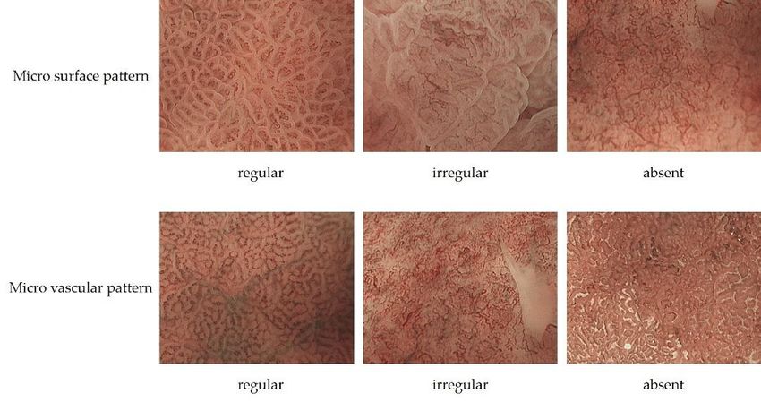
J. Clin. Med. 2021, 10, x FOR PEER REVIEW
J. Clin. Med. 2021, 10, 2918 2 of 12
staining of the esophagus [7,8]. On the other hand, in NBI observation of muc
glandular structures, the light projected onto the marginal crypt epithelium (MC
not reach the blood vessels and causes backward confusion, which is visualized a
structures, the light projected onto the marginal crypt epithelium (MCE) does not reach
border. This is called the white zone (WZ), and the morphology of the glandular s
the blood vessels and causes backward confusion, which is visualized as a white border.
canisbe
This estimated
called the white[9].
zoneFurthermore, when used
(WZ), and the morphology of in
the combination withcanmagnifyin
glandular structure be
copy, the
estimated [9].mucosal capillaries
Furthermore, when used and glandular structures
in combination can be
with magnifying evaluated
endoscopy, the in mo
mucosal
(Figurecapillaries
1). Whileand glandular
various structuresofcan
categories be evaluated in more
image-enhanced detail (Figure
endoscopy have 1).been ap
While various categories of image-enhanced endoscopy have been
clinical practice, NBI, when used in combination with magnifying endoscopy, is applied in clinical
practice, NBI, when used in combination with magnifying endoscopy, is particularly good
atlarly good fine
observing at observing
anatomical fine anatomical
structures structures
and is expected to beand is expected
useful to be useful in
in the endoscopic
doscopic
diagnosis of diagnosis of In
gastric cancer. gastric cancer.
this paper, In this
we review thepaper, we review
basic principles, thestatus,
current basic and
principles
status,
future and future
prospects prospects
of magnifying of magnifying
endoscopy endoscopy with NBI (ME-NBI).
with NBI (ME-NBI).
Figure
Figure 1. Mucosal
1. Mucosal capillaries
capillaries are observed
are observed as low
as low signal signaltocompared
compared to the
the surrounding surrounding
tissue. WZ ti
reflects
reflects thethe morphology
morphology of the of the marginal
marginal crypt epithelium.
crypt epithelium.
2. Basic Observation Methods and Points of ME-NBI for Gastric Cancer
2. Basic
2.1. Observation
ME-NBI Methods Mucosa
for Gastric Non-Cancerous and Points of ME-NBI for Gastric Cancer
2.1.Since
ME-NBI for Gastric
the normal gastricNon-Cancerous
mucosa shows aMucosa wide variety of mucosal patterns due to
differences in glandular areas and inflammatory modifications, knowing these typical
Since the normal gastric mucosa shows a wide variety of mucosal pattern
images is extremely important in ME-NBI diagnosis of gastric cancer.
differences in glandular
In the gastric fundic gland areas and without
mucosa inflammatory modifications,
inflammation, knowing
the gland ducts these ty
are ar-
ages is extremely important in ME-NBI diagnosis of gastric cancer.
ranged regularly in a concave position perpendicular to the mucosal surface. When this is
evaluated by ME-NBI,
In the gastricthe crypt openings
fundic are observed
gland mucosa as brown
without dots, and the surrounding
inflammation, the gland duct
MCE as a WZ. Capillaries of the intervening part around the WZ have a brown mesh
ranged regularly in a concave position perpendicular to the mucosal surface. Wh
pattern (Figure 2a). Inflammatory changes caused by Helicobacter pylori (HP) infection
evaluated
gradually by ME-NBI,
distort the crypt
the gland ducts. openings
The crypt openingsarebecome
observed
oval as brown dots,
or indistinct, and the su
and the
ing which
WZ, MCE isascircular,
a WZ. changes
Capillaries ofshape
to oval the intervening
(Figure 2b). Aspart
thearound the WZ
inflammatory have a brow
changes
pattern (Figure 2a). Inflammatory changes caused by Helicobacter pylori (HP)
progressed, the oval WZ gradually become tubular and the capillaries of the intervening
part are indistinct
gradually distortor coil-shaped
the gland(Figure
ducts.2c).TheThen,
cryptwhen the gastric
openings fundic gland
become disap-
oval or indistinct
pears, the appearance resembles the mucosa of the pyloric gland (Figure 2d). The pyloric
WZ, which is circular, changes to oval shape (Figure 2b). As
gland mucosa shows little change in the ME-NBI image due to inflammation. In the pyloric
the inflammatory
progressed,
gland the oval
mucosa without WZ gradually
inflammation, become
the gland duct istubular
distorted,and
and the capillariespart
the intervening of the inte
part areinto
protrudes indistinct
the mucosalor coil-shaped (Figure
surface in a ridged 2c).When
pattern. Then,it when the gastric
was observed fundic glan
by ME-NBI,
MCE appears as a regular tubular WZ, and coil-shaped capillaries
pears, the appearance resembles the mucosa of the pyloric gland (Figure are observed in the2d). Th
intervening part (Figure 2e) [10,11].
gland mucosa shows little change in the ME-NBI image due to inflammation. In t
ME-NBI of intestinal metaplasia shows a blue-white border at the limbus of the WZ.
ric gland mucosa
This was reported as without
a light blue inflammation,
crest by Uedo et theal.gland
and isduct is distorted,
thought to representand
the the int
part protrudes into the mucosal surface in a ridged pattern. When
difference in reflectance characteristics caused by the projection of 415 nm narrow-band it was observed
NBI,onto
light MCE appears
the brush as of
border a intestinal
regular tubular
metaplasia WZ, and3) coil-shaped
(Figure [12]. capillaries are obs
the intervening part (Figure 2e) [10,11].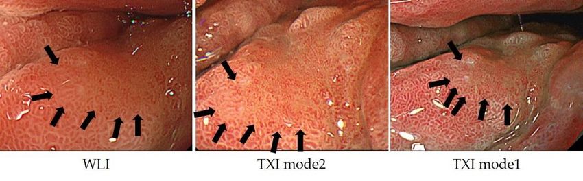
J. Clin.
J. Clin. Med.
Med. 2021, 10,x2918
2021,10, FOR PEER REVIEW 3 of 12 3 of 1
Figure 2. (a) The fundic gland mucosa without inflammation. The glandular structures are regularl
arranged in a mesh pattern; (b) The glandular structure is still circular, but gradually becomes ir
regular in size and shape; (c) The circular glandular structures gradually disappear and tubula
glandular structures are observed; (d) Gastric fundic glands disappear and glandular structure re
sembles that of pyloric glands; (e) The pyloric gland mucosa without inflammation.
Figure2.2.(a)
Figure (a)The
Thefundic
fundic gland
gland mucosa
mucosa without
without inflammation.
inflammation. The glandular
The glandular structures
structures are regularly
are regularly
arranged
arranged ininaamesh
ME-NBI mesh pattern;
of intestinal
pattern; (b)(b) The glandular
metaplasia
The shows
glandular structure is still
a blue-white
structure is still circular,
border
circular, butthe
at gradually
but graduallylimbus
becomesbecomes
of the WZir
regular
This was
irregular in size
in size and
reported shape;
as a(c)
and shape; (c)
light The
The blue circular
crest
circular glandular
by Uedo
glandular structures
et al.gradually
structures gradually
and is thought disappear
disappeartoand
representand tubula
tubular the dif
glandular
glandular
ference in structures
structures
reflectanceareobserved;
are observed; (d)(d)
characteristics Gastric
Gastric fundic
fundic
caused by glands
glands
the disappear
disappear
projection and andnm glandular
glandular
of 415 structure
structure
narrow-band re
ligh
sembles that of pyloric glands; (e) The pyloric gland mucosa without
resembles that of pyloric glands; (e) The pyloric gland mucosa without inflammation. inflammation.
onto the brush border of intestinal metaplasia (Figure 3) [12].
ME-NBI of intestinal metaplasia shows a blue-white border at the limbus of the WZ
This was reported as a light blue crest by Uedo et al. and is thought to represent the dif
ference in reflectance characteristics caused by the projection of 415 nm narrow-band ligh
onto the brush border of intestinal metaplasia (Figure 3) [12].
Figure3.3.The
Figure The intestinal
intestinal metaplasia
metaplasia observed
observed as a blue-white
as a blue-white border border by ME-NBI.
by ME-NBI.
2.2. Diagnostic Algorithm for Early Gastric Cancer Using ME-NBI
2.2. Diagnostic Algorithm for Early Gastric Cancer Using ME-NBI
In 2009, Yao et al. proposed vessel plus surface (VS) classification, a diagnostic
In 2009, Yao et al. proposed vessel plus surface (VS) classification, a diagnostic algo
algorithm for early gastric cancer using ME-NBI [10], and in 2016, Muto et al. proposed a
rithm
magnifying
Figure 3. forThe
early gastricmetaplasia
endoscopy
intestinal cancerdiagnostic
simple using ME-NBI
observed [10],for
algorithm and
as a blue-white inborder
early 2016, Muto
gastric
by cancer et (MESDA-G)
ME-NBI. al. proposed a mag
nifying
based on endoscopy
VS classificationsimple[11].diagnostic
In MESDA-G, algorithm for early
(1) the presence ofgastric cancer line
a demarcation (MESDA-G)
(DL) is base
on VS
checked, classification
and if present,
2.2. Diagnostic Algorithm [11].
(2)for In MESDA-G,
microvascular
Early Gastric (1)
pattern the
Cancer(MVP) presence
UsingorME-NBI of a demarcation
microsurface pattern (MSP) line (DL) i
checked,
will and ifand
be analyzed, present,
if either (2)ormicrovascular pattern
both are irregular, early (MVP) or microsurface
gastric cancer is diagnosed. pattern
The (MSP
In 2009,
anatomical
Yao etused
structuresand
al. proposed vesselare
plus surface (VS) classification, a diagnostic algo
will be analyzed, if in MVPor
either analysis subepithelial
both are irregular, capillaries,
early gastriccollecting
cancer isvenules,
diagnosed. Th
rithm
and for early gastric
pathological cancer using
microvessels. ME-NBI [10], and in 2016, Muto et al. for
proposed
small a mag
anatomical structures used inAMVP pathological
analysismicrovessel is a general
are subepithelial term
capillaries, collecting ven
nifying
blood endoscopy simple diagnostic algorithm for early gastric cancervenules.
(MESDA-G)
The based
ules, vessels that cannot
and pathological be classified
microvessels. as subepithelial
A pathological capillaries or collecting
microvessel is a general term for sma
on VS classification
anatomical structures used[11]. inIntheMESDA-G,
analysis of (1)
MSP thearepresence
the WZ, whichof a demarcation
reflects the MCE, line (DL) i
blood vessels that cannot be classified as subepithelial capillaries or collecting venules
crypt openings, intervening part, and white opaque substance
checked, and if present, (2) microvascular pattern (MVP) or microsurface pattern (WOS) (Figure 4). WOS is a (MSP
The
white anatomical
substance structures
thatand
exists used in
in the surfacethe analysis of MSP are the WZ, which reflects the MCE
will be analyzed, if either or bothofare
intestinal metaplasia,
irregular, adenoma,
early gastric and carcinoma
cancer is diagnosed. The
crypt
of openings,
chronic gastritis intervening
mucosa part,byand
reported Yao white opaque
et al. WOS substance (WOS)
is a microscopic (Figure
lipid droplets that4). WOS i
anatomical structures used in MVP analysis are subepithelial capillaries, collecting ven
a white substance that exists in the surface of intestinal
accumulates in the epithelium and subepithelium. The morphology and distribution ofmetaplasia, adenoma, and carc
ules, and pathological microvessels. A pathological microvessel is a general term for smal
noma
WOS canofbechronic
used togastritis
differentiatemucosa reported
between cancerby andYao et al. WOSlesions.
non-cancerous is a microscopic
The presence lipid drop
blood
of WOS
vessels
also
that cannot
suggests gastric
begastrointestinal
or
classified as subepithelial
mucin phenotype
capillaries
[13–16].
or collecting venules
lets that accumulates in the epithelium and subepithelium. The morphology and distribu
The anatomical structures used in the analysis of MSP are the WZ, which reflects the MCE
tion of WOS can be used to differentiate between cancer and non-cancerous lesions. Th
crypt openings, intervening part, and white opaque substance (WOS) (Figure 4). WOS i
presence of WOS also suggests gastric or gastrointestinal mucin phenotype [13–16].
a white substance that exists in the surface of intestinal metaplasia, adenoma, and carci
noma of chronic gastritis mucosa reported by Yao et al. WOS is a microscopic lipid drop
lets that accumulates in the epithelium and subepithelium. The morphology and distribu
tion of WOS can be used to differentiate between cancer and non-cancerous lesions. The
J. Clin.
J. Clin. Med.
Med.
J. Clin. 2021,
2021,
Med. 10,10,
10,
2021, xx 2918
FOR PEER
FOR PEERREVIEW
REVIEW 4 of 12 4 of 13
Figure 4. White opaque substance.
Figure
Figure 4. 4. White
White opaque
opaque substance.
substance.
MVP morphology can be classified as regular, irregular, or absent. Regular MVPs
haveMVP a uniform morphology
morphology and shape
can be classified for eachirregular,
as regular, vessel, symmetrical distribution,
or absent. Regular MVPs have and reg-
ular
a uniform
MVP morphology
arrangement.
morphology Irregular can
and shapeMVPbe has
for
classified
eacha variety as
vessel, of
regular, distribution,
morphologies,
symmetrical
irregular,
includingor absent.
andlooped,
Regula
regular tortu-
have
ous, a uniform
branched,
arrangement. andmorphology
bizarrely
Irregular MVP has a and
shaped
varietyshape
vessels for
witheach vessel,
asymmetric
of morphologies, symmetrical
distribution
including distribution,
and
looped, tortuous, irregular
ular arrangement.
branched,
arrangement. bizarrelyIrregular
andCancer-specific MVP with
shapedmorphology
vessels has aof variety
asymmetric
irregular ofdistribution
morphologies,
micro vesselsandhas including
irregular ar- loope
been described
rangement.
asous,
dilatation,Cancer-specific
branched, morphology
and bizarrely
heterogeneity shaped
in shape, of irregular
abrupt vessels micro
caliberwith vessels has
asymmetric
alteration, been described
distribution
and tortuousness. asAbsent
and i
dilatation,
MVP heterogeneity
has WOS on in shape, abrupt
the mucosal surface, caliber alteration,
and MVP of cannotand tortuousness.
be adequately Absent
observed.MVP
arrangement. Cancer-specific morphology irregular micro vessels has been d
has WOS on the mucosal surface, and MVP cannot be adequately observed.
MSP morphology
as dilatation, is classified as regular,
heterogeneity irregular, or absent. In regular MSP, WZ is
MSP morphology is classifiedinasshape,
regular,abrupt
irregular,caliber alteration,
or absent. In regularandMSP,tortuousness
WZ
aMVPuniformhaslinear,
WOS curved,
theoval, or circular structure with homogeneous morphology, sym-
is a uniform linear,oncurved, mucosal surface,
oval, or circular and MVP
structure with cannot be adequately
homogeneous morphology, observed
metrical distribution, and regular arrangement. If the WOS is present and the MSP is not
MSP morphology
symmetrical distribution, and is regular
classified as regular,
arrangement. irregular,
If the or absent.
WOS is present and theIn regular
MSP MS
visible,
isa not the MSP
visible, the is regular
MSP is if theifWOS
regular the is regularly
WOS is arranged
regularly arrangedin ainuniform
a uniformform. In irregular
form. In
MSP,
uniform
the MSP,
linear, curved,
morphology
oval, or
of WZs isofan
circular
irregular
structure with
linear, curved,
homogeneous
oval, circular,
morpholo
or villous
irregular the morphology WZs is an irregular linear, curved, oval, circular, or struc-
metrical
ture
villouswith distribution,
heterogeneous
structure
and regular
morphology,
with heterogeneous
arrangement.
asymmetrical
morphology,
If the WOS
distribution,
asymmetrical
is present
and and
distribution, irregular and
irregular
the M
arrange-
visible,
ment.
arrangement.the WOS
When MSP
WhenisisWOS
regular
present, if the
the
is present, WOS
morphology
the is regularly
morphologyof WOS arranged
is non-uniform
of WOS in a uniform
is non-uniform andandthetheform. In
arrange-
MSP, the
arrangement morphology
is irregular; of
this isWZs
called is an irregular
irregular MSP. linear,
Absent MSPcurved,
ment is irregular; this is called irregular MSP. Absent MSP is the state in which the is the oval,
state in circular,
which the or WZ
villo
WZ
and and
tureWOS WOS
with are not visible
areheterogeneous (Figure
not visible (Figure 5, Table
morphology, 1)
5, Table 1) [11]. [11].
asymmetrical distribution, and irregular
ment. When WOS is present, the morphology of WOS is non-uniform and the
ment is irregular; this is called irregular MSP. Absent MSP is the state in which
and WOS are not visible (Figure 5, Table 1) [11].
5. The
Figure 5.
Figure The upper
upperrow
rowisisananexample
example of of
regular MSP,
regular irregular
MSP, MSP,MSP,
irregular and absent MSP. The
and absent MSP.bottom
The bottom
row is an example of regular MVP, irregular MVP, and absent
row is an example of regular MVP, irregular MVP, and absent MVP. MVP.
Table 1. Differences in MSP and MVP between cancer and non-cancer.
Non Cancer Cancer
Figure 5. The upper
morphology: row is an example of regular
homogeneous MSP, irregular
morphology: MSP, and absent MSP. Th
heterogeneous
row is an example of regular MVP,
(uniform linear, curved, oval, circular) irregular MVP, and absent MVP.
(irregular linear, curved, oval, circular)
Micro surface pattern
distribution: symmetric distribution: asymmetric
Tablearrangement:
1. Differencesregular
in MSP and MVP between cancer and non-cancer.
arrangement: irregular
Micro vascular pattern morphology: uniform morphology: heterogeneous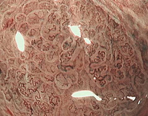
J. Clin. Med. 2021, 10, 2918 5 of 12
Table 1. Differences in MSP and MVP between cancer and non-cancer.
Non Cancer Cancer
morphology: homogeneous morphology: heterogeneous
J. Clin.Micro
Med. 2021, 10, pattern (uniform linear, curved, oval, circular)
x FOR PEER REVIEW (irregular linear, curved, oval, circular) 5 of 13
surface
distribution: symmetric distribution: asymmetric
arrangement: regular arrangement: irregular
morphology: heterogeneous
distribution: symmetric
morphology: uniform (dilatation, heterogeneity
(dilatation, heterogeneityin in
shape,
shape,abrupt
abrupt caliber
caliber
Micro vascular pattern distribution:
arrangement: symmetric
regular alteration,
alteration, tortuousness)
tortuousness)
arrangement: regular distribution: asymmetric
distribution: asymmetric
arrangement: irregular
arrangement: irregular
On
Onthe theother
otherhand,
hand,ME-NBI
ME-NBIfindings
findingsspecific
specifictotoearly
earlygastric
gastriccancer
cancerhave
havealso
alsobeen
been
reported,
reported, including the presence
including the presence ofof white
whiteglobular
globularreservoirs
reservoirsless
lessthan
than 1 mm
1 mm in in
sizesize
be-
beneath
neath the intraepithelial vessels, termed white globe appearance (WGA) by Doyama et al.
the intraepithelial vessels, termed white globe appearance (WGA) by Doyama et al.
(Figure
(Figure6).6).WGA
WGAreflects
reflectsan
anintraglandular
intraglandularnecrotic
necroticdebris,
debris,which
whichhashasbeen
beenreported
reportedas asaa
specific
specifichistopathological
histopathologicalmarker
markerforforcancer,
cancer,and
andisispresent
presentin
in20%
20%ofofearly
earlygastric
gastriccancers,
cancers,
but not in adenomas, and has a high specificity for early gastric cancer
but not in adenomas, and has a high specificity for early gastric cancer [17]. [17].
Figure6.6.White
Figure Whiteglobe
globeappearance.
appearance.
2.3.
2.3.Inference
InferenceofofHistopathological
HistopathologicalType TypeofofGastric
GastricCancer
CancerbybyME-NBI
ME-NBI
Tubular adenocarcinomas proliferate while
Tubular adenocarcinomas proliferate while retaining retaining thethe
morphology
morphology of of
thethe
glandular
glandu-
structure.
lar structure. In well-differentiated adenocarcinomas with relatively straight vertically
In well-differentiated adenocarcinomas with relatively straight and and verti-
concave gland ducts,
cally concave gland the capillaries
ducts, in the intervening
the capillaries part arepart
in the intervening in a reticular pattern, termed
are in a reticular pattern,
the mesh pattern by Yagi et al. (Figure 7). Capillaries in the mesh pattern
termed the mesh pattern by Yagi et al. (Figure 7). Capillaries in the mesh pattern observed observed in early
gastric
in earlycancer show
gastric dilatation,
cancer caliber change,
show dilatation, caliberand uneven
change, andshape,
unevenandshape,
can beand
differentiated
can be dif-
from gastritis. Well-differentiated adenocarcinomas with a
ferentiated from gastritis. Well-differentiated adenocarcinomas with a mesh pattern mesh pattern tend to have
tend
dense
to have dense glandular structures, and the WZ is often not visible, so the DLclearly
glandular structures, and the WZ is often not visible, so the DL can be can be
visualized. DL is also
clearly visualized. DLuseful
is alsoforuseful
differentiating gastritis from
for differentiating gastric
gastritis fromcancer. A cancer.
gastric regular A mesh
reg-
pattern is called a complete mesh pattern and is suggestive of a well-differentiated
ular mesh pattern is called a complete mesh pattern and is suggestive of a well-differenti- mucosal
adenocarcinoma [18]. This pattern
ated mucosal adenocarcinoma [18].isThis
considered
patternto is be synonymous
considered to bewith the fine network
synonymous with the
pattern
fine network pattern reported by Nakayoshi et al. [19]. In tubular adenocarcinomas,gland
reported by Nakayoshi et al. [19]. In tubular adenocarcinomas, where the where
ducts are tortuous and branched, the WZ is irregularly tubular, and the capillaries in the
the gland ducts are tortuous and branched, the WZ is irregularly tubular, and the capil-
intervening part are irregularly looped (Figure 8). This is referred to as the loop pattern
laries in the intervening part are irregularly looped (Figure 8). This is referred to as the
proposed by Yagi et al. [18]. In addition, Kanemitu et al. reported the characteristic findings
loop pattern proposed by Yagi et al. [18]. In addition, Kanemitu et al. reported the char-
of papillary adenocarcinoma visualized using ME-NBI, with vessels within the circular
acteristic findings of papillary adenocarcinoma visualized using ME-NBI, with vessels
intervening part surrounded by circular MCE, which labeled as the vessels within the
within the circular intervening part surrounded by circular MCE, which labeled as the
epithelial circle (VEC) pattern. They reported that 94.3% of early gastric cancers with a VEC
vessels within the epithelial circle (VEC) pattern. They reported that 94.3% of early gastric
pattern showed papillary structures on histopathology, and the frequency of submucosal
cancers with a VEC pattern showed papillary structures on histopathology, and the fre-
invasion was significantly high in early gastric cancers with a VEC pattern [20]. Papillary
quency of submucosal invasion was significantly high in early gastric cancers with a VEC
adenocarcinoma is more biologically aggressive than tubular adenocarcinoma and requires
pattern [20]. Papillary adenocarcinoma is more biologically aggressive than tubular ade-
more attention [21–23].
nocarcinoma and requires more attention [21–23].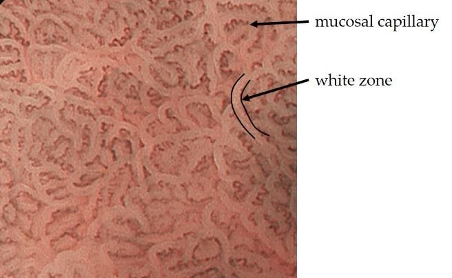
J.J.Clin.
Clin.Med.
Med.2021, 10, x2918
2021, 10, FOR PEER REVIEW 6 of 126 of 13
J. Clin.
J. Clin. Med.
Med. 2021,
2021, 10,
10, xx FOR
FOR PEER
PEER REVIEW
REVIEW 66 of
of 13
13
Figure
Figure
Figure 7.
Figure7.
7. The
7.The
The mesh
Themesh
mesh pattern
meshpattern
pattern suggests
patternsuggests
suggests
suggests tubular adenocarcinomas.
tubular
tubular
tubular adenocarcinomas.
adenocarcinomas.
adenocarcinomas.
Figure 8.
Figure 8. The
The loop
loop pattern
pattern suggests
suggests tubular
tubular adenocarcinomas.
adenocarcinomas.
Figure8.8.The
Figure Theloop
looppattern
patternsuggests tubular
suggests adenocarcinomas.
tubular adenocarcinomas.
The ME-NBI
The
The findings
ME-NBI findings
findingsin ininundifferentiated
undifferentiated carcinomas,
undifferentiatedcarcinomas,
carcinomas, including
including
including poorly differentiated
poorly
poorly differenti-
differentiated
ated The ME-NBIand
adenocarcinomas
adenocarcinomas findings
and in
signet-ring undifferentiated
cell
signet-ring carcinomas,
cell carcinomas,
vary
carcinomas, vary including
depending on
depending thepoorly
degree
on differentiated
the of inva-
degree
adenocarcinomas and signet-ring cell carcinomas, vary depending on the degree of inva-
adenocarcinomas
sion.
of In the
invasion. early
In the and
stage,
early signet-ring
non-cancerous
stage, cellglandular
non-cancerous carcinomas,ducts
glandularvary depending
remain
ducts in the
remain on the
superficial
in the degree
layer
superficialof inva-
and
sion. In the early stage, non-cancerous glandular ducts remain in the superficial layer and
sion.
the
layer In
numberthe early
of cancer
and theofnumber
the number stage,
cancer of non-cancerous
cells is
cancer
cells small; glandular
therefore,
cells therefore,
is small; ME-NBI
is small; therefore,ducts
ME-NBI often remain
often
ME-NBI shows in
often
shows the
only superficial
shows
only subtle
only
subtle subtle and
layer
changes,
changes,
the number
such as
as an
changes,
such ansuch of cancer
enlarged
as an enlarged
enlarged cells
intervening
intervening is small;
intervening
part. therefore,
part. With
Withpart. ME-NBI
further
With
further invasion, often shows
poorly connected,
further invasion,
invasion, poorly only
connected, subtle changes,
shrunken
poorly connected,
shrunken
such as anmicrovessels
microvessels
shrunken
microvessels enlarged
can be
can intervening
be observed.
observed.
can be observed. part. With
Furthermore,
Furthermore, further
when
Furthermore,
when invasion,
undifferentiated
undifferentiatedpoorly connected,
adenocarcinoma
when undifferentiated
adenocarcinoma shrunken
adenocar- in-
in-
vaded
cinoma all layers
invaded of
all the mucosa
observed.
layers of theand the glandular
Furthermore,
mucosa and when
the structure
vaded all layers of the mucosa and the glandular structure disappeared, the WZ disap- in-
microvessels can be disappeared,
undifferentiated
glandular structure the WZ
adenocarcinoma
disappeared, disap-
the
peared,
WZ
vaded
peared, thelayers
all
the density
disappeared,
density theofthe
ofof blood
density
mucosa
blood vessels
of blood
vessels and decreased,
vessels and microvessels
the glandular
decreased, and microvessels
decreased, in the
the form
and microvessels
structure forminof
disappeared,
in ofthe
contracted
the form
WZ of
contracted disap-
surfaces,
contracted
peared, the
surfaces, each
each of which
surfaces,
density
of which eachmeanders
of
of blood
meanderswhich irregularly,
vessels meanders
decreased,
irregularly, were
were andobserved
irregularly, were(Figure
microvessels
observed observed
(Figurein 9).
9).theThis is
(Figure
Thisform referred
9). This to
of contracted
is referred to
is
asreferred
surfaces,
as wavy to as
wavy microvessels wavy
microvessels
each of which microvessels
bymeanders
by Yagi et
Yagi et al. by Yagi
al. irregularly, et
[18]. However,
[18]. However, al.
were [18]. However,
theobserved
the majority(Figure
majority the
of early
of majority
early9). of early
undifferentiated
This is referred to
undifferentiated
undifferentiated
adenocarcinomas
as wavy microvessels
adenocarcinomas adenocarcinomas
are covered by
by Yagibyetaal.
are covered a are covered
non-cancerous by
[18]. However,
non-cancerous a non-cancerous
epithelium, and
the majority
epithelium, and of epithelium,
the ME-NBI
theearly
ME-NBI and theis
image
undifferentiated
image is
ME-NBI
poorly image
altered,
adenocarcinomas
poorly is
altered, limitingpoorly
limiting altered,
the
are covered limiting
diagnosis. the
Capture
by a non-cancerous
the diagnosis. diagnosis.
of slight
Capture of slight Capture
changes
epithelium,
changes like of
like slight changes
enlargement
andenlargement
the ME-NBIofimageoflike
the
the is
enlargement
intervening part
intervening of the
part intervening
warrants
warrants biopsy
biopsy part warrants biopsy evaluation.
evaluation.
evaluation.
poorly altered, limiting the diagnosis. Capture of slight changes like enlargement of the
intervening part warrants biopsy evaluation.
Figure 9.
Figure
Figure 9. No
9. Noglandular
No glandularstructures
glandular structureswere
structures wereobserved,
were observed,and
observed, and
and wavy
wavy
wavy micro
micro
micro vessels
vessels were
were
vessels observed,
observed,
were suggest-
suggesting
observed, suggest-
ing undifferentiated
undifferentiated adenocarcinoma.
adenocarcinoma.
ing undifferentiated adenocarcinoma.
Figure 9. No glandular structures were observed, and wavy micro vessels were observed, suggest-
2.4.
2.4. Efficacy
Efficacy of
of ME-NBI
ME-NBI
ing undifferentiated for
for Early
Early Gastric
adenocarcinoma. Gastric Cancer
Cancer
2.4. Efficacy of ME-NBI for Early Gastric Cancer
2.4.1. ME-NBI in Screening Tests for Gastric Cancer
2.4.1. ME-NBI
2.4.1. ME-NBI in in Screening
Screening Tests
Tests for
for Gastric
Gastric Cancer
Cancer
Yao et al.
2.4. Efficacy of reported for
ME-NBI thatEarly
97%Gastric
of earlyCancer
gastric cancers met the VS classification crite-
Yao et
Yao et al.
al. reported
reported thatthat 97% of of early
early gastric
gastric cancers
cancers met the
the VS
VS classification
classification criteria,
criteria,
ria, and
2.4.1. Ezoe
ME-NBI reported97%
etinal.Screening that the
Tests foraccuracy
Gastric rate of VSmet
Cancer classification for small gastric
and Ezoe et al.
and Ezoe etcancer reported
al. reported that the
that the accuracy
accuracy rate of VS classification
rate of VSprospective
classification for small
for small gastric
gastric depres-
depres-
depression was 96.6% [24]. In a multicenter study, Yao et al. reported
sion cancer
sion cancer wasreported
Yao et was
al. 96.6% [24].
96.6% [24]. In
thatIn aa multicenter
97% multicenter prospective
of early gastric study,
cancersstudy,
prospective met the Yao
VSet
Yao etclassification
al. reported
al. reported criteria,
the
the
and Ezoe et al. reported that the accuracy rate of VS classification for small gastric depres-
sion cancer was 96.6% [24]. In a multicenter prospective study, Yao et al. reported the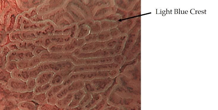
J. Clin. Med. 2021, 10, 2918                                                                                            7 of 12

                               the usefulness of ME-NBI using VS classification in routine examinations. Especially in
                               erythematous/isochromatic mucosal lesions, mainly differentiated adenocarcinoma, the
                               accuracy rate was extremely high at 99.4%, and I think that NBI is an essential technique
                               for routine screening examinations outside of referral centers [25].
                                    Marta et al. performed a meta-analysis to evaluate the diagnostic value of NBI for
                               gastric intestinal metaplasia and early gastric cancer. The pooled sensitivity and specificity
                               of NBI for gastric intestinal metaplasia were 0.79 and 0.91, respectively. The tubulovillous
                               pattern was the most accurate marker for detecting gastric intestinal metaplasia and could
                               be evaluated effectively without the need for high magnification. The pooled sensitivity
                               and specificity of NBI for gastric cancer were 0.87 and 0.97, respectively. The use of
                               magnification improved the performance of NBI in characterizing gastric cancer, especially
                               when VS classification was applied [26].
                                    These reports suggest that NBI is useful in screening tests and is an essential technique
                               in daily practice.

                               2.4.2. ME-NBI in Histological Diagnosis of Gastric Cancer
                                    Nonaka et al. reported the usefulness of ME-NBI in differentiating gastric adenomas
                               from well-differentiated adenocarcinomas. Type I is when the WZ is preserved and rela-
                               tively uniform with distinct MVP; Type II, when the WZ is relatively uniform and the MVP
                               is similar to the surrounding mucosa; Type III is when the WZ is relatively uniform with
                               irregular, darker MVP that is more prominent than the surrounding mucosa; Type IV is
                               when the WZ tends to disappear with presence of irregular MVP; and Type V is when the
                               WZ disappears with presence of irregular MVP. The accuracy rate was 88.5%, with type I
                               and II as adenomas and type III to V as well-differentiated adenocarcinomas [27].
                                    Yao et al. reported that 100% of the lesions with well-formed WOS were adenomas,
                               and 83% of the lesions with irregular WOS were well-differentiated adenocarcinomas [15].
                                    The usefulness of ME-NBI for the histopathological diagnosis of gastric cancer requires
                               further investigation in the future.

                               2.4.3. ME-NBI in Determining the Horizontal Extent of Gastric Cancer
                                     Nonaka et al. examined the concordance rate between DL and histopathologic borders
                               observed in depressed and flat-differentiated adenocarcinomas. The accuracy rate was
                               100%, indicating that ME-NBI is useful for diagnosing the extent of differentiated adeno-
                               carcinoma [28]. Especially in cases where the irregularity of the WZ is clear, or in lesions
                               where the WZ is not visible and the irregularity of the MVP is easily visible, determining
                               the horizontal extent is easy. However, some lesions, such as extremely well-differentiated
                               adenocarcinoma, have only minor irregularities in the WZ and MVP; therefore, caution
                               is required.
                                     Horii et al. examined the rate of negative biopsies from non-cancerous tissue outside
                               the lesion and negative horizontal margins of endoscopic submucosal dissection specimens
                               in early gastric cancer after confirmation of DL using ME-NBI.
                                     The rates of biopsy-negative and negative horizontal margins were 96.7% and 97.9%,
                               respectively, in early gastric cancer. They reported that the risk factors for a positive hori-
                               zontal resection margin were tumor size > 20 mm and moderately or poorly differentiated
                               adenocarcinomas [29].
                                     Horiuchi et al. examined the concordance rate between the DL and histopathological
                               borders in undifferentiated adenocarcinomas. They reported a concordance rate of 81.6%,
                               which was 27.6% higher than that of white light observation alone. In particular, undif-
                               ferentiated adenocarcinoma with little inflammatory cell infiltration could be delineated
                               with high accuracy [30]. However, undifferentiated adenocarcinoma may develop laterally
                               with non-cancerous epithelium, and ME-NBI has limitations in accurately diagnosing its
                               extent. In such cases, a biopsy should be performed in combination with ME-NBI findings
                               to make a decision.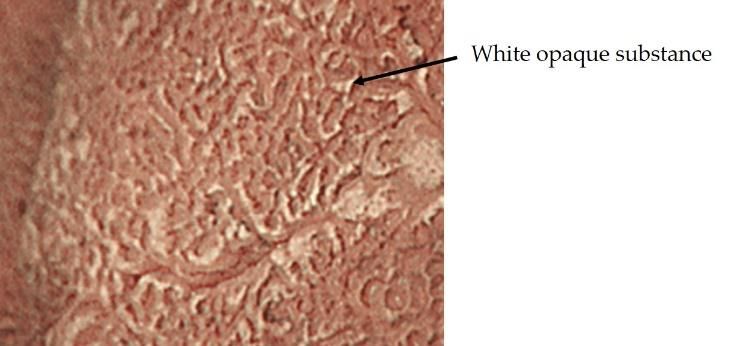
J. Clin. Med. 2021, 10, 2918                                                                                            8 of 12

                                     In recent years, the number of gastric cancers detected after HP eradication has been
                               increasing, and it has been reported that gastric cancers detected after HP eradication
                               may have a gastritis-like surface structure, which may make qualitative diagnosis and
                               determining extent difficult. [31]. Saka et al. reported that the histopathological reason for
                               this was that non-cancerous epithelium and non-cancerous glandular ducts covered the
                               cancerous glandular ducts [32].
                                     Akazawa et al. reported that there were significantly more gastric cancers with unclear
                               DL in the HP eradication group than in the HP infection group (11.8% vs. 1.5%) [33].
                                     Horiguchi et al. investigated the accuracy of determining the horizontal extent of
                               gastric cancer after HP eradication and reported that WLI, chromoendoscopy, and ME-NBI
                               all significantly decreased the reliability of the determining the horizontal extent of gastric
                               cancer after HP eradication compared with non-eradication [34].
                                     The number of gastric cancers detected after HP eradication is expected to increase in
                               the future, and further studies on the margin delineation for gastric cancer detected after
                               eradication are needed.

                               2.4.4. ME-NBI in Estimating the Depth of Invasion of Early Gastric Cancer
                                    Kikuchi et al. reported that the presence of dilated abnormal blood vessels (three
                               times larger than microvessels) in early gastric cancer may correlate with submucosal
                               invasion, but the number of cases was small and the sensitivity was 37.5%, which is not
                               sufficient [35]. Yagi et al. reported that 94.9% of early gastric cancers with a mesh pattern
                               were intramucosal carcinomas and 92.3% of early gastric cancers with an interrupted
                               pattern were submucosal invasive carcinomas, but the number of cases was small, and
                               further studies are needed [18].
                                    The usefulness of ME-NBI in estimating the invasion depth has been reported in recent
                               years, but no definite opinion has been obtained, and this is a subject for further study.

                               3. Comparison of ME-NBI with Other Modalities
                                    Blue laser imaging (BLI) is an observation method that is often compared with NBI.
                               BLI uses two short-wavelength narrow-band lasers of 410 nm and 450 nm. The 410 nm
                               laser mainly highlights the surface blood vessels and structures of the mucosa. The output
                               of the 450 nm laser light was adjusted to excite the phosphor to produce light with a wide
                               wavelength to ensure brightness. Depending on the balance of output power, BLI can be
                               used in two modes: BLI mode, which is suitable for magnification, and BLI-bright mode,
                               which is slightly brighter and suitable for non-magnified observation [36].
                                    Le et al. compared the diagnostic accuracy for gastric cancer of conventional WLI,
                               magnifying endoscopy with WLI (ME-WLI), ME-NBI, and magnifying endoscopy with
                               BLI (ME-BLI) in a meta-analysis including eight prospective studies. They reported that
                               the diagnostic accuracy of ME-WLI, ME-NBI, and ME-BLI was higher than that of conven-
                               tional WLI. They also reported that the diagnostic accuracy of ME-NBI and ME-BLI was
                               significantly higher than that of ME-WLI, but there was no difference between ME-NBI
                               and ME-BLI [37].
                                    Zhou et al. compared the diagnostic efficacy of NBI and BLI in detecting gastric cancer
                               in a meta-analysis of six BLI and 22 NBI reports. The pooled sensitivity of BLI for gastric
                               cancer was 0.89, and the specificity was 0.92. The pooled sensitivity of NBI for gastric
                               cancer was 0.83 and the specificity was 0.95. There was no difference in the diagnostic
                               performance of NBI and BLI, and both groups had high diagnostic performance [38].
                                    The results of the two meta-analyses showed no significant difference between the
                               diagnostic performance of NBI and BLI.

                               4. Future Prospects
                                    Till date, the fundamentals and diagnostic capabilities of NBI for early gastric cancer
                               are outlined. Although ME-NBI has become an essential technique for screening and
                               precision examination of gastric cancer at an earlier stage, it is somewhat complicated,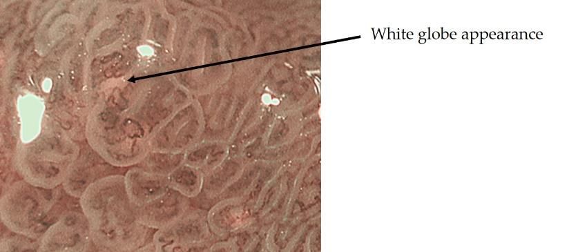
J. Clin. Med. 2021, 10, 2918                                                                                                      9 of 12

                                     requires a higher level of expertise, and is still subjective. Today, artificial intelligence (AI)
                                     has great potential to support decision making in various medical fields, and may be able
                                     to detect abnormalities that are often overlooked by non-experts. Li et al. developed a
                                     new system based on convolutional neural network to analyze the early gastric cancer
                                     observed by ME-NBI. The results showed that the sensitivity, specificity, and accuracy in
                                     diagnosing early gastric cancer were 91.18%, 90.64%, and 90.91%, respectively. In addition,
                                     there was no significant difference in the specificity and accuracy of diagnosis between
                                     their system and experts. Moreover, the diagnostic sensitivity, specificity, and accuracy
                                     of their system were significantly higher than those of the non-experts [39]. Hu et al.
                                     developed a computer-aided diagnostic model for early gastric cancer to analyze and assist
                                     in the diagnosis of early gastric cancer using ME-NBI. They compared the model with
                                     eight endoscopists with varying experience, and found that their model achieved similar
                                     predictive performance to the senior endoscopists (accuracy: 0.770 vs. 0.755, p = 0.355;
                                     sensitivity: 0.792 vs 0.767, p = 0.183; specificity: 0.745 vs. 0.742, p = 0.931) but better than
                                     the junior endoscopists (accuracy: 0.770 vs. 0.728, p < 0.05). After referring to the results,
                                     the average diagnostic ability of the endoscopists was significantly improved in terms of
                                     accuracy, sensitivity, positive and negative predictive value [40].
                                           These results suggest that the combination of ME-NBI and artificial intelligence (AI) is
                                     useful for the diagnosis of early gastric cancer. The combination of AI has the potential
                                     to overcome the weaknesses of NBI in that it is subjective and requires a high level of
                                     expertise, suggesting that it can diagnose non-experts at the same level as experts, and
                                     research in this direction is expected to further enhance the diagnostic capabilities of NBI.
                                           In addition to NBI, a variety of other observation methods have emerged. For the
                                     detection of tumors, observation methods that emphasize color tone and structure, such
                                     as linked color imaging, have been developed and are widely used, and there are reports
                                     on their usefulness [41]. In addition, the development of texture and color enhancement
                                     imaging (TXI), which is a new observation method that emphasizes color tone and struc-
                                     ture, extended depth of field, which is easy to focus, and red dichromatic imaging (RDI),
                                     which is a new narrow-band light observation method that improves the visibility of deep
                                     tissues such as deep blood vessels and improves the visibility of bleeding points, has been
                                     reported. TXI is designed to enhance three image factors in WLI (texture, brightness, and
                                     color) to define subtle tissue difference clearly. TXI has two settings, “mode1” with color
                                     enhancement and “mode2” without color enhancement. TXI mode2 looks more like WLI
                                     color tone (Figure 10) [42]. RDI is a new image-enhanced endoscopy consisting of three
                                     types of illumination with wavelengths of 540, 600, and 630 nm. Hemoglobin strongly
                                     absorbs light at 600 nm and weakly absorbs light at 630 nm. Because of this absorption
                                     property, the light intensity of the reflected light at 600 nm is greatly attenuated compared
                                     to that of the reflected light at 630 nm in areas with high hemoglobin concentration, such
                                     as the bleeding point, and it is observed as an orange color. On the other hand, when
                                     the hemoglobin concentration is low around the bleeding point, the light intensity of
 J. Clin. Med. 2021, 10, x FOR PEER REVIEW
                                     the reflected light at 600 nm and 630 nm is about the same, and it is observed as yellow    10 of 13
                                     (Figure 11) [43].

                                  Figure 10. In TXI mode2, the structure is enhanced compared to WLI, and the relatively dark areas
                                   Figure 10. In TXI mode2, the structure is enhanced compared to WLI, and the relatively dark areas
                                  in
                                   inthe
                                      theback
                                          backare
                                                areadjusted
                                                    adjustedtotobe
                                                                 bebrighter;
                                                                    brighter;ininTXI
                                                                                  TXImode
                                                                                     mode1,1,the
                                                                                              thecolor
                                                                                                  colortone
                                                                                                        toneisisfurther
                                                                                                                 furtherenhanced
                                                                                                                         enhancedand
                                                                                                                                  andthe
                                                                                                                                      the
                                  contrast is more  distinct.
                                   contrast is more distinct.
J. Clin. Med. 2021, 10, 2918 Figure 10. In TXI mode2, the structure is enhanced compared to WLI, and the relatively dark10 areas
of 12
in the back are adjusted to be brighter; in TXI mode 1, the color tone is further enhanced and the
contrast is more distinct.
Figure11.
Figure 11.No
Noglandular
glandularstructures
structureswere
wereobserved,
observed, and
and wavy
wavy micro
micro vessels
vessels were
were observed,
observed, suggest-
suggesting
ing undifferentiated adenocarcinoma.
undifferentiated adenocarcinoma.
Inthis
In thisway,
way,the
thedevelopment
developmentandandimprovement
improvementofofendoscopic
endoscopicequipment
equipmentcontinues
continues
at a rapid pace, and it is important to understand the principles and advantages
at a rapid pace, and it is important to understand the principles and advantages of ofeach
each
observation method before performing an examination.
observation method before performing an examination.
5.5.Conclusions
Conclusions
The
The current
current status of ME-NBI
ME-NBI forforearly
earlygastric
gastriccancer
cancerwas
wasoutlined.
outlined. ME-NBI
ME-NBI hashas
be-
become an essential procedure for screening and
come an essential procedure for screening and detailed detailed examination, but it is somewhat
but it is somewhat
complicated
complicatedand andsubjective
subjective than that
than for for
that esophagus
esophagusandand
colon. In addition,
colon. with the
In addition, increase
with the in-
of gastric
crease cancer cancer
of gastric detected after HP
detected aftereradication, the qualitative
HP eradication, and and
the qualitative extent delinetion
extent of
delinetion
ME-NBI
of ME-NBI maymay
become
becomemore complicated
more in the
complicated infuture. Although
the future. the application
Although of AI of
the application may
AI
solve this problem, it is necessary to constantly feed back the consistency of
may solve this problem, it is necessary to constantly feed back the consistency of ME-NBIME-NBI and
histopathological findings
and histopathological observed
findings in clinical
observed practice
in clinical and and
practice to train oneself
to train in order
oneself to
in order
improve ME-NBI techniques.
to improve ME-NBI techniques.
Author Contributions:Conceptualization,
AuthorContributions: Conceptualization, H.K.,
H.K., K.N.,
K.N., Y.I.,
Y.I., A.Y.,
A.Y.,K.K.,
K.K.,K.Y.
K.Y.and
andH.I.;
H.I.;methodology,
methodology,
H.K.,
H.K., K.N. and H.I.; validation, H.K., K.N. and H.I.; formal analysis, H.K.; investigation, H.K.;H.K.;
K.N. and H.I.; validation, H.K., K.N. and H.I.; formal analysis, H.K.; investigation, writ-
writing—original draft
ing—original draft preparation,
preparation, H.K.;
H.K.; writing—review
writing—review andand editing,
editing, K.N.K.N.
and and
H.I.;H.I.; supervision,
supervision, H.I.;
H.I.; project
project administration,
administration, H.I.
H.I. All All authors
authors haveand
have read read and agreed
agreed to the published
to the published version of version of
the man-
the manuscript.
uscript.
Funding:This
Funding: Thisresearch
researchreceived
receivedno
noexternal
externalfunding.
funding.
Institutional
InstitutionalReview
ReviewBoard Statement:Ethical
BoardStatement: review
Ethical and
review approval
and were
approval waived
were for this
waived for study, due
this study,
to thistoarticle
due is narrative
this article review
is narrative article.
review article.
Informed
InformedConsent Statement:Patient
ConsentStatement: Patientconsent was
consent waived
was duedue
waived to this
to article is narrative
this article reviewreview
is narrative article.
article.
Data Availability Statement: No new data were created or analyzed in this study. Data sharing is
not applicable
Data to this
Availability article. No new data were created or analyzed in this study. Data sharing is
Statement:
not applicable
Conflicts to this article.
of Interest: Author H.I. is a Guest Editor of Journal of Clinical Medicine. The other
6Conflicts
authors declare no Conflict
of Interest: AuthorofH.I.
Interest for this
is a Guest article.
Editor of Journal of Clinical Medicine. The other 6 au-
thors declare no Conflict of Interest for this article.
References
1. Sung, H.; Ferlay, J.; Siegel, R.L.; Laversanne, M.; Soerjomataram, I.; Jemal, A.; Bray, F. Global Cancer Statistics 2020: GLOBOCAN
Estimates of Incidence and Mortality Worldwide for 36 Cancers in 185 Countries. CA Cancer J. Clin. 2021, 71, 209–249. [CrossRef]
[PubMed]
2. Isomoto, H.; Shikuwa, S.; Yamaguchi, N.; Fukuda, E.; Ikeda, K.; Nishiyama, H.; Ohnita, K.; Mizuta, Y.; Shiozawa, J.; Kohno, S.
Endoscopic submucosal dissection for early gastric cancer: A large-scale feasibility study. Gut 2009, 58, 331–336. [CrossRef]
[PubMed]
3. Emura, F.; Saito, Y.; Ikematsu, H. Narrow-band imaging optical chromocolonoscopy: Advantages and limitations. World J.
Gastroenterol. 2008, 14, 4867–4872. [CrossRef] [PubMed]
4. Arima, M.; Tada, M.; Arima, H. Evaluation of microvascular patterns of superficial esophageal cancers by magnifying endoscopy.
Esophagus 2005, 2, 191–197. [CrossRef]
5. Inoue, H.; Kaga, M.; Ikeda, H.; Sato, C.; Sato, H.; Minami, H.; Santi, E.G.; Hayee, B.; Eleftheriadis, N. Magnification endoscopy in
esophageal squamous cell carcinoma: A review of the intrapapillary capillary loop classification. Ann. Gastroenterol. 2015, 28,
41–48. [PubMed]J. Clin. Med. 2021, 10, 2918 11 of 12
6. Oyama, T.; Inoue, H.; Arima, M.; Momma, K.; Omori, T.; Ishihara, R.; Hirasawa, D.; Takeuchi, M.; Tomori, A.; Goda, K. Prediction
of the invasion depth of superficial squamous cell carcinoma based on microvessel morphology: Magnifying endoscopic
classification of the Japan Esophageal Society. Esophagus 2017, 14, 105–112. [CrossRef]
7. Morita, F.H.A.; Bernardo, W.M.; Ide, E.; Rocha, R.S.P.; Aquino, J.C.M.; Minata, M.K.; Yamazaki, K.; Marques, S.B.; Sakai, P.;
Moura, E.G.H. Narrow band imaging versus lugol chromoendoscopy to diagnose squamous cell carcinoma of the esophagus: A
systematic review and meta-analysis. BMC Cancer 2017, 17, 54–68. [CrossRef]
8. Gruner, M.; Denis, A.; Masliah, C.; Amil, M.; Metivier-Cesbron, E.; Luet, D.; Kaasis, M.; Coron, E.; Le Rhun, M.; Lecleire, S.; et al.
Narrow-band imaging versus Lugol chromoendoscopy for esophageal squamous cell cancer screening in normal endoscopic
practice: Randomized controlled trial. Endoscopy 2020. [CrossRef]
9. Yagi, K.; Nozawa, Y.; Endou, S.; Nakamura, A. Diannosis of Early Gastric Cancer by Magnifying Endoscopy with NBI from
Viewpoint of Histological Imaging: Mucosal Patterning in terms of White Zone Visibility and Its Relationship to Histology. Diagn.
Ther. Endosc. 2012, 2012, 954809. [CrossRef]
10. Yao, K.; Anagnostopoulos, G.K.; Ragunath, K. Magnifying endoscopy for diagnosing and delineating early gastric cancer.
Endoscopy 2009, 41, 462–467. [CrossRef]
11. Muto, M.; Yao, K.; Kaise, M.; Kato, M.; Uedo, N.; Yagi, K.; Tajiri, H. Magnifying endoscopy simple diagnostic algorithm for early
gastric cancer (MESDA-G). Dig. Endosc. 2016, 28, 379–393. [CrossRef] [PubMed]
12. Uedo, N.; Ishihara, R.; Iishi, H.; Yamamoto, S.; Yamamoto, S.; Yamada, T.; Imanaka, K.; Takeuchi, Y.; Higashino, K.;
Ishiguro, S.; et al. A new method of diagnosing gastric intestinal metaplasia: Narrow-band imaging with magnifying endoscopy.
Endoscopy 2006, 38, 819–824. [CrossRef] [PubMed]
13. Togo, K.; Ueo, T.; Yao, K.; Wada, K.; Honda, H.; Inoue, S.; Fukuda, M.; Yanai, Y.; Yonemasu, H.; Murakami, K. White opaque
substance visualized by magnifying narrow-band imaging is associated with intragastric acid conditions. Endosc. Int. Open 2018,
7, E830–E837. [CrossRef] [PubMed]
14. Yao, K.; Iwashita, A.; Nambu, M.; Tanabe, H.; Nagahama, T.; Maki, S.; Ishikawa, H.; Matsui, T.; Enjoji, M. Nature of white opaque
substance in gastric epithelial neoplasia as visualized by magnifying endoscopy with narrow-band imaging. Dig. Endosc. 2012,
24, 419–425. [CrossRef]
15. Yao, K.; Iwashita, A.; Tanabe, H.; Nishimata, N.; Nagahama, T.; Maki, S.; Takaki, Y.; Hirai, F.; Hisabe, T.; Nishimura, T.; et al. White
opaque substance within superficial elevated gastric neoplasia as visualized by magnification endoscopy with narrow-band
imaging: A new optical sign for differentiating between adenoma and carcinoma. Gastrointest. Endosc. 2008, 68, 574–580.
[CrossRef]
16. Ueo, T.; Yonemasu, H.; Yao, K.; Ishida, T.; Togo, K.; Yanai, Y.; Fukuda, M.; Motomura, M.; Narita, R.; Murakami, K. Histologic
differentiation and mucin phenotype in white opaque substance-positive gastric neoplasias. Endosc. Int. Open 2015, 03, E597–E604.
[CrossRef]
17. Doyama, H.; Yoshida, N.; Tsuyama, S. The “white globe appearance”(WGA): A novel marker for a correct diagnosis of early
gastric cancer by magnifying endoscopy with narrowband imaging(MNBI). Gastroenterol. Endosc. 2015, 57, 2577. [CrossRef]
18. Yagi, K.; Nakamura, A.; Sekine, A.; Umezu, H. Magnifying endoscopy with narrow band imaging for early differentiated gastric
adenocarcinoma. Dig. Endosc. 2008, 20, 115–122. [CrossRef]
19. Nakayoshi, T.; Tajiri, H.; Matsuda, K.; Kaise, M.; Ikegami, M.; Sasaki, H. Magnifying endoscopy combined with narrow band
imaging system for early gastric cancer: Correlation of vascular pattern with histopathology (including video). Endoscopy 2004,
36, 1080–1084. [CrossRef]
20. Kanemitsu, T.; Yao, K.; Nagahama, T.; Fujiwara, S.; Takaki, Y.; Ono, Y.; Matsushima, Y.; Matsui, T.; Tanabe, H.; Ota, A.; et al. The
vessels within epithelial circle (VEC) pattern as visualized by magnifying endoscopy with narrow-band imaging (ME-NBI) is a
useful marker for the diagnosis of papillary adenocarcinoma: A case-controlled study. Gastric Cancer Off. J. Int. Gastric Cancer
Assoc. Jpn. Gastr. Cancer Assoc. 2014, 17, 469–477. [CrossRef] [PubMed]
21. Kaibara, N.; Kimura, O.; Nishidoi, H.; Makino, M.; Kawasumi, H.; Koga, S. High incidence of liver metastasis in gastric cancer
with medullary growth pattern. J. Surg. Oncol. 1985, 3, 195–198. [CrossRef]
22. Mita, T.; Shimoda, T. Risk factors for lymph node metastasis of submucosal invasive differentiated type gastric carcinoma: Clinical
significance of histological heterogeneity. J. Gastroenterol. 2001, 10, 661–668. [CrossRef] [PubMed]
23. Uefuji, K.; Ichikura, T.; Tamakuma, S. Clinical and prognostic characteristics of papillary clear cell carcinoma of stomach. Jpn. J.
Surg. 1996, 26, 15–163.
24. Ezoe, Y.; Muto, M.; Uedo, N.; Doyama, H.; Yao, K.; Oda, I.; Kaneko, K.; Kawahara, Y.; Yokoi, C.; Sugiura, Y.; et al. Magnifying nar-
rowband imaging is more accurate than conventional white-light imaging in diagnosis of gastric mucosal cancer. Gastroenterology
2011, 141, 2017–2025.e3. [CrossRef]
25. Yao, K.; Doyama, H.; Gotoda, T.; Ishikawa, H.; Nagahama, T.; Yokoi, C.; Oda, I.; Machida, H.; Uchita, K.; Tabuchi, M. Diagnostic
performance and limitations of magnifying narrow-band imaging in screening endoscopy of early gastric cancer: A prospective
multicenter feasibility study. Gastr. Cancer 2014, 17, 669–679. [CrossRef] [PubMed]
26. Rodríguez-Carrasco, M.; Esposito, G.; Libânio, D.; Pimentel-Nunes, P.; Dinis-Ribeiro, M. Image-enhanced endoscopy for gastric
preneoplastic conditions and neoplastic lesions: A systematic review and meta-analysis. Endoscopy 2020, 52, 1048–1065. [CrossRef]You can also read