Functional Calcium Binding Peptides from Pacific Cod (Gadus macrocephalus) Bone: Calcium Bioavailability Enhancing Activity and Anti-Osteoporosis ...
←
→
Page content transcription
If your browser does not render page correctly, please read the page content below
nutrients
Article
Functional Calcium Binding Peptides from Pacific Cod
(Gadus macrocephalus) Bone: Calcium Bioavailability
Enhancing Activity and Anti-Osteoporosis Effects in
the Ovariectomy-Induced Osteoporosis Rat Model
Kai Zhang, Bafang Li, Qianru Chen, Zhaohui Zhang, Xue Zhao and Hu Hou *
 College of Food Science and Engineering, Ocean University of China, No.5, Yu Shan Road,
 Qingdao 266003, China; decond@163.com (K.Z.); bfli@ouc.edu.cn (B.L.); qianru.chen@outlook.com (Q.C.);
 zhangzhh@ouc.edu.cn (Z.Z.); zhaoxue@ouc.edu.cn (X.Z.)
 * Correspondence: houhu@ouc.edu.cn; Fax: +86-532-82031936

 Received: 31 August 2018; Accepted: 17 September 2018; Published: 18 September 2018

 Abstract: Calcium binding peptides from Pacific cod (Gadus macrocephalus) bone have attracted
 attention due to their potential effects on bone health. In this study, calcium binding peptides
 (CBP) were prepared from Pacific cod bone by trypsin and neutral protease. Ultraviolet spectra,
 circular dichroism (CD), and Fourier transform infrared spectroscopy (FTIR) revealed that carboxyl
 and amino groups in CBP could bind to Ca2+ , and form the peptide-calcium complex (CBP-Ca).
 Single-pass intestinal perfusion (SPIP) experiments indicated that the intestinal calcium absorption
 was significantly enhanced (p < 0.01) in CBP-Ca treated Wistar rats. The anti-osteoporosis activity
 of CBP-Ca was investigated in the ovariectomized (OVX) Wistar rat model. The administration
 of CBP-Ca significantly (p < 0.01) improved the calcium bioavailability, trabecular bone structure,
 bone biomechanical properties, bone mineral density, and bone mineralization degree. CBP-Ca notably
 (p < 0.01) increased serum calcium, however, it remarkably (p < 0.01) reduced the levels of osteocalcin
 (OCN), bone alkaline phosphatase (BALP), tartrate-resistant acid phosphatase isoform 5b (TRAP5b),
 and C-telopeptide of type I collagen (CTX-1) in serum. Results suggested that the cod bone derived
 CBP could bind with calcium, improve the intestinal calcium absorption, calcium bioavailability,
 and serum calcium, then reduce the bone turnover rate, and thus ameliorate osteoporosis.

 Keywords: calcium binding peptide; peptide-calcium complex; intestinal calcium absorption; calcium
 bioavailability; anti-osteoporosis activity

1. Introduction
     Osteoporosis is a prevalent chronic disease that may result in fracture [1,2]. It has been characterized
by the undesirable high bone turnover rate, the loss of bone mass, and the malabsorption of calcium [3].
Previous studies suggested that the osteoporosis and bone loss could be ameliorated by restoring
intestinal calcium absorption [4], and many pharmacological compounds (such as strontium ranelate,
teriparatide, raloxifene, alendronate, hormone melatonin, etc.) have been applied in this area [3–5].
However, these medicines do have some limitations such as undesired side effects, drug resistances,
and high prices [3,4,6]. Therefore, health professionals have paid much attention to both safety and
economical food-derived compounds to enhance intestinal calcium absorption [4], especially the calcium
binding peptides [7,8].
     Calcium binding peptides could interact with Ca2+ during the formation of the peptide-calcium
complex [8], which was believed to be a suitable candidate for calcium supplementation [9]. In the
recent decade, calcium binding peptides and the peptide-calcium complex have attracted great

Nutrients 2018, 10, 1325; doi:10.3390/nu10091325                              www.mdpi.com/journal/nutrients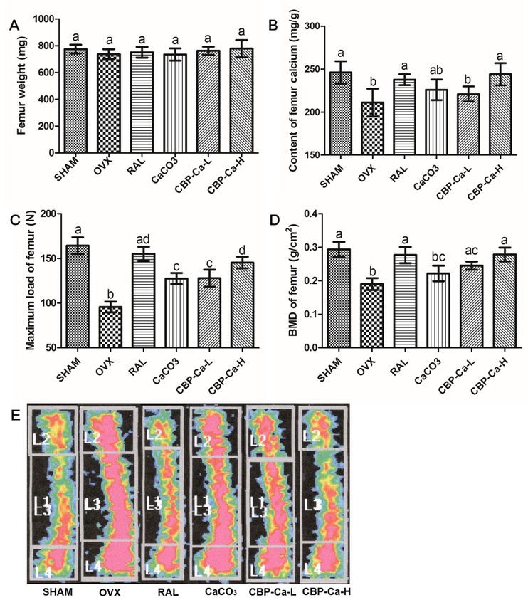
Nutrients 2018, 10, 1325                                                                                2 of 16

attention and many food proteins have been explored to prepare and isolate calcium binding peptides,
including tilapia protein [10], Antarctic krill protein [11], Pacific cod skin protein [12], whey protein [13],
duck egg white [14], etc. Furthermore, previous studies reported that fishbone derived peptides also
possess high calcium binding activity and can improve calcium bioavailability [15,16]. Thus, this
provides a possibility for their anti-osteoporosis activities [17].
      Fishbone has often been discarded directly as the major by-product during aquatic processing [18].
However, it contains a high protein content (about 30 g protein per 100 g dry fishbone) [19]. This makes
fishbone an excellent resource of proteins and peptides [16,18]. Therefore, many researches have been
devoted to the utilization of fishbone derived bioactive peptides [18], especially the calcium binding
peptides and peptide-calcium complex [8,15]. However, studies on the anti-osteoporosis activity
of fish bone peptide-calcium complex have been rare, as most of them have only paid attention to
calcium bioavailability.
      The main purpose herein was to investigate the calcium bioavailability enhancing activity and
the anti-osteoporosis effects of Pacific cod bone derived calcium binding peptides. In the present
study, calcium binding peptides (CBP) were prepared from Pacific cod bone by trypsin and neutral
protease. Then the peptide-calcium complex (CBP-Ca) was prepared and characterized. The intestinal
calcium absorption of CBP-Ca was investigated by using the single-pass intestinal perfusion (SPIP)
experiment. Subsequently, the calcium bioavailability and anti-osteoporosis effects of CBP-Ca were
studied utilizing the ovariectomized rat model. Parameters including calcium apparent absorption
rate, calcium retention rate, bone mass, bone mineral density, bone biomechanical properties, bone
histomorphometry, and bone turnover markers were investigated.

2. Materials and Methods

2.1. Materials
     Pacific cod (Gadus macrocephalus) bone gelatin was prepared according to our previous report [16].
Neutral protease and trypsin were obtained from Pangbo Biotech Co., Ltd. (Nanning, China).
The 4-(2-hydroxyethyl)-1-piperazine-ethanesulphonic acid (HEPES), deuteroxide (D2 O), and raloxifene
were purchased from Sigma-Aldrich (St. Louis, MO, USA). Commercial kits for serum calcium (Ca),
phosphorus (P), osteocalcin (OCN), bone alkaline phosphatase (BALP), and tartrate-resistant acid
phosphatase isoform 5b (TRAP5b) were provided by Nanjing Jiancheng Bioengineering Institute
(Nanjing, China). ELISA assay kit of C-telopeptide of type I collagen (CTX-1) was obtained from
Shanghai Enzyme-linked Biotechnology Co., Ltd. (Shanghai, China). All other chemicals and reagents
were of analytical purity.

2.2. Preparation of CBP
     The peptides with the highest binding affinity for calcium ions were optimized by calcium binding
assay. Finally, the cod bone gelatin solution (2.5% w/w) was hydrolyzed by 0.5% trypsin and neutral
protease (pH 7.2, 52 ◦ C, 90 min) for preparing the calcium binding peptides. The CBP was lyophilized
and stored at −20 ◦ C until use. The calcium binding assay was performed according to the method
described previously [20].

2.3. Amino Acid Profile
     The amino acid composition of CBP was determined according to the method described by Liu
and co-workers [13]. Samples were hydrolyzed in 6 M HCl (130 ◦ C, 4 h), then an amino acid analyzer
(Hitachi, Tokyo, Japan) was adopted to analyze the hydrolysates [13].

2.4. Preparation of CBP-Calcium Complex (CBP-Ca)
     CaCl2 (11.0 g) was mixed with 500 mL 6% (w/v) CBP solution (pH 7.0), and incubated at 50 ◦ C
for 1 h. The resulted solution was subsequently mixed with 8 times the volume of absolute ethanol,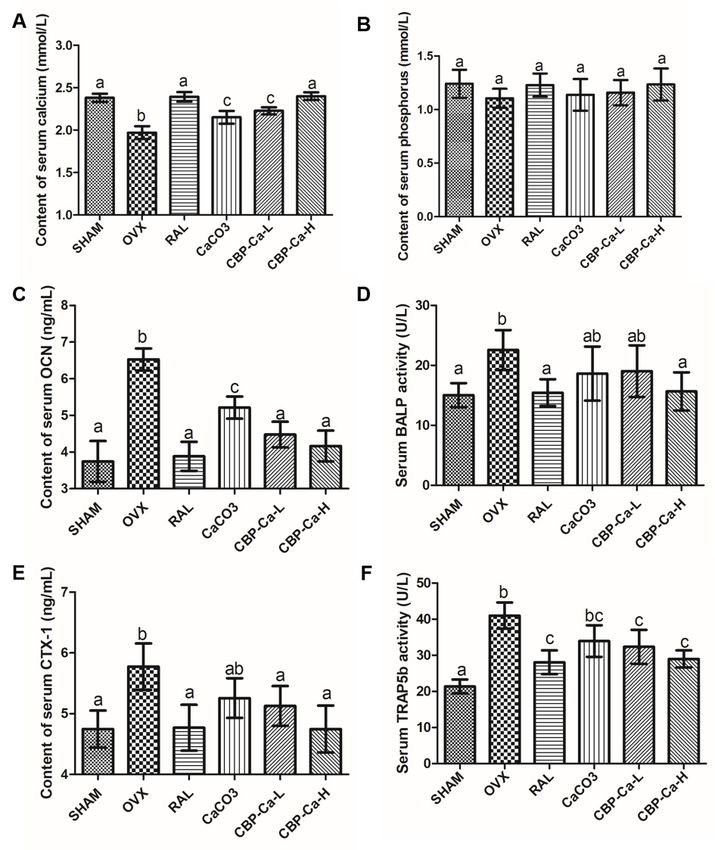
Nutrients 2018, 10, 1325                                                                         3 of 16

precipitated for 30 min, and then centrifuged at 7000× g for 15 min to remove the free Ca2+ . Finally,
the CBP-Ca sedimentation was collected, lyophilized, and stored at −20 ◦ C until use.

2.5. Structural Characterization of CBP and CBP-Ca in Solution

2.5.1. UV Absorption
     The UV absorption spectrum was recorded by a UV-spectrophotometer (Shimadzu UV-2550,
Kyoto, Japan). The sample solution (0.2 mg/mL) was placed into a quartz cell with a path length of
1 cm. The UV spectrum was measured by scanning the wavelength from 190 to 400 nm at a scan speed
of 2 nm/s with an interval of 1 nm.

2.5.2. Circular Dichroism Analysis
     The circular dichroism (CD) spectra of CBP and CBP-Ca were recorded at 25 ◦ C using a Jasco
J-815 spectrophotometer (Japan Spectroscopic Company, Tokyo, Japan). The CBP (1.0 mg/mL) solution
was scanned in the presence of 20 mM HEPES buffer (pH 7.0). The CBP-Ca solution was prepared by
adding 3 µM CaCl2 into 1.0 mg/mL CBP solution (pH 7.0). The mixed solution was incubated at 50 ◦ C
for 1 h before CD analysis. Each scan of the sample was executed after subtracting the spectrum of
20 mM HEPES buffer.

2.5.3. Fourier Transform Infrared (FTIR) Spectroscopy Measurement
     FTIR measurement was implemented using a Nicolet 200SXV infrared spectrophotometer
(Thermo-Nicolet Co., Madison, WI, USA) at 25 ◦ C, according to the method reported by Nara et al. [21].
To obtain reliable infrared spectra in solution, the CBP and CBP-Ca were completely deuterated by
dissolving in deuteroxide at 65 ◦ C for 2 h, respectively. The deuterated samples were then lyophilized
and collected for FTIR analysis. The peptide concentration of each sample for FTIR measurement
was 20 mg/mL. Each FTIR spectrum was scanned at a data acquisition rate of 2 cm−1 per point and
normalized by the Omnic 6.0 software (Thermo-Nicolet Co., Madison, WI, USA).

2.6. In Situ Single-Pass Intestinal Perfusion (SPIP) Study

2.6.1. Animals
     All animal procedures were approved by the ethical committee of animal research in the Ocean
University of China, and complied with the requirements of the National Act on the use of experimental
animals (China). Twelve male Wistar rats (210 ± 10 g; Licensed ID: SCXK 2014-0007) arrived at the
animal chamber at least 7 days before the perfusion study, with free access to distilled water and
pelleted AIN-93 diet. They were randomly assigned into two groups (n = 6), namely group A (CaCl2 )
and group B (CBP-Ca). Prior to the SPIP experiment, all rats were kept with overnight fasting but free
access to water.

2.6.2. Single-Pass Intestinal Perfusion (SPIP) Experiment
     The SPIP experiment was conducted according to previous reports [22,23]. Rats were anesthetized
with sodium pentobarbital (25 mg/kg) and fixed upon a heating pad (37 ◦ C) to keep the body
temperature. The abdominal incision was opened along the belly line (4–5 cm). Four intestine segments
(duodenum, jejunum, ileum, and colon) were identified and carefully exposed for about 10 cm,
followed the method reported previously [23]. Each segment was flushed gently with physiological
saline to remove the residues, and cannulated with a polypropylene tube (diameter: 4 mm) on the
proximal and distal end, respectively. Each inlet tube was then fitted together with a peristaltic pump,
and the outlet tube was connected to a collecting vial. The surgical area was covered with absorbent
cotton saturated with physiological saline (37 ◦ C) to avoid dehydration (Figure 1).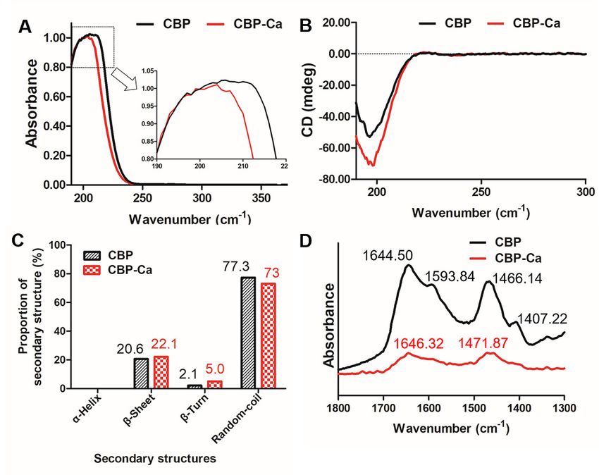
Nutrients 2018, 10, x FOR PEER REVIEW 4 of 16
peristaltic pump, and the outlet tube was connected to a collecting vial. The surgical area was
covered
Nutrients 2018,with absorbent cotton saturated with physiological saline (37 °C) to avoid dehydration
10, 1325 4 of 16
(Figure 1).
Figure
Figure 1. 1.
InInsitu
situsingle-pass
single-passintestinal
intestinal perfusion
perfusion (SPIP)
(SPIP) system.
system.
Prior
Prior to the
to the perfusion
perfusion study,
study, Krebs–Ringer
Krebs–Ringer buffer
buffer (KRB)(KRB)
pH pH 7.4 was
7.4 was perfused
perfused for 30formin
30 min to
to attain
attain system.
a steady a steady After
system. After reaching
reaching the steady-state,
the steady-state, rats of rats of group
group A were A perfused
were perfused
with 10 with
mM 10 CaCl
mM 2
CaCl2 (dissolved
(dissolved in KRB),in KRB),
and the and
ratsthe rats of group
of group B wereBperfused
were perfused
with with CBP-Ca
CBP-Ca solution
solution (containing
(containing 10
10 mM
mM
2+ Ca 2+, dissolved in KRB). In order to simulate real in vivo condition as much as possible, the
Ca , dissolved in KRB). In order to simulate real in vivo condition as much as possible, the CaCl2
andCaCl 2 and CBP-Ca were pre-treated under simulated gastric conditions. The flow rate was 0.2
CBP-Ca were pre-treated under simulated gastric conditions. The flow rate was 0.2 mL/min,
mL/min, and allused
and all solutions solutions
in theused in studies
SPIP the SPIPwere
studies were pre-warmed
pre-warmed at 37 ◦at
C 37
and °Cheated
and heated
in a in a water
water bath
bath
◦ (37 °C) throughout the experiment. The outflow liquid of each outlet tube was quantitatively
(37 C) throughout the experiment. The outflow liquid of each outlet tube was quantitatively collected
collected at 15 min intervals for 90 min. The calcium content was determined by a flame atomic
at 15 min intervals for 90 min. The calcium content was determined by a flame atomic absorption
absorption spectrometry (FAAS). Finally, the rats were euthanized, the length and radius of each
spectrometry (FAAS). Finally, the rats were euthanized, the length and radius of each perfused
perfused intestinal segment were measured.
intestinal segment were measured.
2.6.3. Data Analysis and Equations
2.6.3. Data Analysis and Equations
The calcium percent absorption, calcium absorption rate constant (Ka), and calcium effective
The calcium percent absorption, calcium absorption rate constant (Ka), and calcium effective
permeability (Peff) were calculated using Equations (1)–(3), respectively.
permeability (Peff ) were calculated using Equations (1)–(3), respectively.
Cout Qout
Calcium percent absorption % 1 Cout × Qout 100% (1)
Calcium percent absorption (%) = 1 − Cin Qin × 100% (1)
Cin × Qin
Cout Qout vin
K a 1 Cout × Qout 2vin (2)
Ka = 1 − Cin Qin ×r l 2 (2)
Cin × Qin πr l
CCout
×QQ
−vvinin ×
ln
ln Cout× Q
outout
Pe f f = Cinin inQin (3) (3)
Peff 2πrl
2 rl
where Cout and Cin are the respective calcium concentration in the outlet and inlet perfusate, Qout and
where Cout and Cin are the respective calcium concentration in the outlet and inlet perfusate, Qout and
Qin Qare the respective volumes of outlet and inlet perfusate, v in is
in are the respective volumes of outlet and inlet perfusate, vin
is the
theinlet
inletflow
flowrate
rateofofperfusate, r and
perfusate,r and
l represent the radius and length of perfused intestinal segment after the SPIP experiment.
l represent the radius and length of perfused intestinal segment after the SPIP experiment.
2.7. Calcium Bioavailability and Anti-Osteoporosis Activity of CBP-Ca
2.7.1. Animals and Treatments
Forty-two female Wistar rats aged 2 months (220 ± 10 g; Licensed ID: SCXK 2014-0007) were
purchased from Pengyue Laboratory Animal Breeding Center (Jinan, China) and acclimated for 7 days
to a controlled environment (23 ± 1 ◦ C, 12–12 h light–dark cycle). All rats were individually housed
in stainless cages and allowed free access to pelleted AIN-93 diet and deionized water. The diet was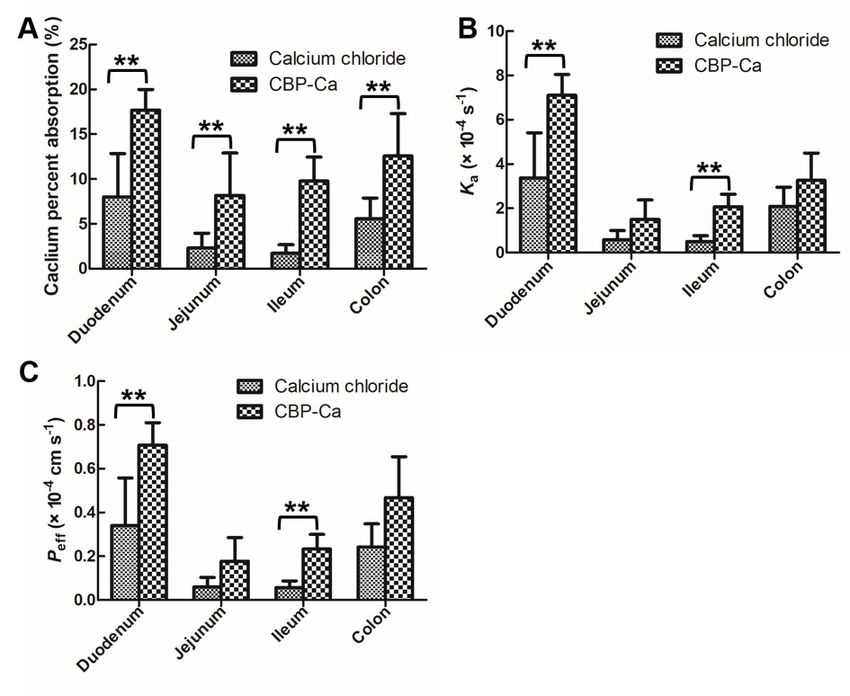
Nutrients 2018, 10, 1325                                                                        5 of 16

adjusted to either normal calcium diet (12.5 g CaCO3 /kg diet) or low calcium diet (2.5 g CaCO3 /kg
diet). All animal experiments were approved by the ethical committee of animal research in the Ocean
University of China.
      Anaesthetized animals were randomly given either bilateral laparotomy (sham ovariectomy
operated) as SHAM group (n = 7), or bilateral ovariectomy (OVX) to establish the osteoporosis
model (n = 35) [24]. The content of serum β-estradiol (E2 ) was determined routinely to confirm the
establishment of the osteoporosis model. Compared with the SHAM group, rats with a significant
decrease in serum E2 content were confirmed as a successful osteoporosis model. Thirty-five
osteoporosis model rats were randomly allocated into five groups (n = 7/group): OVX group (treated
with normal physiological saline as a negative control group, 7 mL/kg/body weight), RAL group
(treated with raloxifene as the positive control model, 3 mg/kg/body weight), CaCO3 treated group
(Ca2+ content: 200 mg/kg/body weight), CBP-Ca-L treated group (Ca2+ content: 100 mg/kg/body
weight) and CBP-Ca-H group (Ca2+ content: 200 mg/kg/body weight). The SHAM group was treated
with 7 mL/kg/body weight normal saline. According to this protocol, each rat was intragastrically
administrated with a volume of 10 mL/kg/body weight once a day for 90 days. To maintain the same
calcium intake, the SHAM, OVX, and RAL groups were fed with a normal calcium diet, but the other
groups were fed with a low calcium diet.

2.7.2. Calcium Bioavailability of CBP-Ca
     To determine the calcium bioavailability, rats were placed in individual metabolic cages for
the last 3 days of the experiment. Parameters including calcium apparent absorption (mg/day),
calcium apparent absorption rate (%), calcium retention (mg/day), and calcium retention rate (%) were
determined according to previous study [16].

2.7.3. Sampling and Analytical Methods
     All rats were fasted for 12 h and anaesthetized, after the 90 days experiment. The blood was
collected from the abdominal aorta and centrifuged to separate the serum. Then the contents of Ca, P,
OCN, and CTX-1, as well as the activities of BALP and the TRAP5b in the serum were determined
using commercial kits. Rats were then sacrificed, and their tibias and femurs were dissected. Right
femurs were weighed, and their calcium contents were determined according to the method described
previously [16]. Left femurs were selected to evaluate the maximum loads using the three-point
bending method [25]. Right tibias were collected for the determinations of bone mineral density
(BMD) and bone mineralization degree [26]. Left tibias were fixed with 10% methanol and decalcified
by EDTA, then the bone histomorphology was analyzed by the hematoxylin–eosin (HE) staining
method [24].

2.8. Statistical Analysis
     Statistical significance between two treatments was assessed by Student’s t-tests using an SPSS
18.0 software (SPSS Inc., Chicago, IL, USA). All data were expressed as mean ± standard deviation
(SD), and differences were considered statistically significant if p < 0.05.

3. Results and Discussion

3.1. Calcium Binding Activity and Amino Acid Composition of CBP
     The calcium binding activity of CBP was determined as 0.43 ± 0.09 µg/mg. It is similar to that of
the Alaska pollack skin derived calcium binding peptide (0.55 µg/mg) [20]. Amino acid analysis of
CBP (shown in Table 1) suggested that it was rich in Gly, Pro, Hyp, Ala, Glu, Ser, Asp, Arg, and His.
The glycine and imino acids (Pro and Hyp) contents of CBP were 322 and 178 residues/1000 residues,
respectively. Similar results were found in enzymatic hydrolysis of tilapia scale collagen [25] and sea
bream bones collagen peptides [27]. In addition, previous reports indicated that the carboxyl groups of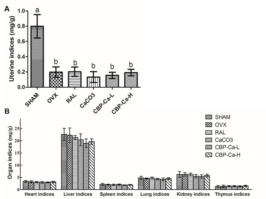
Nutrients 2018, 10, 1325                                                                                     6 of 16

Asp and Glu and the amino groups in Arg and Lys were responsible for the calcium binding activity of
the peptides [8,10]. The Glu, Asp, Arg and Lys contents in CBP were 97, 84, 46, and 14 residues/1000
residues, respectively. Liu, et al. reported that wheat germ protein derived calcium binding peptides
are rich in Glu, Asp, and Arg [13]. Hou et al., suggested that the acidic amino acids, as well as basic
amino acids, contribute to the calcium binding activity of peptides [11]. These are consistent with our
study. Results suggested that the specific amino acid residues might provide suitable calcium binding
sites for CBP, therefore, contribute to its calcium binding activity.

      Table 1. Amino acid composition of calcium binding peptides (CBP) (residues/1000 total amino
      acid residues).

                           Amino Acid           CBP              Amino Acid                CBP
                    Aspartic acid (Asp)          84            Leucine (Leu)                11
                     Threonine (Thr)             22            Tyrosine (Tyr)                3
                        Serine (Ser)             59         Phenylalanine (Phe)             10
                    Glutamic acid (Glu)          97             Lysine (Lys)                14
                       Glycine (Gly)            322           Histamine (His)               29
                       Alanine (Ala)             86            Arginine (Arg)               46
                      Cysteine (Cys)             15             Proline (Pro)              106
                        Valine (Val)            10         Hydroxyproline (Hyp)             72
                     Methionine (Met)             2        Imino acid (Pro + Hyp)          178
                      Isoleucine (Ile)           12                 Total                  1000

3.2. Structural Differences between CBP and CBP-Ca in Solution

3.2.1. UV Absorption Spectrum of CBP and CBP-Ca
     Figure 2A shows that the UV spectra of CBP changed before and after the presence of calcium.
After cooperating with calcium, the absorption peak of CBP shifted from 206 nm to 204 nm, and a
small peak arose in the UV absorption curve of CBP-Ca. A similar phenomenon was also observed
byNutrients
   Zhao, 2018,
            et al.10,inx the Gly-Tyr and Gly-Tyr-Ca [28]. This indicated that CBP could bind with Ca 2+ and
                         FOR PEER REVIEW                                                           7 of 16
form CBP-Ca.

      Figure 2. Structural differences of calcium binding peptide (CBP) and CBP-Ca in solution. (A) UV
        Figure 2. Structural differences of calcium binding peptide (CBP) and CBP-Ca in solution. (A) UV
      absorption
        absorptionspectra; (B)(B)
                     spectra;  circular dichroism
                                   circular       spectroscopic
                                            dichroism            data; data;
                                                        spectroscopic  (C) proportions of secondary
                                                                             (C) proportions        structures;
                                                                                              of secondary
      (D) Fourier  transform  infrared  spectroscopy analysis.
        structures; (D) Fourier transform infrared spectroscopy analysis.

  3.2.2. Secondary Structure of CBP and CBP-Ca in Solution
      CD analysis was implemented to investigate the secondary structure differences between CBP
  and CBP-Ca [29]. Figure 2B showed the full CD spectra (190 to 300 nm) of CBP and CBP-Ca. The CD
Nutrients 2018, 10, 1325                                                                           7 of 16

3.2.2. Secondary Structure of CBP and CBP-Ca in Solution
     CD analysis was implemented to investigate the secondary structure differences between CBP
and CBP-Ca [29]. Figure 2B showed the full CD spectra (190 to 300 nm) of CBP and CBP-Ca. The CD
spectrum of CBP changed significantly after calcium was presented. The random coil was the
predominant structure, followed by β-sheets and β-turns, while the α-helix structure was absent in
both CBP and CBP-Ca (shown in Figure 2C). Compared with CBP, the secondary structure composition
of CBP-Ca changed dramatically: the β-turn content doubled (from 2.1% to 5.0%), the proportion of
β-sheet increased from 20.6% to 22.1%, while the percentage of random-coil decreased from 77.3%
to 73%. These results revealed that a compact structure was formed after Ca2+ interacted with CBP.
A similar result was reported previously [16].

3.2.3. Fourier Transform Infrared (FTIR) Spectra of CBP and CBP-Ca
     As a powerful instrument for investigating the peptide structures [30], the FTIR technique was
employed for the further study of the mechanisms underlying peptide-calcium reactions. The FTIR
spectra of CBP and CBP-Ca in D2 O solution were normalized against the peak intensity, and the major
vibration bands (1800 to 1300 cm−1 ) were identified and associated with the main peptide groups
(Figure 2D).
     The amide-I, amide-II, and –COOH stretching vibration bands were the most valuable and
informative infrared bands in the investigations of peptide secondary structures in solution [30,31].
The infrared absorbance band at 1644.50 and 1466.14 cm−1 of CBP could be attributed to the amide-I
band (C=O stretching vibration) and amide-II band (mainly caused by N–H bending vibration),
respectively. In the infrared spectrum of CBP-Ca, these bands shifted to 1646.32 and 1471.87 cm−1 ,
respectively. Meanwhile, their intensities decreased significantly. These results indicated that the
carboxylate groups and amino groups in CBP contributed to the calcium binding reaction [16,28].
Previous researches suggested that the infrared absorption of amino acid side chains provide a wealth
of information, which could be used to research the peptide reactions mechanism [31]. In the CBP
spectrum, the infrared peak at 1593.84 and 1407.22 cm−1 representing the antisymmetric stretching
vibration and the symmetric stretching vibration of side chains –COOH in CBP, respectively [30,31].
These –COOH variation bands disappeared in the presence of Ca2+ , indicating that –COOH interacted
with calcium ions, therefore, transformed into –COO–Ca [13,16].
     Results demonstrated that the Ca2+ binds to CBP via interactions with amino N atoms and the
carboxyl O atoms in CBP. Chen et al. [25] reported that calcium ions could bind to the carbonyl group
and the amino group in tilapia derived calcium binding peptides. Similarly, Zhao et al. suggested that
–NH3 and –COOH contribute to the calcium binding activity of calcium binding peptides [28,32].

3.3. CBP-Ca Improved the Rat Intestinal Calcium Absorption
      The in situ single-pass intestinal perfusion (SPIP) experiment system (see Figure 1), which
provides the intact intestinal barrier, functional blood circulation, as well as normal secretion of
enzymes and transporters [33], could be used with precision in the forecasting of intestinal calcium
absorption in the human body [22,33]. Calcium absorption percent, calcium absorption rate constant
(Ka), and calcium effective permeability (Peff ) were determined in four intestine segments utilizing the
SPIP study (see Figure 3A–C).
transported rapidly into enterocytes [33,36]. Thus, the calcium in the peptide-bound state is superior
to the free calcium ions in term of intestinal calcium absorption. These results suggested that the
CBP-Ca could improve the intestinal permeability and absorption of calcium [23]. Previous studies
suggested that the peptide-calcium complex is a superior candidate to those of ionized calcium (such
as calcium chloride, calcium carbonate, calcium lactate, and calcium gluconate) for promoting
Nutrients 2018, 10, 1325 8 of 16
calcium absorption in the intestinal tract [8,9]. This is consistent with our results.
Figure 3. Effects of CBP-Ca on intestinal calcium absorption. The calcium percent absorption (A),
calcium absorption rate constant (Ka) (B), and calcium effective permeability (Peff ) (C) comparisons of
CaCl2 and CBP-Ca in four different intestinal segments. All values are expressed as the mean ± SD.
(each n = 6). ** p < 0.01.
Duodenum and ileum are the two most important intestine segments in transcellular and
paracellular calcium transport [34,35], Figure 3C showed that the calcium Peff × 10−4 values of group
A (CaCl2 treated) were 0.341 ± 0.217 and 0.0561 ± 0.031 cm s−1 in duodenum and ileum, respectively.
In group B (CBP-Ca treated), they were increased to 0.708 ± 0.103 and 0.233 ± 0.067 cm s−1 ,
respectively. A similar trend was seen in the Ka values (see Figure 3B). Meanwhile, a significant
increase in calcium absorption percent between the two groups was also observed in each intestine
segment (Figure 3A).
The high calcium permeability of CBP-Ca indicated that, in combination with CBP, the calcium
transported rapidly into enterocytes [33,36]. Thus, the calcium in the peptide-bound state is superior to
the free calcium ions in term of intestinal calcium absorption. These results suggested that the CBP-Ca
could improve the intestinal permeability and absorption of calcium [23]. Previous studies suggested
that the peptide-calcium complex is a superior candidate to those of ionized calcium (such as calcium
chloride, calcium carbonate, calcium lactate, and calcium gluconate) for promoting calcium absorption
in the intestinal tract [8,9]. This is consistent with our results.
3.4. CBP-Ca Improved the Calcium Bioavailability of OVX Rats
The uterine index of the OVX group was significantly lower than that of the SHAM group,
caused by OVX-induced estrogen deficiency (Figure 4A). This indicated that the OVX model had been
established successfully. In addition, part B of Figure 4 showed that there were no statistical differences
of other organic indices between each group. It suggested that the administration of CBP-Ca does not
affect organ health.The uterine index of the OVX group was significantly lower than that of the SHAM group,
caused by OVX-induced estrogen deficiency (Figure 4A). This indicated that the OVX model had
been established successfully. In addition, part B of Figure 4 showed that there were no statistical
differences of other organic indices between each group. It suggested that the administration of
Nutrients
CBP-Ca 2018,
does10, 1325
not affect organ health. 9 of 16
Figure4.4.Effects
Figure EffectsofofCBP-Ca
CBP-Caononthe theuterine
uterineindices
indices(A)
(A)and
andorgan
organindices
indicesofofdifferent
differentinternal
internalorgans
organs
(B). values are presented as mean ±
(B). All values are presented as mean ± SD (n = 7/group). Different lowercases lettersrepresenting
All SD (n = 7/group). Different lowercases letters representing
significant
significantdifferences
differences(p(p
< 0.05). Peng
Chen et al. reported that calcium absorption was improved by tilapia scale
et al. suggested that fish bone peptides can increase the calcium bioavailability in rats [16]. derived peptides [25].In
These findings
another study,are in accordance
Chen withthat
et al. reported our calcium
results inabsorption
the presentwas
study. According
improved to the scale
by tilapia SPIP results
derived
shown in Figure
peptides 3, thefindings
[25]. These intestinalare
permeability
in accordance andwith
intestinal absorption
our results in theofpresent
calcium was significantly
study. According to
improved
the SPIP by CBP-Ca.
results shown Theincomprehensive analysis of
Figure 3, the intestinal the results of
permeability and Figure 3 (SPIP
intestinal experiments
absorption data)
of calcium
and Table 2 (calcium bioavailability results) suggested that CBP-Ca could
was significantly improved by CBP-Ca. The comprehensive analysis of the results of Figure 3 (SPIP improve the intestinal
permeability and absorption of calcium, thus enhance calcium bioavailability.Nutrients 2018, 10, x FOR PEER REVIEW 10 of 16
experiments data) and Table 2 (calcium bioavailability results) suggested that CBP-Ca could
improve the intestinal permeability and absorption of calcium, thus enhance calcium
Nutrients 2018, 10, 1325 10 of 16
bioavailability.
Table 2. Effect
Table 2. Effect of
of CBP-Ca on calcium
CBP-Ca on calcium bioavailability
bioavailability in
in ovariectomized
ovariectomized (OVX)
(OVX) rats.
rats.
Ca Apparent Ca Apparent
Ca Intake Ca Apparent Ca Apparent
Ca Intake Urinary Ca Ca
Urinary FecalFecal
Ca Ca
Absorption Absorption Rate CaCa Retention Ca Ca
Retention Retention
Retention
(mg/day) Absorption Absorption
(mg/day) (mg/day)
(mg/day) (mg/day)
(mg/day) (mg/day) (%)(%)
(mg/day)
(mg/day) Rate
Rate (%)(%)
(mg/day) Rate
SHAM 57.74 ± 4.34 a 1.48 ± 0.28 ac 19.57 ± 2.90 a 38.17 ± 5.37 ac ac 65.90 ± 5.97 a 36.69 ± 5.37 a 63.33 ± 6.15 a
SHAM 57.74 ± 4.34 a 1.48 ±ac0.28 ac 19.57 ± b2.90 a 38.17 ± 5.37 65.90 ± 5.97 ba 36.69 ± 5.37 a b 63.33 ± 6.15 a b
OVX 58.95 ± 4.15 a a1.43 ± 0.28 ac 34.63 ± 3.07 24.32 ± 4.84 b b 41.02 ± 6.41 b 22.96 ± 4.76 38.72 ± 6.41
OVX 58.95 ± 4.15 1.43 ± 0.28 34.63 ± 3.07 b 24.32 ± 4.84 41.02 ± 6.41 a 22.96 ± 4.76 b a 38.72 ± 6.41 b a
RAL 58.18 ± 3.58 a a1.26 ± 0.21 ac ac 19.43 ± 2.08 a 38.75 ± 2.53 a a 66.64 ± 2.56 37.49 ± 2.58 64.46 ± 2.64
RAL 58.18 ± 3.58 1.26 ± 0.21 19.43 ± 2.08 a 38.75 ± 2.53 66.64 ± 2.56 ba 37.49 ± 2.58 a b 64.46 ± 2.64 a b
CaCO3 57.14 ± 3.80 a 1.53 ± 0.09 a 29.13 ± 3.48 b 28.01 ± 5.16 bc 48.79 ± 7.30 b 26.48 ± 5.13 46.11 ± 7.32
CaCO3 57.14 ± 3.80 a 1.53 ± 0.09 a 29.13 ± 3.48 b 28.01 ± 5.16 bc 48.79 ± 7.30 c 26.48 ± 5.13 b b 46.11 ± 7.32 b c
CBP-Ca-L 29.53 ± 2.11 b b0.62 ±0.62
0.10 b b 6.396.39
± 2.10 c 23.13 ± 2.25 b b 78.44 ± 6.64 22.51 ± 2.26 76.34 ± 6.68
CBP-Ca-L 29.53 ± 2.11 ± 0.10 ± 2.10 c 23.13 ± 2.25 78.44 ± 6.64 c 22.51 ± 2.26 b a 76.34 ± 6.68 c
CBP-Ca-H
CBP-Ca-H 61.70
61.70 ± 3.99 a1.10 ±1.10
± 3.99 a 0.16±c 0.16 c 18.1618.16
± 3.34 a
± 3.34 a 43.54 ± 5.60 a
43.54 ± 5.60 a 70.41 ±
70.41 ± 5.65 c
5.65 c 42.44
42.44 ± 5.65
± 5.65 a 68.62
68.62 ± 5.77
± 5.77 ac ac
All valuesare
All values arepresented
presented as as mean
mean ± SD
± SD (n7/group).
(n = = 7/group). Different
Different lowercases
lowercases letters
letters in thein the column
same same column
denote
denote significant
significant differences
differences (p < 0.05). (p < 0.05).
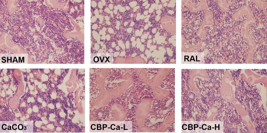
3.5. CBP-Ca Has Protective Effects in Bone Microarchitecture of OVX Rats
The histomorphometry
histomorphometry changes
changesof oftibias
tibiaswere
wereinvestigated
investigatedby byHE HEstaining.
staining.As Asshown
shownininFigure
Figure5,
thethe
5, SHAM
SHAM group
grouppresented
presented with well-formed
with well-formed and competent
and competent trabecular
trabecular bone, which
bone, whichwas normal
was normalin
compactness
in compactness and
anddensity.
density. However,
However, thethetrabecular
trabecularbone
boneininthe
thetibia
tibiaof
ofOVX
OVX group
group rats
rats was poorly
In addition,
observed. In addition, these
these trabecular
trabecular structures
structures were
were significantly
significantly thinning
thinning andand widely
widely separated,
separated,
revealing a severe degree of osteoporosis in the OVX rats. Similar phenomena were also observed in
previous studies [24,37]. Rats in CaCO33 and and CBP-Ca-L
CBP-Ca-L groups
groups have
have aa mild degree of osteoporosis as
moderate recovery of trabecular bone. Meanwhile, the rats
recovery of trabecular bone. Meanwhile, the rats treated treated by raloxifene and high
by raloxifene dose
and CBP-Ca
high dose
exhibitedexhibited
CBP-Ca significant recoveryrecovery
significant with nearlywithcompletely recovered
nearly completely trabecular
recovered structurestructure
trabecular and density.
and
These reults
density. suggested
These that the OVX
reults suggested that induced
the OVXosteoporosis was slightlywas
induced osteoporosis alleviated
slightlyinalleviated
the CaCOin 3 and
the
CBP-Ca-L
CaCO 3 andgroups, and groups,
CBP-Ca-L significantly recovered by recovered
and significantly raloxifene byandraloxifene
high doseand CBP-Ca. However,
high dose the
CBP-Ca.
daily Ca2+ intake
However, of CBP-Ca-H
the daily Ca2+ intake group was equal to
of CBP-Ca-H that of
group the equal
was CaCO3togroup
that of(200
themg/kg/body
CaCO3 group weight).
(200
This indicated
mg/kg/body that CBP-Ca
weight). is superior
This indicated thatto CBP-Ca
CaCO3 in is bone restoration.
superior to CaCO3 in bone restoration.
Figure 5.5.Effect
Effectof of CBP-Ca
CBP-Ca on bone
on bone (left tibias)
(left tibias) histomorphometry.
histomorphometry. Left
Left tibias tibias
were were eliminated
dissected, dissected,
eliminated
the adheringthe adhering
tissues, tissues,
fixed with fixed decalcified,
methanol, with methanol, decalcified,
stained with stained with (HE)
the hematoxylin–eosin the
hematoxylin–eosin
staining technique, and (HE) staining
observed technique,
under an OlympusandBX41
observed
optical under an Olympus
microscope (Olympus,BX41
Tokyo,optical
Japan,
microscope
magnification ×40). Tokyo, Japan, magnification ×40).
(Olympus,
3.6. CBP-Ca Enhanced Bone Properties in OVX Rats
3.6. CBP-Ca Enhanced Bone Properties in OVX Rats
Bone parameters including weight, calcium content, maximum load, BMD, and bone
Bone parameters including weight, calcium content, maximum load, BMD, and bone
mineralization degree were investigated. As shown in Figure 6A, there was no significant difference
mineralization degree were investigated. As shown in Figure 6A, there was no significant difference
in femur weight between each group. Figure 6B revealed that the femur calcium content of the
OVX group was significantly (p < 0.01) lower than that of the SHAM group. Whereas, the femur
calcium content was partly recovered by CaCO3 (200 mg Ca2+ /kg/body weight) and low dose CBP-Caosteoporosis [38,39]. The tibia BMD of OVX group was dramatically decreased to 64.78% compared
with the normal rats in the SHAM group (p < 0.01) (Figure 6D). However, the BMD of the RAL
group, CBP-Ca-L group, and CBP-Ca-H group significantly recovered to 94.34%, 83.53%, and
94.89%, respectively while there was no significant difference between the OVX and CaCO3 group. A
Nutrients 2018,
similar trend 10, was
1325 observed in the bone mineralization degree results (see Figure 6E). 11 of 16
These
suggested that CBP-Ca is superior to CaCO3 in preventing osteoporosis. The results indicated that
CBP-Ca
(100 mg could improve the
Ca2+ /kg/body bone calcium
weight), andrecovered
and fully BMD, strengthen bone mechanical
by raloxifene properties,
and high dose CBP-Caand
(200thus
mg
ameliorate
2+ the
Ca /kg/body weight).OVX induced osteoporosis.
Figure 6. Effects of CBP-Ca on bone properties (n = 7/group). The rats were treated for 12 weeks,
Figure 6. Effects of CBP-Ca on bone properties (n = 7/group). The rats were treated for 12 weeks, then
then their femurs and tibias were dissected for measurements of the weight of the right femurs (A),
their femurs and tibias were dissected for measurements of the weight of the right femurs (A), the
the calcium content of the right femurs (B), the bone biomechanical properties of the left femurs
(C), the BMD of the right tibias (D), and the bone mineralization degree of the right tibias (E).
Different lowercase letters denote statistically significant differences (p < 0.05).
Ovariectomy strongly induces postmenopausal osteoporosis and bone loss, then leads to poor
bone mechanical properties [38]. Mechanical tests indicated that the femur maximum load of the
OVX group was significantly (p < 0.01) reduced to 58.23% when compared with the SHAM group
(Figure 6C). However, it recovered to 94.48%, 77.59%, 77.87%, and 88.44% in RAL, CaCO3 , CBP-Ca-L,
and CBP-Ca-H groups, respectively. This suggested that CBP-Ca could effectively strengthen the
mechanical properties of OVX rats [39,40].
BMD was widely recognized as the most preferred standard for the judgment of bone loss and
osteoporosis [38,39]. The tibia BMD of OVX group was dramatically decreased to 64.78% compared
with the normal rats in the SHAM group (p < 0.01) (Figure 6D). However, the BMD of the RAL
group, CBP-Ca-L group, and CBP-Ca-H group significantly recovered to 94.34%, 83.53%, and 94.89%,
respectively while there was no significant difference between the OVX and CaCO3 group. A similar
trend was observed in the bone mineralization degree results (see Figure 6E). These suggested that
CBP-Ca is superior to CaCO3 in preventing osteoporosis. The results indicated that CBP-Ca couldNutrients 2018, 10, 1325 12 of 16
improve the bone calcium and BMD, strengthen bone mechanical properties, and thus ameliorate the
OVX induced osteoporosis.
3.7. CBP-Ca Reduces the Bone Turnover Rate of OVX Rats
Bone turnover (including bone formation and resorption phase) were regulated by the bone
remodeling process, and any disturbance in this process will lead to osteopathic disease [24,41].
The postmenopausal osteoporosis was recognized as high bone turnover rate, which was characterized
by the increased bone turnover markers [5,26].
Serum Ca, P, OCN, CTX-1, BALP, and TRAP5b are the most important bone turnover markers
used in the evaluation of bone formation and resorption [3]. Changes in these serum biochemical
markers are shown in Figure 7A–F. Results recorded an insignificant difference in the serum P content
between each group (Figure 7B). Whereas, there was a significant decrease in the content of serum
Ca, and significant increases in serum OCN, BALP, CTX-1, and TRAP5b of the OVX group when
comparing with the SHAM group. These changes were ameliorated by the treatment of RAL, CBP-Ca
or CaCO3 , and the most amelioration was obtained in the RAL and CBP-Ca-H group.
The serum calcium content of the OVX group decreased 17.28%, compared with the SHAM
group (Figure 7A). However, the administration of CBP-Ca (200 mg Ca2+ /kg/body weight) markedly
increased the serum Ca to 100.80%. This might result from the outstanding calcium bioavailability of
CBP-Ca, according to the calcium absorption results given in Figure 3 and Table 2. A similar result
was reported by Alkhamees and co-workers [37].
Serum OCN and BALP, specifically secreted by osteoblasts, are widely considered as important
bone formation markers [5]. As seen in Figure 7C,D, compared with the SHAM group, serum OCN
and BALP were significantly increased to 168.38% and 150.19% in the OVX group, respectively.
The increased levels of OCN and BALP were also found in men with insufficient calcium intake [42] as
well as the patients with calcium malabsorption [43]. However, the administration of low dose CBP-Ca
significantly (p < 0.01) reduced the levels of OCN (31.60% reduction) and BALP (15.62% reduction), in
comparison with the OVX group. Treatments with raloxifene, CaCO3 , and high dose CBP-Ca show
similar trends. Wang et al., reported that the Antarctic krill derived peptides could decrease the levels
of bone formation markers in OVX rats [26]. Also, Sato et al. suggested that the milk intake could
reduce bone formation markers [42]. They are in accordance with the results in the present study.
The serum CTX-1 content and TRAP5b activity were investigated to provide the information of
bone resorption degree (see Figure 7E,F). TRAP5b is the enzyme produced by osteoclasts during bone
resorption phase [3,24]. As seen in Figure 7F, the serum TRAP5b activity was significantly increased
(p < 0.01) to 191.93% in the OVX group compared with the normal SHAM group, indicating an
OVX-induced increase of osteoclasts activity and excessive bone resorption [24,42]. The serum TRAP5b
activity was markedly reduced (p < 0.01) by the administration of CBP-Ca, while the CaCO3 treated
group showed no significant change (p > 0.05) compared to the OVX group. A similar phenomenon
was observed in CTX-1, the breakdown product of bone type I collagen during bone resorption, which
is well recognized as the biochemical marker of osteoclast activity. Similar results were reported by
previous studies [24,42]. Results indicated that the bone turnover rate of OVX rats was remarkably
(p < 0.01) reduced by the treatment of CBP-Ca.Nutrients 2018, 10, 1325 13 of 16
Nutrients 2018, 10, x FOR PEER REVIEW 13 of 16
Figure 7. Effects of CBP-Ca on bone turnover markers of OVX rats (n = 7/group). All rats were treated
Figure 7. Effects of CBP-Ca on bone turnover markers of OVX rats (n = 7/group). All rats were treated
for 12 weeks, and the blood was collected. Then the serum was separated for measurements of the
for 12 weeks, and the blood was collected. Then the serum was separated for measurements of the
content of serum calcium (A) and phosphorus (B), osteocalcin (OCN) (C), the serum bone alkaline
content of serum calcium (A) and phosphorus (B), osteocalcin (OCN) (C), the serum bone alkaline
phosphatase (BALP) activity (D), the content of C-telopeptide of type I collagen (CTX-1) (E), and the
phosphatase (BALP) activity (D), the content of C-telopeptide of type I collagen (CTX-1) (E), and the
tartrate-resistant acid phosphatase isoform 5b (TRAP5b) activity (F). Different lowercase letters denote
tartrate-resistant acid phosphatase isoform 5b (TRAP5b) activity (F). Different lowercase letters
statistically significant differences (p < 0.05).
denote statistically significant differences (p < 0.05).
Previous study reported that, in order to maintain the calcium homeostasis, the bone turnover
Previous study reported that, in order to maintain the calcium homeostasis, the bone turnover
process could be accelerated by a decrease of serum calcium content [35,41] and also sufficient calcium
process could be accelerated by a decrease of serum calcium content [35,41] and also sufficient
intestinal absorption is critical for reduction of bone turnover rate and alleviation of osteoporosis [2].
calcium intestinal absorption is critical for reduction of bone turnover rate and alleviation of
Our results indicated that CBP-Ca could significantly increase the intestinal absorption, bioavailability
osteoporosis [2]. Our results indicated that CBP-Ca could significantly increase the intestinal
of calcium, and serum calcium, then decrease bone turnover rate, and thus ameliorate osteoporosis.
absorption, bioavailability of calcium, and serum calcium, then decrease bone turnover rate, and
thus ameliorate osteoporosis.
4. Conclusions
In this study, calcium binding peptides (CBP) were prepared from Pacific cod bone.
4. Conclusions
Structural analyses suggested that the –COOH and –NH3 groups in CBP could bind with calcium
ions In this
and study,
form calcium
CBP-Ca. The binding
in situpeptides (CBP) were
SPIP experiment prepared
indicated from
that the Pacific
CBP-Cacod bone.
could Structural
significantly
analyses
enhance thesuggested that
intestinal the –COOH
absorption and and –NH3 groups
permeability in CBP
of calcium. could bind in
Furthermore, with
vivocalcium
tests inions and
the OVX
form CBP-Ca. The in situ SPIP experiment indicated that the CBP-Ca could significantly
rat model revealed that CBP-Ca could efficiently improve calcium bioavailability, bone mass, bone enhance the
intestinal absorption and permeability of calcium. Furthermore, in vivo tests in
mineral density, bone strength, and bone mineralization degree. The mechanism underlying the the OVX rat model
revealed that CBP-Ca
anti-osteoporosis could
activity efficiently
of CBP-Ca improve
might relate tocalcium bioavailability,
the reduction bone mass,
of bone turnover bone mineral
rate (supported by
density, bone strength, and bone mineralization degree. The mechanism underlying the
anti-osteoporosis activity of CBP-Ca might relate to the reduction of bone turnover rate (supportedNutrients 2018, 10, 1325 14 of 16
the decrease of bone turnover markers), resulting from the increase of calcium intestinal absorption
and bioavailability. Results indicated that the calcium binding peptides from Pacific cod bone could be
employed as functional supplementation in preventing OVX-induced osteoporosis.
Author Contributions: K.Z., B.L. and H.H. conceived and designed the experiments; K.Z., B.L., Q.C., Z.Z., X.Z.
and H.H. performed the experiments; K.Z. and Q.C. analyzed the data; H.H. contributed analysis tools; K.Z. wrote
the paper.
Funding: This work was supported by National Natural Science Foundation of China (Nos. 31772046 and
31471606), Key Research & Development Plan of Shandong Province (Nos. 2017YYSP015, 2016YYSP005 and
2016YYSP017), and National Key R&D Program of China (2018YFC0311200).
Conflicts of Interest: The authors declare no conflict of interest.
References
1. Hwang, Y.-H.; Ha, H.; Kim, R.; Cho, C.-W.; Song, Y.-R.; Hong, H.-D.; Kim, T. Anti-osteoporotic effects of
polysaccharides isolated from persimmon leaves via osteoclastogenesis inhibition. Nutrients 2018, 10, 901.
[CrossRef] [PubMed]
2. Khosla, S.; Hofbauer, L.C. Osteoporosis treatment: Recent developments and ongoing challenges.
Lancet Diabetes Endocrinol. 2017, 5, 898–907. [CrossRef]
3. Eastell, R.; Szulc, P. Use of bone turnover markers in postmenopausal osteoporosis. Lancet Diabetes Endocrinol.
2017, 5, 908–923. [CrossRef]
4. Areco, V.; Rivoira, M.A.; Rodriguez, V.; Marchionatti, A.M.; Carpentieri, A.; Tolosa de Talamoni, N.
Dietary and pharmacological compounds altering intestinal calcium absorption in humans and animals.
Nutr. Res. Rev. 2015, 28, 83–99. [CrossRef] [PubMed]
5. Naylor, K.; Eastell, R. Bone turnover markers: Use in osteoporosis. Nat. Rev. Rheumatol. 2012, 8, 379–389.
[CrossRef] [PubMed]
6. Binkley, N.; Blank, R.D.; Leslie, W.D.; Lewiecki, E.M.; Eisman, J.A.; Bilezikian, J.P. Osteoporosis in crisis: It’s
time to focus on fracture. J. Bone Mine. Res. 2017, 32, 1391–1394. [CrossRef] [PubMed]
7. Bone, H.G.; Wagman, R.B.; Brandi, M.L.; Brown, J.P.; Chapurlat, R.; Cummings, S.R.; Czerwiński, E.;
Fahrleitner-Pammer, A.; Kendler, D.L.; Lippuner, K.; et al. 10 years of denosumab treatment in
postmenopausal women with osteoporosis: Results from the phase 3 randomised FREEDOM trial and
open-label extension. Lancet Diabetes Endocrinol. 2017, 5, 513–523. [CrossRef]
8. Guo, L.; Harnedy, P.A.; Li, B.; Hou, H.; Zhang, Z.; Zhao, X.; FitzGerald, R.J. Food protein-derived chelating
peptides: Biofunctional ingredients for dietary mineral bioavailability enhancement. Trends Food Sci. Technol.
2014, 37, 92–105. [CrossRef]
9. Sun, N.; Wu, H.; Du, M.; Tang, Y.; Liu, H.; Fu, Y.; Zhu, B. Food protein-derived calcium chelating peptides: A
review. Trends Food Sci. Technol. 2016, 58, 140–148. [CrossRef]
10. Charoenphun, N.; Cheirsilp, B.; Sirinupong, N.; Youravong, W. Calcium-binding peptides derived from
tilapia (Oreochromis niloticus) protein hydrolysate. Eur. Food Res. Technol. 2013, 236, 57–63. [CrossRef]
11. Hou, H.; Wang, S.; Zhu, X.; Li, Q.; Fan, Y.; Cheng, D.; Li, B. A novel calcium-binding peptide from Antarctic
krill protein hydrolysates and identification of binding sites of calcium-peptide complex. Food Chem. 2018,
243, 389–395. [CrossRef] [PubMed]
12. Wu, W.; Li, B.; Hou, H.; Zhang, H.; Zhao, X. Isolation and identification of calcium-chelating peptides from
Pacific cod skin gelatin and their binding properties with calcium. Food Funct. 2017, 8, 4441–4448. [CrossRef]
[PubMed]
13. Liu, F.-R.; Wang, L.; Wang, R.; Chen, Z.-X. Calcium-Binding Capacity of Wheat Germ Protein Hydrolysate
and Characterization of Peptide–Calcium Complex. J. Agric. Food Chem. 2013, 61, 7537–7544. [CrossRef]
[PubMed]
14. Hou, T.; Liu, Y.; Kolba, N.; Guo, D.; He, H. Desalted Duck Egg White Peptides Promote Calcium Uptake and
Modulate Bone Formation in the Retinoic Acid-Induced Bone Loss Rat and Caco-2 Cell Model. Nutrients 2017,
9, 490. [CrossRef] [PubMed]
15. Jung, W.-K.; Kim, S.-K. Calcium-binding peptide derived from pepsinolytic hydrolysates of hoki
(Johnius belengerii) frame. Eur. Food Res. Technol. 2007, 224, 763–767. [CrossRef]Nutrients 2018, 10, 1325 15 of 16
16. Peng, Z.; Hou, H.; Zhang, K.; Li, B. Effect of calcium-binding peptide from Pacific cod (Gadus macrocephalus)
bone on calcium bioavailability in rats. Food Chem. 2017, 221, 373–378. [CrossRef] [PubMed]
17. Heo, S.-Y.; Ko, S.-C.; Nam, S.Y.; Oh, J.; Kim, Y.-M.; Kim, J.-I.; Kim, N.; Yi, M.; Jung, W.-K. Fish bone peptide
promotes osteogenic differentiation of MC3T3-E1 pre-osteoblasts through upregulation of MAPKs and Smad
pathways activated BMP-2 receptor. Cell Biochem. Funct. 2018, 36, 137–146. [CrossRef] [PubMed]
18. Harnedy, P.A.; FitzGerald, R.J. Bioactive peptides from marine processing waste and shellfish: A review.
J. Funct. Foods 2012, 4, 6–24. [CrossRef]
19. Toppe, J.; Albrektsen, S.; Hope, B.; Aksnes, A. Chemical composition, mineral content and amino acid
and lipid profiles in bones from various fish species. Comp. Biochem. Physiol. B Biochem. Mol. Biol. 2007,
146, 395–401. [CrossRef] [PubMed]
20. Guo, L.; Harnedy, P.A.; O’Keeffe, M.B.; Zhang, L.; Li, B.; Hou, H.; FitzGerald, R.J. Fractionation and
identification of Alaska pollock skin collagen-derived mineral chelating peptides. Food Chem. 2015,
173, 536–542. [CrossRef] [PubMed]
21. Nara, M.; Tanokura, M. Infrared spectroscopic study of the metal-coordination structures of calcium-binding
proteins. Biochem. Biophys. Res. Commun. 2008, 369, 225–239. [CrossRef] [PubMed]
22. Sun, L.; Liu, Q.; Fan, J.; Li, X.; Zhuang, Y. Purification and characterization of peptides inhibiting MMP-1
activity with C terminate of Gly-Leu from simulated gastrointestinal digestion hydrolysates of tilapia
(Oreochromis niloticus) skin gelatin. J. Agric. Food Chem. 2018, 66, 593–601. [CrossRef]
23. Ma, H.; Chen, H.; Sun, L.; Tong, L.; Zhang, T. Improving permeability and oral absorption of mangiferin by
phospholipid complexation. Fitoterapia 2014, 93, 54–61. [CrossRef] [PubMed]
24. Xia, G.; Zhao, Y.; Yu, Z.; Tian, Y.; Wang, Y.; Wang, S.; Wang, J.; Xue, C. Phosphorylated peptides from antarctic
krill (Euphausia superba) prevent estrogen deficiency induced osteoporosis by inhibiting bone resorption in
ovariectomized rats. J. Agric. Food Chem. 2015, 63, 9550–9557. [CrossRef] [PubMed]
25. Chen, D.; Mu, X.; Huang, H.; Nie, R.; Liu, Z.; Zeng, M. Isolation of a calcium-binding peptide from tilapia
scale protein hydrolysate and its calcium bioavailability in rats. J. Funct. Foods 2014, 6, 575–584. [CrossRef]
26. Wang, Y.; Wang, S.; Wang, J.; Xue, C.; Chang, Y.; Xue, Y. Preparation and anti-osteoporotic activities in vivo
of phosphorylated peptides from Antarctic krill (Euphausia superba). Peptides 2015, 68, 239–245. [CrossRef]
[PubMed]
27. Akagündüz, Y.; Mosquera, M.; Giménez, B.; Alemán, A.; Montero, P.; Gómez-Guillén, M.C. Sea bream bones
and scales as a source of gelatin and ACE inhibitory peptides. LWT Food Sci. Technol. 2014, 55, 579–585.
[CrossRef]
28. Zhao, L.; Huang, Q.; Huang, S.; Lin, J.; Wang, S.; Huang, Y.; Hong, J.; Rao, P. Novel Peptide with a Specific
Calcium-Binding Capacity from Whey Protein Hydrolysate and the Possible Chelating Mode. J. Agric.
Food Chem. 2014, 62, 10274–10282. [CrossRef] [PubMed]
29. Greenfield, N.J. Using circular dichroism spectra to estimate protein secondary structure. Nat. Protoc. 2007,
1, 2876–2890. [CrossRef] [PubMed]
30. Nara, M.; Morii, H.; Tanokura, M. Coordination to divalent cations by calcium-binding proteins studied by
FTIR spectroscopy. Biochim. Biophys. Acta 2013, 1828, 2319–2327. [CrossRef] [PubMed]
31. Barth, A. The infrared absorption of amino acid side chains. Prog. Biophys. Mol. Biol. 2000, 74, 141–173.
[CrossRef]
32. Zhao, L.; Huang, S.; Cai, X.; Hong, J.; Wang, S. A specific peptide with calcium chelating capacity isolated
from whey protein hydrolysate. J. Funct. Foods 2014, 10, 46–53. [CrossRef]
33. Dezani, T.M.; Dezani, A.B.; da Silva Junior, J.B.; dos Reis Serra, C.H. Single-Pass Intestinal Perfusion (SPIP)
and prediction of fraction absorbed and permeability in humans: A study with antiretroviral drugs. Eur. J.
Pharm. Biopharm. 2016, 104, 131–139. [CrossRef] [PubMed]
34. Khanal, R.C.; Nemere, I. Regulation of intestinal calcium transport. Annu. Rev. Nutr. 2008, 28, 179–196.
[CrossRef] [PubMed]
35. Pu, F.; Chen, N.; Xue, S. Calcium intake, calcium homeostasis and health. Food Sci. Hum. Wellness 2016,
5, 8–16. [CrossRef]
36. Chen, Y.; Li, X.; Wei, X.; Gou, J.; Tang, X.; He, H.; Xu, H. Influence of lipid composition on the oral
bioavailability of cinnarizine sub-microemulsions: Oral bioavailability of cinnarizine sub-microemulsions.
Eur. J. Lipid Sci. Technol. 2017, 119, 1600184. [CrossRef]Nutrients 2018, 10, 1325 16 of 16
37. Alkhamees, O.A.; Al-Roujayee, A.S.; Abuohashish, H.M.; Ahmed, M.M. Anti-osteoporotic effects of an
antidepressant tianeptine on ovariectomized rats. Biomed. Pharmacother. 2017, 87, 575–582. [CrossRef]
[PubMed]
38. Johnston, B.D.; Ward, W.E. The Ovariectomized Rat as a model for studying alveolar bone loss in
postmenopausal women. BioMed Res. Int. 2015, 2015, 1–12. [CrossRef] [PubMed]
39. Tan, H.; Furuta, S.; Nagata, T.; Ohnuki, K.; Akasaka, T.; Shirouchi, B.; Sato, M.; Kondo, R.; Shimizu, K.
Inhibitory effects of the leaves of loquat (Eriobotrya japonica) on bone mineral density loss in ovariectomized
mice and osteoclast differentiation. J. Agric. Food Chem. 2014, 62, 836–841. [CrossRef] [PubMed]
40. Ma, J.; Granton, P.V.; Holdsworth, D.W.; Turley, E.A. Oral administration of hyaluronan reduces bone
turnover in ovariectomized rats. J. Agric. Food Chem. 2013, 61, 339–345. [CrossRef] [PubMed]
41. Drissi, H.; Sanjay, A. The multifaceted osteoclast; far and beyond bone resorption. J. Cell. Biochem. 2016,
117, 1753–1756. [CrossRef] [PubMed]
42. Sato, Y.; Iki, M.; Fujita, Y.; Tamaki, J.; Kouda, K.; Yura, A.; Moon, J.-S.; Winzenrieth, R.; Iwaki, H.; Ishizuka, R.;
et al. Greater milk intake is associated with lower bone turnover, higher bone density, and higher bone
microarchitecture index in a population of elderly Japanese men with relatively low dietary calcium intake:
Fujiwara-kyo Osteoporosis Risk in Men (FORMEN) Study. Osteoporos. Int. 2015, 26, 1585–1594. [CrossRef]
[PubMed]
43. Duggan, S.N.; Purcell, C.; Kilbane, M.; O’Keane, M.; McKenna, M.; Gaffney, P.; Ridgway, P.F.; Boran, G.;
Conlon, K.C. An association between abnormal bone turnover, systemic inflammation, and osteoporosis in
patients with chronic pancreatitis: A case-matched study. Am. J. Gastroenterol. 2015, 110, 336–345. [CrossRef]
[PubMed]
© 2018 by the authors. Licensee MDPI, Basel, Switzerland. This article is an open access
article distributed under the terms and conditions of the Creative Commons Attribution
(CC BY) license (http://creativecommons.org/licenses/by/4.0/).You can also read