Original Article An interspecies study of lipid profiles and atherosclerosis in familial hypercholesterolemia animal models with low-density ...
←
→
Page content transcription
If your browser does not render page correctly, please read the page content below
Am J Transl Res 2019;11(5):3116-3127
www.ajtr.org /ISSN:1943-8141/AJTR0086659

Original Article
An interspecies study of lipid profiles and
atherosclerosis in familial hypercholesterolemia animal
models with low-density lipoprotein receptor deficiency
Kunxiang He1*, Jinjie Wang1*, Haozhe Shi1, Qiongyang Yu1, Xin Zhang2, Mengmeng Guo1, Huijun Sun3, Xiao
Lin1, Yue Wu4, Luya Wang4, Yuhui Wang1, Xunde Xian5, George Liu1
1
 Institute of Cardiovascular Sciences and Key Laboratory of Molecular Cardiovascular Sciences, Ministry of
Education, Peking University, Beijing 100191, China; 2Hebei Invivo Biotech Co, Shijiazhuang, China; 3College
of Pharmacy, Dalian Medical University, Dalian 116044, China; 4The Key Laboratory of Remodeling-Related
Cardiovascular Diseases, Ministry of Education, Beijing An Zhen Hospital Affiliated to Capital Medical University,
Beijing Institute of Heart, Lung and Blood Vessel Diseases, Beijing 100029, China; 5Department of Molecular
Genetics, UT Southwestern Medical Center, Dallas 75390, Texas, USA. *Equal contributors.
Received October 12, 2018; Accepted April 25, 2019; Epub May 15, 2019; Published May 30, 2019

Abstract: Small rodents, especially mice and rats, have been widely used in atherosclerosis studies even though
humans exhibit completely different lipoprotein metabolism and atherosclerotic characteristics. Until recently, vari-
ous rodent models of human familial hypercholesterolemia (FH) have been created, including mice, rats, and golden
Syrian hamsters. Although hamsters reportedly possess metabolic features similar to humans, there is no system-
atic characterization of the properties of circulating lipids and atherosclerotic lesions in these rodent models. We
used three FH animal species (mice, rats, and hamsters) with low-density lipoprotein receptor (Ldlr) deficiency to
fully assess lipoprotein metabolism and atherosclerotic characteristics. Compared to chow diet-fed mice and rats,
Ldlr knockout (KO) hamsters showed increased cholesterols in LDL fractions similar to human FH patients. Upon
12-week high-cholesterol/high-fat diet feeding, both heterozygous and homozygous Ldlr KO hamsters displayed
hyperlipidemic phenotypes, whereas only homozygous Ldlr KO mice and rats showed only moderate increases in
plasma lipid levels. Moreover, rats were resistant to diet-induced atherosclerosis compared to mice, and hamsters
showed more atherosclerotic lesions in the aortas and coronary arteries. Further morphological study revealed that
only hamsters developed atherosclerosis in the abdominal segments, which is highly similar to FH patients. This
unique animal model will provide insight into the translational study of human atherosclerosis and could be useful
for developing novel treatments for FH patients.

Keywords: Mouse, rat, hamster, low-density lipoprotein receptor, familial hypercholesterolemia, atherosclerosis

Introduction                                                   Although the molecular mechanisms underlying
                                                               FH pathogenesis have been extensively investi-
Familial hypercholesterolemia (FH) is an auto-                 gated using genetically manipulated small ro-
somal-dominant inherited condition that ca-                    dents like LDLR-deficient mice and rats, these
uses very high levels of cholesterol in the blood,             species are not ideal FH models because of dif-
mainly low-density lipoprotein LDL cholesterol                 ferences in lipoprotein metabolism among mi-
(LDL-C), which leads to an increased incidence                 ce, rats, and humans [3, 4]. Mice and rats lack
of premature cardiovascular disease (CVD) [1].                 endogenous cholesteryl ester transfer protein
Genetic studies have established the associa-                  (CETP) but have higher hepatic LDLR expres-
tion of FH and variants in three genes: LDL                    sion levels and display high ApoB editing activi-
receptor (LDLR), apolipoprotein B (APOB), and                  ty in both the intestine and liver, which contrib-
proprotein convertase subtilisin/kexin type 9                  ute to a circulating lipoprotein profile with ele-
(PCSK9). Among these, loss-of-function muta-                   vated high-density lipoprotein (HDL) cholesterol
tions in LDLR account for approximately 90% of                 (HDL-C), making these two species resistant to
cases [1, 2].                                                  diet-induced hypercholesterolemia [5, 6]. Hy-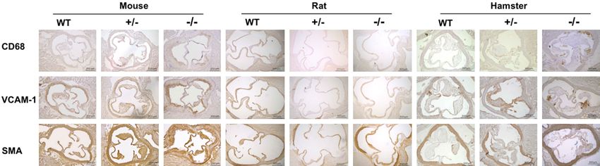
Small rodent animal models for FH study

percholesterolemia can reportedly be induced           specific pathogen-free conditions with a 14:10-
in homozygous Ldlr-deficient mice by the Paigen        h light-dark cycle for hamsters and a 12:12-h
diet containing 1.25% cholesterol and 0.5%             light-dark cycle for mice and rats. Animals were
cholate. However, this treatment elicits severe        fed either a regular chow diet (20% protein and
liver toxicity and leads to weight loss and sick-      4% fat; Beijing Ke’ao company, Beijing, China)
ness in mice, indicating the Paigen diet should        or a high-cholesterol/high-fat (HCHF) diet (0.5%
be avoided in experimental animal studies [7,          cholesterol and 15% fat) for 12 weeks. Plasma
8]. Moreover, FH is a genetic disease with a           was collected after overnight fasting. In our
gene dosage-dependent effect. Homozygous               studies, male animals aged 10-12 weeks were
and heterozygous FH patients show very high            used. All experiments were performed under
and moderately increased cholesterol levels in         the principle of experimental animal health
blood, respectively, but the risk of CVD is signifi-   (NIH released no. 85Y231996 Revision) and
cantly increased in both groups. In contrast to        approved by the laboratory animal ethics com-
FH patients, heterozygous Ldlr-deficient mice          mittee of Peking University (LA2010-059).
exhibit unexpectedly normal plasma cholester-
ol concentrations, suggesting that one copy of         Clinical characterization of FH patients
the Ldlr gene is sufficient to maintain normal
plasma cholesterol levels in mice. Thus, hetero-       Plasma samples of six patients with familial
zygous Ldlr KO mice have not been used to              hypercholesterolemia and three normal sub-
study hypercholesterolemia and atherosclero-           jects (male, 0-40 years old) were gifts from An
sis. Ldlr KO rats have also been generated and         Zhen Hospital, Beijing. Patient diagnoses were
characterized [9, 10], but information on simi-        made based on genetic analyses and clinical
larities of Ldlr KO rats and FH patients is still      manifestations. The patients were divided into
lacking, and it is unknown whether heterozy-           heterozygotes and homozygotes according Ldlr
gous Ldlr KO rats can mimic heterozygous FH.           gene mutations [15, 16].

Golden Syrian hamsters possess similar lipo-           Analysis of plasma lipids, lipoproteins, and
protein metabolism to humans [11-13], so we            apolipoproteins in different species
used the CRISPR/Cas9 system to generate a
                                                       Plasma ApoE, ApoB, and ApoA1 were detected
hamster model with Ldlr deficiency and found
                                                       by western blotting using methods described
that Ldlr KO hamsters display hyperlipidemia
                                                       previously [17]. Briefly, 1 μL of plasma was sub-
and atherosclerosis like humans [14]. To better
                                                       jected to 6% or 12% sodium dodecyl sulfate
understand the similarities and differences of
                                                       polyacrylamide gel electrophoresis for ApoB or
lipid profiles and atherosclerosis among three
                                                       ApoA1/ApoE, then transferred to a polyvinyli-
species with Ldlr deficiency, in the present st-
                                                       dene fluoride membrane for immunoblotting
udy we used wild-type (WT), heterozygous, and
                                                       with rabbit anti-ApoA1 (Calbiochem, San Diego,
homozygous animals in a systematical evalua-
                                                       CA, USA), goat anti-ApoE (Calbiochem), or goat
tion. Compared to mice and rats, both heterozy-
                                                       anti-ApoB (Calbiochem, California, USA) poly-
gous and homozygous Ldlr KO hamsters repli-
                                                       clonal antibody. Mouse anti-ApoB monoclonal
cate the phenotypes of hyperlipidemia and
                                                       antibody (Santa Cruz Biotechnology, Dallas, TX,
mimic the atherosclerotic plaque distribution
                                                       USA), rabbit anti-ApoA1 (Santa Cruz Biotech-
observed in the aortic roots, coronary arteries,
                                                       nology), and goat anti-ApoE (Calbiochem) were
and abdominal segments of FH patients.
                                                       used for human samples. Proteins were visual-
Materials and methods                                  ized by incubation with horseradish peroxidase-
                                                       conjugated secondary antibodies, followed by
Animals and diets                                      enhanced chemiluminescence detection (Mo-
                                                       lecular Imager Gel Doc XR System, Bio-Rad,
Ldlr KO hamsters were created with CRISPR/             Hercules, CA, USA). Plasma total cholesterol
Cas9 in our lab as described previously [14].          (TC) and triglyceride (TG) were measured using
WT and Ldlr KO rats were purchased from Gene           enzymatic commercial kits (Sigma-Aldrich, St.
Biotechnology Company (Beijing, China), and            Louis, MO, USA). Plasma lipoprotein profiles
mice were obtained from the Experimental               were analyzed by fast protein liquid chromatog-
Animal Center of Peking University Health Sci-         raphy (FPLC). Briefly, 200 μl of pooled plasma
ence Center. All animals were housed under             from each genotype was applied to Tricorn

3117                                                          Am J Transl Res 2019;11(5):3116-3127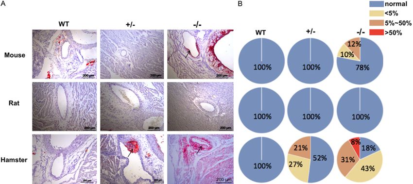
Small rodent animal models for FH study

high-performance Superose S-6 10/300 GL              tion (PBS containing 10% goat serum) for 30
column (Amersham Biosciences, Little Chal-           min, then incubated with primary antibody over-
font, UK), and then eluted with phosphate-buff-      night at 4°C and washed three times with PBS
ered saline (PBS) at a flow rate of 0.25 mL/min.     followed by an incubation with the appropriate
Cholesterol contents in each fraction (500 μL/       biotinylated secondary antibodies (1:200, ABC
fraction) were determined by the same com-           Vectastain; Vector Laboratories, Burlingame,
mercial kit.                                         CA, USA) and visualized using 3.3’-diaminoben-
                                                     zidine (DAB; Vectastain, Vector Laboratories)
Lipid extraction                                     [19].
Lipids were extracted according to modified          For Sirius red staining, hepatic tissues were
method of Bligh and Dyer [18]. Briefly, 100 mg       fixed with 4% paraformaldehyde solution over-
liver tissues were homogenized with 1 mL cold        night. Fixed liver tissues were embedded in par-
PBS. Then lipids were extracted by adding chlo-      affin and then cut into 4-μm sections and then
roform/methanol (v:v=2:1). Ten-milliliter glass      stained with Picrosirius Red Stain kit (Polysci-
tubes were used to avoid polymer contamina-          ences, Inc., Warrington, PA). Briefly, slices were
tion. Samples were vortexed for 2 min and then       deparaffinized and washed with double dis-
incubated for 20 min at room temperature, fol-       tilled (DI) water, then stained in hematoxylin for
lowed by centrifugation at 1000 rpm for 5 min.       8 min and rinsed well in DI water. The slices
The chloroform layer was then transferred with       were placed in solution A for 2 min and rinsed
a glass syringe and dried under nitrogen. Lipid      in DI water, followed by an incubation with solu-
samples were stored in -80°C freezer for future      tion B for 60 min and then solution C for 2 min.
use. TC and TG contents were measured using          Next, 70% ethanol was applied to the slices for
enzymatic commercial kits (Sigma-Aldrich).           45 s, and then samples were dehydrated to
                                                     mount. For hematoxylin-eosin (HE) staining,
Pathological analysis                                samples were immersed in xylene and alcohol,
                                                     then stained with hematoxylin for 5 min, fol-
Animals were sacrificed at the indicated times
                                                     lowed by another incubation with eosin for 3
following different diet interventions. Heart tis-
                                                     min. Afterward, slices were re-immersed in
sues and whole aortas were harvested after
                                                     alcohol and xylene, and then mounted. Fatty
perfusion with 30 mL 0.01 M PBS and placed in
                                                     liver severity was assessed by HE and Sirius
4% paraformaldehyde solution for 1 week, fol-
                                                     red staining as previously defined [20].
lowed by an overnight incubation in 20% su-
crose solution. The heart tissues were embed-        Statistical analysis
ded in Optimal Cutting Temperature (OCT) com-
pound and then cryo-sectioned (7-μm sections).       Group comparisons were performed using two-
The atherosclerotic plaques in aortic roots and      way analysis of variance followed by Tukey’s
whole aortas were analyzed with 0.3% Oil Red         test. GraphPad Prism 7.0 software (GraphPad
O (ORO) staining (Sigma-Aldrich). Coronary ath-      Software, La Jolla, CA, USA) was used for all
erosclerosis was scored by the plaques con-          statistical analyses. Differences at P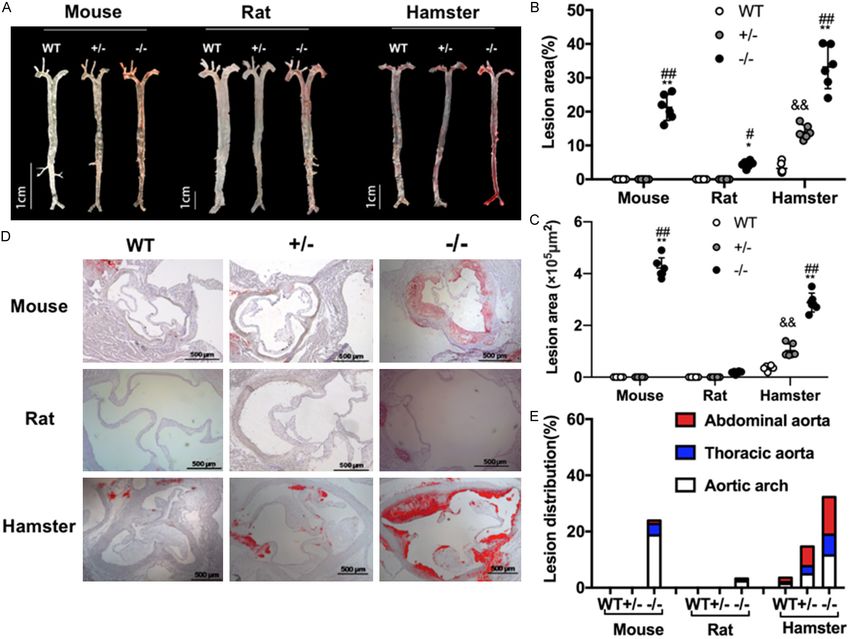
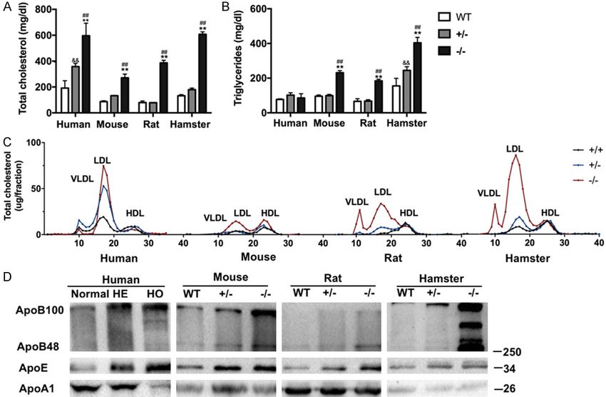
Small rodent animal models for FH study

Figure 1. Plasma lipids and lipoprotein profiles in different species fed a regular chow diet. (A) Plasma total cho-
lesterol and (B) triglycerides were measured from WT, Ldlr+/-, and Ldlr-/- rats, mice, and hamsters and in normal
subjects and heterozygous and homozygous FH patients. (C) FPLC analysis of 200 µl of pooled plasma lipoprotein
profiles from WT, Ldlr+/-, and Ldlr-/- rats, mice, and hamsters and FH patients. (D) Western blot analysis of plasma
ApoB, ApoE and ApoA1 levels from WT, Ldlr+/-, and Ldlr-/- rats, mice, and hamsters and FH patients. Data are
shown as mean ± SEM. n=3-6 per group. Significance was determined by two-way analysis of variance. *P
Small rodent animal models for FH study

Figure 2. Plasma lipids in different species upon 12-week HCHF diet feeding. (A) Plasma TC and (B) TG from WT,
Ldlr+/- and Ldlr-/- rats, mice, and hamsters on an HCHF diet for 12 weeks (n=6 per group). Data are shown as mean
± SEM. **P
Small rodent animal models for FH study

3121                                             Am J Transl Res 2019;11(5):3116-3127
Small rodent animal models for FH study

Figure 3. The characteristics of HCHF diet-induced atherosclerosis in different species. (A) Representative images
of Oil Red O stained en face aortas from WT, Ldlr KO+/-, and Ldlr KO-/- rats, mice, and hamsters with 12-week
HCHF diet treatment. (B) Quantification of atherosclerotic lesion sizes in the whole aortas from (A). (C) Analysis
of the plaque distribution in WT, Ldlr KO+/-, and Ldlr KO-/- rats, mice, and hamsters after a 12-week HCHF diet.
Aortic arch: aortic root to below the left subclavian. Thoracic aorta: the region between the end of the arch and the
last intercostal branch. Abdominal aorta: the region between the end of the thoracic aorta segment and the iliac
bifurcation. (D) Representative images of aortic root sections with Oil Red O staining from WT, Ldlr KO+/-, and Ldlr
KO-/- rats, mice, and hamsters with 12-week HCHF diet treatment. Scale bar =500 μm. (E) Quantification of lesion
areas in aortic roots. Data are expressed as mean ± SEM. n=6 per group. *P
Small rodent animal models for FH study

Figure 4. Analysis of coronary atherosclerosis in animals after 12-week HCHF diet feeding. (A) Representative images of coronary arteries stained with ORO from
WT, Ldlr+/-, and Ldlr-/- mice, rats, and hamsters with 12-week HCHF diet treatment. (B) Semi-quantification of coronary atherosclerosis in each group from (A). Ar-
rows indicate atherosclerotic lesions in the coronary arteries. WT: wild type, Ldlr+/-: heterozygous of low-density lipoprotein receptor, Ldlr-/-: low-density lipoprotein
receptor knockout, HCHF: high-cholesterol/high-fat.

3123                                                                                                                       Am J Transl Res 2019;11(5):3116-3127
Small rodent animal models for FH study

Mouse and rat VLDL/IDL particles contain both          hood in HoFH patients. Unlike HeFH patients,
ApoB100 and -48. Since ApoB48-containing               Ldlr+/- mice exhibit unchanged plasma choles-
lipoproteins possess a high fractional catabolic       terol levels compared to WT controls and do not
rate and HDL-C cannot be transferred to VLDL           develop atherosclerosis, indicating that they
due to a lack of CETP, HDL is the major lipopro-       are not a good model for human atherosclero-
tein in mice and rats. On an HCHF diet, these          sis study. Furthermore, atherosclerotic lesions
dietary lipids are assembled into very large lipo-     are often induced in the Ldlr-/- mice with the
proteins and are still effectively removed from        Paigen diet, which causes severe liver toxicity.
circulation in Ldlr+/- mice and rats. In contrast,     To avoid confounding effects of this diet and
there is no hepatic Apobec-1 expression in             investigate accelerated atherosclerotic devel-
hamsters and humans, but there are high lev-           opment in different species, we placed all three
els of CETP activity [24, 25], both of which con-      species on an HCHF diet for 12 weeks and
tribute to the stability of large lipoprotein parti-   found that the atherosclerotic lesions were
cles in plasma. In addition, reduced LDLR pro-         restricted to the aortic arch and thoracic aorta
tein in the liver is insufficient to clear accumu-     in Ldlr-/- mice, while there were no lesions in
lated lipoproteins in Ldlr+/- hamsters, predis-        Ldlr+/- mice. Interestingly, both Ldlr+/- and Ldlr-
posing them to diet-induced hyperlipidemia.            /- hamsters showed atherosclerotic lesions in
Therefore, Ldlr+/- hamsters could serve as an          coronary arteries and whole aortas, including
ideal model to study the molecular mecha-              the thoracic and abdominal segments, with
nisms involved in LDLR regulation and assess           more severe lesions in whole aortas and coro-
drug efficacy.                                         nary artery occlusion in Ldlr-/- hamsters. Con-
                                                       sistent with this, previous reports propose that
It is important to notice that compared to other       atherosclerosis begins as fatty streak deposits
species, complete loss of the Ldlr gene in ham-        in the right coronary artery and abdominal
sters caused increased baseline TG levels and          aorta of young persons, indicating that these
exacerbated HTG with severe hypercholesterol-          two sites are critical for atherosclerosis devel-
emia, and these animals developed combined             opment [30, 31]. Additionally, FH patients have
hyperlipidemia in response to a 12-week HCHF           an atherosclerosis distribution in the carotid,
diet. However, this combination is rare in FH          thoracic, and abdominal arteries [32-34]. It was
patients, and whether HTG can be induced is            surprising that Ldlr KO rats on an HCHF diet for
unknown because an HCHF diet intervention              12 weeks did not develop discernable athero-
could not be considered for clinical FH patients.      sclerotic lesions, which is similar to the absence
We previously demonstrated that Ldlr deficien-         of this finding in Ldlr-/- rats on a Western diet
cy did not alter lipoprotein lipase (LPL) enzyme       (WD) for 16 weeks. However, when Ldlr-/- rats
activity in hamsters, which is required to clear       were fed a WD for 52 or 72 weeks, atheroscle-
plasma TG-rich large lipoproteins. Moreover,           rotic lesions were found in the whole aortas
chylomicron remnants from LPL-mediated lipol-          and coronary arteries [10]. These observations
ysis accumulated in Ldlr-/- mice, suggesting           suggest that Ldlr-/- rats are more resistant to
that the LDLR pathway is critical for the remov-       diet-induced hyperlipidemia and ASCHD than
al of TG as well as cholesterol [26]. Our western      Ldlr-/- hamsters. This could be an ideal rodent
blot results further confirmed that ApoB100/48         animal model to mimic the central features of
and ApoE, two key components of large lipopro-         FH.
teins, were accumulated in all Ldlr mutant spe-
cies in a gene dose-dependent manner. Since            Porcine models of Ldlr mutations recently ga-
hamsters possess the highest CETP activity             ined attention for FH studies because the size
and CETP inhibitors have been reported to              is close to that of humans. Davis and colleagues
lower plasma TG, it will be interesting to test        generated homozygous Ldlr-targeted Yucatan
the effect of CETP inhibition on TG metabolism         miniature pigs showing hypercholesterolemia
in our Ldlr KO hamsters.                               (300 mg/
patients and as early as adolescence or child-         dL), which differed from FH patients [35] and

3124                                                           Am J Transl Res 2019;11(5):3116-3127
Small rodent animal models for FH study
our heterozygous hamster model. Importantly, of Molecular Cardiovascular Sciences, Ministry of
there is high variability in the genetic back- Education, Peking University, Beijing 100191, China.
grounds of Ldlr-targeted Yucatan miniature pigs E-mail: georgeliu@bjmu.edu.cn
and a long-term HCHF diet is required for ath-
erosclerosis studies using these animals. The References
validity of this porcine model was questioned
[1] Mytilinaiou M, Kyrou I, Khan M, Grammato-
by Li and colleagues in a study where Ldlr gene poulos DK and Randeva HS. Familial hypercho-
was deleted in domestic pigs. The Ldlr-deficient lesterolemia: new horizons for diagnosis and
domestic pigs on a 4-month HCHF diet showed effective management. Front Pharmacol 2018;
human-like advanced coronary plaque that was 9: 707.
attenuated by prophylactic statin treatment [2] Ito MK and Watts GF. Challenges in the diagno-
[36]. Unfortunately, data regarding atheroscle- sis and treatment of homozygous familial hy-
rosis and CHD in heterozygous Ldlr-deficient percholesterolemia. Drugs 2015; 75: 1715-
domestic pigs were not presented by the 1724.
[3] Emini Veseli B, Perrotta P, De Meyer GRA, Roth
authors. The unchanged plasma lipid levels in
L, Van der Donckt C, Martinet W and De Meyer
the heterozygotes suggest that domestic pigs GRY. Animal models of atherosclerosis. Eur J
are not an ideal animal model for FH, particu- Pharmacol 2017; 816: 3-13.
larly for translational studies on HeFH. [4] Poledne R and Jurcikova-Novotna L. Experi-
mental models of hyperlipoproteinemia and
Although data from Ldlr KO hamsters suggest atherosclerosis. Physiol Res 2017; 66: 69-75.
that this species could replicate the major fea- [5] Russell JC and Proctor SD. Small animal mod-
tures of FH, including lipoprotein profiles with els of cardiovascular disease: tools for the
LDL-C dominance and the atherosclerotic le- study of the roles of metabolic syndrome, dys-
sion distribution, there are two important ques- lipidemia, and atherosclerosis. Cardiovasc Pa-
tions that need to be answered in future stud- thol 2006; 15: 318-330.
[6] Barter PJ, Brewer HB Jr, Chapman MJ, Henne-
ies. Firstly, why do Ldlr-/- hamsters show incr-
kens CH, Rader DJ and Tall AR. Cholesteryl es-
eased VLDL-C and is this increase in VLDL-C ter transfer protein: a novel target for raising
associated with high CETP activity? Secondly, HDL and inhibiting atherosclerosis. Arterioscler
will a chronic HCHF diet (e.g., 52 weeks) exacer- Thromb Vasc Biol 2003; 23: 160-167.
bate atherosclerotic development in Ldlr KO [7] Getz GS and Reardon CA. Diet and murine at-
hamsters? Taken together, the results from this herosclerosis. Arterioscler Thromb Vasc Biol
extensive interspecies comparison study dem- 2006; 26: 242-249.
onstrate that Ldlr KO hamsters could provide [8] Nishina PM, Verstuyft J and Paigen B. Synthetic
new insight into FH and human atherosclero- low and high fat diets for the study of athero-
sclerosis in the mouse. J Lipid Res 1990; 31:
sis.
859-869.
[9] Wang HY, Quan C, Hu C, Xie B, Du Y, Chen L,
Acknowledgements
Yang W, Yang L, Chen Q, Shen B, Hu B, Zheng
Z, Zhu H, Huang X, Xu G and Chen S. A lipido-
NSFC 31520103909 and 81270367 to G.L., mics study reveals hepatic lipid signatures as-
NSFC81570787 to Y.W.; National Key Research sociating with deficiency of the LDL receptor in
and Development Program of China (2016YF- a rat model. Biol Open 2016; 5: 979-986.
E0126000) to Y.W. G.L. is a fellow at the Co- [10] Sithu SD, Malovichko MV, Riggs KA, Wickra-
llaborative Innovation Center for Cardiovascu- masinghe NS, Winner MG, Agarwal A, Hamed-
lar Disease Translational Medicine, Nanjing Me- Berair RE, Kalani A, Riggs DW, Bhatnagar A
dical University. and Srivastava S. Atherogenesis and metabol-
ic dysregulation in LDL receptor-knockout rats.
Disclosure of conflict of interest JCI Insight 2017; 2.
[11] Dorfman SE, Smith DE, Osgood DP and Lich-
None. tenstein AH. Study of diet-induced changes in
lipoprotein metabolism in two strains of Go-
Address correspondence to: Xunde Xian, Depart- lden-Syrian hamsters. J Nutr 2003; 133: 4183-
4188.
ment of Molecular Genetics, UT Southwestern
[12] Nistor A, Bulla A, Filip DA and Radu A. The hy-
Medical Center, Dallas 75390, Texas, USA. E-mail: perlipidemic hamster as a model of experi-
xunde.xian@utsouthwestern.edu; George Liu, Insti- mental atherosclerosis. Atherosclerosis 1987;
tute of Cardiovascular Sciences and Key Laboratory 68: 159-173.
3125 Am J Transl Res 2019;11(5):3116-3127Small rodent animal models for FH study
[13] Remillard P, Shen G, Milne R and Maheux P. expression in rat liver and intestine. Arterioscler
Induction of cholesteryl ester transfer protein Thromb Vasc Biol 1998; 18: 1079-1092.
in adipose tissue and plasma of the fructose- [25] Reaves SK, Wu JY, Wu Y, Fanzo JC, Wang YR,
fed hamster. Life Sci 2001; 69: 677-687. Lei PP and Lei KY. Regulation of intestinal apo-
[14] Guo X, Gao M, Wang Y, Lin X, Yang L, Cong N, lipoprotein B mRNA editing levels by a zinc-de-
An X, Wang F, Qu K, Yu L, Wang Y, Wang J, Zhu ficient diet and cDNA cloning of editing protein
H, Xian X and Liu G. LDL receptor gene-ablated in hamsters. J Nutr 2000; 130: 2166-2173.
hamsters: a rodent model of familial hypercho- [26] Ishibashi S, Perrey S, Chen Z, Osuga J, Shimada
lesterolemia with dominant inheritance and M, Ohashi K, Harada K, Yazaki Y and Yamada
diet-induced coronary atherosclerosis. EBio- N. Role of the low density lipoprotein (LDL) re-
Medicine 2018; 27: 214-224. ceptor pathway in the metabolism of chylomi-
[15] Wu WF, Sun LY, Pan XD, Yang SW and Wang LY. cron remnants. A quantitative study in knock-
Use of targeted exome sequencing in genetic out mice lacking the LDL receptor, apolipopro-
diagnosis of Chinese familial hypercholesterol- tein E, or both. J Biol Chem 1996; 271: 22422-
emia. PLoS One 2014; 9: e94697. 22427.
[16] Jiang L, Sun LY, Pan XD, Chen PP, Tang L, Wang [27] Stone NJ, Fredrickson DS, Verter J and Levy RI.
W, Zhao LM, Yang SW and Wang LY. Charac- Coronary-artery disease in 116 kindred with
terization of the unique Chinese W483X muta- familial type-Ii hyperlipoproteinemia. Circula-
tion in the low-density lipoprotein-receptor tion 1974; 49: 476-488.
gene in young patients with homozygous famil- [28] Humphries SE, Cooper JA, Seed M, Capps N,
ial hypercholesterolemia. J Clin Lipidol 2016; Durrington PN, Jones B, McDowell IFW, Soran
10: 538-546. H, Neil HAW; Simon Broome Familial Hyper-
[17] Xian X, Ding Y, Dieckmann M, Zhou L, Plattner lipidaemia Register Group. Coronary heart dis-
F, Liu M, Parks JS, Hammer RE, Boucher P, Tsai ease mortality in treated familial hypercholes-
S and Herz J. LRP1 integrates murine macro- terolaemia: update of the UK Simon Broome
phage cholesterol homeostasis and inflamma- FH register. Atherosclerosis 2018; 274: 41-46.
tory responses in atherosclerosis. Elife 2017; [29] Marks D, Thorogood M, Neil HA and Humphries
6. SE. A review on the diagnosis, natural history,
[18] Bligh EG and Dyer WJ. A rapid method of total and treatment of familial hypercholesterolae-
lipid extraction and purification. Can J Biochem mia. Atherosclerosis 2003; 168: 1-14.
Physiol 1959; 37: 911-917. [30] McGill HC, McMahan CA, Herderick EE, Ma-
[19] Liu XJ, Wu XY, Wang H, Wang SX, Kong W, lcom GT, Tracy RE, Strong JP and Ather PD.
Zhang L, Liu G and Huang W. Renal injury in Origin of atherosclerosis in childhood and ado-
Seipin-deficient lipodystrophic mice and its re- lescence. Am J Clin Nutr 2000; 72 Suppl:
versal by adipose tissue transplantation or 1307S-1315S.
leptin administration alone: adipose tissue- [31] McGill HC Jr, McMahan CA, Herderick EE, Tracy
kidney crosstalk. FASEB J 2018; 32: 5550- RE, Malcom GT, Zieske AW and Strong JP.
5562. Effects of coronary heart disease risk factors
[20] Miyaoka Y, Jin D, Tashiro K, Komeda K, Ma- on atherosclerosis of selected regions of the
subuchi S, Hirokawa F, Hayashi M, Takai S and aorta and right coronary artery. PDAY research
Uchiyama K. Chymase inhibitor prevents the group. Pathobiological determinants of athero-
development and progression of non-alcoholic sclerosis in youth. Arterioscler Thromb Vasc
steatohepatitis in rats fed a high-fat and high- Biol 2000; 20: 836-845.
cholesterol diet. J Pharmacol Sci 2017; 134: [32] Roberts WC, Won VS, Weissenborn MR, Khalid
139-146. A and Lima B. Massive diffuse calcification of
[21] Hartgers ML, Ray KK and Hovingh GK. New ap- the ascending aorta and minimal focal calcifi-
proaches in detection and treatment of fam- cation of the abdominal aorta in heterozygous
ilial hypercholesterolemia. Curr Cardiol Rep familial hypercholesterolemia. Am J Cardiol
2015; 17: 109. 2016; 117: 1381-1385.
[22] Baum SJ, Soifer D and Duell PB. Emerging [33] Ribeiro P, Shapiro LM, Gonzalez A, Thompson
treatments for heterozygous and homozygous GR and Oakley CM. Cross sectional echocar-
familial hypercholesterolemia. Rev Cardiovasc diographic assessment of the aortic root and
Med 2016; 17: 17-25. coronary ostial stenosis in familial hypercho-
[23] Getz GS and Reardon CA. Do the Apoe(-/-) and lesterolaemia. Br Heart J 1983; 50: 432-437.
Ldlr(-/-) mice yield the same insight on athero- [34] Aggeli C, Kazazaki C, Felekos I, Roussakis G,
genesis? Arterioscler Thromb Vasc Biol 2016; Lagoudakou S, Skoumas J, Toutouzas K, Tou-
36: 1734-1741. soulis D, Pitsavos C and Stefanadis C. Role of
[24] Greeve J, Axelos D, Welker S, Schipper M and real time-3D transesophageal echocardiogra-
Greten H. Distinct promoters induce APOBEC-1 phy in evaluating the atheromatous burden of
3126 Am J Transl Res 2019;11(5):3116-3127Small rodent animal models for FH study
thoracic aorta in patients with heterozygous bon S, Nakai M, Kojima M, Iwamoto M, Hashi-
familial hypercholesterolemia: comparison wi- moto M, Yoda S, Kunimoto S, Hiro T, Matsumoto
th 2D transesophageal study. Int J Cardiol T, Mitsumata M, Sugitani M, Saito S, Hirayama
2011; 150: 92-93. A and Onishi A. Development of human-like ad-
[35] Davis BT, Wang XJ, Rohret JA, Struzynski JT, vanced coronary plaques in low-density lipo-
Merricks EP, Bellinger DA, Rohret FA, Nichols protein receptor knockout pigs and justifica-
TC and Rogers CS. Targeted disruption of LDLR tion for statin treatment before formation of
causes hypercholesterolemia and atheroscle- atherosclerotic plaques. J Am Heart Assoc
rosis in Yucatan miniature pigs. PLoS One 2016; 5: e002779.
2014; 9: e93457.
[36] Li YX, Fuchimoto D, Sudo M, Haruta H, Lin QF,
Takayama T, Morita S, Nochi T, Suzuki S, Sem-
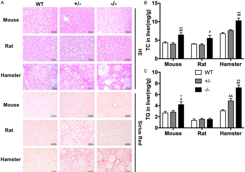
3127 Am J Transl Res 2019;11(5):3116-3127Small rodent animal models for FH study Figure S1. An HCHF diet induced NAFLD in hamsters, but not in mice or rats. (A) Representative staining with HE (upper panel) and Sirius red (lower panel) in liver tissue from WT, Ldlr KO+/-, and Ldlr KO-/- rats, mice, and hamsters with 12-week HCHF diet treatment. Scale bar =50 μm. (B, C) Hepatic TC (B) and TG (C) contents were measured and normalized to liver weight (n=4/group). Data are shown as mean ± SEM. *P
Small rodent animal models for FH study

Figure S2. Analysis of atherosclerotic plaque components in animals after 12-week HCHF diet feeding. Atherosclerotic lesion components in WT, Ldlr KO+/-, and
Ldlr KO-/- rats, mice, and hamsters on a 12-week HCHF diet were analyzed with immunohistochemical staining using antibodies against CD68, VCAM-1, and α-SMA.
Representative images of aortic root sections stained for CD68 (upper panel), VCAM-1 (middle panel), and α-SMA (lower panel). Scale bar =200/500 μm. WT: wild
type, Ldlr+/-: heterozygous of low-density lipoprotein receptor, Ldlr-/-: low-density lipoprotein receptor knockout, HCHF: high-cholesterol/high-fat.

2
You can also read