Induced oral mucositis in Wistar rats treated with different drugs: Preventive potential in cytokine production
←
→
Page content transcription
If your browser does not render page correctly, please read the page content below
MOLECULAR AND CLINICAL ONCOLOGY 14: 127, 2021

       Induced oral mucositis in Wistar rats treated with different
           drugs: Preventive potential in cytokine production
          MARIA INÊS DA CRUZ CAMPOS1, CELSO NEIVA CAMPOS2, JOSÉ OTÁVIO AMARAL CORRÊA3,
              FERNANDO MONTEIRO AARESTRUP4 and BEATRIZ JULIÃO VIEIRA AARESTRUP1

  1
   Laboratory of Immunopathology and Experimental Pathology, Reproductive Biology Center, Department of Morphology,
Institute of Biological Sciences, Federal University of Juiz de Fora; 2School of Dentistry Clinic, Department of Dental Clinic,
           3
             Laboratory of Pharmacology, Department of Immunohistochemistry, Federal University of Juiz de Fora;
 4
  Laboratory of Immunopathology and Experimental Pathology, Reproductive Biology Center, Department of Dental Clinic,
       Faculty of Medical Sciences and Juiz de Fora Health‑SUPREMA, Juiz de Fora, Minas Gerais 36036‑330, Brazil

                                            Received July 11, 2019; Accepted April 1, 2021

                                                      DOI: 10.3892/mco.2021.2289

Abstract. The aim of the present study was to investigate               expression levels of TNF‑α during all stages of the experi‑
the preventive potential of pentoxifylline, atorvastatin and            ment. Treatment with trans‑caryophyllene modulated serum
trans‑caryophyllene in oral mucositis through histopatho‑               IFN‑γ levels negatively, whereas treatment with atorvastatin
logical analysis of wounds in the oral mucosa of Wistar rats            and trans‑caryophyllene maintained lower levels of IFN‑ γ
treated with 5‑FU, and to evaluate the immunomodulatory                 compared with the control group.
effect of these drugs on serum nitrite production, in situ IFN‑γ,
TNF‑α and TGF‑β, and TNF‑α in tissues. A total of 32 male               Introduction
Wistar rats with an average age of 9 weeks and an average
body weight of 250 g were divided into four treatment groups:           Oral mucositis affects 40‑80% of patients undergoing chemo‑
Saline, trans‑caryophyllene, pentoxifylline and atorvastatin.           therapy and almost all patients undergoing head and neck
Oral mucositis was then induced. On days 3 and 4, the mucosa            radiotherapy (1‑3). Oral mucositis causes dysphagia, dysarthria,
of the mouth of eight pre‑treated animals in each group was             and odynophagia and is a possible gateway for opportunistic
bilaterally scarified twice with the tip of a sterile needle, with      infections; therefore, it leads to a decreased quality of life and
an anesthetic solution. Mucosal samples from animals treated            is considered an important non‑hematological complication of
with trans‑caryophyllene preserved a thin epithelial lining             antitumor treatment (4‑8).
associated with focal perivascular inflammatory infiltrates.                Typical macroscopic and microscopic characteristics of
Pentoxifylline‑treated animals exhibited total epithelial loss          oral mucositis reflect a natural history following radiation
in oral wounds with severe inflammatory infiltrates and                 therapy or the chemotherapy cycle, in which the initial wound
mild re‑epithelialization associated with mild and diffuse              of an erythematous plaque evolves into isolated ulcerations
inflammatory infiltrates. Samples from atorvastatin‑treated             that converge and result in more extensive and deeper wounds,
animals exhibited no epithelial dissolution, with preserved             with symptoms ranging from burning to severe pain (4). The
thin lining and mild diffuse inflammatory infiltrates. The              erythematous wound histopathologically represents epithelial
analysis of TNF‑α expression revealed improved results in               hypoplasia associated with inflammatory reaction on the
trans‑caryophyllene animals. The analysis of TGF‑β expres‑              underlying lamina propria caused by an antitumor drug that
sion revealed positive mononuclear cells. Preventive treatment          secondarily inhibits normal epithelial renewal; the healing
with atorvastatin was demonstrated to modulate the serum                phase occurs with the end of the antineoplastic cycle, showing
                                                                        spontaneous resolution of the ulcers (7,9‑11).
                                                                            The in situ cytokine production by oral inflammatory
                                                                        cells influences the establishment and remission of ulcers.
                                                                        Parkin and Cohen (12) suggested that the cytokine profile of
Correspondence to: Professor Maria Inês da Cruz Campos,                 the lamina propria cells affects the mitotic and apoptotic activi‑
Laboratory of Immunopathology and Experimental Pathology,
                                                                        ties of oral epithelial tissue. Notable among these cytokines,
Reproductive Biology Center, Department of Morphology,
Institute of Biological Sciences, Federal University of Juiz de Fora,
                                                                        are tumor necrosis factor‑alpha (TNF‑α), transforming growth
Lindalva de Paula Ribeiro, 240 Bosque Imperial, Juiz de Fora,           factor‑beta (TGF‑β), and interferon‑gamma (IFN‑γ). TNF‑α is
Minas Gerais 36036‑330, Brazil                                          a pro‑inflammatory cytokine that is primarily produced by acti‑
E‑mail: marimurucci@gmail.com                                           vated macrophages. TGF‑β enhances the proliferation of several
                                                                        cells of mesenchymal origin and increases extracellular matrix
Key words: leukopenia, oral mucositis, trans‑caryophyllene,             synthesis by T‑lymphocytes and platelets. IFN‑γ is produced
atorvastatin, chemotherapy                                              by Th1 lymphocytes and natural killer cells; it regulates the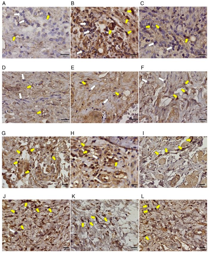
2                                    DA CRUZ CAMPOS et al: ORAL MUCOSITIS IN WISTAR RATS

proliferation and differentiation of various cell types and has     nitrite production, in situ IFN‑γ and TNF‑α, and TGF‑β and
the ability to modulate the immune system (13‑15).                  TNF‑α in tissues.
    Besides cytokines, nitric oxide is one of the ten smallest
molecules existing in nature and is a highly soluble reactive       Materials and methods
free radical produced by the NO synthase (NOS) enzyme from
L‑arginine. This molecule remains in the tissue for only a few      Approval and separation of animals. The entire experimental
seconds, but its presence can be indirectly detected through        protocol was approved by the Animal Research Committee of
NOS based on its tissue expression namely eNOS (endothelial),       the Federal University of Juiz de Fora (Notion No. 062/2011).
nNOS (neuronal), and iNOS (inducible) (16‑18). Experimental             Male Wistar rats (Rattus norvegicus) (N=32) from the
and clinical studies suggest that both overproduction and           Animal Facility of the Reproductive Biology Center (CBR) of
inhibition of NO are associated with greater potential for tissue   the Federal University of Juiz de Fora were used for this study,
damage or maintenance of a chronic response, respectively (17).     with an average age of 9 weeks and average body weight of
    Nitric oxide also has a destructive effect on invading          250 g. The rats were allowed ad libitum access to food and water.
microorganisms and is therefore released into the inflamed site         Fifteen days before induction of oral mucositis, the
by neutrophils and macrophages (19).                                animals were divided into four groups: Group I‑saline‑treated
    Some immunomodulatory drugs have been employed to               animals with chemotherapy‑induced mucositis (n=8), Grou
control the exacerbation of oral mucositis wounds (20‑22).          p II‑trans‑caryophyllene‑treated animals with chemo‑
Among these, pentoxifylline (PTX) is an anti‑bleeding and           therapy‑induced mucositis (n=8), Group III‑pentoxifylline‑t
anti‑thrombogenic agent with the ability to inhibit the TNF‑α       reated animals with chemotherapy‑induced mucositis (n=8),
expression by inhibiting its genetic transcription (23,24).         and Group IV‑atorvastatin‑treated animals with chemo‑
Atorvastatin has an anti‑inflammatory and anti‑thrombogenic         therapy‑induced mucositis (n=8).
action that is capable of reducing C‑reactive protein levels and
TNF‑α expression in situ (20,25).                                   Drug delivery protocol. Trans‑caryophyllene gavage admin‑
    Studies conducted over the past decade have also                istration began two days before 5‑FU treatment and was
demonstrated the effectiveness of the anti‑inflammatory             continued for 2 days after medication (totalling 9 days of treat‑
action of trans‑caryophyllene, a molecule isolated from             ment) with a dose of 50 mg/kg (0.102 ml) (31‑33).
the copaiba oil‑resin of the Brazilian medicinal plant                  Pentoxifylline dissolved in sterile saline of 0.9% at a
Copaifera langsdorffii (25‑28).                                     concentration of 100 mg/kg/day that was administered intra‑
    Although the current control of oral wounds is non‑specific     peritoneally for 15 consecutive days (34).
and palliative‑preventing their establishment is critical to            Atorvastatin was administered intraperitoneally
cancer prognosis, since severe wounds limit and disrupt treat‑      at 10 mg/kg/day for 1 week before 5‑FU treatment (20).
ment (4,6,29,30).                                                       The drug delivery schedule is outlined in Fig. 1.
    In our previous study, utilizing the same experimental
design as the current study, we determined the influence of         Induction of oral mucositis. Oral mucositis was induced by the
these drugs on white blood cell counts of animals undergoing        administration of the chemotherapeutic agent 5‑FU (Eurofarma,
chemotherapy with 5‑fluorouracil (5‑FU). The results demon‑         São Paulo, Brazil) via intraperitoneal injection on Day 0
strated that atorvastatin significantly prevented leukocyte         (100 mg/kg) and on Day 2 (60 mg/kg) of the experiment (35).
reduction in comparison to other experimental groups, thus              On Days 3 and 4, the mouth's mucosa of 8 pre‑treated
demonstrating excellent potential for the prevention of leuko‑      animals from each group, after being anesthetized with
penia (25).                                                         Ketamine (100 mg/kg) and Xylazine (10 mg/kg), was bilater‑
    Considering the increase in the number of patients              ally scarified twice by the same operator using the tip of a
submitted to chemotherapy and/or radiotherapy treatments for        sterile needle (35,36).
cancer control and, consequently, sequelae caused by those
treatments, this article discusses the need to control these        Determination of when the animals should be sacrificed.
manifestations with the objective of improving the quality of       The animals were separated into groups to be euthanized
life of the aforementioned patients. The large number of cancer     at 8, 11 and 15 days after induction of oral mucositis. However,
cases makes the use of such therapies to increase even further,     clinical evaluation standards were used that could cause the
as they are considered the only way to control the development      anticipation of deaths: Marked weight loss, ocular or nasal
of the disease. As a consequence of the use of these therapies,     hemorrhage, decreased consumption of water and feed, loss of
oral mucositis might appear, which leads to poor nutrition of       movement in the cage, demonstration of pain.
the patient, difficulties to swallow and, generally, causes the
abandonment of treatment due to lack of control. Given the          Euthanasia method. The method used was deep anestesia with
fact that there is little knowledge on the control and preven‑      the combination of Xylazine 10 mg/kg + 100 mg/kg Ketamine,
tion of such oral ulcers, new studies are necessary to unveil       mixed in the same syringe, intraperitoneally.
information that might be useful in such situations.                    The death of the animals was confirmed through cardiac
    The aim of the present study was to investigate the             and respiratory arrest, absence of reflexes (seen through the
preventive potential of pentoxifylline, atorvastatin and            hind legs), drop in body temperature.
trans‑caryophyllene based on histopathological analysis of              After the drug is applied, exsanguination is performed,
wounds in the oral mucosa induced in 5‑FU‑treated Wistar            performed by means of cardiac puncture or large blood vessels,
rats, and to evaluate their immunomodulatory effect on serum        when rodent serum is obtained.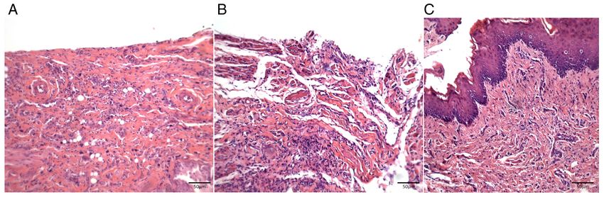
MOLECULAR AND CLINICAL ONCOLOGY 14: 127, 2021                                                                 3

Figure 1. Drug delivery schedule by group of animals and days of euthanasia. Day 0: Day of mucositis induction; Day 15: Initial preventive treatment; Days 8,
11, and 15: Days of euthanasia with oral mucosa and serum sampling. Group V includes two animals from each drug group without mucositis. D, day.

Sample collection and analysis. After deep anesthesia with
Ketamine (100 mg/kg) and Xylazine (10 mg/kg), via intraperi‑
toneal injection, followed by euthanasia, the mouth's mucosa
was excised.
    The schedule of euthanasia, as well as the drug delivery, is
outlined in Fig. 1.
    The excised mouth's mucosa was dissected to obtain the
mucositis‑affected areas. The samples were immediately
fixed in 10% buffered formalin and submitted to routine
histological processing for H&E staining.
    The sections were analyzed with a x400 magnification Zeiss
microscope (Hallbergmoos) in three fields of each sample by                     Figure 2. Body weight variation in animals upon euthanasia on different days
an Axion Cam ICC 5 (Zeiss) computer system digital capture                      of the experiment. Group I animals exhibited marked weight loss of ~50%
performed by means of a digital camera attached to an optical                   on the 11th day and slight recovery at the end of the experiment. Group II
microscope. The captured images were processed in an auto‑                      animals showed slight initial weight loss and recovery >50% by the end.
                                                                                Group III showed weight loss leading to cachexia on the 11th day and slight
matic morphometry Zen 2012 (Blue Edition) program, in which                     recovery until the 15th day. Group IV animals showed a weight gain >50% on
semi‑automatic microscopic field morphometry of leukocytes                      all days of the experiment. White column, saline; grey column, trans‑cario‑
and vascular sections was performed on the lamina propria                       phyllene; horizontal lines column, pentoxifylline; black column, atorvastatin.
underlying the epithelial wound.

Body weight. Animals should not lose more than 30% of their                     concentrations between 3.12 and 100 µM. The results were
initial body weight. For this, both body weight and feed intake                 expressed in moles/ml. The Griess method or test is a chemical
should were evaluated daily. As the animals were controlled                     reaction that detects the presence of organic nitrites through its
daily, when there was excessive weight loss, they were eutha‑                   reaction with sulfanilamide in an acidic medium. To measure
nized.                                                                          nitrite production, 100 µl aliquots of samples were incubated
                                                                                with 100 µl of the Griess reagent (50 µl 1% sulfanilamide
In situ detection of TNF‑ α and TGF‑β expression. The Starr                     solution and 50 µl 0.1% N‑naphthyl ethylenediamine dihy‑
Trek Universal HRP Detection System was used to analyze                         drochloride solution in 2.5% H2PO4) at room temperature for
the expression of TGF‑β and TNF‑ α in the lamina propria                        10 min. This reaction produces a bright red staining compound
cells that were considered positive by intracytoplasmic brown                   that can be estimated from 10 min to 2 h after mixing (38).
pigmentation. Three field counts were averaged, in addition to
total counts for each group.                                                    Statistical analysis. The data are presented as mean ± standard
                                                                                deviation of the mean (SEM). For comparison between
TNF‑ α and IFN‑γ serum levels. TNF‑α and IFN‑γ (both from                       groups, we used a Kruskal‑Wallis test with Dunn's Multiple
PeproTech Inc.) levels were examined by the ELISA (Enzyme                       Comparison post‑hoc test. In all instances, the significance
Linked ImmunoSorbent Assay) method, which is based on                           level was set at 5% (P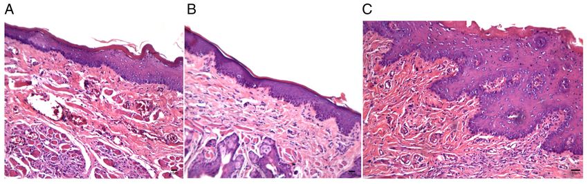
4 DA CRUZ CAMPOS et al: ORAL MUCOSITIS IN WISTAR RATS
Figure 3. Photomicrographs of histological sections of oral mucosa samples from Wistar rats subjected to chemotherapy. (A) Control group (day 8), with
diffuse inflammatory infiltrate and loss of epithelial continuity. (B) Control group (day 11), showing that oral wounds progressed to ulcers with severe
diffuse inflammatory infiltrate. (C) Control group (day 15) with mild re‑epithelialization associated with underlying inflammatory infiltrate still present. H&E
staining. Magnification, x100.
Figure 4. Photomicrographs of histological sections of oral mucosa samples from Wistar rats subjected to chemotherapy. (A) Trans‑caryophyllene group
(day 8), showing preserved thin epithelial lining associated with focal perivascular inflammatory infiltrate. Areas were occasionally identified with no leuko‑
cyte infiltration. (B) Trans‑caryophyllene group (day 11), no inflammation in the samples and decreased amount of vascular sections. (C) Trans‑caryophyllene
group (day 15), with keratinized epithelial restoration and recovery of the number of vascular sections. H&E staining. Magnification, x100.
Group I animals showed marked weight loss of around infiltrates and occasional necrotic areas; on Day 15 slight
50% on the 11th day and slight recovery at the end of the re‑epithelialization associated with a mild and diffuse inflam‑
experiment; Group II animals showed slight initial weight matory infiltrate was observed. The number of blood vessels
loss and recovery greater than 50% by the end of it; Group III decreased in animals from this group on Day 11 before it
showed weight loss leading to cachexia on the 11th day and increased again on Day 15 (Fig. 5A‑C).
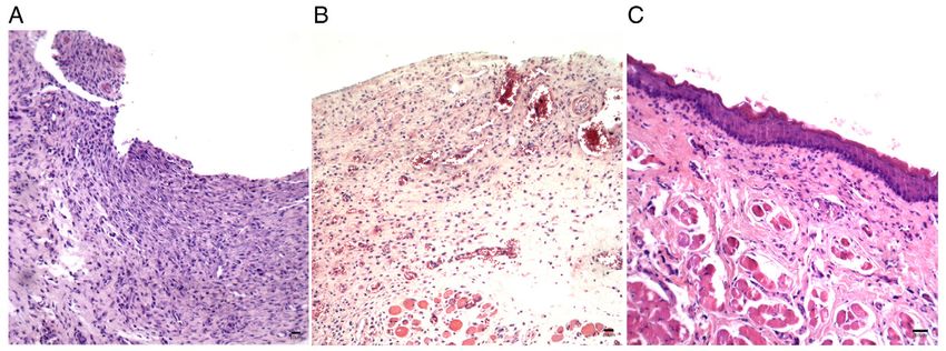
slight recovery until the 15th day; Group IV animals showed On Day 8, samples from atorvastatin‑treated animals
a weight gain greater than 50% on all days of the experiment. (Group IV) displayed no epithelial dissolution, with
preserved thin lining and mild diffuse inflammatory infil‑
Histopathological and histomorphometric analysis. At 8 days trates; on Days 11 and 15 there was a progressive increase
after delivery of the chemotherapeutic drug, samples from in inflammatory infiltrates, but with epithelial preservation.
the saline‑treated control group of animals (Group I) showed Similar to other groups, recovery of the number of vascular
loss of epithelium in the oral mucosa with mild inflamma‑ sections was observed on Day 15, after a decrease on
tory mononuclear infiltrates. At 11 days after chemotherapy Day 11 (Fig. 6A‑C).
in these animals, the oral wounds progressed to ulcers with The morphometric analysis of the inflammatory infiltrates
severe diffuse inflammatory infiltrates and at 15 days after and the wound area vascularization is shown in Figs. 7 and 8.
chemotherapy, the wounds showed mild re‑epithelialization
associated with an underlying inflammatory infiltrate still In situ analysis of TNF‑ α and TGF‑β expression. The analysis
present (Fig. 3A‑C). of TNF‑ α expression revealed many positive mononuclear
Mucosal samples in trans‑caryophyllene‑treated animals cells, mainly in samples from trans‑caryophyllene‑treated
(Group II) showed a preserved thin epithelial lining associated animals (Fig. 9). Atorvastatin negatively modulated the levels
with focal perivascular inflammatory infiltrates on Day 8 and of this cytokine during mucositis development at all stages
areas with no leukocyte infiltration were occasionally identi‑ (P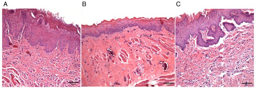
MOLECULAR AND CLINICAL ONCOLOGY 14: 127, 2021                                                                    5

Figure 5. Photomicrographs of histological sections of oral mucosa samples from Wistar rats subjected to chemotherapy. (A) Pentoxifylline group (day 8)
with total epithelial loss of oral wounds with severe inflammatory infiltrate. Necrotic areas were occasionally observed. (B) Pentoxifylline group (day 11)
continued total epithelial loss of oral wounds with severe inflammatory infiltrate and decreased number of blood vessels. (C) Pentoxifylline group (day 15),
mild re‑epithelialization associated with mild and diffuse inflammatory infiltrate. Increased vascular sections. H&E staining. Magnification, x100.

Figure 6. Photomicrographs of histological sections of oral mucosa samples from Wistar rats subjected to chemotherapy. (A) Atorvastatin group (day 8), no
epithelial dissolution, with preserved thin lining and mild diffuse inflammatory infiltrate. (B) Atorvastatin group (day 11), with progressive increase of inflam‑
matory infiltrate, but with epithelial preservation and reduced number of vascular sections. (C) Atorvastatin group (day 15), with recovered number of vascular
sections and increased epithelial thickness. H&E staining. Magnification, x100.

Figure 7. Number of inflammatory cells per group on different days of the          Figure 8. Number of vascular sections per group on different days of the
experiment. White column, saline; grey column, trans‑cariophyllene; hori‑          experiment. White column, saline; grey column, trans‑cariophyllene; hori‑
zontal lines column, pentoxifylline; black column, atorvastatin. *P
6 DA CRUZ CAMPOS et al: ORAL MUCOSITIS IN WISTAR RATS
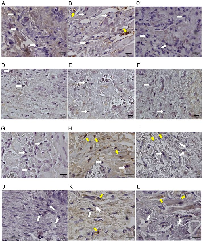
Figure 9. Photomicrographs of histological sections of oral mucosa samples from Wistar rats subjected to chemotherapy. (A) Animal in the control group,
without medication, euthanized on day 8. (B) Animal in the control group, without medication, euthanized on day 11. (C) Animal in the control group,
without medication, euthanized on the 15th day. (D) Animal that received trans‑karyophylene, euthanized on day 8. (E) Animal that received euthanized
trans‑karyophylene on day 11. (F) Animal that received trans‑karyophylene, euthanized on the 15th day. (G) Animal that received pentoxifylline, euthanized
on day 8. (H) Animal that received pentoxifylline, euthanized on day 11. (I) Animal that received pentoxifylline, euthanized on day 15. (J) Animal that received
atorvastatin, euthanized on the 8th day. (K) Animal that received atorvastatin, euthanized on the 11th day. (L) Animal that received atorvastatin, euthanized
on day 15. Magnification, x400. White arrows show negative cells and yellow arrows indicate positive cells. Anti‑TNF‑ α immunohistochemical reaction.
Discussion
Oral mucositis has been the focus of several experimental
and clinical studies because its control allows more effective
anti‑cancer radiation therapy and/or chemotherapy. However,
despite its importance, no control or preventive treatment for
oral mucositis has been established or filed so far (6,7,9,39‑44).
To this end, several experimental models have been developed
to investigate the mechanisms associated with the oral mucositis
development, and to assess the effects of different therapeutic
agents on the evolution of oral ulcers (1,8,9,20,22,30,35,45).
Figure 10. Comparison of TNF‑α levels on different days of the experiment. In this study, histopathological and histomorphometric
White column, saline; grey column, trans‑cariophyllene; horizontal lines analyses of oral mucosa samples demonstrated that treat‑
column, pentoxifylline; black column, atorvastatin. **PMOLECULAR AND CLINICAL ONCOLOGY 14: 127, 2021 7 Figure 11. Photomicrographs of histological sections of oral mucosa samples from Wistar rats subjected to chemotherapy. (A) Animal in the control group, without medication, euthanized on day 8. (B) Animal in the control group, without medication, euthanized on day 11. (C) Animal in the control group, without medication, euthanized on the 15th day. (D) Animal that received trans‑karyophylene, euthanized on day 8. (E) Animal that received euthanized trans‑karyophylene on day 11. (F) Animal that received trans‑karyophylene, euthanized on the 15th day. (G) Animal that received pentoxifylline, euthanized on day 8. (H) Animal that received pentoxifylline, euthanized on day 11. (I) Animal that received pentoxifylline, euthanized on day 15. (J) Animal that received atorvastatin, euthanized on the 8th day. (K) Animal that received atorvastatin, euthanized on the 11th day. (L) Animal that received atorvastatin, euthanized on day 15. Magnification, x400. White arrows indicate negative cells and yellow arrows indicate positive cells for the anti‑TGF‑β immunohistochemical reaction. Figure 12. Comparison of TNF‑α levels on different days of the experiment. Figure 13. Comparison of TGF‑β levels on different days of the experiment. White column, saline; grey column, trans‑cariophyllene; horizontal lines White column, saline; grey column, trans‑cariophyllene; horizontal lines column, pentoxifylline; black column, atorvastatin. **P
8                                           DA CRUZ CAMPOS et al: ORAL MUCOSITIS IN WISTAR RATS

                                                                                 that this cytokine does not influence exacerbation (Day 11)
                                                                                 of the ulcers directly. Other studies were performed using
                                                                                 trans‑caryophyllene, which focused on the immunomodula‑
                                                                                 tory effects of the drug (49‑51).
                                                                                     Wright et al (52) performed jejunal integrity studies in rats
                                                                                 using the herbal extract‑Iberogast‑ and concluded that this
                                                                                 plant partially improved the histopathological features of 5‑FU
                                                                                 induced mucositis, but conferred no significant protection.
                                                                                     In 2014, Cheah et al (53) investigated the effects of grape
                                                                                 seed extract in colon cancer and it demonstrated reduced
                                                                                 severity of intestinal mucositis in patients undergoing 5‑FU
                                                                                 chemotherapy.
Figure 14. Comparison of IFN‑γ levels on different days of the experiment.           In addition, preventive treatment with atorvastatin
White column, saline; grey column, trans‑cariophyllene; horizontal lines
column, pentoxifylline; black column, atorvastatin. *P
MOLECULAR AND CLINICAL ONCOLOGY 14: 127, 2021 9
MIDCC, CNC, BJVA and FMA designed the experiments. 16. Aarestrup B: In situ evaluation of mediators of the inflammatory
response and apoptosis process in chronic periodontitis in patients
MIDCC, BJVA and FMA performed pathologic anatomy. with AIDS. Niteroi, Universidade Federal Fluminense, 2006.
MIDCC and JOAC performed the immunohistochemistry 17. Baliga RS, Chaves AA, Jing L, Ayers LW and Bauer JA:
experiments. MIDCC and JOAC were involved in ELISA. AIDS‑related vasculopathy: Evidence for oxidative and inflam‑
matory pathways in murine and human AIDS. Am J Physiol
MIDCC, CNC, BJVA and FMA wrote the text. All authors Heart Circ Physiol 289: H1373‑H1380, 2005.
read and approved the final manuscript. 18. Duda A, Stange A, Lüftenegger D, Stanke N, Westphal D,
Pietschmann T, Eastman SW, Linial ML, Rethwilm A and
Lindemann D: Prototype foamy virus envelope glycoprotein leader
Ethics approval and consent to participate peptide processing is mediated by a Furin‑like cellular protease,
but cleavage is not essential for viral infectivity. J Virol 78:
The experimental research was carried out on rodents 13865‑13870, 2004.
19. Consolaro A: Cellular stress: Precedes and is present in inflam‑
(Rattus novergicus) having been approved by the Animal mation. In: Inflammation and Repair. Dental Press, Maringá,
Research Ethics Committee of the Federal University of Juiz 2009.
de Fora under number 062/2011. All stages of the research were 20. Azevedo IM, Kumakura HS, Alloufa SL, Mourão TS, Souza PM,
Carvalho MDF, Medeiros VB, Araújo‑Filho I, Rêgo ACM and
explained in the proper form and, after being verified by three Medeiros AC: Effect of simvastatin in attenuation of mucositis
evaluators, it was approved without changes or considerations. induced by methotrexate in rats. J Surg Clin Res 1: 22‑32, 2010.
21. Flores D and Lisart R: Effectiveness of palifermin in the preven‑
tion of oral mucositis in patients with haematological cancers.
Patient consent for publication Farm Hosp 34: 163‑169, 2010 (In Spanish).
22. Spielberger R, Stiff P, Bensinger W, Gentile T, Weisdorf D,
Not applicable. Kewalramani T, Shea T, Yanovich S, Hansen K, Noga S, et al:
Palifermin for oral mucositis after intensive therapy for hemato‑
logic cancers. N Engl J Med 351: 2590‑2598, 2004.
Competing interests 23. Schmidt‑Choudhury A, Furuta GT, Lavigne JA, Galli SJ and
Wershil BK: The regulation of tumor necrosis factor‑alpha
production in murine mast cells: Pentoxifylline or dexametha‑
The authors declare that they have no competing interests. sone inhibits IgE‑dependent production of TNF‑alpha by distinct
mechanisms. Cell Immunol 171: 140‑1446, 1996.
References 24. Raber‑Durlacher JE, von Bültzingslöwen I, Logan RM, Bowen J,
Al‑Azri AR, Everaus H, Gerber E, Gomez JG, Pettersson BG,
Soga Y, et al: Systematic review of cytokines and growth factors
1. Kostler WJ, Hejna M, Wenzel C and Zielinski CC: Oral mucositis for the management of oral mucositis in cancer patients. Support
complicating chemotherapy and/or radiotherapy: Options for Care Cancer 21: 343‑355, 2013.
prevention and treatment. CA Cancer J Clin 51: 290‑315, 2001. 25. Campos MI, Vieira WD, Campos CN, Aarestrup FM and
2. Sonis ST: Mucositis as a biological process: A new hypothesis for Aarestrup BJ: Atorvastatin and trans‑caryophyllene for the
the development of chemotherapy‑induced stomatotoxicity. Oral prevention of leukopenia in an experimental chemotherapy
Oncol 34: 39‑43, 1998. model in Wistar rats. Mol Clin Oncol 3: 825‑828, 2015.
3. Sonis ST: New thoughts on the initiation of mucositis. Oral 26. Basile AC, Ser tie JA, Freit as PC a nd Za n in i AC:
Dis 16: 597‑600, 2010. Anti‑inflammatory activity of oleoresin from Brazilian Copaifera.
4. Sonis ST: The pathobiology of mucositis. Nat Rev Cancer 4: J Ethnopharmacol 22: 101‑109, 1988.
277‑284, 2004. 27. Pereira FJ, Martins FT, Corrêa RS, Moreira ME, Costa A, Santos MH,
5. Sonis ST and Costello KA: A database for mucositis induced by Polo M and Barbosa LC: Isolation, chemical composition and
cancer chemotherapy. Eur J Cancer B Oral Oncol 31B: 258‑260, anti‑inflammatory activity of Copaifera langsdorffii Desf. fruit
1995. peels essential oil according to successive hydrodistillations. Acta
6. Stone R, Fliedner MC and Smiet AC: Management of oral Farm Bonaer 27: 369‑374, 2008.
mucositis in patients with cancer. Eur J Oncol Nurs 9 (Suppl 1): 28. Vilanova CM, Ribeiro SM, Machado RC, Vieira SM, Lima SG,
S24‑S32, 2005. Nunes PH and Martins MC: Evaluation of oil‑resin activity of
7. Trotti A, Bellm LA, Epstein JB, Frame D, Fuchs HJ, Gwede CK, Copaifera sp. On gastric emptying in Rattus novergicus. Emir J
Komaroff E, Nalysnyk L and Zilberberg MD: Mucositis inci‑ Food Agricult 25: 394‑397, 2013.
dence, severity and associated outcomes in patients with head 29. Siddiqui MA and Wellington K: Palifermin: In myelotoxic
and neck cancer receiving radiotherapy with or without chemo‑ therapy‑induced oral mucositis. Drugs 65: 2139‑2149, 2005.
therapy: A systematic literature review. Radiother Oncol 66: 30. Sonis ST: Efficacy of palifermin (keratinocyte growth factor‑1)
253‑262, 2003. in the amelioration of oral mucositis. Core Evid 4: 199‑205, 2009.
8. Trucci VM, Veeck EB and Morosolli ARC: Current strategies 31. Fernandes ES, Passos GF, Medeiros R, da Cunha FM, Ferreira J,
for the management of oral mucositis induced by radiotherapy or Campos MM, Pianowski LF and Calixto JB: Anti‑inflammatory
chemotherapy. Rev Odonto Cienc 24: 309‑314, 2009. effects of compounds alpha‑humulene and (‑)‑trans‑caryophyl‑
9. Lalla RV, Sonis ST and Peterson DE: Management of oral mucositis lene isolated from the essential oil of Cordia verbenacea. Eur
in patients who have cancer. Dent Clin North Am 52: 61‑77, 2008. J Pharmacol 569: 228‑236, 2007.
10. Peterson DE: New strategies for management of oral mucositis 32. Leandro LM, Vargas Fde S, Barbosa PC, Neves JK, da Silva JA
in cancer patients. J Support Oncol 4 (Suppl 1): S9‑S13, 2006. and da Veiga‑Junior VF: Chemistry and biological activi‑
11. Yeoh AS, Gibson RJ, Yeoh EE, Bowen JM, Stringer AM, ties of terpenoids from copaiba (Copaifera spp.) oleoresins.
Giam KA and Keefe DM: A novel animal model to investigate Molecules 17: 3866‑3889, 2012.
fractionated radiotherapy‑induced alimentary mucositis: The 33. Paiva LA, de Alencar Cunha KM, Santos FA, Gramosa NV,
role of apoptosis, p53, nuclear factor‑kappaB, COX‑1, and COX‑2. Silveira ER and Rao VS: Investigation on the wound healing
Mol Cancer Ther 6: 2319‑2327, 2007. activity of oleo‑resin from Copaifera langsdorffi in rats.
12. Parkin J and Cohen B: An overview of the immune system. Phytother Res 16: 737‑739, 2002.
Lancet 357: 1777‑1789, 2001. 34. Ward A and Clissold SP: Pentoxifylline. A review of its pharma‑
13. Goldman L and Ausiello D (eds): Principles of cancer treatment. codynamic and pharmacokinetic properties, and its therapeutic
In: Internal Medicine Treaty. Campus Elsevier, Rio de Janeiro, efficacy. Drugs 34: 50‑97, 1987.
2005. 35. Sonis ST, Tracey C, Shklar G, Jenson J and Florine D: An animal
14. Kolios G, Petoumenos C and Nakos A: Mediators of inflamma‑ model for mucositis induced by cancer chemotherapy. Oral Surg
tion: Production and implication in inflammatory bowel disease. Oral Med Oral Pathol 69: 437‑443, 1990.
Hepatogastroenterology 45: 1601‑1609, 1998. 36. Scully C, Epstein J and Sonis S: Oral mucositis: A challenging
15. Mitchell R, Kumar V, Abbas A and Fausto N: Robbins and complication of radiotherapy, chemotherapy, and radiochemo‑
Cotran Pathologic Basis of Diseases. 7th edition. Elsevier, therapy: Part 1, pathogenesis and prophylaxis of mucositis. Head
Rio de Janeiro, 2006. Neck 25: 1057‑1070, 2003.10 DA CRUZ CAMPOS et al: ORAL MUCOSITIS IN WISTAR RATS
37. Cai Y, Wang Z, Li J, Li N, Wei F and Liu Q: Evaluation of an 48. Allen R, Rapecki S and Higgs G: The role of IL‑10 in the inhi‑
indirect ELISA using recombinant granule antigen Gra7 for sero‑ bition of LPS‑mediated TNF release from human PBMCs by
diagnosis of Toxoplasma gondii infection in cats. J Parasitol 101: phosphodiesterase 4 (PDE4) inhibitors. Inflamm Res 46: 218,
37‑40, 2015. 1997.
38. Grisham MB, Jourd'Heuil D and Wink DA: Nitric oxide. I. 49. Dias DS, Fontes LB, Crotti AE, Aarestrup BJ, Aarestrup FM,
Physiological chemistry of nitric oxide and its metabolites: da Silva Filho AA and Corrêa JO: Copaiba oil suppresses
Implications in inflammation. Am J Physiol 276: G315‑G321, 1999. inflammatory cytokines in splenocytes of C57Bl/6 mice induced
39. Fekrazad R and Chiniforush N: Oral mucositis prevention and with experimental autoimmune encephalomyelitis (EAE).
management by therapeutic laser in head and neck cancers. Molecules 19: 12814‑12826, 2014.
J Lasers Med Sci 5: 1‑7, 2014. 50. Guo K, Mou X, Huang J, Xiong N and Li H: Trans‑caryophyllene
40. Herrstedt J: Prevention and management of mucositis in patients suppresses hypoxia‑induced neuroinflammatory responses by
with cancer. Int J Antimicrob Agents 16: 161‑163, 2000. inhibiting NF‑κ B activation in microglia. J Mol Neurosci 54:
41. Lara RN, da Guerra EN and de Melo NS: Macroscopic and 41‑48, 2014.
microscopic effects of GaAIAs diode laser and dexamethasone 51. Veiga Junior VF, Rosas EC, Carvalho MV, Henriques MG and
therapies on oral mucositis induced by fluorouracil in rats. Oral Pinto AC: Chemical composition and anti‑inflammatory activity
Health Prev Dent 5: 63‑71, 2007. of copaiba oils from Copaifera cearensis Huber ex Ducke,
42. Pico JL, Avila‑Garavito A and Naccache P: Mucositis: Its occur‑ Copaifera reticulata Ducke and Copaifera multijuga Hayne‑a
rence, consequences, and treatment in the oncology setting. comparative study. J Ethnopharmacol 112: 248‑254, 2007.
Oncologist 3: 446‑451, 1998. 52. Wright TH, Yazbeck R, Lymn KA, Whitford EJ, Cheah KY,
43. Silverman S Jr: Diagnosis and management of oral mucositis. Butler RN, Feinle‑Bisset C, Pilichiewicz AN, Mashtoub S and
J Support Oncol 5: 13‑21, 2007. Howarth GS: The herbal extract, Iberogast, improves jejunal
44. Wardill HR, Bowen JM and Gibson RJ: New pharmacotherapy integrity in rats with 5‑fluorouracil (5‑FU)‑induced mucositis.
options for chemotherapy‑induced alimentary mucositis. Expert Cancer Biol Ther 8: 923‑929, 2009.
Opin Biol Ther 14: 347‑354, 2014. 53. Cheah KY, Howarth GS and Bastian SE: Grape seed extract
45. Vanderhoof JA, Park JH, Mohammadpour H and Blackwood D: dose‑responsively decreases disease severity in a rat model of
Effects of dietary lipids on recovery from mucosal injury. mucositis; concomitantly enhancing chemotherapeutic effective‑
Gastroenterology 98: 1226‑1231, 1990. ness in colon cancer cells. PLoS One 9: e85184, 2014.
46. Lima V, Vidal FD, Rocha FA, Brito GA and Ribeiro RA: Effects
of tumor necrosis factor‑alpha inhibitors pentoxifylline and
thalidomide on alveolar bone loss in short‑term experimental
periodontal disease in rats. J Periodontol 75: 162‑168, 2004. This work is licensed under a Creative Commons
47. Jain P, Keservani R and Dahima R: In‑vivo characterization Attribution-NonCommercial-NoDerivatives 4.0
of hydrogel for treatment of chemo‑radiotherapy induced oral International (CC BY-NC-ND 4.0) License.
mucositis. Pharmacol Online 1: 1016‑1025, 2010.You can also read