Association of Antibody-Dependent Neutrophil Phagocytosis With Distinct Antibody Glycosylation Profiles Following Typhoid Vaccination
←
→
Page content transcription
If your browser does not render page correctly, please read the page content below
ORIGINAL RESEARCH
                                                                                                                                           published: 16 September 2021
                                                                                                                                           doi: 10.3389/fitd.2021.742804

                                            Association of Antibody-Dependent
                                            Neutrophil Phagocytosis With
                                            Distinct Antibody Glycosylation
                           Edited by:
            Sushant Sahastrabuddhe,
                                            Profiles Following Typhoid
        International Vaccine Institute,
                          South Korea
                                            Vaccination
                     Reviewed by:
                                            Mari Johnson 1,2*, Lisa Stockdale 1,2†, Noortje de Haan 3, Manfred Wuhrer 3, Jan Nouta 3,
              Malcolm Scott Duthie,
          HDT Biotech Corporation,
                                            Carolien A. M. Koeleman 3, Jenny Clarke 1,2, Spyridoula Marinou 1,2, Mila Shakya 4,
                      United States         Rachel Colin-Jones 1,2, Katherine Theiss-Nyland 1,2, Merryn Voysey 1,2, Celina Jin 1,2,
                     Yun Shan Goh,          Dikshya Pant 4, Elizabeth Jones 1,2, Sarah Kelly 1,2, Sabina Dongol 4, Abhilasha Karkey 4,
     Singapore Immunology Network           Shrijana Shrestha 4, Buddha Basnyat 5, Jennifer Hill 1,2‡ and Andrew J. Pollard 1,2‡
                (A*STAR), Singapore
                                            1 Oxford Vaccine Group, Department of Paediatrics, University of Oxford, Oxford, United Kingdom, 2 National Institute for
                 *Correspondence:
                                            Health Research (NIHR) Oxford Biomedical Research Centre, Oxford, United Kingdom, 3 Center for Proteomics and
                      Mari Johnson
                                            Metabolomics, Leiden University Medical Center, Leiden, Netherlands, 4 Oxford University Clinical Research Unit, Patan
          mari.johnson@lmh.ox.ac.uk
                                            Academy of Health Sciences, Kathmandu, Nepal, 5 Centre for Tropical Medicine and Global Health, University of Oxford,
                           †
                             ORCID:         Oxford, United Kingdom
                      Lisa Stockdale
     orcid.org/0000-0002-2576-8783
         ‡
                                            Typhoid Vi-conjugate vaccines (Vi-TCV) have been developed to control typhoid fever in
          These authors share senior
                         authorship         children in endemic regions. Previously, in a human challenge model of typhoid, Vi-TCV
                                            was administered prior to deliberate ingestion of Salmonella Typhi by healthy adult
                  Specialty section:        volunteers in the UK. Vi-specific antibody-dependent neutrophil phagocytosis (ADNP)
        This article was submitted to
      Vaccines for Tropical Diseases,       was associated with protection against enteric fever in this model, but it is not known if
              a section of the journal      ADNP is induced by vaccination of children. We measured ADNP in a cohort of Nepalese
       Frontiers in Tropical Diseases
                                            children receiving a Vi-TCV in a field study to investigate whether functional antibody
            Received: 16 July 2021
        Accepted: 31 August 2021
                                            responses were also present in children in an endemic setting. Furthermore, we
     Published: 16 September 2021           investigated relationships between the functional antibody measures and other
                             Citation:      properties of the antibody response, including Vi-IgG and IgA titres, and Fc region
Johnson M, Stockdale L, de Haan N,          glycosylation. Antibody-dependent neutrophil phagocytosis significantly increased in
Wuhrer M, Nouta J, Koeleman CAM,
     Clarke J, Marinou S, Shakya M,         children aged 9 months to 15 years between the day of vaccination and 28 days
    Colin-Jones R, Theiss-Nyland K,         following administration of Vi-TCV (D28). The magnitude of ADNP was also comparable
  Voysey M, Jin C, Pant D, Jones E,
        Kelly S, Dongol S, Karkey A,
                                            with the levels of ADNP induced by plasma from vaccinated UK adults. Neither IgG nor IgA
   Shrestha S, Basnyat B, Hill J and        antibody titres significantly correlated with ADNP scores at D28; however, increased
    Pollard AJ (2021) Association of        vaccine-induced ADNP was associated with decreased levels of IgG1 sialylation. These
     Antibody-Dependent Neutrophil
 Phagocytosis With Distinct Antibody        data suggest that vaccination with Vi-TCV produces functional antibody responses in
     Glycosylation Profiles Following        children, which associate with specific glycosylation patterns of the Fc region.
                 Typhoid Vaccination.
          Front. Trop. Dis. 2:742804.       Keywords: vaccination, typhoid fever, glycosylation, Fc-mediated effector function, correlates of protection,
      doi: 10.3389/fitd.2021.742804          antibody-dependent neutrophil phagocytosis, Salmonella

Frontiers in Tropical Diseases | www.frontiersin.org                                1                                       September 2021 | Volume 2 | Article 742804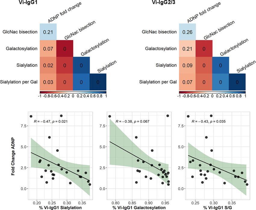
Johnson et al.                                                                                                         ANDP in Typhoid Vaccination

INTRODUCTION                                                               example, afucosylated IgG has been shown to associate with
                                                                           severe COVID disease and increased macrophage inflammation
Typhoid fever is a gastrointestinal infection caused by the                (16, 17). Whilst the manipulation of Fc glycoforms to alter
bacterium Salmonella enterica serovar Typhi (S. Typhi), which              function has long been utilised in monoclonal therapeutics, the
invades through the gastrointestinal mucosa to cause systemic              implications of Fc glycosylation following vaccination is only just
disease. Typhoid fever predominately affects children in low and           being investigated (18, 19). In 2016, Mahan et al. showed that
middle-income countries where access to clean drinking water is            HIV vaccination induced more inflammatory IgG glycoforms,
limited (1, 2). In 2017 there were an estimated 10.9 million cases         including agalactosylation and asialylation, that were associated
of typhoid fever resulting in approximately 116,000 deaths (3).            with higher antibody-dependent cellular cytotoxicity (ADCC)
Within Nepal, S. Typhi is the dominant cause of bloodstream                (20). However, it is not yet well understood how glycosylation
bacterial infections (4), and annual incidence has been estimated          influences ADNP in either infection or vaccine induced responses.
up to 449 cases per 100,000 (95% CI, 383 to 521) (3). Antibiotic               In this study we measure the capacity of vaccine induced
resistance for S. Typhi is on the rise in several South Asian              antibodies to mediate neutrophil phagocytosis in a subset of
countries, with nearly 65% of S. Typhi strains in Pakistan being           children aged 9 months - 15 years of age in the Phase 3 TyVAC
reported as extensively drug-resistant (XDR) between 2012 and              Nepal Study who are to be targeted for Vi-TCV vaccination (10).
2018 (5, 6). Given the high prevalence of typhoid fever, and               In addition, we compare antibody driven neutrophil responses in
increasing antimicrobial resistance, effective vaccination                 this cohort with those adults vaccinated in the UK to see if there
campaigns are needed now more than ever (7, 8). In 2018, a                 are differences in ADNP post-vaccination between populations.
Vi-conjugate vaccine (Vi-TCV), which targets the Vi                        Furthermore, in order to understand factors which may influence
polysaccharide capsule of S. Typhi, was approved by the                    ADNP, we examine the relationships between participant age, Vi-
WHO, and recently demonstrated clinical efficacy of 81.6% in                antibody titres and Fc antibody glycosylation, and ADNP.
a field study involving over 20,000 Nepalese children (9, 10).
    If correlates of protection can be identified, smaller
immunogenicity cohorts can be used to bridge likely vaccine                MATERIALS AND METHODS
efficacy to new populations, bypassing larger scale efficacy studies
and aiding new product development and vaccine deployment                  Study Design
(11). In a human challenge model of typhoid fever, potential               Vi-TCV Vaccination in Nepalese Children
correlates of protection were assessed, including; Vi-antibody             To examine the efficacy of the Vi-TCV vaccine amongst
levels, cellular responses to vaccination, antibody-antigen                populations endemic for typhoid fever, children aged 9
binding, and Fc-mediated (fragment crystallisable) antibody                months-16 years were vaccinated with Vi-TCV (Typbar-TCV,
effector functions (12, 13). A higher fold change in antibody-             Bharat Biotech) in a Phase 3, double-blind, randomized
dependent phagocytosis (ADNP) was measured in participants                 controlled trial. For full details of the study see Shakya et al.
who appeared protected from typhoid fever when compared with               (CTR: ISRCTN43385161) (10). Vaccinations took place in
diagnosed individuals, from baseline to 28 and 114 days post-Vi-           Lalitpur Metropolitan City, Kathmandu Valley, Nepal between
TCV vaccination (p=0.06, and p=0.047 respectively) (11). An                the 20th of November 2017 and the 9th of April 2018. Children
increase in the neutrophil oxidative burst, which occurs                   enrolled in the study were randomised in a 1:1 ratio to receive
downstream of phagocytosis, was also significantly higher in                either the Vi-TCV or control meningitis vaccine (MenAfriVac,
protected individuals at D28 (p=0.014), suggesting that                    Serum Institute of India PVT Ltd). Blood sampling was
antibody-induced neutrophil function may contribute to                     performed at day 0 (pre-vaccination), and 28 days following
vaccine-mediated protection.                                               vaccination for participants in the immunogenicity cohort. For
    Fc-mediated effector functions, such as ADNP, are                      our study we randomly selected 77 participants from the
determined in part by antibody subclass and isotype, which                 immunogenicity cohort who received a Vi-TCV vaccine. These
allows the selective binding of antibodies to Fc receptors. The            participants also included 24 children who had glycosylation
Fc region is modified co- and post-translationally by the                   profiles measured who had previously been randomly selected
glycosylation pathway which induces a large structural and                 for analysis.
functional diversification of the proteome. For IgG antibodies,
N-linked glycosylation occurs on the conserved asparagine                  Vi-TCV Vaccination in UK Adults
located in position 297 of the constant heavy domain (14). IgG             Healthy UK-based adults aged between 18 and 60 years with no
glycosylation gives rise to a diverse range of possible IgG                history of typhoid fever or vaccination, were given a single dose of
glycoforms, including diantennary glycans with varying levels              either a Vi tetanus conjugate vaccine (Typbar-TCV, Bharat
of galactosylation, fucosylation, sialylation and bisecting N-             Biotech), Vi-PS vaccine (Typhim Vi; Sanofi Pasteur), or a control
acetylglucosamine (GlcNAc).                                                (MenACWY) vaccine (MENVEO; GlaxoSmithKline), randomised
    Through conformational changes, Fc glycosylation can                   1:1:1 in a double-blind trial as previously described (CTR:
influence the binding strength of antibodies to Fc receptors,               NCT02324751) (21). In order to assess efficacy of the Vi-TCV
and thus the magnitude of downstream cellular activity (13, 14).           vaccine, 28 days after vaccination, participants were challenged with
Generally, a loss of galactose, sialic acid and fucose is associated       a dose of 1-5 x 10⁴ CFU of S. Typhi in bicarbonate solution and
with increased inflammation and disease severity (15). For                  monitored for a diagnosis of typhoid fever.

Frontiers in Tropical Diseases | www.frontiersin.org                   2                                September 2021 | Volume 2 | Article 742804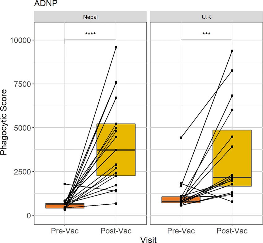
Johnson et al.                                                                                                    ANDP in Typhoid Vaccination

    Participants were diagnosed with typhoid infection on               international Standard Vi polysaccharide of C. freundii NIBSC
presentation with a continuous fever of 38°C or higher for 12           code: 12/244) at 1.25 μg/ml in 0.5X PBS. After washing three
h or longer, or by positive S. Typhi blood culture within 14 days       times with 200 μl 0.5X PBS, 200 μl of 1:20 diluted (in 0.5X PBS)
after challenge. Blood samples were collected for serum and             plasma was incubated for 1 hour at room temperature and then
plasma separation at day 0 (pre-vaccination) and 28 days                the sample transferred to another Vi-coated blank well for
following vaccination (D28), before S. Typhi challenge. Thirty-         another 1 hour incubation. After each incubation, wells were
seven participants received the Vi-conjugate vaccine. For the           washed once with 200 μl 0.5X PBS and three times with 200 μl
ADNP assay in our study, we randomly selected plasma from a             freshly made 25 mM ammonium bicarbonate (Sigma). Following
subset of 16 Vi-TCV vaccinated individuals who were not                 the washes, 50 μl of 100 mM formic acid (Sigma) was added and
diagnosed with typhoid fever during the study.                          incubated at room temperature for 5 min with agitation. After
                                                                        transferring the 50 μl eluate to a clean V-bottom 96-well plate,
Vi-IgG and IgA ELISA                                                    another 50 μl 100 mM formic acid was added, incubated for
Anti-Vi IgG titers were measured using a commercial vaccine             5 min with agitation and the 2 eluates from the same sample
enzyme-linked immunosorbent assay (ELISA) kit (VaccZyme,                were pooled.
Binding Site) according to the manufacturer’s instructions. Anti-           Samples were dried using a vacuum centrifuge for 2-3 hours at
IgA titres were measured using a modification to the same                50/60°C, and shipped to LUMC for processing as below for total
commercially available kit, replacing the Typhi Vi-IgG                  IgG. Briefly, samples were resolubilised in 20 ml 50 mM
conjugate with a Vi-IgA conjugate. ELISA titres were measured           ammonium bicarbonate (Sigma) while shaking for 5 min.
in plasma obtained on day 0 (pre-vaccination), and 28 days post-        Twenty ml of 0.05 mg/ml tosyl phenylalanyl chloromethyl ketone
vaccination and the data has been published previously (21, 22).        (TPCK)-treated trypsin (Sigma) in ice-cold purified water was
                                                                        added per well, and the samples were incubated at 37°C overnight.
Bead-Coupling to Vi-Antigen
2.5 μl of biotinylated 1 mg/ml Vi-antigen (NIBSC) was added to          Total IgG Glycopeptide Analysis
10 μl of red fluorescent NeutrAvidin™-Labelled microspheres              For IgG purification, 2 ml of Protein G Sepharose 4 Fast Flow
(Thermofisher) per plate and incubated overnight at 4°C, or for          beads (GE Healthcare) were added per well on an Orochem filter
two hours at 37°C. Prior to the assay, the beads are washed twice       plate and washed three times with PBS. 20 ml of 1:20 diluted
with 0.1%, and 5% BSA, before being resuspended in 1 ml of              plasma (in PBS) was added to each well. Plates were incubated
RPMI per plate.                                                         for 1 hour with shaking at 750rpm. Using a vacuum manifold,
                                                                        the samples were washed three times by adding 400 ml PBS,
Antibody Dependent Neutrophil                                           followed by three times 400 ml of purified water. Antibodies were
Phagocytosis Assays                                                     eluted from the beads by addition of 100 ml of 100 mM formic
Plasma samples were first heat inactivated at 56°C for 30 min.           acid (Sigma) incubating for 5 min with shaking, and centrifuging
Plasma was diluted in RPMI at 1:320 for assessment of antibody-         at 100 × g for 2 min. Samples were dried for 2 hours at 60°C in a
mediated neutrophil phagocytosis. Samples were then incubated           vacuum centrifuge before resolubilisation in 20 ml 50 mM
for 2 hours with Vi-coupled red fluorescent microspheres                 ammonium bicarbonate (Sigma) while shaking for 5 min.
(Thermofisher) in 96-well plates.                                        Twenty ml of 0.05 mg/ml tosyl phenylalanyl chloromethyl
   Leukocytes, including neutrophils were isolated from sodium          ketone (TPCK)-treated trypsin (Sigma) in ice-cold purified
heparin treated blood from healthy anonymised UK donors                 water was added per well, incubated at 37°C overnight, and
using ammonium-chloride-potassium (ACK) lysis buffer                    stored at -20°C until MS analysis.
(Thermofisher). 5 x 10⁴ leukocytes were added to each well of
antibody-opsonised beads. Cells were pelleted and stained with          NanoLC-ESI-QTOF-MS Analysis
APC-Cy7 anti-CD14 (Clone MyP9, BD Biosciences, Franklin                 Samples were thawed and thoroughly mixed. Two hundred nl of
Lakes, USA), Alexa Fluor 700 anti-CD3 (Clone UCHT1700, BD               total IgG or 5 ml of Vi-specific IgG glycopeptide samples were
Biosciences, Franklin Lakes, USA) and Pacific Blue anti-CD66b            injected into an Ultimate 3000 RSLCnano system (Dionex/
(Clone G10F5, Biolegend, San Diego, USA).                               Thermo Scientific, Breda, the Netherlands) coupled to a
   Leukocytes were washed and analysed using a BD                       quadrupole-TOF-MS (MaXis HD; Bruker Daltonics, Bremen,
LSRFortessa X-20 for flow cytometry. FlowJo v10.6 software               Germany). The LC system was equipped with an Acclaim
was used for gating cell populations. The phagocytic score was          PepMap 100 trap column (particle size 5 mm, pore size 100 Å,
calculated as the mean fluorescent intensity (MFI) of red beads          100 mm × 20 mm, Dionex/Thermo Scientific) and an Acclaim
within gated neutrophils (CD14, CD3 negative, CD66b positive),          PepMap C18 nano analytical column (particle size 2 mm, pore
multiplied by the percentage of bead-positive gated neutrophils.        size 100 Å, 75 mm × 150 mm, Dionex/Thermo Scientific).
                                                                        Glycopeptides were identified based on their m/z, retention
Vi-Specific IgG Capture and                                              time and literature (23).
Glycopeptide Analysis
Vi-specific IgG was captured using NeutrAvidin plates                    Statistical Analysis
(Thermofisher), coated for 2 hours at room temperature with              Data from this study was analysed using R version 4.00.
100 μl biotinylated Vi (commercially biotinylated WHO                   Phagocytosis data were first tested for normality using

Frontiers in Tropical Diseases | www.frontiersin.org                3                              September 2021 | Volume 2 | Article 742804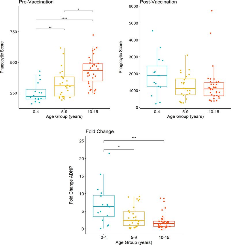
Johnson et al.                                                                                                                           ANDP in Typhoid Vaccination

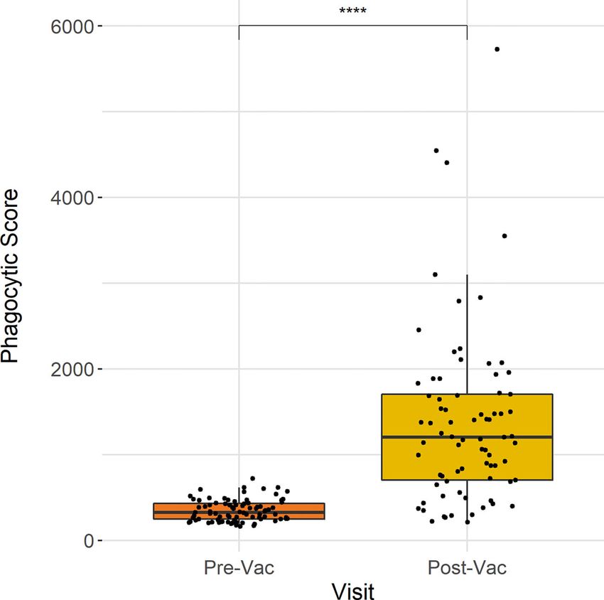
Shapiro-Wilk normality test. The Wilcoxon paired test used for                               neutrophil phagocytosis at baseline and 28 days post-vaccination
comparisons between non-parametric groups, including                                         (n = 77). These data show that ADNP significantly increased 28
comparisons between time points and the Wilcoxon test                                        days following Vi-conjugate vaccination (p
Johnson et al.                                                                                                                           ANDP in Typhoid Vaccination

 FIGURE 2 | Comparison of ADNP between Nepalese and UK vaccines. Anti-Vi ADNP was measured pre, and 28 days post Vi-TCV vaccination in participants from
 the TyVAC Nepal study (n =17) and adults vaccinated in the UK (n = 16). The phagocytic scores were calculated from bead+ neutrophils, multiplied by the MFI of the
 red fluorescent beads. Samples were run on the same day and in technical duplicate. Presented are the mean phagocytic score per participant, pre and 28-days
 post vaccination. Box plot shows the median and interquartile ranges for each time point, and the Wilcoxon paired test was used to determine statistical significance
 between time points, **** = (p < 0.0001), *** = (p < 0.001).

children when compared with UK adults (Figure 2). In addition,                        prior to vaccination, ADNP activity was higher along with
whilst there were higher baseline ADNP levels in the UK adults                        increasing participant age (Figure 3A); the oldest children, 10-
compared with Nepalese children (p=0.02), the fold changes                            15 years of age, had significantly higher levels of ADNP when
between baseline ADNP and D28 were not significantly different                         compared with children aged 5-9 (p
Johnson et al.                                                                                                                           ANDP in Typhoid Vaccination

                                 A                                                    B

                                                            C

 FIGURE 3 | Differences in ADNP by age of vaccinated children in Nepal. (A, B) Participants were separated into groups aged 0-4 (n= 21), 5-9 (n= 24) and 10-15
 years of age (n= 32). Phagocytic scores were then calculated from bead+ neutrophils, multiplied by the MFI of the red fluorescent beads, at pre and post-vaccination
 time points, respectively. Box plots display the median and interquartile range for each age group, and differences in anti-Vi ADNP responses between age groups
 were compared using the Wilcoxon test, **** = (p < 0.0001), *** = (p < 0.001). ** = (p < 0.01) * = (p < 0.05). Samples were run in technical duplicate and displayed
 are the mean phagocytic scores (C) The fold change in anti-Vi ADNP calculated for children in each age group following Vi-TCV vaccination. Fold change was
 calculated using the mean phagocytic scores pre and 28-days post Vi-TCV vaccination.

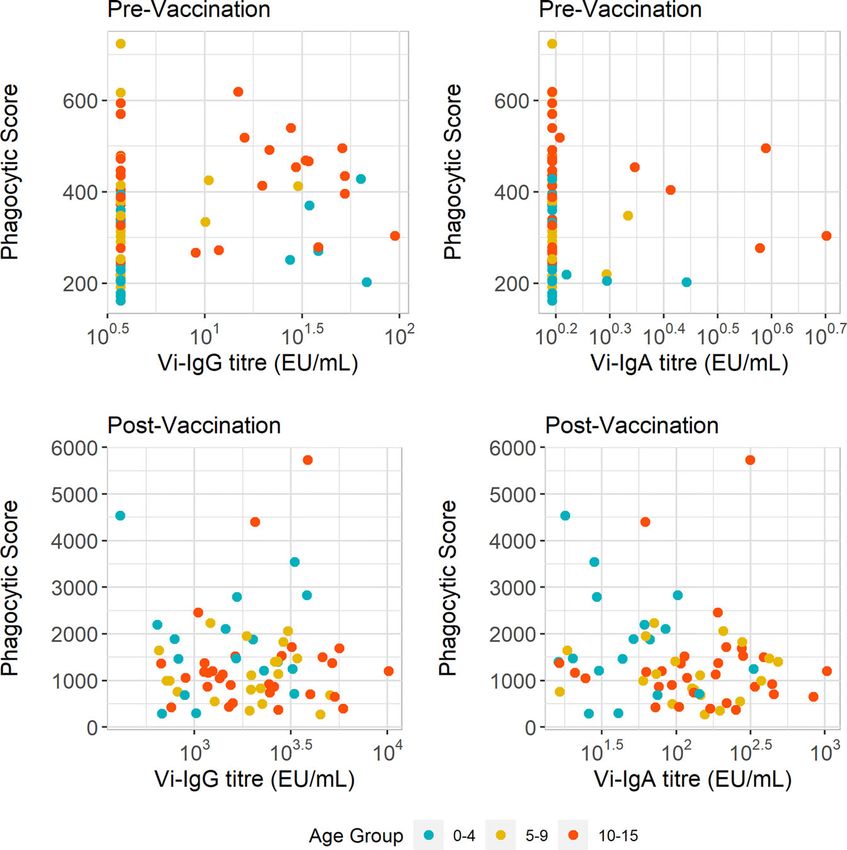
Vi-IgG titres and ADNP responses pre-vaccination was weak                                 Vi-Specific Antibody Fc Glycosylation
(R =0.24, p=0.044) (Figure 4A). Additionally we did not observe                           Associates With ADNP Following
a significant difference in baseline Vi-IgA titres between children as                     Vaccination
they aged in our cohort of children (Supplementary Figure 2B). No                         Glycosylation profiles including galactosylation, sialyation and
correlation was observed between baseline ADNP and Vi-IgA titres                          fucosylation, were measured in a randomly selected subset (n
(R =-0.075, p=0.54) (Figure 4B).                                                          =24) of plasma samples from Nepalese children aged 9 months –
    Following vaccination, we investigated the relationship                               15 years. Due to the similarities in amino acid sequence in the Fc
between Vi-antibody titre and ADNP. Twenty eight days after                               region proximal to the asparagine at position 297, IgG2 and IgG3
vaccination there is both an increase in Vi-antibody titres and                           cannot be distinguished from each other, and were therefore
phagocytosis scores (Supplementary Figure 3). Vi-IgG titre also                           recorded together. LC-MS was used to measure Fc glycopeptides,
increased with age (Supplementary Figure 2D). Yet as with pre-                            providing IgG subclass-specific glycosylation profiles for both Vi-
vaccination ADNP, there was no correlation between ADNP                                   specific antibodies and the total IgG antibody compartment (26).
and Vi-IgG (R =0.018, p=0.89) and Vi-IgA (R =0.17, p=0.16)                                   We first assessed the correlation between Fc glycan traits of
antibody titres (Figures 4C, D). Therefore, differences in ADNP                           Vi-specific IgG1 and Vi-IgG2/3 at D28 with ADNP fold change
magnitude at baseline and following vaccination are not solely                            from baseline (Figures 5A, B). We observed that lower
due to antibody quantity and may be driven by other changes in                            percentages of sialylated Vi-IgG1 antibodies correlated with
antibody quality such as Fc receptor binding or modulation by                             higher levels of ADNP induced at D28 from baseline (R =
Fc glycosylation.                                                                         0.47, p=0.021), (Figure 5C). A lower percentage of terminal

Frontiers in Tropical Diseases | www.frontiersin.org                              6                                     September 2021 | Volume 2 | Article 742804
Johnson et al. ANDP in Typhoid Vaccination
A B
C D
FIGURE 4 | ADNP does not correlate with Vi-IgG or Vi-IgA titre. (A, B) Scatterplot displaying anti-Vi phagocytic scores with Vi-IgG, and Vi-IgA titres at baseline
(pre-vaccination), and 28 days post-vaccination. (C, D) Scatterplot displaying anti-Vi phagocytic scores and Vi-IgG and Vi-IgA and titres 28 days post-vaccination,
respectively. The phagocytic scores were calculated from bead+ neutrophils, multiplied by the MFI of the red fluorescent beads. Samples were run in technical
duplicates and presented are the mean phagocytic scores and ELISA units (EU/ml) per participant (n = 77).
galactose residues, on the Fc domain of Vi-IgG1 28 days post- we investigated if sialyation and galactosylation of Vi-specific
vaccination, also correlated with ADNP at D28 (R = 0.38, antibodies correlated. We observed a strong correlation between
p=0.067) (Figure 5D), however this was not statistically the percentage of galactosylation and sialyation of Vi-IgG1
significant. Moreover, when breaking down galactosylation levels (pJohnson et al. ANDP in Typhoid Vaccination
A B
C D E
FIGURE 5 | Correlations of Vi-IgG glycosylation states with ADNP fold change. Spearman’s correlation of the percentage of Vi-IgG1 (A), and Vi-IgG2/3 (B)
sialylation, galactosylation, and GlcNAc bisection at day 28, with the fold change in ADNP between baseline and D28, as well as the participant age (n =24). The
colour of the square indicates the strength of the correlation (Rho) and the p-value is noted within each box. Correlation of the percentage of Vi-IgG1 sialylation (C),
galactosylation (D), and sialic acid per galactose residues (S/G) (E) at day 28, respectively, with the fold change in ADNP between baseline and D28 (n =24). Shown
for each graph are the Spearman’s Rho and p-values for significance. In addition, the black line indicates the linear regression, and the shaded area shows 95%
confidence intervals for the model.
(Supplementary Figure 5). We also examined if age was a factor in Encouragingly, 28 days after vaccination, ADNP activity
driving glycosylation states at D28 and associations with ADNP, as measured in plasma from these children was present at similar
glycosylation states at baseline have been known to vary with the age levels to ADNP activity measured in plasma from UK adults. In
of children and adolescents (28). Therefore, we tested for addition, we found that lower abundances of sialic acid of Vi-
interactions between age and Vi-IgG1 galactosylation and specific IgG1 correlated with higher levels of ADNP 28 days
sialylation in a linear model to predict ADNP fold change. This following immunisation.
model showed that the interaction between the two glycosylation Antibody-induced neutrophil effector functions provide
profiles with age, respectively, was non-significant (p=0.95, p=0.64). important immune responses against S. Typhimurium infections,
localizing Salmonellae to the gut (29). Conversely, during infection
with S. Typhi, the Vi capsule enables the bacterium to evade
DISCUSSION antibody-mediated neutrophil responses (30, 31), and in vitro
models of S. Typhi have shown its ability to evade the oxidative
The capacity of antibodies to induce neutrophil activity, burst and phagosome maturation within monocytes and
including phagocytosis, has been shown to associate with neutrophils after phagocytosis (32, 33), ultimately resulting in
protection following Vi-TCV vaccination prior to S. Typhi bacterial dissemination. Vaccination with Vi-TCV however
exposure in a UK based human challenge model (21). Our induces gut homing plasma cells that produce Vi-specific
findings demonstrate that Vi-TCV vaccination also gives rise antibodies (34). Given the increase in ADNP following
to a significant increase in capacity for Vi-specific antibody- vaccination, these antibodies may have a role in activating Fc
dependent neutrophil phagocytosis amongst Nepalese children receptors on neutrophils to bring about phagocytosis, thus
aged 9 months - 15 years. We found that the level of ADNP overcoming the natural ability of S. Typhi to evade neutrophil-
activity reached after vaccination was unaffected by age, despite mediated responses. Consequently, ADNP may be playing a
differences in pre-vaccination Vi-IgG titres and ADNP levels. mechanistic role in vaccine-mediated protection.
Frontiers in Tropical Diseases | www.frontiersin.org 8 September 2021 | Volume 2 | Article 742804Johnson et al. ANDP in Typhoid Vaccination
In order to better understand which antibody isotypes may be future studies investigating the role of IgA Fc glycosylation and
driving neutrophil phagocytosis, we correlated Vi-IgA and Vi- downstream effector functions post-vaccination could help
IgG titres with ADNP. Previous analyses demonstrated that Vi- understand the potential relationship between IgA and ADNP
IgA antibody levels were associated with vaccine-mediated further. Our study also used neutrophils from healthy adults in
protection (13). Neutrophils express the highest levels of the the UK. Neutrophil function in target vaccine populations may
IgA receptor, CD89, of any phagocytic immune cell type (35, 36) differ, as neutropenia, for example, may be more common in
and IgA has also been shown to be the primary isotype African and South Asian populations where HIV prevalence is
responsible for monoclonal-induced phagocytosis of tumour high (53). Monitoring vaccine efficacy in these subgroups should
cells by neutrophils (37). In the context of SARS-CoV-2 therefore be considered when deploying the Vi-conjugate
infection, high levels of IgA and ADNP have both been vaccine in different regions.
associated with increased inflammation (38), highlighting the Due to the low abundance of Vi-antibodies at baseline, we did
potentially synergistic effect these pathways may play in disease not examine changes in glycosylation between baseline and Day
as well as protection. It is therefore possible that Vi-IgA and 28 with ADNP in this study. Several studies have shown that
ADNP could be acting in tandem, with Vi-IgA mediating ADNP antigen-specific IgG Fc sialylation and galactosylation increases
following vaccination. Whilst there is an increase in ADNP from after vaccination (54, 55). Future work evaluating the impact of
baseline following vaccination, together with increasing Vi-IgA Vi-TCV vaccination on Fc glycosylation states from baseline will
and IgG titres, neither Vi-IgA nor Vi-IgG titres correlated therefore help understand the influence vaccination has on
strongly with post-vaccination or baseline anti-Vi ADNP this response.
responses in Nepalese children. In this study, we show that vaccination increases ADNP in
These data suggest that qualitative antibody properties such Nepalese children, and that ADNP associated with Vi-IgG1 Fc
as Fc glycosylation may be affecting the magnitude of antibody- sialyation measured at Day 28. Fc-mediated antibody functions
dependent neutrophil phagocytosis. We observed distinct Fc are increasingly recognised in evaluating vaccine immunity to
glycan characteristics of Vi-specific IgG1 antibodies measured many infections, including typhoid fever, HIV, and SARS-CoV-2
28 days following vaccination, which associated with an increase (40, 56). Furthermore, recent research has shown that the adjuvant
in ADNP post-vaccination, featuring a lower abundance of sialic and site of vaccine delivery can alter antibody glycosylation
acid on the Vi-IgG1 Fc domain. Furthermore, an increase in the profiles, and thus function (57). Decoding factors driving Fc-
number of sialic acid residues per galactose, also correlated with mediated effector functions could therefore provide pathways for
reducing ADNP levels. Sialylation of IgG Fc has been associated targeted vaccine design and implementation, including emerging
with reduced FcgRIIIa binding and ADCC function of IgG1 (39, typhoid and paratyphoid vaccine candidates (58).
40), and sialylation is also responsible for anti-inflammatory
effects of intravenous immunoglobulin treatments (41).
As well as IgG sialylation, antigen-specific Fc galactosylation has DATA AVAILABILITY STATEMENT
been demonstrated to influence Fc-mediated antibody effector
functions against pathogens such as HIV (42), and agalactosylation The raw data supporting the conclusions of this article will be
has been implicated in increasing inflammation within active made available by the authors, without undue reservation.
tuberculosis (43, 44). In this context we show that both Vi-IgG
sialylation, and, to a lesser extent, galactosylation levels associate with
ADNP activity after Vi-TCV vaccination. Removal of sialic acid and ETHICS STATEMENT
galactose on IgG Fc has been shown to induce a conformational
The TyVAC Nepal study was approved by the Oxford Tropical
change in the IgG Fc hinge region, resulting in antibody structures
Research Ethics Committee and the Nepal Health Research
that are more accessible to Fc receptors, and thus stronger binding of
Council. The VAST human challenge study was approved by
IgG to Fc receptors (45). Through greater cross-linking of Fc
the sponsor (University of Oxford), the South Central Oxford A
receptors, increased binding of immunoglobulin to Fc receptors has
Ethics Committee (14/SC/1427), and the Medicines and
been shown to impact responses downstream from ADNP in other
Healthcare Products Regulatory Agency (Eudract 2014-002978-
studies, including the oxidative burst (46, 47). We did not find
36). Written informed consent to participate in this study was
significant associations with Vi-IgG2 and 3 sialylation with ADNP at
provided by the participants’ legal guardian/next of kin.
Day 28, however glycosylation states displayed a similar relationship
as seen in with Vi-IgG1. IgG1 has the highest affinity for Fc g
receptors, therefore it is possible that this is a greater driver of ADNP AUTHOR CONTRIBUTIONS
compared with Vi-IgG2 and 3 (48).
While our study did not measure IgA Fc glycosylation, MJ, LS, NH, MW, JH, and AP conceived and designed the work.
reduced sialylation on IgA has been demonstrated to increase Methodology was developed by MJ, LS, JH, NH, MW, JN, CK.
Fc-mediated neutrophil and monocyte responses within CJ, EJ, JH, and AP carried out the VAST clinical trial. MS, RC-J,
inflammatory rheumatoid arthritis (49, 50). Given the role IgA KT-N, MV, DP, SK, SD, AK, SS, BB, and AP carried out the
plays in limiting S. Typhi infection, and its associations with TyVAC clinical trial. MJ, LS, NH, JN, CK, JC, SM, and EJ
vaccine mediated-protection and neutrophil responses (51, 52), acquired data for the study. MJ performed statistical analysis of the
Frontiers in Tropical Diseases | www.frontiersin.org 9 September 2021 | Volume 2 | Article 742804Johnson et al. ANDP in Typhoid Vaccination
study, and MV also provided statistical support and database SUPPLEMENTARY MATERIAL
management. MJ wrote the original draft of the manuscript. LS,
NH, MW, JN, MV, CJ, SS, JH, and AP edited and contributed to the The Supplementary Material for this article can be found online at:
manuscript. LS, JH, and AP provided project supervision. All authors https://www.frontiersin.org/articles/10.3389/fitd.2021.742804/
contributed to the article and approved the submitted version. full#supplementary-material
REFERENCES 18. Golay J, Da Roit F, Bologna L, Ferrara C, Leusen JH, Rambaldi A, et al.
Glycoengineered CD20 Antibody Obinutuzumab Activates Neutrophils and
1. Ochiai RL, Acosta CJ, Danovaro-Holliday MC, Baiqing D, Bhattacharya SK, Mediates Phagocytosis Through CD16B More Efficiently Than Rituximab.
Agtini MD, et al. A Study of Typhoid Fever in Five Asian Countries: Disease Blood (2013) 122:3482–91. doi: 10.1182/blood-2013-05-504043
Burden and Implications for Controls. Bull World Health Organ (2008) 19. Kuhns S, Shu J, Xiang C, De Guzman R, Zhang Q, Bretzlaff W, et al.
86:260–8. doi: 10.2471/BLT.06.039818 Differential Influence on Antibody Dependent Cellular Phagocytosis by
2. Antilló n M, Warren JL, Crawford FW, Weinberger DM, Kürüm E, Pak GD, Different Glycoforms on Therapeutic Monoclonal Antibodies. J Biotechnol
et al. The Burden of Typhoid Fever in Low- and Middle-Income Countries: (2020) 317:5–15. doi: 10.1016/j.jbiotec.2020.04.017
A Meta-Regression Approach. PloS Negl Trop Dis (2017) 11:e0005376. 20. Mahan AE, Jennewein MF, Suscovich T, Dionne K, Tedesco J, Chung AW.
doi: 10.1371/journal.pntd.0005376 Antigen-Specific Antibody Glycosylation Is Regulated via Vaccination. PLoS
3. Stanaway JD, Reiner RC, Blacker BF, Goldberg EM, Khalil IA, Troeger CE, Pathog (2016) 12(3):e1005456. doi: 10.1371/journal.ppat.1005456
et al. The Global Burden of Typhoid and Paratyphoid Fevers: A Systematic 21. Jin C, Gibani MM, Moore M, Juel HB, Jones E, Meiring J, et al. Efficacy and
Analysis for the Global Burden of Disease Study. Lancet Infect Dis (2019) 19 Immunogenicity of a Vi-Tetanus Toxoid Conjugate Vaccine in the Prevention
(4):369–81. doi: 10.1016/S1473-3099(18)30685-6 of Typhoid Fever Using a Controlled Human Infection Model of Salmonella
4. Zellweger RM, Basnyat B, Shrestha P, Prajapati KG, Dongol S, Sharma PK, Typhi: A Randomised Controlled, Phase 2b Trial. Lancet (2017) 390:2472–80.
et al. A 23-Year Retrospective Investigation of Salmonella Typhi and doi: 10.1016/S0140-6736(17)32149-9
Salmonella Paratyphi Isolated in a Tertiary Kathmandu Hospital. PloS Negl 22. Voysey M, Pollard AJ. Seroefficacy of VI Polysaccharide–Tetanus Toxoid
Trop Dis (2017) 11(11):e0006051. doi: 10.1371/journal.pntd.0006051 Typhoid Conjugate Vaccine (Typbar TCV). Clin Infect Dis (2018) 67:18–24.
5. Dyson ZA, Klemm EJ, Palmer S, Dougan G. Antibiotic Resistance and doi: 10.1093/cid/cix1145
Typhoid. Clin Infect Dis (2019) 68:S165–70. doi: 10.1093/cid/ciy1111 23. Falck D, Jansen BC, de Haan N, Wuhrer M. High-Throughput Analysis of Igg
6. Umair M, Siddiqui SA. Antibiotic Susceptibility Patterns of Salmonella Typhi Fc Glycopeptides by LC-MS. In: Methods in Molecular Biology, vol. 1503. New
and Salmonella Paratyphi in a Tertiary Care Hospital in Islamabad. Cureus York: Humana Press Inc. (2017). p. 31–47.
(2020) 12(9):e10228. doi: 10.7759/cureus.10228 24. Watson CH, Baker S, Lau CL, Rawalai K, Taufa M, Coriakula J, et al. A Cross-
7. Marchello CS, Birkhold M, Crump JA. Complications and Mortality of Sectional Seroepidemiological Survey of Typhoid Fever in Fiji. PloS Negl Trop
Typhoid Fever: A Global Systematic Review and Meta-Analysis. J Infect Dis (2017) 11(7):e0005786. doi: 10.1371/journal.pntd.0005786
(2020) 81:902–10. doi: 10.1016/j.jinf.2020.10.030 25. House D, et al. Antibodies to the Vi Capsule of Salmonella Typhi in the Serum
8. O’Reilly PJ, Pant D, Shakya M, Basnyat B, Pollard AJ. Progress in the Overall of Typhoid Patients and Healthy Control Subjects From a Typhoid Endemic
Understanding of Typhoid Fever: Implications for Vaccine Development. Region. J Infect Dev Ctries (2008) 2:308–12. doi: 10.3855/jidc.227
Expert Rev Vaccines (2020) 19:367–82. doi: 10.1080/14760584.2020.1750375 26. Chandler KB, Mehta N, Leon DR, Suscovich TJ, Alter G, Costello CE. Multi-
9. World Health Organization. Typhoid Vaccines: WHO Position Paper, March Isotype Glycoproteomic Characterization of Serum Antibody Heavy Chains
2018 – Recommendations. Vaccine (2019) 37:214–6. doi: 10.1016/ Reveals Isotype- and Subclass-Specific N-Glycosylation Profiles. Mol Cell
j.vaccine.2018.04.022 Proteomics (2019) 18:686–703. doi: 10.1074/mcp.RA118.001185
10. Shakya M, Colin-Jones R, Theiss-Nyland K, Voysey M, Pant D, Smith N, et al. 27. Van de Geijn FE, Wuhrer M, Selman MHJ, Willemsen SP, de Man YA,
Phase 3 Efficacy Analysis of a Typhoid Conjugate Vaccine Trial in Nepal. Deelder AM, et al. Immunoglobulin G Galactosylation and Sialylation Are
N Engl J Med (2019) 381:2209–18. doi: 10.1056/NEJMoa1905047 Associated With Pregnancy-Induced Improvement of Rheumatoid Arthritis
11. Blohmke CJ, O’Connor D, Pollard AJ. The Use of Systems Biology and and the Postpartum Flare: Results From a Large Prospective Cohort Study.
Immunological Big Data to Guide Vaccine Development. Genome Med (2015) Arthritis Res Ther (2009) 11:R193. doi: 10.1186/ar2892
7:114. doi: 10.1186/s13073-015-0236-1 28. de Haan N, Reiding KR, Driessen G, van der Burg M, Wuhrer M. Changes in
12. Jin C, Hill J, Gunn BM, Yu W-H, Dahora LC, Jones E, et al. Vi-Specific Healthy Human Igg Fc-Glycosylation After Birth and During Early Childhood.
Serological Correlates of Protection for Typhoid Fever. J Exp Med (2020) 218 J Proteome Res (2016) 15:1853–61. doi: 10.1021/acs.jproteome.6b00038
(2):e20201116. doi: 10.1084/jem.20201116 29. Broz P, Ohlson MB, Monack DM. Innate Immune Response to Salmonella
13. Dahora LC, Jin C, Spreng RL, Feely F, Mathura R, Seaton KE, et al. Iga and Typhimurium, a Model Enteric Pathogen. Gut Microbes (2012) 3:62–70. doi:
Igg1 Specific to Vi Polysaccharide of Salmonella Typhi Correlate With 10.4161/gmic.19141
Protection Status in a Typhoid Fever Controlled Human Infection Model. 30. Wangdi T, Lee CY, Spees AM, Yu C, Kingsbury DD, Winter SE, et al. The Vi
Front Immunol (2019) 10:2582. doi: 10.3389/fimmu.2019.02582 Capsular Polysaccharide Enables Salmonella Enterica Serovar Typhi to Evade
14. Rudd PM, Leatherbarrow RJ, Rademacher TW, Dwek RA. Diversification of Microbe-Guided Neutrophil Chemotaxis. PloS Pathog (2014) 10(8):e1004306.
the Igg Molecule by Oligosaccharides. Mol Immunol (1991) 28:1369–78. doi: doi: 10.1371/journal.ppat.1004306
10.1016/0161-5890(91)90039-M 31. Hiyoshi H, Wangdi T, Lock G, Saechao C, Raffatellu M, Cobb BA, et al.
15. Irvine EB, Alter G. Understanding the Role of Antibody Glycosylation Mechanisms to Evade the Phagocyte Respiratory Burst Arose by Convergent
Through the Lens of Severe Viral and Bacterial Diseases. Glycobiol (2020) Evolution in Typhoidal Salmonella Serovars. Cell Rep (2018) 22:1787. doi:
30:241–53. doi: 10.1093/glycob/cwaa018 10.1016/j.celrep.2018.01.016
16. Chakraborty S, Gonzalez J, Edwards K, Mallajosyula V, Buzzanco AS, 32. Vazquez-Torres A, Xu Y, Jones-Carson J, Holden DW, Lucia SM, Dinauer
Sherwood R, et al. Proinflammatory Igg Fc Structures in Patients With MC, et al. Salmonella Pathogenicity Island 2-Dependent Evasion of the
Severe COVID-19. Nat Immunol (2021) 22:67–73. doi: 10.1038/s41590-020- Phagocyte NADPH Oxidase. Sci (80-) (2000) 287:1655–8. doi: 10.1126/
00828-7 science.287.5458.1655
17. Hoepel W, Chen H-J, Geyer CE, Allahverdiyeva S, Manz XD, de Taeye SW, 33. Crawford RW, Wangdi T, Spees AM, Xavier MN, Tsolis RM, Bäumler AJ.
et al. High Titers and Low Fucosylation of Early Human Anti-SARS-Cov-2 Igg Loss of Very-Long O-Antigen Chains Optimizes Capsule-Mediated Immune
Promote Inflammation by Alveolar Macrophages. Sci Transl Med (2021) 13 Evasion by Salmonella Enterica Serovar Typhi. MBio (2013) 4(4):e00232–13.
(596):eabf8654. doi: 10.1126/scitranslmed.abf8654 doi: 10.1128/mBio.00232-13
Frontiers in Tropical Diseases | www.frontiersin.org 10 September 2021 | Volume 2 | Article 742804Johnson et al. ANDP in Typhoid Vaccination
34. Cross DL, Verheul MK, Leipold MD, Obermoser G, Jin C, Jones E, et al. Vi- 51. Michetti P, Mahan MJ, Slauch JM, Mekalanos JJ, Neutra MR. Monoclonal
Vaccinations Induce Heterogeneous Plasma Cell Responses That Associate Secretory Immunoglobulin a Protects Mice Against Oral Challenge With the
With Protection From Typhoid Fever. Front Immunol (2020) 11:3020. doi: Invasive Pathogen Salmonella Typhimurium. Infect Immun (1992) 60:1786–
10.3389/fimmu.2020.574057. 92. doi: 10.1128/iai.60.5.1786-1792.1992
35. Cheeseman HM, Carias AM, Evans AB, Olejniczak NJ, Ziprin P, King DFL, 52. Wahid R, Pasetti MF, Maciel M, Simon JK, Tacket CO, Levine MM, et al. Oral
et al. Expression Profile of Human Fc Receptors in Mucosal Tissue: Priming With Salmonella Typhi Vaccine Strain CVD 909 Followed by
Implications for Antibody-Dependent Cellular Effector Functions Targeting Parenteral Boost With the s. Typhi Vi Capsular Polysaccharide Vaccine
HIV-1 Transmission. PloS One (2016) 11(5):e0154656. doi: 10.1371/ Induces CD27+Igd- s. Typhi-Specific Iga and Igg B Memory Cells in
journal.pone.0154656 Humans. Clin Immunol (2011) 138:187–200. doi: 10.1016/j.clim.2010.11.006
36. Morton HC, Brandtzaeg P. CD89: The Human Myeloid Iga Fc Receptor. Arch 53. Shi X, Sims MD, Hanna MM, Xie M, Gulick PG, Zheng Y-H, et al. Neutropenia
Immunol Ther Exp (2001) 49:217–29. During HIV Infection: Adverse Consequences and Remedies. Int Rev Immunol
37. Heemskerk N, van Egmond M. Monoclonal Antibody-Mediated Killing of (2014) 33:511. doi: 10.3109/08830185.2014.893301
Tumour Cells by Neutrophils. Eur J Clin Invest (2018) 48 Suppl 2(Suppl Suppl 54. Selman MHJ, de Jong SE, Soonawala D, Kroon FP, Adegnika AA, Deelder AM,
2):e12962. doi: 10.1111/eci.12962 et al. Changes in Antigen-Specific Igg1 Fc N-Glycosylation Upon Influenza and
38. Zohar T, Loos C, Fischinger S, Atyeo C, Wang C, Slein MD, et al. Tetanus Vaccination. Mol Cell Proteomics (2012) 11(4):M111.014563. doi:
Compromised Humoral Functional Evolution Tracks With SARS-Cov-2 10.1074/MCP.M111.014563
Mortality. Cell (2020) 183:1508–19.e12. doi: 10.1016/j.cell.2020.10.052 55. Vestrheim AC, Moen A, Egge-Jacobsen W, Reubsaet L, Halvorsen TG, Bratlie
39. Roberts JT, Patel KR, Barb AW. Site-Specific N-Glycan Analysis of Antibody- DB, et al. A Pilot Study Showing Differences in Glycosylation Patterns of Igg
Binding Fc g Receptors From Primary Human Monocytes. Mol Cell Subclasses Induced by Pneumococcal, Meningococcal, and Two Types of
Proteomics (2020) 19:362–74. doi: 10.1074/mcp.RA119.001733 Influenza Vaccines. Immun Inflamm Dis (2014) 2(2):76–91. doi: 10.1002/
40. Barrett JR, Belij-Rammerstorfer S, Dold C, Ewer KJ, Folegatti PM, Gilbride C, iid3.22
et al. Phase 1/2 Trial of SARS-Cov-2 Vaccine Chadox1 Ncov-19 With a 56. Arnold KB, Chung AW. Prospects From Systems Serology Research. Immunol
Booster Dose Induces Multifunctional Antibody Responses. Nat Med (2020) (2018) 153:279–89. doi: 10.1111/imm.12861
27:279–88. doi: 10.1038/s41591-020-01179-4 57. Bartsch YC, Eschweiler S, Leliavski A, Lunding HB, Wagt S, Petry J, et al. Igg
41. Ahmed AA, Giddens J, Pincetic A, Lomino JV, Ravetch JV, Wang LX, et al. Fc Sialylation Is Regulated During the Germinal Center Reaction Following
Structural Characterization of Anti-Inflammatory Immunoglobulin G Fc Immunization With Different Adjuvants. J Allergy Clin Immunol (2020)
Proteins. J Mol Biol (2014) 426:3166–79. doi: 10.1016/j.jmb.2014.07.006 146:652–66.e11. doi: 10.1016/j.jaci.2020.04.059
42. Muenchhoff M, Chung AW, Roider J, Dugast A-S, Richardson S, Kløverpris 58. Howlader DR, Koley H, Maiti S, Bhaumik U, Mukherjee P, Dutta S. A Brief
H, et al. Distinct Immunoglobulin Fc Glycosylation Patterns Are Associated Review on the Immunological Scenario and Recent Developmental Status of
With Disease Nonprogression and Broadly Neutralizing Antibody Responses Vaccines Against Enteric Fever. Vaccine (2017) 35:6359–66. doi: 10.1016/
in Children With HIV Infection. mSphere (2020) 5(6):e00880–20. doi: j.vaccine.2017.09.066
10.1128/mSphere.00880-20
43. Pilkington C, Yeung E, Isenberg D, Lefvert AK, Rook GAW. Agalactosyl Igg Author Disclaimer: The views expressed in this article do not necessarily
and Antibody Specificity in Rheumatoid Arthritis, Tuberculosis, Systemic represent the views of DHSC, JCVI, NIHR or WHO.
Lupus Erythematosus and Myasthenia Gravis. Autoimmunity (1995) 22:107–
11. doi: 10.3109/08916939508995306 Conflict of Interest: AP is Chair of UK Dept. Health and Social Care’s (DHSC)
44. Lu LL, et al. Antibody Fc Glycosylation Discriminates Between Latent and Active Joint Committee on Vaccination & Immunisation (JCVI). He is a member of the
Tuberculosis. J Infect Dis (2020) 222:2093–102. doi: 10.1093/infdis/jiz643 WHO’s SAGE. AP is an NIHR Senior Investigator. Oxford University has entered
45. Krapp S, Mimura Y, Jefferis R, Huber R, Sondermann P. Structural Analysis of a joint COVID19 vaccine development partnership with Astra Zeneca.
Human Igg-Fc Glycoforms Reveals a Correlation Between Glycosylation and
Structural Integrity. J Mol Biol (2003) 325:979–89. doi: 10.1016/S0022-2836 The remaining authors declare that the research was conducted in the absence of
(02)01250-0 any commercial or financial relationships that could be construed as a potential
46. Strohmeier GR, Brunkhorst BA, Seetoo KF, Bernardo J, Weil GJ, Simons ER. conflict of interest.
Neutrophil Functional Responses Depend on Immune Complex Valency.
J Leukoc Biol (1995) 58:403–14. doi: 10.1002/jlb.58.4.403 Publisher’s Note: All claims expressed in this article are solely those of the authors
47. Nesspor TC, Raju TS, Chin C-N, Vafa O, Brezski RJ. Avidity Confers Fcgr and do not necessarily represent those of their affiliated organizations, or those of
Binding and Immune Effector Function to Aglycosylated Immunoglobulin the publisher, the editors and the reviewers. Any product that may be evaluated in
G1. (2012) 25:147–54. doi: 10.1002/jmr.2155 this article, or claim that may be made by its manufacturer, is not guaranteed or
48. Gunn BM, Alter G. Modulating Antibody Functionality in Infectious Disease endorsed by the publisher.
and Vaccination. Trends Mol Med (2016) 22:969–82. doi: 10.1016/
j.molmed.2016.09.002 Copyright © 2021 Johnson, Stockdale, de Haan, Wuhrer, Nouta, Koeleman, Clarke,
49. Steffen U, Koeleman CA, Sokolova MV, Bang H, Kleyer A, Rech J, et al. Iga Marinou, Shakya, Colin-Jones, Theiss-Nyland, Voysey, Jin, Pant, Jones, Kelly, Dongol,
Subclasses Have Different Effector Functions Associated With Distinct Karkey, Shrestha, Basnyat, Hill and Pollard. This is an open-access article distributed
Glycosylation Profiles. Nat Commun (2020) 11(1):120. doi: 10.1038/s41467- under the terms of the Creative Commons Attribution License (CC BY). The use,
019-13992-8 distribution or reproduction in other forums is permitted, provided the original author(s)
50. Breedveld A, Van Egmond M. Iga and Fcari: Pathological Roles and and the copyright owner(s) are credited and that the original publication in this journal is
Therapeutic Opportunities. Front Immunol (2019) 10:553. doi: 10.3389/ cited, in accordance with accepted academic practice. No use, distribution or
fimmu.2019.00553 reproduction is permitted which does not comply with these terms.
Frontiers in Tropical Diseases | www.frontiersin.org 11 September 2021 | Volume 2 | Article 742804You can also read


