Novel Endosymbionts in Rhizarian Amoebae Imply Universal Infection of Unrelated Free-Living Amoebae by Legionellales - Frontiers
←
→
Page content transcription
If your browser does not render page correctly, please read the page content below
ORIGINAL RESEARCH
                                                                                                                                               published: 08 March 2021
                                                                                                                                        doi: 10.3389/fcimb.2021.642216

                                             Novel Endosymbionts in Rhizarian
                                             Amoebae Imply Universal Infection of
                                             Unrelated Free-Living Amoebae by
                                             Legionellales
                                             Marcel Dominik Solbach , Michael Bonkowski and Kenneth Dumack *

                                             Terrestrial Ecology Group, Institute of Zoology, University of Cologne, Cologne, Germany

                                             Legionellales-infected water is a frequent cause of local outbreaks of Legionnaires’
                                             disease and Pontiac fever. Decontaminations are difficult because Legionellales
                                             reproduce in eukaryotic microorganisms (protists). Most often, Legionellales have been
                                             isolated from amoebae; however, the culture-based sampling methods are taxonomically
                                             biased. Sequencing studies show that amoebae in the cercozoan class Thecofilosea are
                            Edited by:
                                             dominant in soils and wastewater treatment plants, prompting us to screen their capability
                      Dongsheng Zhou,
  Beijing Institute of Microbiology and      to serve as potential hosts of endosymbiotic bacteria. Environmental isolates of
                   Epidemiology, China       Thecofilosea contained a surprising richness of endosymbiotic Legionellales, including
                         Reviewed by:        Legionella. Considering the widespread dispersal of Legionellales in apparently unrelated
                        Hwan Su Yoon,
            Sungkyunkwan University,
                                             amoeboid protist taxa, it appears that the morphotype and not the evolutionary origin of
                            South Korea      amoebae determines their suitability as hosts for Legionellales. We further provide a
                    Eric D. Cambronne,
                                             protocol for gnotobiotic cultivation of Legionellales and their respective hosts, facilitating
          University of Texas at Austin,
                          United States      future genomic and transcriptomic research of host–symbiont relationships.
                  *Correspondence:           Keywords: Legionella, endosymbiotic bacteria, pneumonia, free-living amoebae, biofilms, Coxiellaceae, Cercozoa,
                    Kenneth Dumack           gnotobiotic protist culture
        kenneth.dumack@uni-koeln.de

                    Specialty section:
          This article was submitted to      INTRODUCTION
                     Bacteria and Host,
                a section of the journal     Legionellales are common intracellular bacteria in a variety of eukaryotes (Boamah et al., 2017; Duron
               Frontiers in Cellular and     et al., 2018). Their function, i.e. whether they act as exploitatory parasites or mutualistic
                 Infection Microbiology      endosymbionts, in most hosts is unexplored, but infections of humans often cause serious diseases
       Received: 15 December 2020            including Legionnaires’ disease, Pontiac fever, or Q fever (Cunha et al., 2016; Beauté and Robesyn,
        Accepted: 03 February 2021           2017; Eldin et al., 2017). Humans are usually infected through inhalation of contaminated aerosols or
         Published: 08 March 2021            aspiration of contaminated water (Shelton et al., 2010). The symptoms vary from common cold-like
                                 Citation:   symptoms to fatal pneumonia. Confirmed cases of Legionnaires’ disease range in several thousand
       Solbach MD, Bonkowski M and           cases per 100,000 population per year, but the exact number of cases per year is difficult to estimate
                 Dumack K (2021) Novel
                                             since official bodies assume widespread underdiagnosis (Beauté and Robesyn, 2017; Shah et al., 2019).
Endosymbionts in Rhizarian Amoebae
 Imply Universal Infection of Unrelated
                                             The European Center for Disease Prevention and Control reports a fourfold increased notification rate
Free-Living Amoebae by Legionellales.        of Legionnaires’ disease in the EU/EEA between the years 1995 to 2015 (Beauté and Robesyn, 2017).
Front. Cell. Infect. Microbiol. 11:642216.   Due to a 5.5-fold increase of cases between the years 2000 and 2017 in the US, the U.S. Center for
       doi: 10.3389/fcimb.2021.642216        Disease Control and Prevention classifies Legionnaires’ disease as “on the rise” (The Centers for

Frontiers in Cellular and Infection Microbiology | www.frontiersin.org              1                                         March 2021 | Volume 11 | Article 642216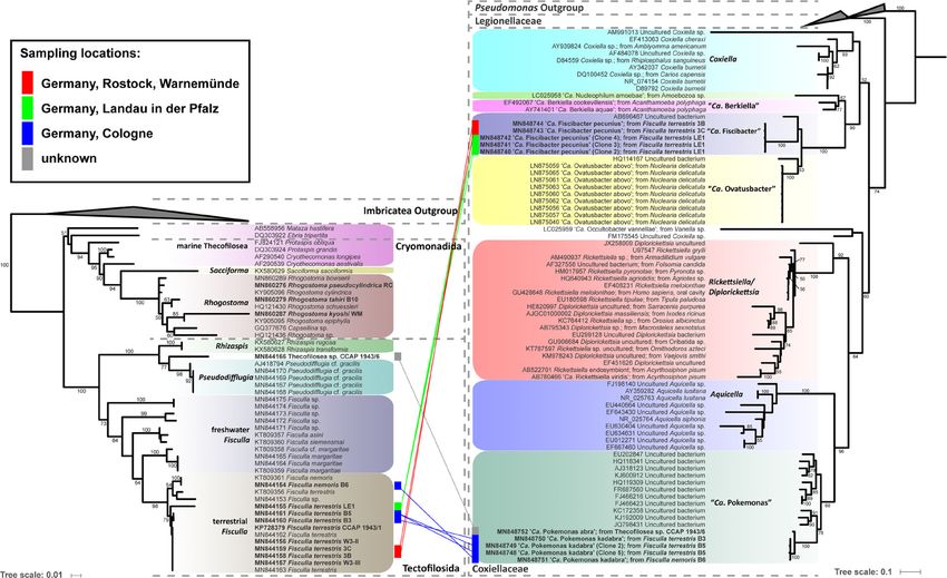
Solbach et al.                                                                                          Thecofilosean Breeding Ground for Legionellales

Disease Control and Prevention (CDC)). Since Legionellales are               accommodating Legionellales bacteria and only one of the 13
common in the environment, an increased local abundance may                  strains accommodating a non-Legionellales endosymbiotic
thus quickly lead to fatal local disease outbreaks (Morton et al.,           bacterium. Four different species of Legionellales bacteria
1986; van Loenhout et al., 2012). Under natural conditions,                  including species of the potentially pathogenic genus Legionella
Legionellales bacteria rely on a eukaryotic host to proliferate              and two novel Legionellales genera were detected. The
(Fields et al., 2002; Declerck et al., 2009; Declerck, 2010).                incongruence of the phylogenies of the amoeboid hosts and their
Accordingly, fluctuations in the abundance of Legionellales                   bacterial endosymbionts suggests a promiscuous distribution of
bacteria are strongly linked to fluctuations of the abundance of              Legionellales in amoeboid hosts of wide evolutionary origin. The
their hosts in the environment (Scheikl et al., 2014; Amaro et al.,          amoeboid lifestyle appears as the only common denominator in
2015). However, surprisingly little is still known on their host             this relationship.
ranges. As parasites in humans and animals, Legionellales infect
the amoeba-like alveolar macrophages (Escoll et al., 2013; Duron
et al., 2018). In the environment, Legionellales were found to               MATERIAL AND METHODS
reside and reproduce in protists, strikingly often in various kinds
of amoebae (Fields et al., 2002; Duron et al., 2018).                        Sampling and Cultivation
    It is hence commonly stated that Legionellales proliferate               Samples were taken in several different habitats including soil,
within amoebae; this statement, however, is not trivial. Amoebae             freshwater, and plant leaves. Leaves and soil samples were
taxonomy has been in permanent change during the last decades.               submerged in Waris-H+Si (Mcfadden and Melkonian, 1986)
Initially, the lack of clear morphological characters in amoeboid            and incubated for at least one day before analyses to enable
microorganisms hampered their classification. With the                        microscopic observation of attached protists. The samples were
application of molecular tools, the eukaryotic tree of life was              screened weekly for up to three weeks after collection for
reshaped and ‘amoeba’-like morphology turned out as a                        Thecofilosea-like cells with a Nikon Eclipse TS100 inverted
polyphyletic trait. It is now widely accepted that the term                  microscope (up to 400× magnification, phase contrast). Single
‘amoebae’ describes an assemblage of morphologically similar,                cells were picked with a glass micropipette and transferred into a
but only very distantly related microeukaryotes (Adl et al., 2019;           well of a 24-well plate (Sarstedt, Nümbrecht, Germany),
Burki et al., 2019). By far most recognized hosts of Legionellales           containing 2 ml of the respective culture medium and food
are amoebae in the higher-order Amorphea, including host genera              source. Subculturing was repeated until a monoclonal culture
like Acanthamoeba, Vermamoeba (former Hartmannella                           free of other eukaryotes was obtained. Depending on the species,
vermiformis), Arcella, Vannella, Cochliopodium, and Nuclearia                strains were cultured in either Waris-H+Si (Mcfadden and
(Santos et al., 2003; Dirren and Posch, 2016; Mehari et al., 2016;           Melkonian, 1986), WC-Medium (Guillard and Lorenzen, 1972)
Tsao et al., 2017; Gomaa et al., 2018; Galindo et al., 2019). Non-           or Wheat Grass (WG)-Medium (Bonkowski, 2019). For a
amorphean amoeba hosts seem rare, although Legionellales were                list of respective culture media and food sources refer to
also detected in the distantly related amoeba genus Naegleria                Supplementary Table S1.
(Excavata; (Delafont et al., 2013)).                                             To obtain gnotobiotic Fisculla strains, wells of 96-well plates
    The Rhizaria are rather unrelated to the previously mentioned            were filled with 50 µl sterile Waris-H+Si medium and axenic
taxa, but independently evolved a huge variety of amoeboid taxa.             Saccharomyces cerevisiae. Single Fisculla cells were sorted by size
The Cercozoa (Rhizaria) are particularly species-rich and                    (approximately 10–20 µm diameter) via flow cytometry (BD
dominant in terrestrial and aquatic habitats (Lentendu et al.,               FACSVantage, Becton, Dickinson and Company, Franklin Lakes,
2014; Grossmann et al., 2016). Despite their commonness, not a               New Jersey). Plates were incubated for approximately five days at
single amoeba belonging to the Cercozoa has yet been                         room temperature before further analysis to allow cultures to
investigated for putative endosymbiotic relationships with                   grow. The 96-well plates were then screened for wells free of
bacteria like the Legionellales. Hereinafter, we use the term                environmental bacteria and containing only cells of Fisculla spp.,
“endosymbiosis” in its most general definition encompassing                   and the remaining S. cerevisiae. Respective wells were used for
the broad range of intracellular associations, ranging from                  subsequent inoculation of 1.25% Waris-H+Si agar. Waris-H+Si
mutualism to parasitism.                                                     agar (1.25%) was obtained by autoclaving 2.5 g of agar (Sigma-
    We aimed to investigate whether Rhizaria may function as                 Aldrich, Munich, Germany) in 200 ml Waris-H+Si. Plates were
hosts for Legionellales; as a starting point, we chose to screen the         poured under a sterile bench. Once the agar dried, axenic
amoeboid Thecofilosea (Cercozoa, Rhizaria) since molecular                    S. cerevisiae cells were distributed with a cell scraper. Plates
investigations indicate an occasional dominance of these small               were stored at approximately 14–16°C in the dark.
and inconspicuous microorganisms in soils, wastewater
treatment plants, and water filters (Matsunaga et al., 2014;                  Sequencing and Cloning
Remmas et al., 2016a; Remmas et al., 2016b; Seppey et al.,                   To reduce the number of environmental bacteria (if present)
2017; Fernandez, 2019; Öztoprak et al., 2020).                               before amplification and sequencing of bacterial DNA, amoeba
    We screened 13 strains of Thecofilosea for potential bacterial            cells were preferably separated from environmental bacteria. Single
endosymbionts and found a surprising taxonomic richness of                   Fisculla cells were sorted via flow cytometry to Waris-H+Si
endosymbiotic Legionellales with eight of 13 screened amoebae                containing axenic S. cerevisiae as food organism and collected

Frontiers in Cellular and Infection Microbiology | www.frontiersin.org   2                                  March 2021 | Volume 11 | Article 642216
Solbach et al.                                                                                                Thecofilosean Breeding Ground for Legionellales

when all food cells were consumed (approx. after one week of                 medium was added to the ligation reactions. LB agar plates
incubation). Three hundred Rhogostoma cells of each strain were              were obtained by dissolving 9.6 g of LB-Agar—Powder according
sorted via flow cytometry and collected without further incubation            to Miller (AppliChem, Darmstadt, Germany) in 300 ml water.
for DNA extraction. The Thecofilosea sp. CCAP 1943/6 cells were               The mixture was autoclaved, then 175 µl Ampicillin (100 mg/ml),
too large and too low in density to be sorted and thus were filtered          300 µl 100 mM IPTG (Thermo Fisher Scientific, Dreieich,
(filter pore size: 30 µm). The filter was given in medium and                  Germany), and 600 µl X-Gal Solution, ready-to-use (Thermo
directly afterwards 150–200 cells were collected with a glass                Fisher Scientific, Dreieich, Germany) were added to the liquid
micropipette. DNA extraction of the collected cells was                      agar once it cooled down to approximately 50°C. Plates were
performed with a DNEasy Blood and Tissue kit (Qiagen, Venlo,                 poured under a sterile bench. For every strain 50 µl and 150 µl of
Netherlands). Extracted DNA was stored at −21°C.                             the transformation cultures were plated onto duplicate LB agar
    DNA amplification of the hypervariable region V4 of the SSU               plates. Plates with transformation cultures were incubated
rDNA of the amoeba strains was performed. Preferably single                  overnight at 37°C. White colonies were picked with a sterile
individuals with approximately 1 µl of medium were transferred               toothpick and directly added to 10 µl PCR mix. The mixture
into PCR-tubes, containing 17 µl PCR mixture. The mixture                    included 0.3 µl 10 µM forward and 0.3 µl 10 µM reverse primer,
included 1.7 µl 10 µM forward and 1.7 µl 10 µM reverse primer,               0.3 µl 10 mM dNTPs, 1 µl Thermo Scientific Dream Taq Green
0.34 µl 10 mM dNTPs, 1.7 µl Thermo Scientific Dream Taq                       Buffer, 0.1 µl DreamTaq polymerase, and 8 µl water. DNA
Green Buffer, 0.17 µl DreamTaq polymerase (Thermo Fisher                     amplification of the colonies was performed using the primers
Scientific, Dreieich, Germany), and 11.4 µl water. Sequences of               M13-40 and M13R. The following PCR conditions were used:
the SSU rDNA were obtained in two consecutive steps. First, a                initial denaturation at 95°C for 2 min, 30 cycles (denaturation at
sequence with a length of approximately 347 bases was amplified               95°C for 30 s, annealing at 52°C for 30 s, elongation at 72°C for
with the Cercozoa-specific primers S616F_Cercomix and                         2 min), terminal extension at 72°C for 5 min, and cooling at 4°C.
S963R_Cerco (Fiore-Donno et al., 2018). In the second step,                  Purification and sequencing of the PCR products were
semi-nested reamplifications were performed with a PCR                        performed with the same settings as above using the bacterial
mixture containing the same components as above and the                      primers 27F and 1492R. GenBank accession numbers of
primer pairs S947R_Cerco and S616F_Cercomix (Fiore-Donno                     obtained thecofilosean and bacterial sequences are given in
et al., 2018). Two µl of the first PCR product was used as                    Supplementary Table S1.
template. The following PCR conditions were used: initial
denaturation at 95°C for 2 min, 30 cycles (denaturation at 95°C              Probe Design and Fluorescence In Situ
for 30 s, annealing at 52°C for 30 s, elongation at 72°C for 30 s),          Hybridization
terminal extension at 72°C for 5 min, and cooling at 4°C.                    An alignment of the obtained bacterial sequences and their top ten
    DNA amplification of the 16S rDNA of bacteria was                         hits from a BLAST search (Basic Local Alignment Tool) and close
performed with 2 µl of extracted DNA as template with a PCR                  relatives was assembled in SeaView (Gouy et al., 2010). The
mixture containing the same components as above and the                      alignment was screened for short sequence segments with
bacterial primers 27F and 1492R (Weisburg et al., 1991; Jiang                lengths of 16–20 bases in which the respective bacteria group
et al., 2006). The following PCR conditions were used: initial               differed from all the others. The short sequence segments were
denaturation at 95°C for 2 min, 30 cycles (denaturation at 95°C              searched against the SILVA database (Pruesse et al., 2007) using
for 30 s, annealing at 48°C for 30 s, elongation at 72°C for 90 s),          the Probe Match and Evaluation Tool TestProbe 3.01 until
terminal extension at 72°C for 5 min, and cooling at 4°C. PCR                sequence segments as specific as possible to the desired bacteria
products were purified by adding 0.15 µl of Exonuclease, 0.9 µl               group were found. Short sequences fulfilling these requirements
FastAP and 1.95 µl water to 8 µl PCR product, then heating for               were tested with the OligoEvaluator tool (Sigma-Aldrich)2 to find
30 min at 37°C, and subsequently for 20 min at 85°C. The Big dye             sequences without secondary structures to avoid self-binding or
Terminator Cycle sequencing Kit (Thermo Fisher Scientific,                    folding of the FISH probes. Reverse compliment FISH probes were
Dreieich, Germany) and an ABI PRISM automatic sequencer                      ordered from biomers.net GmbH3 as oligonucleotides with either
were used for sequencing.                                                    Cy3 or FITC as 5′-modification. The resulting probes and their
    When direct sequencing of the bacteria did not result in a               numbers of matches in the SILVA database are displayed in
distinct sequence, 100 µl PCR products were subjected to                     Supplementary Table S2.
cloning. Two µl of extracted DNA was used as template. PCR                       Between 20 and 200 µl of cultures were air-dried on 0.1%
products were purified using the GeneJET PCR Purification Kit                  gelatine-coated glass slides and dehydrated in an increasing
(Thermo Fisher Scientific, Dreieich, Germany) and eluted in                   ethanol series (50, 80, and 96%) for 3 min each and finally air-
20 µl elution buffer. Ligation and transformation were performed             dried again. Subsequently, 100 µl hybridization buffer (18 µl 5M
according to the manufacturer’s instructions using the pGEM®-                NaCl solution, 2 µl 1M TrisHCl solution, 35 µl formamide, 45 µl
T Easy Vector System with JM109 Competent Cells (Promega                     water, 0.1 µl 10% SDS) was mixed with 12.5 µl (100 µM) of the
GmbH, Mannheim, Germany), with the following changes to the
protocol: Ligation was conducted overnight at 5°C using 2.5 µl
2× Rapid Ligation Buffer, 0.5 µl pGEM®-T Easy Vector (50 ng),                1
                                                                               https://www.arb-silva.de/search/testprobe/
0.5 µl T4 DNA Ligase, 0.5 µl H2O, and 1 µl of the purified PCR                2
                                                                               http://www.oligoevaluator.com/
                                                                             3
product. In step 6 of the instructions, only 250 µl of SOC                     https://www.biomers.net/

Frontiers in Cellular and Infection Microbiology | www.frontiersin.org   3                                         March 2021 | Volume 11 | Article 642216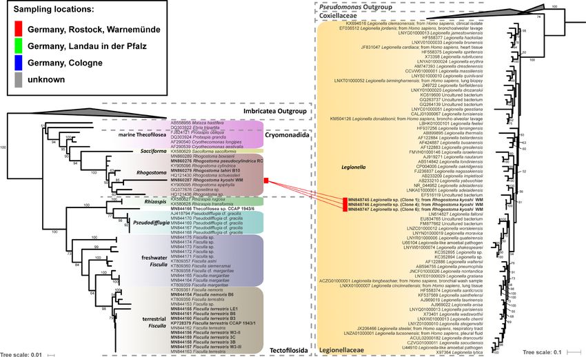
Solbach et al. Thecofilosean Breeding Ground for Legionellales
general bacteria probe EUB338 (Amann and Fuchs, 2008) tagged percentages obtained through 200 non-parametric bootstraps
with FITC and a specific probe tagged with Cy3 (Supplementary under the GTRCAT model.
Table S2), applied to the samples, and incubated for 90 min (48°C
in a moist chamber). Subsequently, samples were incubated in
preheated washing buffer (700 µl 5M NaCl solution, 1,000 µl 1M RESULTS
TrisHCl solution, 50 ml water, and 50 µl 10% SDS) for 25 min at
48°C. Air-dried slides were stained with DAPI for 10 min, Endosymbiont Detection and Phylogeny
washed, air-dried, and subsequently mounted with 85% glycerol Eleven thecofilosean cultures from environmental samples and two
and 15% phosphate-buffered saline (PBS) [after Manz et al. from the CCAP culture collection were screened for endosymbiotic
(1992); Hugenholtz et al. (2001); Hess et al. (2016)]. The bacteria (Supplementary Table S1). Eight of the 13 thecofilosean
general probes EUB338, ALF968, BET42a, and GAM42a amoebae cultures contained Legionellales endosymbionts
(Amann and Fuchs, 2008) and DAPI staining were used as confirmed by sequencing and fluorescence in situ hybridization
controls before taxon-specific probes were applied. Pictures of (FISH). From these, we obtained in total 13 Legionellales 16S
the processed cells were taken with a Nikon digital sight DS-U2 sequences, three of which could be assigned to a novel Legionella
camera (program: NIS-Elements V4.13.04) with a Nikon Eclipse isolate. For the remaining sequences the novel candidate species
90i upright microscope (up to 600× magnification). “Ca. Fiscibacter pecunius”, “Ca. Pokemonas kadabra”, and “Ca.
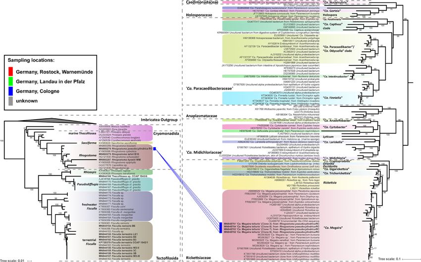
Pokemonas abra” (Figures 1, 2; Supplementary Material) were
Phylogenetic Analyses erected. “Ca. Fiscibacter pecunius” was detected in three different
All partial sequences were checked for sequencing errors in strains of Fisculla terrestris (LE1, 3B, 3C), “Ca. Pokemonas
Chromas (V2.6.6, Technelysium Pty. Ltd., Australia) before they kadabra” in two strains of Fisculla terrestris (B3, B5) and one
were assembled into one sequence contig using SeaView [V4.6, strain of Fisculla nemoris (B6), and “Ca. Pokemonas abra” in one
(Gouy et al., 2010)]. For the thecofilosean alignment, representative strain of Thecofilosea sp. (CCAP 1943/6) (Figure 1). The
sequences of major thecofilosean subphyla were added to the Legionella sp. was found in a strain of Rhogostoma kyoshi (WM)
sequences of the Thecofilosea from this study. Ten selected (Figure 2). Only one non-Legionellales endosymbiont was
Imbricatea sequences were used as outgroup. The probes detected in a strain of Rhogostoma pseudocylindrica (RC), and
LE1_10_Gam, Ft_Chryseo, 5_Sedimini did not result in positive based on 16S rDNA phylogeny it could be assigned to the genus
staining when applied to cells of the respective cultures. Therefore, “Ca. Megaira” in Rickettsiales (Figure 3).
only bacteria successfully stained with the probes LE1_3B_3C_Gam, “Ca. Fiscibacter pecunius”, “Ca. Pokemonas abra” and “Ca.
3_5_6_Thec_Gam, WM_Legio, or RC_Rick, respectively, have been Pokemonas kadabra” formed distinct clades within the
used for phylogenetic analyses. Accordingly, two separate Coxiellaceae (Figure 1). “Ca. Fiscibacter” branches are highly
phylogenetic analyses have been performed for endosymbiotic supported close to “Ca. Ovatusbacter abovo” which was described
bacteria belonging to the Legionellales (Gammaproteobacteria) by Dirren and Posch (2016) as a member of Gammaproteobacteria.
and Rickettsiales (Alphaproteobacteria). Dirren and Posch (2016) already noticed high sequence similarities
To create the datasets for phylogenetic analyses, the top ten hits of “Ca. Ovatusbacter abovo” to “Ca. Berkiella”, but did not assign a
of each verified endosymbiotic bacterium were obtained from the family. In our analysis, they represent a clade within the family
NCBI GenBank database (last date of accession: July 26th, 2019) by Coxiellaceae. “Ca. Pokemonas” represents a highly supported sister
using the blastn search algorithm (blastn 2.3.0) with default group to Aquicella. The closest described relative to the
parameters. For the Legionellales dataset, all available Legionella endosymbiotic Legionella sp. (Legionellaceae, Legionellales) was
sequences were downloaded from the SILVA database (https:// Legionella adelaidensis with a bootstrap value of 56% (Figure 2).
www.arb-silva.de/) and ordered by species names. Sequences of Phylogenetic analyses of the hosts and endosymbionts show
undescribed Legionella spp. were removed, and only one sequence no clear co-evolutionary pattern, indicating a promiscuous
of every described species was maintained. Based on the overview of dispersal of endosymbiotic bacteria in the Thecofilosea (Figure
the order Legionellales by Duron et al. (2018), selected sequences of 1). No endosymbionts were detected in four of the 13 screened
the major subphyla were added to the dataset, including sequences amoeba strains, indicating that the identified endosymbiotic
of ten Diplorickettsia, ten Rickettsiella, ten Aquicella, ten Coxiella, bacteria in Thecofilosea are facultative endosymbionts for the
two “Ca. Berkiella”, “Ca. Nucleophilum amoebae” and “Ca. host. In all but one thecofilosean amoeba, the endosymbionts
Occultobacter vannellae”. 13 selected Pseudomonas sequences were dispersed throughout the whole cell body, except in the
were used as outgroup. For the phylogenetic analyses of the nucleus and the filopodia (Figure 4). Only in one host strain
Rickettsiales sequences from Hess et al. (2016) were obtained and (Thecofilosea sp. 1943/6), the Legionellales endosymbionts were
additional “Candidatus Megaira” sequences from Schrallhammer concentrated in one large vacuole (Figure 4G). The vacuole was
et al. (2013) were added. Sequences were aligned in MAFFT using present in all individuals and usually larger than the hosts’
the L-INS-i algorithm (Katoh and Toh, 2010) and ambiguously nucleus. In all strains, the endosymbionts were transmitted
aligned sequence segments were manually cut using SeaView. The vertically by cell division (see a dividing cell of Fisculla
model GTR + I + G was used and maximum likelihood (ML) terrestris in Figure 4E). Extracellular transmission of bacteria
phylogenetic trees were constructed using RAxML (Randomized was not observed.
Axelerated Maximum Likelihood, Version 8, (Stamatakis, 2014)). For the first time, long-term stable cultures consisting of the
The best scoring tree was used to report the confidence values as Fisculla spp., their bacterial symbiont (Legionellales spp.), and
Frontiers in Cellular and Infection Microbiology | www.frontiersin.org 4 March 2021 | Volume 11 | Article 642216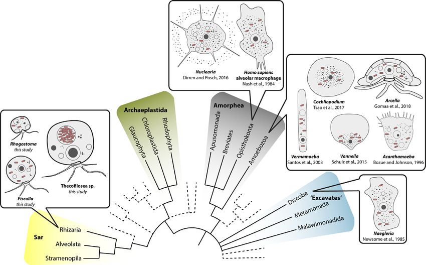
Frontiers in Cellular and Infection Microbiology | www.frontiersin.org

                                                                                                                                                                                                                                                                                        Solbach et al.
5

                                                                                                                                                                                                                                                                                        Thecofilosean Breeding Ground for Legionellales
March 2021 | Volume 11 | Article 642216

                                                                         FIGURE 1 | Phylogenetic trees of thecofilosean amoebae and their respective endosymbionts of the Coxiellaceae (Legionellales, Gammaproteobacteria). Lines connect the thecofilosean hosts and their respective
                                                                         endosymbionts; line color indicates sampling locations (see legend). Only confidence values >50 are displayed.
Frontiers in Cellular and Infection Microbiology | www.frontiersin.org
Solbach et al.
6
Thecofilosean Breeding Ground for Legionellales
March 2021 | Volume 11 | Article 642216
FIGURE 2 | Phylogenetic trees of thecofilosean amoebae and their respective endosymbionts of the Legionellaceae (Legionellales, Gammaproteobacteria). Lines connect the thecofilosean hosts and their respective
endosymbionts; line color indicates sampling locations (see legend). Only confidence values >50 are displayed.Frontiers in Cellular and Infection Microbiology | www.frontiersin.org
Solbach et al.
7
Thecofilosean Breeding Ground for Legionellales
March 2021 | Volume 11 | Article 642216
FIGURE 3 | Phylogenetic trees of thecofilosean amoebae and their respective endosymbionts of the Rickettsiales (Alphaproteobacteria). Lines connect the thecofilosean hosts and their respective endosymbionts;
line color indicates sampling locations (see legend). The tree contains no outgroup, but was rooted to resemble the tree from Hess et al. (2016). Only confidence values >50 are displayed.Solbach et al. Thecofilosean Breeding Ground for Legionellales
FIGURE 4 | FISH pictures. (A–C): Strains containing “Ca. Fiscibacter pecunius”. (A) F. terrestris (strain LE1); (B) F. terrestris (strain 3B); (C) F. terrestris (strain 3C).
(D–F): Strains containing “Ca. Pokemonas kadabra”. (D) F. terrestris (strain B3); (E) F. terrestris (strain B5); (F) F. nemoris (strain B6). (G) Thecofilosea sp. (strain
CCAP 1943/6) containing “Ca. Pokemonas abra”. (H) Rhogostoma kyoshi (strain WM) containing Legionella sp. (I) Rhogostoma pseudocylindrica (strain RC)
containing “Ca. Megaira telluris”. 1: Difference interference contrast (DIC); 2: DAPI staining; 3: staining with general bacteria probe EUB338; 4: staining with specific
probe (see Supplementary Table S2). Scale bars indicate 10 mm. Nu, nucleus; f, filopodia; env, environmental bacteria; end, endosymbiotic bacteria.
the eukaryotic prey (S. cerevisisae) were established (see Material very distantly related Rhizaria (Figure 5). From an evolutionary
and Methods). These cultures can be specified as gnotobiotic, i.e. perspective, it is crucial to trace back Legionellales’ diversity to their
all organisms in the culture (host, endosymbiont, and food respective hosts to understand and predict the bacterial adaptations
organisms) are known. All nine established Fisculla spp. to the immune system of their respective hosts, a prerequisite for the
cultures were free of environmental bacteria and stable over a development of antimicrobial measures/treatments (Molmeret
year, the whole runtime of the project. et al., 2005; Duron et al., 2018; Gomez-Valero and Buchrieser,
2019; Mondino et al., 2020).
Despite numerous publications on diverse bacterial
DISCUSSION endosymbionts of protists, there are few reports of Legionellales.
The ciliate protist Euplotes is commonly used as a model for
Legionellales Proliferate in Amoebal Hosts endosymbiosis research; however, none of the yet determined
of Widespread Evolutionary Origin dozens of different endosymbionts of Euplotes belonged to
Our data on Thecofilosea clearly show that amoebae in the phylum Legionellales (Boscaro et al., 2019). Euplotes differs in morphology
Cercozoa accommodate a variety of bacterial endosymbionts in from the majority of known potential Legionellales hosts which are
Legionellales. The fact that Legionellales are not restricted to amoeboid (Figure 5). It seems that the evolutionary origin of the
Amorphea has important applied implications because our data potential host does not indicate the susceptibility for infection by
expand the potential host range of Legionellales to the evolutionary Legionellales since potential hosts belong to rather unrelated
Frontiers in Cellular and Infection Microbiology | www.frontiersin.org 8 March 2021 | Volume 11 | Article 642216Solbach et al. Thecofilosean Breeding Ground for Legionellales
FIGURE 5 | Overview of eukaryotic diversity based on (Burki et al., 2019). Former eukaryotic supergroups are highlighted, Opisthokonta and Amoebozoa are
combined into Amorphea, and novel or minor groups are reduced. Depicted is the known diversity of amoebal Legionellales hosts. Note that prior to this study most
Legionellales were found in amorphean amoebae and only one excavate taxon.
eukaryotes. Instead, it is striking that amoebae, a morphological Legionellales indicating that Legionellales may form stable and
term and not an evolutionary lineage, function exceptionally often long term symbiosis with their host (Gomaa et al., 2018).
as hosts for Legionellales. Even in humans, the infected cells are Subsequently, this means that not necessarily the biofilms, but
much alike an amoeba since the infected alveolar macrophages of the amoebae within, form a reservoir for Legionellales.
the human immune system are surface-attached grazing cells While the pathogenic potential of the novel described “Ca.
(Duron et al., 2018). Pokemonas” and “Ca. Fiscibacter pecunius” cannot be inferred by
Legionellales are long known to be associated with biofilms, our data, the found Legionella sp. is closely related to Legionella
i.e. they are more frequent on submerged surfaces than in water adelaidensis that belongs to a group with many waterborne human
columns (Rogers et al., 1994; Abu Khweek and Amer, 2018). Our pathogenic species. The found Legionella sp. was detected in a
results consolidate the comprehension of why and how strain of Rhogostoma kyoshi that was obtained from biological soil
Legionellales are associated with biofilms. By far most amoebae crusts, i.e. surface soil (Khanipour Roshan et al., 2021).
swim only occasionally; most often they are substrate-attached, Rhogostoma represents a genus of thecofilosean amoebae that is
accordingly, amoebae are among the most abundant grazers on especially abundant in soils (Degrune et al., 2019; Fiore-Donno
biofilms, where they engulf prey, including whole batches of et al., 2019; Öztoprak et al., 2020), wastewater treatment plants
bacteria (Bonkowski, 2019). Other protist groups, like flagellates (Matsunaga et al., 2014; Remmas et al., 2016a; Remmas et al.,
and ciliates often preferentially predate freely swimming prey 2016b; Öztoprak et al., 2020), and water filters (Domingo
(Verni and Gualtieri, 1997; Boenigk and Arndt, 2002), and as we Fernandez, 2019), and their potential as a reservoir for
hypothesize that is why they come in less contact with substrate Legionellales requires further investigation.
attached Legionellales and are thus only rarely or never infected. The nature of the relationship between the reported endosymbionts
To our knowledge, it is still questionable whether Legionellales and their hosts, whether being mutualistic or parasitic, was not
replicate under natural conditions without a eukaryotic investigated in this study, but the gnotobiotic cultivation method of
host (Fields et al., 2002). It was shown that a lack of amoebae Fisculla spp. facilitates future genomic and transcriptomic studies,
prevents growth of the substrate-attached Legionella pneumophila helping to answer these open research questions.
(Declerck et al., 2009; Declerck, 2010). Legionella can be grown
axenically in the lab, but for this, exact conditions have to be met.
It was proposed that, even if such conditions are met DATA AVAILABILITY STATEMENT
environmentally, other bacteria would quickly outcompete
potentially freely replicating Legionellales (Fields et al., 2002). The datasets presented in this study can be found in online
Although some amoebal hosts are lysed by their endosymbionts repositories. The names of the repository/repositories
(La Scola et al., 2004) our and other studies did not find any and accession number(s) can be found in the article/
evidence for lysis of the host cell after replication of the Supplementary Material.
Frontiers in Cellular and Infection Microbiology | www.frontiersin.org 9 March 2021 | Volume 11 | Article 642216Solbach et al. Thecofilosean Breeding Ground for Legionellales
AUTHOR CONTRIBUTIONS Kleemann for the provision of environmental samples for
isolation of the Thecofilosea strains. We thank Andreas
KD conceived the project. KD and MB administrated the project. Suthaus for his help with fluorescence in situ hybridization,
MS and KD conducted the amplifications, sequencing, FISH Christoph Göttlinger for sorting our cultures with FACS, and the
staining, and phylogenetic analyses. MS wrote the initial members of the Cologne Center for Genomics (CCG) for their
manuscript, KD and MB revised it. All authors contributed to sequencing services.
the article and approved the submitted version.
SUPPLEMENTARY MATERIAL
ACKNOWLEDGMENTS
The Supplementary Material for this article can be found online
We are very grateful to Hüsna Öztoprak, Samira Khanipour at: https://www.frontiersin.org/articles/10.3389/fcimb.2021.
Roshan, Christopher Kahlich, Ferry Siemensma, and Benedikt 642216/full#supplementary-material
REFERENCES Duron, O., Doublet, P., Vavre, F., and Bouchon, D. (2018). The Importance of
Revisiting Legionellales Diversity. Trends Parasitol. 34, 1027–1037.
Abu Khweek, A., and Amer, A. O. (2018). Factors mediating environmental doi: 10.1016/j.pt.2018.09.008
biofilm formation by Legionella pneumophila. Front. Cell. Infect. Microbiol. 8, Eldin, C., Mé lenotte, C., Mediannikov, O., Ghigo, E., Million, M., Edouard, S., et al.
38. doi: 10.3389/fcimb.2018.00038 (2017). From Q fever to Coxiella burnetii infection: A paradigm change. Clin.
Adl, S. M., Bass, D., Lane, C. E., Lukeš, J., Schoch, C. L., Smirnov, A., et al. (2019). Microbiol. Rev. 30, 115–190. doi: 10.1128/CMR.00045-16
Revisions to the Classification, Nomenclature, and Diversity of Eukaryotes. Escoll, P., Rolando, M., Gomez-Valero, L., and Buchrieser, C. (2013). From
J. Eukaryot. Microbiol. 66, 4–119. doi: 10.1111/jeu.12691 Amoeba to Macrophages: Exploring the Molecular Mechanisms of
Amann, R., and Fuchs, B. M. (2008). Single-cell identification in microbial Legionella pneumophila Infection in Both Hosts. Curr. Top. Microbiol.
communities by improved fluorescence in situ hybridization techniques. Nat. Immunol. 376, 1–34. doi: 10.1007/82_2013_351
Rev. Microbiol. 6, 339–348. doi: 10.1038/nrmicro1888 Domingo Fernandez, O. (2019). Microbial Ecology of E. coli Removal
Amaro, F., Wang, W., Gilbert, J. A., Roger Anderson, O., and Shuman, H. A. Mechanisms and Drinking Water Production in Slow Sand Filters Exposed
(2015). Diverse protist grazers select for virulence-related traits in Legionella. To Emerging Contaminants. Available at: http://hdl.handle.net/10379/15110.
ISME J. 9, 1607–1618. doi: 10.1038/ismej.2014.248 Fields, B. S., Benson, R. F., and Besser, R. E. (2002). Legionella and Legionnaires’
Beauté , J., and Robesyn, E. (2017). European Centre for Disease Prevention and Disease: 25 Years of Investigation. Clin. Microbiol. Rev. 15, 506–526.
Control. Legionnaires’ disease in Europe 2015. ECDC. doi: 10.2900/692621 doi: 10.1128/CMR.15.3.506–526.2002
Boamah, D. K., Zhou, G., Ensminger, A. W., and O’Connor, T. J. (2017). From Fiore-Donno, A. M., Rixen, C., Rippin, M., Glaser, K., Samolov, E., Karsten, U., et al.
many hosts, one accidental pathogen: The diverse protozoan hosts of (2018). New barcoded primers for efficient retrieval of cercozoan sequences in high-
Legionella. Front. Cell. Infect. Microbiol. 7, 477. doi: 10.3389/fcimb.2017.00477 throughput environmental diversity surveys, with emphasis on worldwide
Boenigk, J., and Arndt, H. (2002). Bacterivory by heterotrophic flagellates: biological soil crusts. Mol. Ecol. Resour. 18, 229–239. doi: 10.1111/1755-0998.12729
community structure and feeding strategies. Antonie Van Leeuwenhoek 81, Fiore-Donno, A. M., Richter-Heitmann, T., Degrune, F., Dumack, K., Regan, K.
465–480. doi: 10.1023/A:1020509305868 M., Marhan, S., et al. (2019). Functional Traits and Spatio-Temporal Structure
Bonkowski, M. (2019). “Microcosm Approaches to Investigate Multitrophic of a Major Group of Soil Protists (Rhizaria: Cercozoa) in a Temperate
Interactions between Microbial Communities in the Rhizosphere of Plants,” Grassland. Front. Microbiol. 10, 1332. doi: 10.3389/fmicb.2019.01332
in Methods in Rhizosphere Biology Research. Eds. D. Reinhardt and A. K. Galindo, L. J., Torruella, G., Moreira, D., Eglit, Y., Simpson, A. G. B., Völcker, E.,
Sharma (Singapore: Springer), 255–270. doi: 10.1007/978-981-13-5767-1_14 et al. (2019). Combined cultivation and single-cell approaches to the
Boscaro, V., Husnik, F., Vannini, C., and Keeling, P. J. (2019). Symbionts of the phylogenomics of nucleariid amoebae, close relatives of fungi. Phil. Trans. R.
ciliate Euplotes: diversity, patterns and potential as models for bacteria – Soc. B 374, 20190094. doi: 10.1098/rstb.2019.0094
eukaryote endosymbioses. Proc. R. Soc. B 286, 20190693. doi: 10.1098/ Gomaa, F., Gersh, M., and Cavanaugh, C. M. (2018). Diverse Legionella-Like
rspb.2019.0693 Bacteria Associated with Testate Amoebae of the Genus Arcella (Arcellinida:
Burki, F., Roger, A. J., Brown, M. W., and Simpson, A. G. B. (2019). The New Tree Amoebozoa). J. Eukaryot. Microbiol. 65, 661–668. doi: 10.1111/jeu.12511
of Eukaryotes. Trends Ecol. Evol. 35 (1), 1–13. doi: 10.1016/j.tree.2019.08.008 Gomez-Valero, L., and Buchrieser, C. (2019). Intracellular parasitism, the driving
Cunha, B. A., Burillo, A., and Bouza, E. (2016). Legionnaires ‘ disease. Lancet 387, force of evolution of Legionella pneumophila and the genus Legionella. Genes
376–385. doi: 10.1016/S0140-6736(15)60078-2 Immun. 20, 394–402. doi: 10.1038/s41435-019-0074-z
Declerck, P., Behets, J., Margineanu, A., van Hoef, V., De Keersmaecker, B., and Gouy, M., Guindon, S., and Gascuel, O. (2010). Sea view version 4: A
Ollevier, F. (2009). Replication of Legionella pneumophila in biofilms of water multiplatform graphical user interface for sequence alignment and
distribution pipes. Microbiol. Res. 164, 593–603. doi: 10.1016/j.micres.2007. phylogenetic tree building. Mol. Biol. Evol. 27, 221–224. doi: 10.1093/
06.001 molbev/msp259
Declerck, P. (2010). Biofilms: The environmental playground of Legionella Grossmann, L., Jensen, M., Heider, D., Jost, S., Glücksman, E., Hartikainen, H.,
pneumophila. Environ. Microbiol. 12, 557–566. doi: 10.1111/j.1462-2920. et al. (2016). Protistan community analysis: key findings of a large-scale
2009.02025.x molecular sampling. ISME J. 10, 2269–2279. doi: 10.1038/ismej.2016.10
Degrune, F., Dumack, K., Fiore-Donno, A. M., and Bonkowski, M. (2019). Distinct Guillard, R. R. L., and Lorenzen, C. J. (1972). Yellow-Green Algae with
communities of Cercozoa at different soil depths in a temperate agricultural Chlorophyllide C. J. Phycol. 8, 10–14. doi: 10.1111/j.1529-8817.1972.tb03995.x
field. FEMS Microbiol. Ecol. 95 (4), fiz041. doi: 10.1093/femsec/fiz041 Hess, S., Suthaus, A., and Melkonian, M. (2016). “Candidatus Finniella”
Delafont, V., Brouke, A., Bouchon, D., Moulin, L., and Hé chard, Y. (2013). (Rickettsiales, Alphaproteobacteria), novel endosymbionts of viridiraptorid
Microbiome of free-living amoebae isolated from drinking water. Water Res. amoeboflagellates (Cercozoa, Rhizaria). Appl. Environ. Microbiol. 82, 659–
47, 6958–6965. doi: 10.1016/j.watres.2013.07.047 670. doi: 10.1128/AEM.02680-15
Dirren, S., and Posch, T. (2016). Promiscuous and specific bacterial symbiont Hugenholtz, P., Tyson, G. W., and Blackall, L. L. (2001). Design and Evaluation of
acquisition in the amoeboid genus Nuclearia (Opisthokonta). FEMS Microbiol. 16S rRNA-Targeted Oligonucleotide Probes for Fluorescence In Situ
Ecol. 92, 1–16. doi: 10.1093/femsec/fiw105 Hybridization. Gene Probes 176, 029–042. doi: 10.1385/1-59259-238-4:029
Frontiers in Cellular and Infection Microbiology | www.frontiersin.org 10 March 2021 | Volume 11 | Article 642216Solbach et al. Thecofilosean Breeding Ground for Legionellales
Jiang, H., Dong, H., Zhang, G., Yu, B., Chapman, L. R., and Fields, M. W. (2006). formation and growth of Legionella pneumophila in a model potable water
Microbial diversity in water and sediment of Lake Chaka, an athalassohaline system containing complex microbial flora. Appl. Environ. Microbiol. 60, 1585–
lake in northwestern China. Appl. Environ. Microbiol. 72, 3832–3845. 1592. doi: 10.1128/AEM.60.5.1585-1592.1994
doi: 10.1128/AEM.02869-05 Santos, P., Pinhal, I., Rainey, F. A., Empadinhas, N., Costa, J., Fields, B., et al.
Katoh, K., and Toh, H. (2010). Parallelization of the MAFFT multiple sequence (2003). Gamma-Proteobacteria Aquicella lusitana gen. nov., sp. nov., and
alignment program. Bioinformatics 26, 1899–1900. doi: 10.1093/bioinformatics/ Aquicella siphonis sp. nov. Infect Protozoa and Require Activated Charcoal
btq224 for Growth in Laboratory Media. Appl. Environ. Microbiol. 69, 6533–6540.
Khanipour Roshan, S., Dumack, K., Bonkowski, M., Leinweber, P., Karsten, U., doi: 10.1128/AEM.69.11.6533-6540.2003
and Glaser, K. (2021). Taxonomic and Functional Diversity of Heterotrophic Scheikl, U., Sommer, R., Kirschner, A., Rameder, A., Schrammel, B., Zweimüller, I.,
Protists (Cercozoa and Endomyxa) from Biological Soil Crusts. et al. (2014). Free-living amoebae (FLA) co-occurring with legionellae in
Microorganisms 9 (2), 205. doi: 10.3390/microorganisms9020205 industrial waters. Eur. J. Protistol. 50, 422–429. doi: 10.1016/j.ejop.2014.04.002
La Scola, B., Birtles, R. J., Greub, G., Harrison, T. J., Ratcliff, R. M., and Raoult, D. Schrallhammer, M., Ferrantini, F., Vannini, C., Galati, S., Schweikert, M., Görtz,
(2004). Legionella drancourtii sp. nov., a strictly intracellular amoebal H. D., et al. (2013). ‘Candidatus Megaira polyxenophila’ gen. nov., sp. nov.:
pathogen. Int. J. Syst. Evol. Microbiol. 54, 699–703. doi: 10.1099/ijs.0.02455-0 Considerations on Evolutionary History, Host Range and Shift of Early
Lentendu, G., Wubet, T., Chatzinotas, A., Wilhem, C., Buscot, F., and Schlegel, M. Divergent Rickettsiae. PLoS One 8 (8), e72581. doi: 10.1371/journal.pone.
(2014). Effects of long-term differential fertilization on eukaryotic microbial 0072581
communities in an arable soil: a multiple barcoding approach. Mol. Ecol. 23, Seppey, C. V. W., Singer, D., Dumack, K., Fournier, B., Belbahri, L., Mitchell, E. A.
3341–3355. doi: 10.1111/mec.12819 D., et al. (2017). Distribution patterns of soil microbial eukaryotes suggests
Manz, W., Amann, R., Ludwig, W., Wagner, M., and Schleifer, K. H. (1992). widespread algivory by phagotrophic protists as an alternative pathway for
Phylogenetic Oligodeoxynucleotide Probes for the Major Subclasses of nutrient cycling. Soil Biol. Biochem. 112, 68–76. doi: 10.1016/j.soilbio.2017.
Proteobacteria: Problems and Solutions. Syst. Appl. Microbiol. 15, 593–600. 05.002
doi: 10.1016/S0723-2020(11)80121-9 Shah, P., Barskey, A., Binder, A., Edens, C., Lee, S., Smith, J., et al. (2019).
Matsunaga, K., Kubota, K., and Harada, H. (2014). Molecular Diversity of Eukaryotes Legionnaires’ Disease Surveillance Summary Report, United States. Centers
in Municipal Wastewater Treatment Processes as Revealed by 18S rRNA Gene Dis. Control Prev. Available at: https://www.cdc.gov/legionella/health-depts/
Analysis. Microbes Environ. 29, 401–407. doi: 10.1264/jsme2.ME14112 surv-reporting/surveillance-reports.html.
Mcfadden, G., and Melkonian, M. (1986). Use of Hepes buffer for micro algal Shelton, B. G., Kerbel, W., Witherell, L., Millar, J. D., Shelton, B. G., Kerbel, W.,
culture media and fixation for electron microscopy. Phycologia 25, 551–557. et al. (2010). Review of Legionnaires’ Disease. AIHAJ - Am. Ind. Hyg. Assoc. 61
doi: 10.2216/i0031-8884-25-4-551.1 (5), 738–742. doi: 10.1080/15298660008984585
Mehari, Y. T., Hayes, B. J., Redding, K. S., Mariappan, P. V. G., Gunderson, J. H., Stamatakis, A. (2014). RAxML version 8: A tool for phylogenetic analysis and
Farone, A. L., et al. (2016). Description of ‘Candidatus Berkiella aquae’ and post-analysis of large phylogenies. Bioinformatics 30, 1312–1313. doi: 10.1093/
‘Candidatus Berkiella cookevillensis’, two intranuclear bacteria of freshwater bioinformatics/btu033
amoebae. Int. J. Syst. Evol. Microbiol. 66, 536–541. doi: 10.1099/ijsem.0.000750 The Centers for Disease Control and Prevention (CDC). Legionella (Legionnaires'
Molmeret, M., Horn, M., Wagner, M., Santic, M., and Kwaik, Y. A. (2005). Disease and Pontiac Fever). Available at: https://www.cdc.gov/legionella/about/
Amoebae as Training Grounds for Intracellular Bacterial Pathogens. Appl. history.html.
Environ. Microbiol. 71, 20–28. doi: 10.1128/AEM.71.1.20–28.2005 Tsao, H.-F., Scheikl, U., Volland, J.-M., Köhsler, M., Bright, M., Walochnik, J.,
Mondino, S., Schmidt, S., Rolando, M., Escoll, P., Gomez-Valero, L., and et al. (2017). ‘Candidatus Cochliophilus cryoturris’ (Coxiellaceae), a symbiont
Buchrieser, C. (2020). Legionnaires’ Disease: State of the Art Knowledge of of the testate amoeba Cochliopodium minus. Sci. Rep. 7, 3394. doi: 10.1038/
Pathogenesis Mechanisms of Legionella. Annu. Rev. Pathol. Mech. Dis. 15, s41598-017-03642-8
439–466. doi: 10.1146/annurev-pathmechdis-012419-032742 van Loenhout, J. A. F., Paget, W. J., Vercoulen, J. H., Wijkmans, C. J., Hautvast,
Morton, S., Bartlett, C. L. R., Bibby, L. F., Hutchinson, D. N., Dyer, J. V., and J. L. A., and van der Velden, K. (2012). Assessing the long-term health impact
Dennis, P. J. (1986). Outbreak of legionnaires’ disease from a cooling water system of Q-fever in the Netherlands: a prospective cohort study started in 2007 on the
in a power station. Br. J. Ind. Med. 43, 630–635. doi: 10.1136/oem.43.9.630 largest documented Q-fever outbreak to date. BMC Infect. Dis. 12, 2–7.
Öztoprak, H., Walden, S., Heger, T., Bonkowski, M., and Dumack, K. (2020). doi: 10.1186/1471-2334-12-280
What drives the diversity of the most abundant terrestrial cercozoan family Verni, F., and Gualtieri, P. (1997). Feeding behavior in ciliated protists. Micron 28,
(Rhogostomidae, cercozoa, rhizaria)? Microorganisms 8, 1–16. doi: 10.3390/ 487–504. doi: 10.1016/S0968-4328(97)00028-0
microorganisms8081123 Weisburg, W. G., Barns, S. M., Pelletier, D. A., and Lane, D. J. (1991). 16S
Pruesse, E., Quast, C., Knittel, K., Fuchs, B. M., Ludwig, W., Peplies, J., et al. (2007). Ribosomal DNA Amplification for Phylogenetic Study. J. Bacteriol. 173, 697–
SILVA: A comprehensive online resource for quality checked and aligned 703. doi: 10.1128/jb.173.2.697-703.1991
ribosomal RNA sequence data compatible with ARB. Nucleic Acids Res. 35,
7188–7196. doi: 10.1093/nar/gkm864 Conflict of Interest: The authors declare that the research was conducted in the
Remmas, N., Melidis, P., Katsioupi, E., and Ntougias, S. (2016a). Effects of high absence of any commercial or financial relationships that could be construed as a
organic load on amoA and nirS gene diversity of an intermittently aerated and potential conflict of interest.
fed membrane bioreactor treating landfill leachate. Bioresour. Technol. 220,
557–565. doi: 10.1016/j.biortech.2016.09.009 Copyright © 2021 Solbach, Bonkowski and Dumack. This is an open-access article
Remmas, N., Melidis, P., Paschos, G., Statiris, E., and Ntougias, S. (2016b). distributed under the terms of the Creative Commons Attribution License (CC BY).
Protozoan indicators and extracellular polymeric substances alterations in an The use, distribution or reproduction in other forums is permitted, provided the
intermittently aerated membrane bioreactor treating mature landfill leachate. original author(s) and the copyright owner(s) are credited and that the original
Environ. Technol. 3330, 53–64. doi: 10.1080/09593330.2016.1190792 publication in this journal is cited, in accordance with accepted academic practice.
Rogers, J., Dowsett, A. B., Dennis, P. J., Lee, J. V., and Keevil, C. W. (1994). No use, distribution or reproduction is permitted which does not comply with
Influence of temperature and plumbing material selection on biofilm these terms.
Frontiers in Cellular and Infection Microbiology | www.frontiersin.org 11 March 2021 | Volume 11 | Article 642216You can also read