Contributions and Prognostic Values of N6-Methyladenosine RNA Methylation Regulators in Hepatocellular Carcinoma - Frontiers
←
→
Page content transcription
If your browser does not render page correctly, please read the page content below
ORIGINAL RESEARCH
                                                                                                                                                published: 15 January 2021
                                                                                                                                          doi: 10.3389/fgene.2020.614566

                                              Contributions and Prognostic Values
                                              of N6-Methyladenosine RNA
                                              Methylation Regulators in
                                              Hepatocellular Carcinoma
                                              Li-Wen Qi 1 , Jian-Hui Jia 3 , Chen-Hao Jiang 2 and Jian-Ming Hu 2*
                                              1
                                               Department of Clinical Oncology, Liaoning Cancer Hospital, Graduate School of Dalian Medical University, Dalian, China,
                                              2
                                               Department of Pathology, The First Affiliated Hospital, Shihezi University School of Medicine, Shihezi, China, 3 Department
                                              of Gastrointestinal Tumor, Liaoning Cancer Hospital, Cancer Hospital of China Medical University, Shenyang, China

                                              Introduction: The methylation at position N6 of adenine is called N6-methyladenosine
                                              (m6A). This transcriptional RNA modification exerts a very active and important role
                           Edited by:
                                              in RNA metabolism and in other biological processes. However, the activities of m6A
                          Nejat Dalay,        associated with malignant liver hepatocellular carcinoma (LIHC) are unknown and are
            Istanbul University, Turkey       worthy of study.
                       Reviewed by:
           Justin Jong Leong Wong,            Materials and Methods: Using the data of University of California, Santa Cruz (UCSC),
        Centenary Institute of Cancer         the expression of M6A methylation regulators in pan-cancer was evaluated as a
     Medicine and Cell Biology, Royal
      Prince Alfred Hospital, Australia
                                              screening approach to identify the association of M6A gene expression and 18 cancer
                        Albert Mellick,       types, with a specific focus on LIHC. LIHC datasets of The Cancer Genome Atlas
      University of New South Wales,
                                              (TCGA) were used to explore the expression of M6A methylation regulators and their
                              Australia
                                              clinical significance. Gene Ontology (GO) analysis and Gene Set Enrichment Analysis
                  *Correspondence:
                        Jian-Ming Hu          (GSEA) were used to explore the underlying mechanism based on the evaluation of
              jianming.120@163.com            aberrant expression of m6A methylation regulators.
                   Specialty section:         Results: The expression alterations of m6A-related genes varied across cancer
         This article was submitted to
                                              types. In LIHC, we found that in univariate Cox regression analysis, up-regulated
        Epigenomics and Epigenetics,
               a section of the journal       m6A modification regulators were associated with worse prognosis, except for
                  Frontiers in Genetics       ZC3H13. Kaplan–Meier survival curve analysis indicated that higher expression of
        Received: 06 October 2020             methyltransferase-like protein 3 (METTL3) and YTH N6-methyladenosine RNA binding
      Accepted: 07 December 2020
       Published: 15 January 2021
                                              protein 1 (YTHDF1) genes related to the worse survival rate defined by disease-related
                             Citation:
                                              survival (DSS), overall survival (OS), progression-free interval (PFI), and disease-free
      Qi L-W, Jia J-H, Jiang C-H and          interval (DFI). Up-regulated m6A methylation regulator group (cluster2) obtained by
         Hu J-M (2021) Contributions
                                              consensus clustering was associated with poor prognosis. A six-gene prognostic
                and Prognostic Values
          of N6-Methyladenosine RNA           signature established using the least absolute shrinkage and selection operator (LASSO)
                Methylation Regulators        Cox regression algorithm performed better in the early (I + II; T1 + T2) stages than in the
         in Hepatocellular Carcinoma.
             Front. Genet. 11:614566.
                                              late (III + IV; T3 + T4) stages of LIHC. Using the gene signature, we constructed a risk
    doi: 10.3389/fgene.2020.614566            score and found that it was an independent predictive factor for prognosis. Using GSEA,

Frontiers in Genetics | www.frontiersin.org                                          1                                          January 2021 | Volume 11 | Article 614566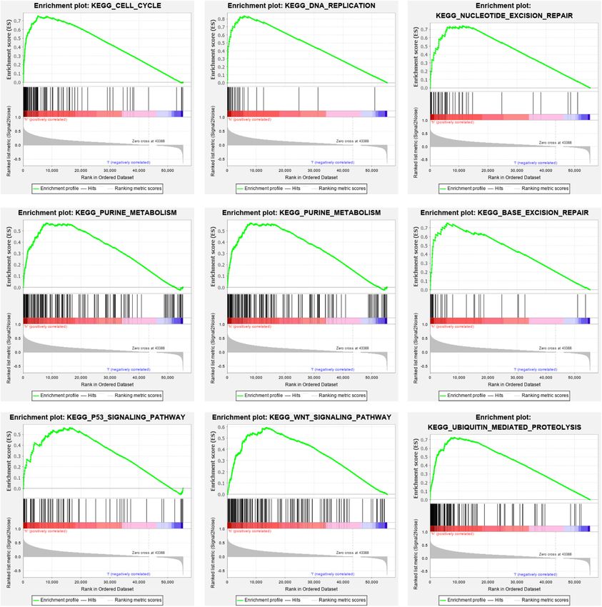
Qi et al.                                                                                                                    m6A RNA Methylation in HCC

                                              we identified processes involved in DNA damage repair and several biological processes
                                              associated with malignant tumors that were closely related to the high-risk group.
                                              Conclusion: In summary, our study identified several genes associated with m6A in
                                              LIHC, especially METTL3 and YTHDF1, and confirmed that a risk signature comprised
                                              of m6A-related genes was able to forecast prognosis.
                                              Keywords: consensus clustering, gene signature, hepatocellular carcinoma, TCGA, UCSC, METTL3, YTHDF1

INTRODUCTION                                                                      uveal melanoma (UM), higher expression of ALKBH5, KIA1429,
                                                                                  and YTHDF1 was found to be associated with worse prognosis
RNAs such as long non-coding RNAs (lncRNAs), transfer RNAs                        (Tang et al., 2020). Conversely, some m6A genes have been found
(tRNAs), messenger RNAs (mRNAs), microRNAs (miRNAs),                              to act as tumor suppressor genes or oncogenes, for example,
and ribosomal RNAs (rRNAs) have been reported to be subjected                     METTL3 and METTL14 (Cui et al., 2017), in neurological
to over 100 types of chemical modifications (Saletore et al.,                     tumors such as glioblastoma (GBM). In addition, a number of
2012; Huang et al., 2020b). Among them, N6-methyladenosine                        other tumors have also been found to be associated with M6A
(m6A) was first identified in 1974. M6A is a reversible post-                     methylation regulators (Zhao et al., 2020).
transcriptional modification and is considered to be the most                         Therefore, the identification of changes in m6A expression
common methylation site of eukaryotic mRNAs (Desrosiers                           in pan-cancer was identified as the starting point of the
et al., 1974; Wei et al., 1975). M6A methyltransferases (also called              study. Among these neoplasms, m6A in LIHC stood out
“writers”) responsible for this type of RNA modification include                  because of its close relationship with prognosis, and thus,
KIAA1429, zinc finger CCCH domain-containing protein                              it became the main focus of our study. LIHC ranks sixth
13 (ZC3H13), methyltransferase-like protein 3 (METTL3),                           among global cancer incidence and ranks fourth in cancer-
METTL14, METTL16, RNA-binding motif protein (RBM15),                              related deaths (Bray et al., 2018). The prognosis of LIHC is
and Wilms Tumor 1-associated protein (WTAP) (Akichika                             unsatisfactory because of the easy recurrence of the tumor
et al., 2019). The α-ketoglutarate-dependent dioxygenase alkB                     after treatment (Llovet et al., 2016). Thus, there is a need to
homolog 5 (ALKBH5) and fat mass and obesity-associated                            identify prognostic markers able to improve the therapeutic
protein (FTO) were called m6A demethylases (also called                           effects. In previous studies on LIHC, some scholars found
“erasers”) and detached m6A (Fu et al., 2013; Zheng et al.,                       that the relationship between M6A methylation regulators and
2013). M6A-binding proteins (also called “readers”) include                       prognosis is not clear and controversial. For example, METTL14
YTHDC1-2, insulin-like growth factor 2 mRNA-binding                               plays an oncogenic role in LIHC (Lin et al., 2016), while
proteins (IGF2BPs), heterogeneous nuclear ribonucleoproteins                      Ma et al. (2017) have proved that METTL14 is an anti-
(HNRNPs), and the YTH N6-methyladenosine RNA binding                              metastatic factor. Similarly, some studies have shown that
proteins 1 to 3 (YTHDF1–3) (Wang et al., 2014; Liao et al., 2018).                YTHDF2 inhibits the development of LIHC (Zhong et al., 2019),
    Except its effects on the synthesis/metabolism of RNA                         but others have found that the overexpression of YTHDF2 is
(Roignant and Soller, 2017), effects on the immune                                related to the poor prognosis of LIHC (Chen et al., 2018).
response, metabolism, viral replication, cancer development,                      In addition, because of the interaction between M6A-related
embryogenesis, and other biological processes have been found                     genes, there is still no criterion for whether the combination
to be associated with modification of m6A (Muthusamy, 2020).                      of these genes can better predict the prognosis of patients.
Some studies have shown that aberrant m6A modification may                        To gain a better comprehensive and accurate understanding
act to induce or inhibit cancer progression in malignant tumors                   of m6A methylation regulators in LIHC with prognosis, we
(Chen X. Y. et al., 2019; Sun et al., 2019), as in, for example,                  did this research.
the hematological malignancy acute myeloid leukemia (AML).
Some studies (Kwok et al., 2017) have revealed that alterations in
m6A regulatory genes confer a worse survival. As oncogenes of                     MATERIALS AND METHODS
AML, METTL3 (Vu et al., 2017), METTL14 (Weng et al., 2018),
WTAP (Bansal et al., 2014), FTO (Li et al., 2017), YTHDF2                         Cancer Datasets
(Li Z. et al., 2018), and IGF2BP1 (Zhou et al., 2017) participate                 All pan-cancerous gene expression datasets (RNA-seq) and
in tumor processes through a variety of pathways, including                       survival information were obtained from The Cancer Genome
the promotion of the growth of cancer cells and inhibition                        Atlas (TCGA1 ). UCSC Xena is a database maintained by the
of apoptosis. In urological tumors, such as prostate cancer                       University of California, Santa Cruz. It contains public datasets
(PCA), studies (Ji et al., 2020) have shown that the expression of                including TCGA, ICGC, TARGET, and other databases and
IGF2BP3, HnRNPA2B1, METTL14, and ALKBH5 was associated                            standardizes the data to make it easier for follow-up analysis
with recurrence-free survival. METTL3 (Cai et al., 2019) and                      (Cline et al., 2013). We analyzed 33 different TCGA projects, each
YTHDF2 (Li J. et al., 2018) as oncogenes promoted PCA cell
proliferation and migration. In neoplasms of the skin, such as                    1
                                                                                      https://xena.ucsc.edu/

Frontiers in Genetics | www.frontiersin.org                                   2                                  January 2021 | Volume 11 | Article 614566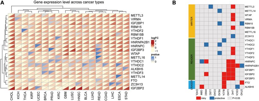
Qi et al.                                                                                                               m6A RNA Methylation in HCC

project represented a specific cancer type, including kidney renal         used as the cutoff value; higher than this value is considered
papillary cell carcinoma (KIRP); kidney chromophobe (KIC);                 as high expression, and lower than this value is considered
brain lower grade glioma (LGG); stomach adenocarcinoma                     as low expression.
(STAD); breast cancer (BRCA); lung adenocarcinoma (LUAD);                      Liver hepatocellular carcinoma samples were grouped by
rectum adenocarcinoma (READ); colon adenocarcinoma                         category using “ConsensusClusterPlus”(Wilkerson and Hayes,
(COAD); acute myeloid leukemia (AML); testicular germ cell                 2010), and the number of groups was denoted by “k.”
tumors (TGCT); liver hepatocellular carcinoma (LIHC); uterine              The LIHC datasets were grouped into distinct and non-
carcinosarcoma (UCS); ovarian serous cystadenocarcinoma                    overlapping groups according to the consistent expression of
(OV); head and neck squamous carcinoma (HNSC); lung                        m6A genes. An optimal prediction model for determination of
squamous cell carcinoma (LUSC); thyroid carcinoma (THCA);                  prognosis was constructed using the least absolute shrinkage
lymphoid neoplasm diffuse large b-cell lymphoma (DLBC);                    and selection operator (LASSO) Cox regression algorithm
prostate adenocarcinoma (PRAD); skin cutaneous melanoma                    (Sauerbrei et al., 2007). According to the cutoff value, patients
(SKCM); bladder urothelial carcinoma (BLCA); uterine corpus                were grouped into high-risk and low-risk groups based on
endometrial carcinoma (UCEC); glioblastoma multiforme                      the risk score (using the risk signature). Using the GLMNET
(GBM); cervical squamous cell carcinoma and endocervical                   package in R to perform the LASSO regression, the risk
                                                                                     n
adenocarcinoma (CESC); adrenocortical carcinoma (ACC);
                                                                                       Coefj∗ xj. In the formula, Coefj and xj, respectively,
                                                                                    P
                                                                           score =
sarcoma (SARC); pancreatic adenocarcinoma (PAAD);                                       j=1
pheochromocytoma and paraganglioma (PCPG); esophageal                      symbolize the coefficient and the z-score-normalized related
carcinoma (ESCA); thymoma (THYM); mesothelioma (MESO);                     expression levels of each gene. Genes with a high correlation
kidney renal clear cell carcinoma (KIRC); uveal melanoma                   were chosen and shrunk to prevent over-fitting, and factors
(UVM); and cholangiocarcinoma (CHOL). In order to use the                  with fairly low association with were removed from the model
most recent data available, liver cancer data in the final study           (Sauerbrei et al., 2007). Finally, an optimal prognostic model
were obtained from TCGA2 database, including gene expression               was constructed using m6A regulatory genes, and each patient
datasets (RNA-seq) belonging to 374 patients with liver cancer             received a risk score. In the display of the heat map, each
and 50 normal controls, as well as the clinical and pathological           small square represents each gene, and the color represents
data in the database.                                                      the expression level of each gene. The greater the expression
                                                                           level, the darker the color (red up-regulation and green
Screening of M6A Methylation                                               down-regulation).
Regulatory Genes                                                              Gene Ontology (GO) analysis was used to explore differences
We identified 21 m6A regulators from recently published review             in biological pathways. Background data of R software “org.Hs.
papers in PubMed (Han and Choe, 2020; Wang T. et al., 2020;                eg.db” was used to obtain the gene ID (Entrez gene ID)
Zhao et al., 2020), including eleven reader, eight writer, and two         of potential targets, and then, the package (Bioconductor)
eraser genes. Among these, 20 m6A methylation regulators were              was used to analyze the GO function enrichment of these
selected for this study, and all were present in the gene expression       potential targets. The correlation with m6A-related genes was
datasets (RNA-seq).                                                        determined by Spearman’s correlation. The string database3
                                                                           is used to generate the interactive network of these genes
Bioinformatics Analysis                                                    (Szklarczyk et al., 2017). Next, the biological process was
All data analysis was based on R (v3.4.1). In the first step, all          divided by the risk score of the high- and the low-risk group,
samples were analyzed for differential expression of m6A in                and Gene Set Enrichment Analysis (GSEA) was used in the
different normal and tumor tissues, excluding tumor types in               LIHC cohort. KEGG gene sets, phenotypic tags (high) and
which the normal group had less than five samples. Wilcoxon’s              (low) expression files were loaded into GSEA (v4.0.3; Broad
rank sum test was used to find the divergence across m6A                   Institute, Cambridge, MA) software and the permutation test was
genes. The identification criteria for m6A having differential             run 1,000 times.
expression for each tumor type was a p-value < 0.05 and at
least a two-fold change in expression. A statistical analysis was          Statistics
needed to evaluate the relationship between survival and M6A-              The differences in continuous variables and categorical variables
related genes in tumors, univariate Cox regression analysis.               among different groups were compared by means of the Student’s
The risk or protective genes correspond to the hazard ratio                t-test and the χ2 test. The differences in survival rates between
of (HR) > 1 or < 1, respectively. The relevance with m6A-                  groups were derived from the Kaplan–Meier survival curve and
related genes and disease-related survival (DSS), overall survival         were verified by the two-sided logrank test. The prognostic
(OS), disease-free interval (DFI), and progression-free interval           capability of the resulting risk score was assessed by singular
(PFI) was shown by Kaplan–Meier survival curves, obtained                  and multiple Cox regression analysis. A p-value < 0.05 was
by the “survival” and “survminer” R package. In survival                   considered to be statistically significant. R (v3.4.14 ) was used for
analysis, genes are ranked from high to low in terms of                    all statistical analysis.
expression level, and the “median” of the expression level is
                                                                           3
                                                                               http://www.string-db.org/
2                                                                          4
    https://portal.gdc.cancer.gov/                                             https://www.r-project.org/

Frontiers in Genetics | www.frontiersin.org                            3                                    January 2021 | Volume 11 | Article 614566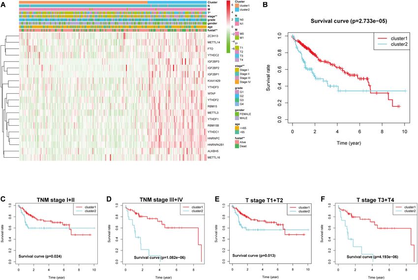
Qi et al.                                                                                                                                 m6A RNA Methylation in HCC

RESULTS                                                                                The Prognostic Role of m6A
                                                                                       Alterations in m6A are prevalent in 18 types of cancers. The
M6A-Related Gene Expression                                                            relationship between the 20 m6A genes and survival time in
In TCGA, we selected the RNA expression datasets relative to 20                        patients in the latter 11 tumor types was assessed by univariate
m6A-related genes in our study (“writers”: METTL3, METTL14,                            Cox analysis. Genes were mainly up-regulated. HR > 1 or HR < 1
METTL16, WTAP, KIAA1429, RBM15, RBM15B, and ZC3H13;                                    corresponded to damaging or protective genes, respectively. We
“readers”: YTHDC1, YTHDC2, YTHDF1, YTHDF2, YTHDF3,                                     found that the tumor survival rates studied were all related to at
HNRNPC, HNRNPA2B1, IGF2BP1, IGF2BP2, and IGF2BP3;                                      least one of the m6A methylation regulators.
and “erasers”: FTO and ALKBH5). The expression changes                                    Some m6A methylation regulators were considered to be
of m6A-related genes are shown as heat maps, with red and                              risk genes, such as the insulin-like growth factor 2 mRNA-
yellow representing up-regulated and down-regulated genes,                             binding proteins (IGF2BPs), including IGF2BP1, IGF2BP2, and
respectively. In the pan-cancer data of 33 cancer types, excluding                     IGF2BP3. Poor survival rates in patients were associated with
the tumor types whose normal samples were less than 5, a                               the increased expression of these genes across cancer types,
total of 18 kinds of tumors were clustered into two categories                         such as IGF2BP1 (HR = 1.324527, P = 2.38E-06) in LUAD
according to the dysregulated expression of m6A-related genes.                         and IGF2BP2 (HR = 1.198617, P = 0.006603) and IGF2BP3
The first seven were mainly genitourinary system tumors, such as                       (HR = 1.570331, P = 0.000198) in LIHC. In contrast, we found
KIRC, UCEC, BRCA, PRAD, KICH, and KIRP. The remaining                                  that several m6A regulators were protective genes for tumors,
11 were mainly respiratory, digestive, and head and neck system                        such as in READ, where the high expression of m6A regulators
tumors and included CHOL, ESCA, STAD, LIHC, COAD,                                      YTHDF2, YTHDC2, RBM15, and METTL14 was significantly
READ, LUSC, LUAD, HNSC, and GBM. Compared with the                                     correlated with better survival (Figure 1B). Among these, we
first seven tumors, the 20 m6A-related genes in the latter 11                          found that m6A in LIHC was associated with the largest number
tumors were mainly up-regulated (METTL3, VIRMA, RBM15,                                 of genes associated with survival, including METTL3, WTAP,
RBM15B, YTHDF1, YTHDF2, IGF2BPs, and HnRNP family)                                     KIAA1429, RBM15, ZC3H13, YTHDF1, YTHDF2, HNRNPC,
and were mainly “writers” and “readers.” However, although                             HNRNPA2B1, IGF2BP1, IGF2BP2, and IGF2BP3. Except for
METTL14 and ZC3H13 are “writer” genes, they were down-                                 ZC3H13, the high expression of these genes was related
regulated in the first seven kinds of tumors listed above. Similarly,                  to the worse survival rate of LIHC; thus, we focused on
an up-regulated gene expression was found in upper digestive                           LIHC in this study.
system tumors including ESCA, STAD, HIHC, and CHOL. In                                    The relationship between the m6A regulators in LIHC
addition, we found that IGF2BP3 was up-regulated in 17 tumors                          and PFI, DSS, OS, and DFI was determined by the Kaplan–
except for THCA (Figure 1A). Based on these findings, we                               Meier method. The OS of patients with higher expression of
preliminarily found that changes in the expression of 20 m6A-                          METTL3, YTHDF2, YTHDF1, IGF2BP3, RBM15B, RBM15, and
related genes may vary across cancer types. These results also                         HNRNPA2B1 was worse than the low-expression group. The
reveal the highly heterogeneous expression changes of m6A                              DSS of patients with higher expression of METTL3, YTHDF1,
in different cancer types, suggesting that the dysregulation of                        METTL16, HNRNPC, and RBM15 was significantly poorer
m6A regulatory factors may play an important role in different                         than that of patients with low expression. The DFI of patients
cancer environments.                                                                   with higher gene expression of METTL3, YTHDF1, HNRNPC,

  FIGURE 1 | The expression of m6A-related genes in the pan-cancer analysis. (A) Gene expression variation of m6A genes across 18 cancer types. The heatmap
  shows the fold changes. Red represents overexpression genes and yellow represents lower expression genes. P < 0.05 was considered as statistically significant.
  (B) The relationship between higher expression of m6A-associated genes and patient survival, with red and blue representing worse and better survival, respectively.
  Only P-values < 0.05 are shown.

Frontiers in Genetics | www.frontiersin.org                                        4                                         January 2021 | Volume 11 | Article 614566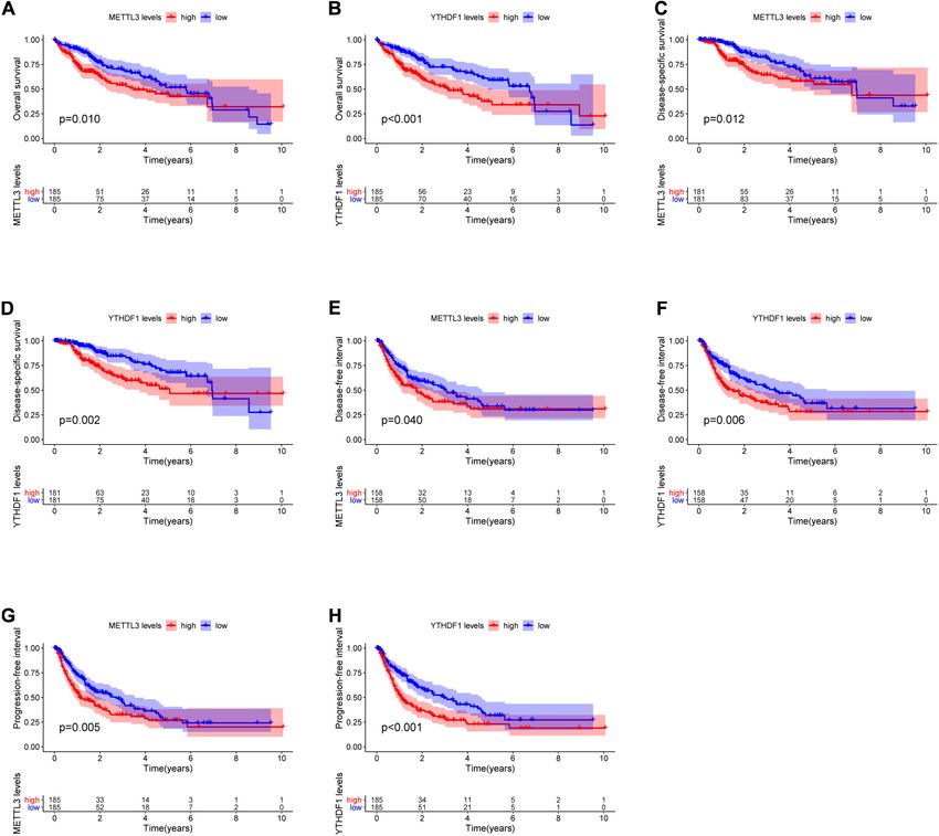
Qi et al. m6A RNA Methylation in HCC
FIGURE 2 | The Kaplan–Meier curves for OS, DSS, DFI, and PFI in LIHC. Kaplan–Meier survival curves showing differences in OS (A,B) and DSS (C,D) stratified
according to METTL3 and YTHDF1 expression. Kaplan–Meier survival curves showing difference in DFI (E,F) and PFI (G,H) stratified according to METTL3 and
YTHDF1 expression. LIHC, liver hepatocellular carcinoma; OS, overall survival; DSS, disease-associated survival; DFI, disease-free interval; PFI, progression-free
interval.
and HNRNPA2B1 was significantly lower than that in the when K = 3, CDF is in a position of slow growth and the
low-expression group. The PFI of patients with higher gene clustering analysis result is the most reliable at this time. The
expression of METTL3, YTHDF1, WTAP, IGF2BP3, YTHDC1, delta area plot (Figure 3B) shows the relative changes in the
RBM15B, RBM15, and HNRNPA2B1 was worse than the area under the CDF curve compared to k and K - 1. The
low-expression genes. Overall, we found that the combined first point represents the total area under the CDF curve at
higher expression of METTL3 and YTHDF1 correlated with K = 2, not the relative change of the area, because there is no
worse prognosis of patients in terms of OS, DSS, DFI, and K = 1. When k = 4, the area under the curve increases only
PFI (Figure 2). slightly, so 3 is the appropriate k value. Figure 3C shows the
matrix heat map when k = 2: the rows and columns of the
matrix represent samples; the values of the consistency matrix are
Subgroup Identification Based on represented by white to dark blue from 0 (impossible to cluster
Consensus Clustering together) to 1 (always clustered together); and the consistency
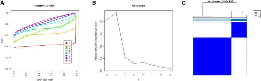
In order to deeper investigate the clinical correlation of 20 matrix is arranged according to the consistency classification
m6A related-genes in LIHC, we used the class discovery tool (the tree above the heat map). The category is divided by the
“ConsensusClusterPlus” to group LIHC samples according to long bar between the tree and the heat map. According to
gene expression patterns and used “k” to indicate the number the CDF and the delta area plot, we can temporarily consider
of subgroups. The cumulative distribution function (CDF) graph grouping patients into three groups. However, the matrix heat
(Figure 3A) shows the cumulative distribution function when map showed that when k = 3, the sample size of one of the groups
“k” takes different values (k = 2–9). As shown in the figure, was too small and the correlation between groups was high. In
Frontiers in Genetics | www.frontiersin.org 5 January 2021 | Volume 11 | Article 614566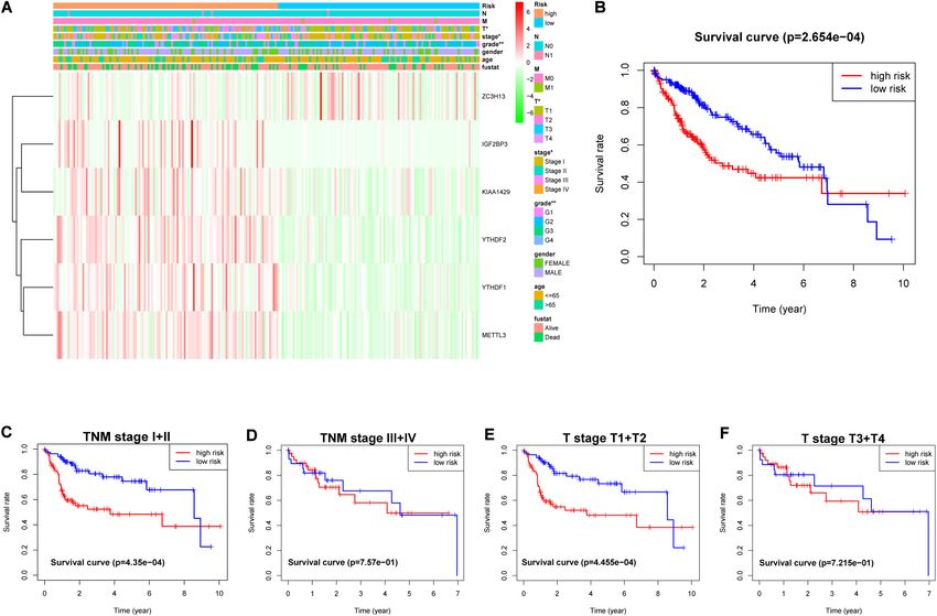
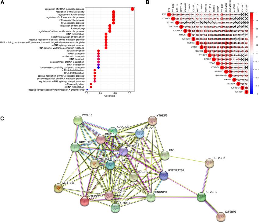
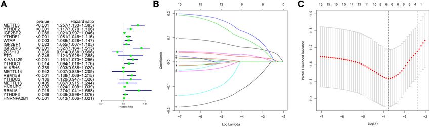
Qi et al.                                                                                                                             m6A RNA Methylation in HCC

  FIGURE 3 | Consensus clustering of m6A-related genes. (A) Cumulative distribution function (CDF) for LIHC. (B) The area under the CDF curve in LIHC.
  (C) Consensus clustering matrix for LIHC. LIHC, liver hepatocellular carcinoma.

summary, in accordance with the m6A-related gene expression                          existed in protein–protein interaction networks, especially in
approach for consensus clustering, when k = 2, the LIHC cohort                       writers (Figure 5C).
could be separated into two subgroups which were different and
did not overlap.                                                                     Building the Prognostic Signatures
   According to the heat map obtained relative to the gene                           Cox regression analysis and Kaplan–Meier survival curves were
expression characteristics of the two groups after observation                       used in univariate analysis to determine the correlation between
consensus clustering, we found that the genes in the cluster2                        genes and prognosis, and consensus clustering was used to
group were generally highly expressed. Next, we investigated                         further explore clinical correlations. Through protein–protein
whether there was a distinction between the clinical and                             interaction network analysis and Spearman’s correlation analysis,
pathological features between the two groups. The outcomes                           we found the functions of these m6A methylation regulators were
showed obvious differences between tumor T stage and clinical                        not isolated, and there was cooperation among writers, erasers,
stage (Figure 4A). According to the correlation between grouping                     and readers. Therefore, in order to improve the predictive ability
and clinical data, we found that the OS of cluster1 (n = 261)                        of m6A in LICH, we used the LASSO Cox regression algorithm
group was better than the cluster2 group (n = 109) (Figure 4B).                      to eliminate genes that did not meet our requirements in order to
Next, we determined that the expression patterns of 20 m6ARNA                        establish suitable prognostic gene markers. This method allowed
methylation regulatory genes could predict clinical outcomes                         us to compute a patient’s risk score by combining the level of gene
of LICH subgroups stratified by clinical stage. This consensus                       expression with the risk coefficient.
clustering based on expression pattern was capable of predicting                         The genes analyzed by univariate Cox analysis were screened
prognosis in both early stage (I + II; T1 + T2) (Figures 4C,E) and                   according to the standard of P < 0.1 by the LASSO Cox
late stage (III + IV; T3 + T4) (Figures 4D,F).                                       regression algorithm (Figure 6A). Fifteen genes that met the
   We further annotated these genes according to GO terms                            requirements were substituted into the model, and we chose
and discovered that they were mainly involved in mRNA                                and shrunk the genes with high correlation to prevent over-
processing-related pathways, containing RNA modification,                            fitting (Figures 6B,C). As a result, LASSO regression produced
regulation of RNA splicing, metabolism, transport, and                               a six-gene signature, including YTHDF2, YTHDF1, METTL3,
stability, which were consistent with the RNA modification                           IGF2BP3, KIAA1429, and ZC3H13. The resulting risk score
function of m6A (Figure 5A). In addition, m6A methylation                            divided the LIHC patients into the low- and high-risk groups
regulators do not work in isolation. Previous evidence has                           of OS. We continued to observe whether there were differences
shown that cooperation among writers, readers, and erasers                           in clinical and pathological features between the two groups.
is the background of carcinogenesis (Panneerdoss et al.,                             The results showed obvious differences in T stage, pathological
2018). Using Spearman’s correlation analysis to calculate                            stage, and clinical stage (Figure 7A). The OS in the low-risk
the correlation of these genes in LIHC, we identified m6A-                           (n = 185) group was significantly better than that in the high-
related genes in the same functional category showing                                risk group (n = 185) (Figure 7B). We also examined whether
highly interrelated expression patterns, which overlapped                            the high-risk or low-risk score could predict clinical outcome in
those of authors, erasers, and readers. For example, the                             LICH subgroup stratified by clinical stage. The results showed
expression levels of METTL3, YTHDF1, RBM15B, YTHDF2,                                 the gene expression in stages I + II out-performed prognosis
RBM15, WTAP, YTHDC1, HNRNPC, HNRNPA2B1, and                                          prediction over gene expression in stages III + IV in LICH
KIAA1429 genes closely associated with each other, while the                         (Figures 7C,D), and was better in calculating prognosis for
expression level of ZC3H13 gene was weakly correlated or not                         stages T1 + T2 than for stages T3 + T4 (Figures 7E,F). Next,
associated with other genes except for METTL14 and YTHDC1                            we performed single and multiple Cox analysis. The resulting
(Figure 5B). In addition, the interplay of these genes also                          risk score was confirmed to be an independent prognostic

Frontiers in Genetics | www.frontiersin.org                                      6                                        January 2021 | Volume 11 | Article 614566
Qi et al.                                                                                                                                 m6A RNA Methylation in HCC

  FIGURE 4 | Heatmap and clinical features in (A) cluster1 and cluster2, stratified by the m6A-related gene consensus analysis. (B) Kaplan–Meier survival curves for
  groups of clusters in LIHC. Kaplan–Meier OS curves for patients with (C) stage I + II and (D) III + IV in LIHC. Kaplan–Meier OS curves for patients with (E)
  stageT1 + T2 and (F) T3 + T4 in LIHC. LIHC, liver hepatocellular carcinoma; OS, overall survival.

factor for LIHC and showed good sensitivity and specificity,                           DISCUSSION
as demonstrated by the receiver operating characteristic (ROC)
curves (Figure 8).                                                                     M6A is the most universal chemical modification in RNA.
                                                                                       Although previous studies have shown that m6A is involved in
                                                                                       many biological processes (Han et al., 2019; Shi et al., 2019; Zhong
Signal Pathways and Cellular Processes                                                 et al., 2019), the role of m6A modification in cancer and clinical
Related to M6A Regulators                                                              exploration is still in infancy (Lan et al., 2019).
In order to further investigate the effects of m6A regulators                             A variety of studies have shown that the role of M6A in
on signal pathways and cellular processes, we turned to GSEA                           different tumors is not consistent, and even in the same tumor,
to inspect the signal pathways involved in the high-risk and                           the conclusion is opposite. In order to more systematically
low-risk prognosis groups. We found that the high-risk group                           study the role of M6A in tumors, we have conducted a
was characterized by the following biological processes/signal                         systematic study on a variety of tumors. We found that in pan-
pathways. Cell cycle (Nes = 2.13, Fdr = 0.001), DNA replication                        cancer, the expression of m6A methylation regulators is often
(Nes = 1.97, p = 0.006), pyrimidine metabolism (Nes = 2.12,                            dysregulated, but presented specific characteristics. Through
p = 0.000), nucleotide excision repair (NER) (Nes = 2.05,                              cluster analysis, we found that there were distinctions in
FDR = 0.001), base excision repair (BER) (Nes = 2.05, p = 0.000),                      the overall expression characteristics in m6A-related genes in
WNT signaling pathway (Nes = 1.99, p = 0.000), purine                                  the seven genitourinary cancers and in 11 cancers involving
metabolism (Nes = 2.08, p = 0.000), p53 signaling pathway                              the respiratory, digestive, and head and neck systems. Could
(Nes = 1.88, p = 0.000), and ubiquitin-mediated proteolysis                            this difference provide a perspective for new research? For
(Nes = 2.02, p = 0.000). The loss of control of the following                          example, future studies may address the different effects of
processes correlated with oncogenesis: cell cycle regulation,                          multiple m6A methylation regulators in multiple human cancers,
WNT signaling pathway, p53 signaling pathway, and ubiquitin-                           rather than investigating a single m6A regulator for each
mediated proteolysis (Figure 9).                                                       tumor as the object of study as in the past. Our findings

Frontiers in Genetics | www.frontiersin.org                                        7                                         January 2021 | Volume 11 | Article 614566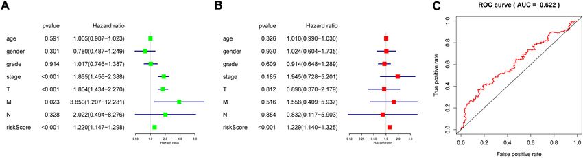
Qi et al.                                                                                                                                  m6A RNA Methylation in HCC

  FIGURE 5 | Spearman’s correlation analysis and functional annotations of 20 m6A-related genes. (A) GO annotation. (B) Spearman correlation analysis in 20 m6A
  modification regulators genes in LIHC. (C) String protein–protein interaction network. LIHC, liver hepatocellular carcinoma.

  FIGURE 6 | Screening of variables. (A) The hazard ratios (HR) and 95% confidence intervals (CI) of 20 m6A modification regulators in LIHC was computed by
  univariate Cox regression. In the 15 genes, high correlation genes were chosen and shrunk to prevent over-fitting (B,C) and finally produced a six-gene signature in
  LASSO regression. LIHC, liver hepatocellular carcinoma; LASSO, least absolute shrinkage and selection operator.

showed that the expression of m6A-related genes was associated                          a direction for future research. In addition, we found that
with changes in gene expression in upper digestive system                               IGF2BP3 was up-regulated in all 17 tumors investigated,
tumors, such as ESCA, STAD, HIHC, and CHOL, which                                       except for THCA, which was basically consistent with that
were all up-regulated. At present, there has been no attempt                            shown by Li et al. (2019).
to compare m6A-related genes in different human cancers                                    On univariate Cox regression analysis, we found that in
and their clinical correlations, and these findings will provide                        LIHC, with the exception of ZC3H13, higher m6A-related gene

Frontiers in Genetics | www.frontiersin.org                                         8                                         January 2021 | Volume 11 | Article 614566
Qi et al.                                                                                                                                  m6A RNA Methylation in HCC

  FIGURE 7 | (A) Heatmap and clinicopathological characteristics of the subgroup classified by the six-gene prognostic signature in LIHC. (B) Kaplan–Meier survival
  curves of LIHC subgroups defined by the six-gene signature based on the LASSO regression. Kaplan–Meier OS curves for patients with (C) stage I + II and
  (D) III + IV LIHC. Kaplan–Meier OS curves for patients with stage (E) T1 + T2 and (F) T3 + T4 LIHC. LIHC, liver hepatocellular carcinoma; OS, overall survival.

  FIGURE 8 | The role of risk score in prognosis. Single (A) and multiple (B) factors used in the Cox analysis to develop the risk scores in LIHC. (C) ROC curve
  showing the supporting the performance of the model. LIHC, liver hepatocellular carcinoma; ROC, receiver operating characteristic.

expression was associated with worse OS. In the Kaplan–Meier                            WTAP contributes to m6A modification contributing to the
survival curve analysis, we found that the up-regulation of m6A                         development of LIHC (Chen Y. et al., 2019). The overexpression
regulators was associated with worse PFI, DFI, DSS, and OS.                             of KIAA1429 induces tumor growth and metastasis by inducing
Among these, the combined high expression of METTL3 and                                 the separation of the HuR binding and degradation of GATA3
YTHDF1 genes correlated with OS, DSS, DFI, and PFI and                                  pre-mRNA (Wang M. et al., 2020). It can also facilitate the
indicated worse overall prognosis of patients. Some studies have                        migration and invasion of tumor cells by inhibiting ID2 mRNA
found that increased gene expression in LIHC may be implicated                          (Cheng et al., 2019). Through regulation of Snail, a key translator
in the development of cancer. For example, WTAP is significantly                        of EMT, METTL3 and YTHDF1 became adverse prognostic
up-regulated in LIHC. Through the HuR-ETS1-p21/p27 axis,                                factors for OS in patients with LIHC (Lin et al., 2019). Copy

Frontiers in Genetics | www.frontiersin.org                                         9                                         January 2021 | Volume 11 | Article 614566
Qi et al.                                                                                                                             m6A RNA Methylation in HCC

  FIGURE 9 | Cellular processes and pathways in LIHC subsets, defined by risk scores. GSEA showed that the low survival subgroup was obviously correlated with
  processes such as the cell cycle, DNA replication, WNT signal pathway, and protein degradation. GSEA, Gene Set Enrichment Analysis; LIHC, liver hepatocellular
  carcinoma.

number variations (CNV) and DNA methylation were found to                            (Zhao et al., 2018). YTHDF2 induces proliferation, migration,
be the main causes of aberrant up-regulation of METTL3, which                        and colony formation of LIHC cells by promoting METTL3-
was found to be an independent prognostic factor in the relapse-                     mediated SOCS2 m6A modification (Chen et al., 2018). In
free survival rate (RFS) and OS rate (Liu G. M. et al., 2020).                       contrast, some studies have shown that YTHDF2 could inhibit
This report was consistent with our findings on the relationship                     the progression of liver cancer by stabilizing EGFR or IL-11
between up-regulation of METTL3 and YTHDF1 and their                                 mRNA (Hou et al., 2019; Zhong et al., 2019). These results
association with OS, DSS, DFI, PFI, and worse prognosis of                           indicate that the aberrant expression of m6A methylation
patients. Among the m6A reader genes, the up-regulation of                           regulators in LIHC is common, although the controversial results
YTHDF1 has been found related to worse prognosis of LIHC                             and the potential mechanisms involved are worthy of further

Frontiers in Genetics | www.frontiersin.org                                     10                                        January 2021 | Volume 11 | Article 614566
Qi et al.                                                                                                             m6A RNA Methylation in HCC

study. Nonetheless, these findings provide further evidence of                The high- and low-risk groups constructed using the
the functional role and potential mechanisms involving a single            m6A methylation regulators stimulated our investigation of
m6A RNA modification regulation in tumors, although other                  the potential mechanisms involved. GSEA analysis showed
m6A methylation regulators that may also play a relatively minor           several significant signaling pathways and cellular processes
role are often ignored. We further analyzed the expression and             in the high-risk group were associated with poor prognosis,
prognostic significance of multiple genes in LIHC.                         including processes involving the cell cycle, NER, DNA
   We comprehensively analyzed the role and prognostic value               replication, purine metabolism, pyrimidine metabolism, BER,
of 20 m6A RNA modification regulators in LIHC. According to                WNT signal pathway, p53 signaling pathway, and ubiquitin-
the consensus clustering of m6A RNA modification regulators,               mediated proteolysis. The cellular processes involved in
LIHC patients could be stratified into two subgroups in terms              DNA replication, purine metabolism, BER, and NER were
of OS. The clustering analysis revealed that all m6A-related               consistent with previous reports, indicating DNA damage
genes were highly expressed in the poor prognosis group                    was regulated by m6A-induced methylation (Xiang et al.,
(cluster2). Univariate analysis indicated that the up-regulated            2017). The response of m6A in DNA damage is transient.
expression of a single gene is related to the poor prognosis               This modification is regulated by N6-methyltransferase-like
of patients, while consensus clustering suggested that there               protein 3 (METTL3) and obesity-associated protein (FTO)
may be some relationship between these genes, which also                   and is mostly found in poly(A) transcripts. Previous studies
require further study. Next, we used the LASSO Cox regression              have shown that M6A plays an important role in the DNA
algorithm to establish a prognostic gene signature for OS. In              damage response, because the lack of METTL3 leads to
accordance with the significant differences in clinical stages             delayed cell repair and increased sensitivity to ultraviolet light
and T stages of the high- and low-risk groups, we found that               (Xiang et al., 2017). Several biological processes related to
the predictive model constituted by the expression profiles of             malignant tumors are noteworthy, including regulation of
six genes was a stronger predictor of prognosis of patients in             cell cycle, WNT signaling pathway, p53 signaling pathway,
stages I + II than in patients in stages III + IV and was also             and ubiquitin-mediated proteolysis. It has been reported that
a better predictor of patients in stages T1 + T2 than those in             the RNA METTL3 and miR-186 regulate hepatoblastoma
stages T3 + T4. The risk score results were an independent                 progression through the Wnt/β-catenin signaling pathway
prognostic factor of LIHC.                                                 (Cui et al., 2020). Overall, this evidence indicated that the
   Among available studies, we found that those evaluating the             characteristic expression of m6A methylation regulators
combination of m6A-related genes were based on 13 major                    has great prospect as prognostic indicators of LIHC. These
M6A genes, including METTL3, METTL14, WTAP, KIAA1429,                      genes and their relative pathways may be latent treatment
RBM15, ZC3H13, YTHDC1, YTHDC2, YTHDF1, YTHDF2,                             objectives for LIHC.
HNRNPC, FTO, and ALKBH5. In our study, we investigated                        However, our research still has some limitations. First
a total of 20 m6A-related genes that have been identified so               of all, most of the LIHC patients we studied were white
far with available data in TCGA. This includes seven genes                 and Asian. However, it is not clear about the geographical
that have not been previously investigated, including METTL16,             location of the specific life of these Asians, so whether
RBM15B, YTHDF3, HNRNPA2B1, IGF2BP1, IGF2BP2, and                           the final results of the study are also applicable to the
IGF2BP3. In our investigation of the correlation of m6A-                   Chinese population needs to be further verified by clinical
related genes, we found that there is a significant relationship           sampling in Chinese patients. Second, the sample size may be
between the 20 M6A genes, and in particular, IGF2BP3                       insufficient because many cases lacked clinical information.
and HNRNPA2B1, RBM15B, and YTHDF2. We think these                          Third, several significant prognostic factors for LIHC were
associations may affect the results of single-factor, multi-factor,        not evaluated in the study, such as hepatitis B virus infection
and cluster analyses and the construction of the prognostic                and abnormal liver function, which may lead to changes in
model; thus, the remaining seven genes should not be excluded.             the correlation between m6A-related genes and prognosis.
In previous studies (Huang et al., 2020a; Liu J. et al., 2020;             Fourth, the description of the potential mechanism of the
Wu et al., 2020), the following five genes have been shown to              role of these up-regulated genes in prognosis was not based
be independent prognostic risk factors, including KIAA1429,                on experimental evidence; thus, further confirmation is
METTL3, YTHDF1, YTHDF2, and ZC3H13. Some of them                           required in the future.
constructed a prognostic model containing five genes. After
taking into account the interaction between 20m6A genes,
our findings newly showed that IGF2BP3 could be included                   CONCLUSION
in the model as an independent risk factor for LIHC, and
the risk score resulted to be an independent prognostic                    Based on an integrated bioinformatics analysis, the present
factor, which was not found in the previously reported gene                study identified several genes associated with m6A in LIHC.
combination analysis. This provides a direction for our further            In particular, METTL3 and YTHDF1 expression were found
experimental research on IGF2BP3 in the future. We also                    to be correlated with an increased risk and were included
found that the subgroups stratified according to consensus                 in an m6A-related gene signature for predicting prognosis of
clustering and gene signature were applicable to the early stages          LIHC. The poor prognosis group was closely associated with
I + II and T1 + T2.                                                        a poor response to DNA damage repair and several biological

Frontiers in Genetics | www.frontiersin.org                           11                                  January 2021 | Volume 11 | Article 614566
Qi et al.                                                                                                                                               m6A RNA Methylation in HCC

processes associated with malignant tumors. In the                                               AUTHOR CONTRIBUTIONS
pan-cancer analysis used in our preliminary study,
we found that changes in m6A-related genes occurred                                              L-WQ and J-MH designed the study. L-WQ and C-HJ collected
across different cancer types. This provides an important                                        the literature. L-WQ, J-HJ, and J-MH performed statistical
rationale to guide future cross-cancer studies. The                                              analyses. L-WQ, J-HJ, J-MH, and C-HJ analyzed the data.
mechanisms indicated for the role played by up-                                                  L-WQ wrote the manuscript. All authors listed have made
regulated genes were only theoretical; thus, functional                                          substantial, direct and intellectual contribution to the work and
experiments and prospective clinical studies are needed to                                       approved it for publication.
validate our findings.

                                                                                                 FUNDING
DATA AVAILABILITY STATEMENT
                                                                                                 This work was supported by the National Natural Science
The original contributions presented in the study are included                                   Foundation of China (Nos. 81760428, 81960435, 81460363,
in the article/supplementary material, further inquiries can be                                  and 81860518) and the Start-up Project of High-level Talents
directed to the corresponding author/s.                                                          Scientific Research in Shihezi University (RCZK2018C19).

REFERENCES                                                                                       Han, D., Liu, J., Chen, C., Dong, L., Liu, Y., Chang, R., et al. (2019). Anti-tumour
                                                                                                     immunity controlled through mRNA m(6)A methylation and YTHDF1 in
Akichika, S., Hirano, S., Shichino, Y., Suzuki, T., Nishimasu, H., and Ishitani, R.                  dendritic cells. Nature 566, 270–274. doi: 10.1038/s41586-019-0916-x
   (2019). Cap-specific terminal -methylation of RNA by an RNA polymerase                        Han, S. H., and Choe, J. (2020). Diverse molecular functions of mA mRNA
   II-associated methyltransferase. Science 363:eaav0080. doi: 10.1126/science.                      modification in cancer. Exp. Mol. Med. 52, 738–749. doi: 10.1038/s12276-020-
   aav0080                                                                                           0432-y
Bansal, H., Yihua, Q., Iyer, S. P., Ganapathy, S., Proia, D. A., and Penalva,                    Hou, J., Zhang, H., Liu, J., Zhao, Z., Wang, J., Lu, Z., et al. (2019). YTHDF2
   L. O. (2014). WTAP is a novel oncogenic protein in acute myeloid leukemia.                        reduction fuels inflammation and vascular abnormalization in hepatocellular
   Leukemia 28, 1171–1174. doi: 10.1038/leu.2014.16                                                  carcinoma. Mol. Cancer 18:163. doi: 10.1186/s12943-019-1082-3
Bray, F., Ferlay, J., Soerjomataram, I., Siegel, R. L., Torre, L. A., and Jemal, A.              Huang, H., Bai, Y., Lu, X., Xu, Y., Zhao, H., and Sang, X. (2020a). N6-
   (2018). Global cancer statistics 2018: GLOBOCAN estimates of incidence and                        methyladenosine associated prognostic model in hepatocellular carcinoma.
   mortality worldwide for 36 cancers in 185 countries. CA Cancer J. Clin. 68,                       Ann. Transl. Med. 8:633. doi: 10.21037/atm-20-2894
   394–424. doi: 10.3322/caac.21492                                                              Huang, H., Weng, H., and Chen, J. (2020b). The biogenesis and precise control of
Cai, J., Yang, F., Zhan, H., Situ, J., Li, W., and Mao, Y. (2019). RNA m(6)A                         RNA mA methylation. Trends Genet. 36, 44–52. doi: 10.1016/j.tig.2019.10.011
   methyltransferase METTL3 promotes the growth of prostate cancer by                            Ji, G., Huang, C., He, S., Gong, Y., Song, G., Li, X., et al. (2020). Comprehensive
   regulating hedgehog pathway. OncoTargets Ther. 12, 9143–9152. doi: 10.2147/                       analysis of m6A regulators prognostic value in prostate cancer. Aging 12,
   ott.S226796                                                                                       14863–14884. doi: 10.18632/aging.103549
Chen, M., Wei, L., Law, C. T., Tsang, F. H., Shen, J., and Cheng, C. L.                          Kwok, C. T., Marshall, A. D., Rasko, J. E., and Wong, J. J. (2017). Genetic
   (2018). RNA N6-methyladenosine methyltransferase-like 3 promotes liver                            alterations of m(6)A regulators predict poorer survival in acute myeloid
   cancer progression through YTHDF2-dependent posttranscriptional silencing                         leukemia. J. Hematol. Oncol. 10:39. doi: 10.1186/s13045-017-0410-6
   of SOCS2. Hepatology 67, 2254–2270. doi: 10.1002/hep.29683                                    Lan, Q., Liu, P. Y., Haase, J., Bell, J. L., Hüttelmaier, S., and Liu, T. (2019). The
Chen, X. Y., Zhang, J., and Zhu, J. S. (2019). The role of mA RNA methylation in                     critical role of RNA m(6)A methylation in cancer. Cancer Res. 79, 1285–1292.
   human cancer. Mol. Cancer 18:103. doi: 10.1186/s12943-019-1033-z                                  doi: 10.1158/0008-5472.Can-18-2965
Chen, Y., Peng, C., Chen, J., Chen, D., Yang, B., and He, B. (2019). WTAP facilitates            Li, J., Meng, S., Xu, M., Wang, S., He, L., Xu, X., et al. (2018). Downregulation
   progression of hepatocellular carcinoma via m6A-HuR-dependent epigenetic                          of N(6)-methyladenosine binding YTHDF2 protein mediated by miR-493-
   silencing of ETS1. Mol. Cancer 18:127. doi: 10.1186/s12943-019-1053-8                             3p suppresses prostate cancer by elevating N(6)-methyladenosine levels.
Cheng, X., Li, M., Rao, X., Zhang, W., Li, X., and Wang, L. (2019). KIAA1429                         Oncotarget 9, 3752–3764. doi: 10.18632/oncotarget.23365
   regulates the migration and invasion of hepatocellular carcinoma by altering                  Li, Y., Xiao, J., Bai, J., Tian, Y., Qu, Y., Chen, X., et al. (2019). Molecular
   m6A modification of ID2 mRNA. OncoTargets Ther. 12, 3421–3428. doi: 10.                           characterization and clinical relevance of m(6)A regulators across 33 cancer
   2147/ott.S180954                                                                                  types. Mol. Cancer 18:137. doi: 10.1186/s12943-019-1066-3
Cline, M. S., Craft, B., Swatloski, T., Goldman, M., Ma, S., and Haussler, D. (2013).            Li, Z., Qian, P., Shao, W., Shi, H., He, X. C., Gogol, M., et al. (2018). Suppression of
   Exploring TCGA pan-cancer data at the UCSC cancer genomics browser. Sci.                          m(6)A reader Ythdf2 promotes hematopoietic stem cell expansion. Cell Res. 28,
   Rep. 3:2652. doi: 10.1038/srep02652                                                               904–917. doi: 10.1038/s41422-018-0072-0
Cui, Q., Shi, H., Ye, P., Li, L., Qu, Q., Sun, G., et al. (2017). m(6)A RNA methylation          Li, Z., Weng, H., Su, R., Weng, X., Zuo, Z., Li, C., et al. (2017). FTO plays an
   regulates the self-renewal and tumorigenesis of glioblastoma stem cells. Cell                     oncogenic role in acute myeloid leukemia as a N(6)-Methyladenosine RNA
   Rep. 18, 2622–2634. doi: 10.1016/j.celrep.2017.02.059                                             demethylase. Cancer Cell 31, 127–141. doi: 10.1016/j.ccell.2016.11.017
Cui, X., Wang, Z., Li, J., Zhu, J., Ren, Z., Zhang, D., et al. (2020). Cross talk between        Liao, S., Sun, H., and Xu, C. (2018). YTH domain: a family of N(6)-
   RNA N6-methyladenosine methyltransferase-like 3 and miR-186 regulates                             methyladenosine (m(6)A) readers. Genomics Proteomics Bioinformatics 16,
   hepatoblastoma progression through Wnt/β-catenin signalling pathway. Cell                         99–107. doi: 10.1016/j.gpb.2018.04.002
   Prolif. 53:e12768. doi: 10.1111/cpr.12768                                                     Lin, S., Choe, J., Du, P., Triboulet, R., and Gregory, R. I. (2016). The m (6) a
Desrosiers, R., Friderici, K., and Rottman, F. (1974). Identification of methylated                  methyltransferase METTL3 promotes translation in human Cancer cells. Mol.
   nucleosides in messenger RNA from Novikoff hepatoma cells. Proc. Natl. Acad.                      Cell 62, 335–345. doi: 10.1016/j.molcel.2016.03.021
   Sci. U.S.A. 71, 3971–3975. doi: 10.1073/pnas.71.10.3971                                       Lin, X., Chai, G., Wu, Y., Li, J., Chen, F., Liu, J., et al. (2019). RNA m(6)A
Fu, Y., Jia, G., Pang, X., Wang, R. N., Wang, X., Li, C. J., et al. (2013). FTO-                     methylation regulates the epithelial mesenchymal transition of cancer cells
   mediated formation of N6-hydroxymethyladenosine and N6-formyladenosine                            and translation of Snail. Nat. Commun. 10:2065. doi: 10.1038/s41467-019-0
   in mammalian RNA. Nat. Commun. 4:1798. doi: 10.1038/ncomms2822                                    9865-9

Frontiers in Genetics | www.frontiersin.org                                                 12                                             January 2021 | Volume 11 | Article 614566
Qi et al. m6A RNA Methylation in HCC Liu, J., Sun, G., Pan, S., Qin, M., Ouyang, R., Li, Z., et al. (2020). The cancer Wang, T., Kong, S., Tao, M., and Ju, S. (2020). The potential role of RNA genome atlas (TCGA) based mA methylation-related genes predict prognosis N6-methyladenosine in Cancer progression. Mol. Cancer 19:88. doi: 10.1186/ in hepatocellular carcinoma. Bioengineered 11, 759–768. doi: 10.1080/21655979. s12943-020-01204-7 2020.1787764 Wang, X., Lu, Z., Gomez, A., Hon, G. C., Yue, Y., Han, D., et al. (2014). N6- Liu, G. M., Zeng, H. D., Zhang, C. Y., and Xu, J. W. (2020). Identification of methyladenosine-dependent regulation of messenger RNA stability. Nature METTL3 as an adverse prognostic biomarker in hepatocellular carcinoma. Dig. 505, 117–120. doi: 10.1038/nature12730 Dis. Sci. doi: 10.1007/s10620-020-06260-z Wei, C. M., Gershowitz, A., and Moss, B. (1975). Methylated nucleotides block Llovet, J. M., Zucman-Rossi, J., Pikarsky, E., Sangro, B., Schwartz, M., Sherman, 5’ terminus of HeLa cell messenger RNA. Cell 4, 379–386. doi: 10.1016/0092- M., et al. (2016). Hepatocellular carcinoma. Nat. Rev.Dis. Primers 2:16018. 8674(75)90158-0 doi: 10.1038/nrdp.2016.18 Weng, H., Huang, H., Wu, H., Qin, X., Zhao, B. S., Dong, L., et al. (2018). Ma, J. Z., Yang, F., Zhou, C. C., Liu, F., Yuan, J. H., Wang, F., et al. (2017). METTL14 mettl14 inhibits hematopoietic stem/progenitor differentiation and promotes suppresses the metastatic potential of hepatocellular carcinoma by modulating leukemogenesis via mRNA m(6)A modification. Cell Stem Cell 22, 191.e9– N -methyladenosine-dependent primary MicroRNA processing. Hepatology 65, 205.e9. doi: 10.1016/j.stem.2017.11.016 529–543. doi: 10.1002/hep.28885 Wilkerson, M. D., and Hayes, D. N. (2010). ConsensusClusterPlus: a class discovery Muthusamy, S.J. (2020). m6 A mRNA methylation: a pleiotropic regulator of tool with confidence assessments and item tracking. Bioinformatics 26, 1572– cancer. Gene 736:144415. doi: 10.1016/j.gene.2020.144415 1573. doi: 10.1093/bioinformatics/btq170 Panneerdoss, S., Eedunuri, V. K., Yadav, P., Timilsina, S., Rajamanickam, S., Wu, X., Zhang, X., Tao, L., Dai, X., and Chen, P. (2020). Prognostic value Viswanadhapalli, S., et al. (2018). Cross-talk among writers, readers, and erasers of an m6A RNA methylation regulator-based signature in patients with of mA regulates cancer growth and progression. Sci. Adv. 4:eaar8263. doi: hepatocellular carcinoma. BioMed Res. Int. 2020:2053902. doi: 10.1155/2020/20 10.1126/sciadv.aar8263 53902 Roignant, J. Y., and Soller, M. (2017). m(6)A in mRNA: an ancient mechanism for Xiang, Y., Laurent, B., Hsu, C. H., Nachtergaele, S., Lu, Z., Sheng, W., et al. (2017). fine-tuning gene expression. Trends Genet. 33, 380–390. doi: 10.1016/j.tig.2017. RNA mA methylation regulates the ultraviolet-induced DNA damage response. 04.003 Nature 543, 573–576. doi: 10.1038/nature21671 Saletore, Y., Meyer, K., Korlach, J., Vilfan, I. D., Jaffrey, S., and Mason, Zhao, X., Chen, Y., Mao, Q., Jiang, X., Jiang, W., Chen, J., et al. (2018). C. E. (2012). The birth of the epitranscriptome: deciphering the function Overexpression of YTHDF1 is associated with poor prognosis in patients with of RNA modifications. Genome Biol. 13:175. doi: 10.1186/gb-2012-13-10- hepatocellular carcinoma. Cancer Biomark. 21, 859–868. doi: 10.3233/cbm- 175 170791 Sauerbrei, W., Royston, P., and Binder, H. (2007). Selection of important Zhao, Y., Shi, Y., Shen, H., and Xie, W. (2020). m6 A-binding proteins: the emerging variables and determination of functional form for continuous predictors in crucial performers in epigenetics. J. Hematol. Oncol. 13:35. doi: 10.1186/s13045- multivariable model building. Stat. Med. 26, 5512–5528. doi: 10.1002/sim. 020-00872-8 3148 Zheng, G., Dahl, J. A., Niu, Y., Fedorcsak, P., Huang, C. M., Li, C. J., et al. (2013). Shi, Y., Fan, S., Wu, M., Zuo, Z., Li, X., Jiang, L., et al. (2019). YTHDF1 links ALKBH5 is a mammalian RNA demethylase that impacts RNA metabolism and hypoxia adaptation and non-small cell lung cancer progression. Na. Commun. mouse fertility. Mol. Cell 49, 18–29. doi: 10.1016/j.molcel.2012.10.015 10:4892. doi: 10.1038/s41467-019-12801-6 Zhong, L., Liao, D., Zhang, M., Zeng, C., Li, X., Zhang, R., et al. (2019). YTHDF2 Sun, T., Wu, R., and Ming, L. (2019). The role of m6A RNA methylation in cancer. suppresses cell proliferation and growth via destabilizing the EGFR mRNA in Biomed.Pharmacother. 112:108613. doi: 10.1016/j.biopha.2019.108613 hepatocellular carcinoma. Cancer Lett. 442, 252–261. doi: 10.1016/j.canlet.2018. Szklarczyk, D., Morris, J. H., Cook, H., Kuhn, M., Wyder, S., Simonovic, M., 11.006 et al. (2017). The STRING database in 2017: quality-controlled protein-protein Zhou, J., Bi, C., Ching, Y. Q., Chooi, J. Y., Lu, X., Quah, J. Y., et al. (2017). Inhibition association networks, made broadly accessible. Nucleic Acids Res. 45, D362– of LIN28B impairs leukemia cell growth and metabolism in acute myeloid D368. doi: 10.1093/nar/gkw937 leukemia. J. Hematol. Oncol. 10:138. doi: 10.1186/s13045-017-0507-y Tang, J., Wan, Q., and Lu, J. (2020). The prognostic values of m6A RNA methylation regulators in uveal melanoma. BMC Cancer 20:674. doi: 10.1186/ Conflict of Interest: The authors declare that the research was conducted in the s12885-020-07159-8 absence of any commercial or financial relationships that could be construed as a Vu, L. P., Pickering, B. F., Cheng, Y., Zaccara, S., Nguyen, D., Minuesa, G., potential conflict of interest. et al. (2017). The N(6)-methyladenosine (m(6)A)-forming enzyme METTL3 controls myeloid differentiation of normal hematopoietic and leukemia cells. Copyright © 2021 Qi, Jia, Jiang and Hu. This is an open-access article distributed Nat. Med. 23, 1369–1376. doi: 10.1038/nm.4416 under the terms of the Creative Commons Attribution License (CC BY). The use, Wang, M., Yang, Y., Yang, J., Yang, J., and Han, S. (2020). circ_KIAA1429 distribution or reproduction in other forums is permitted, provided the original accelerates hepatocellular carcinoma advancement through the mechanism author(s) and the copyright owner(s) are credited and that the original publication of mA-YTHDF3-Zeb1. Life Sci. 257:118082. doi: 10.1016/j.lfs.2020.11 in this journal is cited, in accordance with accepted academic practice. No use, 8082 distribution or reproduction is permitted which does not comply with these terms. Frontiers in Genetics | www.frontiersin.org 13 January 2021 | Volume 11 | Article 614566
You can also read