Exposure to the Gram-Negative Bacteria Pseudomonas aeruginosa Influences the Lung Dendritic Cell Population Signature by Interfering With CD103 ...
←
→
Page content transcription
If your browser does not render page correctly, please read the page content below
ORIGINAL RESEARCH
                                                                                                                                                  published: 06 July 2021
                                                                                                                                         doi: 10.3389/fcimb.2021.617481

                                             Exposure to the Gram-Negative
                                             Bacteria Pseudomonas aeruginosa
                                             Influences the Lung Dendritic Cell
                                             Population Signature by Interfering
                                             With CD103 Expression
                                             Julyanne Brassard , Joanny Roy , Anne-Marie Lemay , Marie-Josée Beaulieu ,
                             Edited by:      Emilie Bernatchez , Marc Veillette , Caroline Duchaine and Marie-Renée Blanchet *
                         Chang H. Kim,
                                             Institut Universitaire de Cardiologie et de Pneumologie de Québec, Université Laval, QC, Canada
                 University of Michigan,
                          United States

                       Reviewed by:          Lung dendritic cells (DCs) are divided into two major populations, which include
                       Even Fossum,
                                             CD103+XCR1+ cDC1s and CD11b+Sirpa+ cDC2s. The maintenance of their relative
     Oslo University Hospital, Norway
                         Allan Mowat,        proportions is dynamic and lung inflammation, such as caused by exposure to
               University of Glasgow,        lipopolysaccharide (LPS), a component of the outer membrane of Gram-negative
                      United Kingdom
                       Susan Kovats,
                                             bacteria, can have a significant impact on the local cDC signature. Alterations in the
         Oklahoma Medical Research           lung cDC signature could modify the capacity of the immune system to respond to various
      Foundation, Oklahoma Medical
                                             pathogens. We consequently aimed to assess the impact of the Gram-negative bacteria
                Research Foundation
                                             Pseudomonas aeruginosa on lung cDC1 and cDC2 populations, and to identify the
                  *Correspondence:
                Marie-Renée Blanchet        mechanisms leading to alterations in cDC populations. We observed that exposure to P.
               Marie-Renee.Blanchet@         aeruginosa decreased the proportions of CD103+XCR1+ cDC1s, while increasing that of
                      criucpq.ulaval.ca
                                             CD11b+ DCs. We identified two potential mechanisms involved in this modulation of lung
                   Specialty section:        cDC populations. First, we observed an increase in bone marrow pre-DC IRF4 expression
         This article was submitted to       suggesting a higher propensity of pre-DCs to differentiate towards the cDC2 lineage. This
       Microbes and Innate Immunity,
                                             observation was combined with a reduced capacity of lung XCR1+ DC1s to express
               a section of the journal
                    Frontiers in Cellular    CD103. In vitro, we demonstrated that GM-CSF-induced CD103 expression on cDCs
           and Infection Microbiology        depends on GM-CSF receptor internalization and RUNX1 activity. Furthermore, we
          Received: 14 October 2020          observed that cDCs stimulation with LPS or P. aeruginosa reduced the proportions of
            Accepted: 15 June 2021
            Published: 06 July 2021          intracellular GM-CSF receptor and decreased RUNX1 mRNA expression. Altogether,
                                 Citation:   these results suggest that alterations in GM-CSF receptor intracellular localization and
       Brassard J, Roy J, Lemay A-M,         RUNX1 signaling could be involved in the reduced CD103 expression on cDC1 in
           Beaulieu M-J, Bernatchez E,
                                             response to P. aeruginosa. To verify whether the capacity of cDCs to express CD103
            Veillette M, Duchaine C and
        Blanchet M-R (2021) Exposure         following P. aeruginosa exposure impacts the immune response, WT and Cd103-/- mice
        to the Gram-Negative Bacteria        were exposed to P. aeruginosa. Lack of CD103 expression led to an increase in the
               Pseudomonas aeruginosa
          Influences the Lung Dendritic       number of neutrophils in the airways, suggesting that lack of CD103 expression on cDC1s
           Cell Population Signature by      could favor the innate immune response to this bacterium.
   Interfering With CD103 Expression.
Front. Cell. Infect. Microbiol. 11:617481.   Keywords: dendritic cell (DC), Pseudomonas aeruginosa, Gram-negative bacteria, lung inflammation,
       doi: 10.3389/fcimb.2021.617481        lipopolysaccharide, CD103, granulocyte-macrophage colony-stimulating factor

Frontiers in Cellular and Infection Microbiology | www.frontiersin.org               1                                           July 2021 | Volume 11 | Article 617481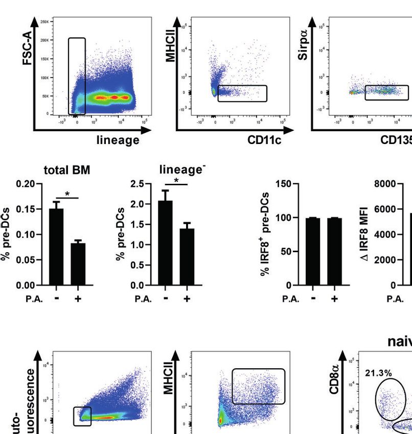
Brassard et al.                                                                                           Bacterial Exposure and Lung Dendritic Cells

INTRODUCTION                                                                 interesting data suggest that, as observed in several other
                                                                             types of immune responses, cDC1s and cDC2s elicit
Conventional dendritic cells (cDCs) play an important role in                distinctive functions in the fight against bacteria. Indeed, in
both innate and adaptive immune responses. In the lungs,                     most bacterial infections, polarization of naive T cells into TH1
cDCs are critical sentinel cells that capture, process                       improves bacterial clearance and lung function, which suggests
and present antigens to activate naive T cells in lymph                      a beneficial role for cDC1s (Moser et al., 1997; Moser et al.,
nodes. In addition, cDCs are involved in the innate immune                   2002). In accordance, in a mouse model of lung infection with
response via cytokine and chemokine production (Macri et al.,                the Gram-negative bacteria Chlamydia muridarum, CD103+
2018). Lung conventional DCs comprise a variety of subsets                   cDC1s induced a stronger TH1 polarization compared to
that are typically subdivided into two sub-populations named                 cDC2s, and CD103 + cDC1 injection improved bacterial
cDC1s and cDC2s (Guilliams et al., 2014; Guilliams et al.,                   clearance (Shekhar et al., 2018). It should be noted, however,
2016). cDC1s express the surface marker XCR1, and the                        that cDC1s are usually involved in infections to intracellular
expression of IRF8 and BATF3 transcription factors is                        bacteria, and that their role in response to Pseudomonas
required for their development. Additionally, they can                       aeruginosa is not well-described. cDC2s, on the other hand,
express the alpha-E integrin CD103 in non-lymphoid organs                    could be important for the innate immune response. Indeed, in
like the lung, while they express CD8a in lymphoid tissues.                  response to LPS lung exposure, lung CD11b+ DCs produce
cDC2s are characterized by IRF4, Sirpa and CD11b                             more KC (CXCL1) and MIP–2 (CXCL2), two chemokines
expression (Crozat et al., 2011; Guilliams et al., 2014; Gurka               involved in neutrophil recruitment, compared to CD103+
et al., 2015).                                                               cDC1s (Beaty et al., 2007).
   The majority of cDC development occurs in the bone                            These distinctive roles for cDC1s and cDC2s in the response
marrow and requires the presence of the FMS-like tyrosine                    to bacteria suggest that the tight balance between cDC lung
kinase 3 ligand (FLT3L) cytokine (Ginhoux et al., 2009).                     subsets is important to support effective local immune
During this process, the commitment of cDC precursors to                     responses. Recently, we demonstrated that lung exposure to
the cDC1 or cDC2 lineage happens relatively early in cDC                     lipopolysaccharide (LPS), which induces a strong local and
development (Grajales-Reyes et al., 2015; Schlitzer et al.,                  peripheral inflammatory response, modulates the proportions
2015). cDC precursors then leave the bone marrow at the                      of cDC populations by decreasing the percentage of CD103+
pre-DC stage and migrate through the bloodstream to various                  cDC1s and increasing CD11b+ DC proportions (Brassard et al.,
organs such as the lung (Liu et al., 2009). Pre-DCs committed                2019). Since LPS is one of multiple bacterial components that
to the cDC1 lineage do not express CD103 (Brassard et al.,                   may influence cDC populations and with the potential crucial
2019). The exact mechanisms by which cDC1s acquire CD103                     role of cDC1s in the fight against bacterial infections in the
expression upon their entrance in the lung remain unclear, but               lung, the impact of whole bacteria on the lung cDC signature
in vivo and in vitro studies suggest that exposure to GM-CSF,                remained an important unanswered question. We therefore set
present in the lung, is a potent inducer of cDC CD103                        out to analyze the influence of an acute exposure to the
expression (King et al., 2010; Greter et al., 2012; Mayer                    Gram–negative bacteria P. aeruginosa on the local lung cDC
et al., 2014). To date, there is no information concerning                   signature. P. aeruginosa is ubiquitously found in nature and
transcription factors involved in cDC CD103 expression.                      causes opportunistic acute and chronic infections in
However, the RUNX family of transcription factors is                         immunocompromised patients, such as those suffering of
involved in the induction of CD103 expression in T cells                     cystic fibrosis (Green et al., 1974; Lyczak et al., 2000;
(Grueter et al., 2005). The only reported ligand of aE                       Moradali et al., 2017). We observed that P. aeruginosa
integrin (CD103) is E-cadherin, which is expressed by                        modulated the proportions of lung cDC1 and cDC2
epithelial cells (Corps et al., 2001). While the role of CD103               populations in favor of cDC2s, which was in part explained
expression on cDC1s remains unclear, reports demonstrate                     by a higher propensity of bone marrow cDC precursors to
that T cells CD103 expression facilitates lymphocyte                         differentiate towards the cDC2 lineage, and by an incapacity of
localization and induces intracellular signaling (Pauls et al.,              lung cDC1s to fully express CD103 in response to GM-CSF.
2001; Franciszkiewicz et al., 2013).                                         The latter was linked to reduced GM–CSF receptor internal
   The two cDC subpopulations have distinct and often                        localization, following exposure to P. aeruginosa and LPS, and
opposite functions (Schlitzer et al., 2015; Guilliams et al.,                alterations in RUNX1 expression, which regulate CD103
2016). cDC1s are particularly important for IL-12 production,                expression. Finally, we report that the lack of CD103
antigen cross-presentation to CD8 T cells and CD4 T cell                     expression on cDCs leads to an exacerbated airways
polarization into TH1 (Hildner et al., 2008; Mashayekhi et al.,              neutrophilia, supporting the idea that the absence of CD103
2011; Martı́nez-Ló pez et al., 2015). The specific function of               expression on cDC1s promotes the lung innate response to P.
cDC2s is more controversial, but some studies suggested that                 aeruginosa. We therefore shed a light on a possible mechanism
they are important for TH2 polarization (Gao et al., 2013;                   demonstrating that the blockade of cDC CD103 expression by
Plantinga et al., 2013). To date, there is no consensus                      Gram-negative bacteria is a crucial step in promoting the initial
regarding the roles of cDC1s or cDC2s in the efficacy of                      innate immune response to this potentially infectious agent in
antibacterial immune responses. Nevertheless, some                           the lung.

Frontiers in Cellular and Infection Microbiology | www.frontiersin.org   2                                   July 2021 | Volume 11 | Article 617481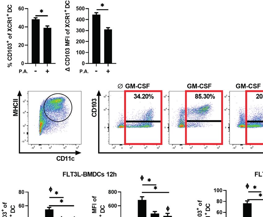
Brassard et al.                                                                                            Bacterial Exposure and Lung Dendritic Cells

MATERIAL AND METHODS                                                         Spleen-Isolated cDCs
                                                                             To expand cDC populations in vivo, WT mice were
Mice                                                                         subcutaneously injected in the lower back with 5 x 10⁵ FLT3L-
Cd103−/− (B6.129S2(C)-Itgaetm1Cmp/J) and wild-type (WT) mice                 producing B16 melanoma cells, previously grown in DMEM
were purchased from Jackson Laboratories and kept in a specific               media (Wisent) supplemented with 10% FBS. When the tumor
pathogen-free animal unit (Centre de recherche de l’Institut                 reached 1 cm diameter, mice were euthanized and the spleen
Universitaire de Cardiologie et de Pneumologie de Qué bec,                  collected. Spleen leukocytes were isolated as described in
Laval University, Qué bec, QC, Canada) for the duration of the              leukocyte isolation section. cDCs were purified by negative
experiments. Cd103−/− and WT mice were not co-housed during                  selection using the EasySep Mouse pan-DC Enrichment Kit
the duration of experiments. Experiments were approved by local              (StemCell Technologies, Vancouver, BC, Canada).
ethics committees and followed Canadian animal care guidelines.
                                                                             DCs In Vitro Stimulation
Intranasal Instillation With P. aeruginosa                                   106 cells/ml of splenic or FLT3L-BMDCs were stimulated with
and LPS                                                                      10 ng/ml Granulocyte-macrophage colony-stimulating factor
Non-mucoid P. aeruginosa, strain Boston 41501 (ATCC #27853,                  (GM-CSF) (Peprotech, catalog no. 315-03), 10 ng/ml LPS
Manassas, VA, USA) was incubated overnight in tryptic soy                    (Sigma-Aldrich) or P. aeruginosa at a ratio of 1 cDC: 1 P.
broth (TSB) (Wisent, St-Bruno, QC, CA) at 37°C in a rotating                 aeruginosa for spleen-isolated cDC or 10 DCs: 1 P. aeruginosa
shaker and 1 ml of the suspension was re-incubated in new TSB                for FLT3L-BMDCs in RPMI 1640 supplemented with 10% FBS
media for 2 h. Bacteria were washed and diluted in saline, and the           and 50 µM b-mercaptoethanol for 12, 18 or 48 h. For some
desired concentration was adjusted by spectrophotometry                      experiments, FLT3L-BMDCs were pre-treated with 40, 80 or 120
according to a reference curve. Bacterial concentration was                  mM of the dynamin inhibitor Dynasore (Sigma-Aldrich) or with
systematically verified by quantitative culture of the inoculum.              10, 25 or 50 mM of CBFb-Runx1 Inhibitor II (Sigma-Aldrich).
Age- and sex-matched WT and Cd103-/- mice received a 50 mL                   Following stimulation, CD103, GM-CSFRa and RUNX1 protein
intranasal (i.n.) instillation of 5 x 10⁵ or 5 x 10⁶ colony forming          or mRNA expression were analyzed by flow cytometry and qRT-
units (CFU) of P. aeruginosa or 350 ng of LPS (Sigma-Aldrich,                PCR respectively.
St. Louis, MO USA). Mice were euthanized at 2, 6 or 18 h
following LPS or P. aeruginosa exposure. Bronchoalveolar
lavages (BAL) were obtained via three injections/aspirations of
                                                                             Flow Cytometry
                                                                             BAL leukocytes, tissue-isolated leukocytes or in vitro-stimulated
1 mL of saline, in mice euthanized at 2 or 6 h post i.n. Total BAL
                                                                             cDCs were stained with TruStain FcX anti-mouse CD16/32
cells of LPS-treated mice were counted and differential counts
                                                                             antibody (BioLegend, San Diego, CA, USA) and CD45-APC-
were determined on Giemsa stained cytospins (HemaStain Set,
                                                                             Cy7, CD103-PE, CD11b-PeCy7, CD11c-BV711, I-A/I-E (MHC
Thermo Fisher Scientific, Waltham, MI, USA). The BAL
                                                                             II)-Pacific Blue, I-A/I-E (MHC II)-PERCP, CD172a (Sirpa)-
composition of P. aeruginosa-exposed mice was analyzed by
                                                                             APC-Cy7, CD19-biotin, CD90.2-biotin, IRF4-PE, Ly-6G-PE,
flow cytometry and neutrophils were identified as auto-
                                                                             XCR1-APC, CD8a-APC-Cy7, Lineage antibody cocktail-
fluorescence-, CD45+, Ly-6G+ and CD11b+ and macrophages
                                                                             Pacific Blue, CD135 (FLT3)-biotin, Streptavidin-PERCP
were identified as auto-fluorescence+, CD45+, CD11c+ and
                                                                             (BioLegend), NK1.1-biotin (ablab, Vancouver, BC, CA),
Siglec-F+. For flow cytometry analysis, the lung, spleen, femur
                                                                             CD11c-APC, Siglec-F-BV711 (BD Biosciences, San Jose,
and tibia were collected in phosphate buffered saline (PBS) 18 h
                                                                             USA), IRF8-APC (Miltenyi Biotec, Bergisch Gladbach,
after lung i.n. instillation with P. aeruginosa.
                                                                             Allemagne) and Streptavidin-Pe-Cy7 (eBioscience, Thermo
Leukocyte Isolation                                                          Fisher Scientific), GM–CSFRa-APC (R&D system,
Lung leukocytes were obtained by the digestion of lung tissue                Minneapolis, MN, USA). Total, neutrophils and macrophages
with 200 U/ml collagenase IV (Sigma-Aldrich) for 45 min at 37°C.             BAL number were determined using precision count beads
Digested lungs and spleens were pressed through a 70-mm cell                 (BioLegend). Intracellular staining was performed using the
strainer. Bone marrow cells were isolated by flushing the cells from          True-Nuclear™ Transcription Factor Buffer Set (BioLegend)
tibias and femurs using a 25 Gauge needle with PBS. Red blood cells          according to the manufacturer’s instructions. Cells were
were lysed with ammonium chloride and cDC or cDC precursors                  analyzed using a BD LSR Fortessa cytometer (BD Biosciences)
were analyzed by flow cytometry.                                              and FlowJo software V10 (BD, Franklin Lakes, NJ, USA). Mean
                                                                             fluorescence intensity (MFI) data were analyzed as D MFI,
FLT3L-BMDCs                                                                  which corresponds to the MFI of the antigen-positive
Bone marrow cells were isolated as described in the leukocyte                population minus the MFI of the fluorescence minus one
isolation section. Cells were cultured at 1.5 x 10⁶ cells/ml for 7           (FMO) control of this population.
days in RPMI 1640 media (Wisent) supplemented with 10% FBS
(Wisent), 50 µM b-mercaptoethanol, antibiotic-antimycotic                    Real-Time PCR Analysis
(Wisent) and 100 ng/ml FMS-like tyrosine kinase 3 ligand                     1.5 x 10⁶ FLT3L-BMDCs were stimulated with GM-CSF, LPS or
(FLT3L) (peprotech, Rocky Hill, NJ, USA, catalog no. 250-                    P. aeruginosa for 12 h and RNA was isolated using RNAspin
31L). On day 7, BMDCs were harvested for stimulation.                        Mini Kit (GE Healthcare Life Sciences, Chicago, USA) and

Frontiers in Cellular and Infection Microbiology | www.frontiersin.org   3                                    July 2021 | Volume 11 | Article 617481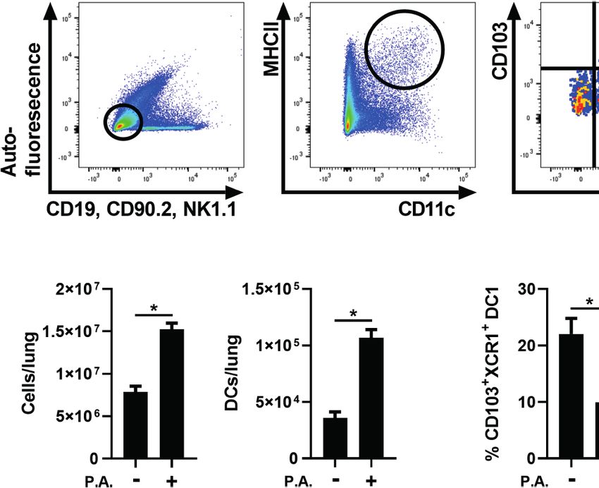
Brassard et al.                                                                                                         Bacterial Exposure and Lung Dendritic Cells

reverse transcribed with an iScript cDNA Synthesis Kit (Bio-rad,                    RESULTS
Mississauga, Ontario, CA). Real-time PCR analysis was
performed for CD103 (Itgae), GM-CSFRa (Csf2ra) and                                  Lung Exposure to the Gram-Negative
RUNX1 using the Rotor-Gene 6000TM (Qiagen, Valencia, CA,                            Bacteria Pseudomonas aeruginosa Leads
USA) in Sso Advanced Universal SYBR Green Supermix (Bio-                            to a Major Modulation of DC Populations
rad). The following primers (IDT, Coralville, USA) were used:                       We first aimed to verify whether lung exposure to whole Gram-
Itgae (forward: 5’-AGGTCATAGATACGGTCAGGT-3’,                                        negative bacteria influences the CD103+ cDC1 and cDC2
reverse: 5’-GGTTAGATTTCAATGGCGATGG-3’), GM-                                         population ratios in the lung. To test this, lung cDC
CSFRa (forward: 5’-CCTCACCATCCATCGCA-3’, reverse: 5’-                               populations were analyzed 18 h following i.n. exposure with P.
GAAGCAGTAGCGTGGAGAAG -3’), RUNX1 (forward:                                          aeruginosa. Lung cDCs were identified as auto-fluorescence-,
5’-GTAGCGAGATTCAACGACCTC-3’, reverse: 5’-                                           CD19-, CD90.2-, NK1.1-, MHC IIHi and CD11c+. CD103+ cDC1
TCTATGGTAGGTGGCAACTTG-3’). Expression was                                           characterization was based on CD103 and XCR1 expression,
normalized to the Gnb and Rplp0 mRNA expression validated                           whereas CD11b and Sirpa markers were used to identify cDC2s
for stability of expression in this model.                                          (Figure 1A and Supplementary Figure 1 for full gating strategy).
                                                                                    We first observed that P. aeruginosa induced an important
Enumeration of Colony Forming Unit in                                               increase in lung total cells, which was accompanied by a two-
BAL and Lung                                                                        fold increase in cDC numbers (Figure 1B). We also report a
Lungs were homogenized in 1 ml of saline using the Polytron                         decrease in CD103+XCR1+ cDC1 proportions and, in return, an
Tissue Homogenizer (Kinematica, Luzern, Switzerland).                               increase in CD11b+Sirpa+ cDC2 proportions (Figure 1C). These
Homogenates and BALs were subjected to 10-fold serial                               results indicate that, as observed previously with LPS (Brassard
dilutions in saline and cultured in tryptic soy agar (Wisent) at                    et al., 2019), P. aeruginosa modulates lung cDC populations in
37°C and CFUs were counted 18 to 24 h later.                                        favor of the DC2/monocyte-derived DC population.

Statistics                                                                          Lung Exposure to Pseudomonas
Data are presented as mean ± SEM. Graphpad Prism version 8                          aeruginosa Influences Bone Marrow
(San Diego, CA, USA) was used to analyze all data. Statistical                      cDC Precursors
analysis for multiple comparisons was performed using an                            The commitment to the cDC1 or cDC2 lineage is defined before
ANOVA table followed by Tukey’s multiple comparison tests.                          the pre-DCs stage, and can be influenced by peripheral
Non-multiple comparisons were analyzed using paired or                              inflammation (Schlitzer et al., 2015; Grajales-Reyes et al., 2015;
unpaired T-tests. Statistical significance was determined at                         Meyer et al., 2018; Beshara et al., 2018; Brassard et al., 2019).
p < 0.05.                                                                           Thus, we hypothesized that following i.n. instillation with

                       A

                       B                                                        C

 FIGURE 1 | Lung exposure to P. aeruginosa altered the proportions of cDC1s and cDC2s in favor of cDC2s. WT mice were exposed to a single i.n. instillation of 5 x
 105 CFU of P. aeruginosa (P.A.) and mice were euthanized 18 h later. Lung cDC populations were analyzed by flow cytometry. (A) Sequential gating strategy used to
 identify total cDCs (auto-fluorescence-, CD19-, CD90.2-, NK1.1-, MHC IIHi and CD11c+), cDC1 (CD103+XCR1+) and cDC2 (CD11b+Sirpa+). (B) Number of total lung
 cells and cDCs (C) Percentage of CD103+XCR1+ cDC1 and CD11b+Sirpa+ cDC2 in lung cDCs. Data are presented as mean ± SEM; n = 8-9 mice per group
 combined from two separate experiments *p < 0.05 using unpaired t-test.

Frontiers in Cellular and Infection Microbiology | www.frontiersin.org          4                                          July 2021 | Volume 11 | Article 617481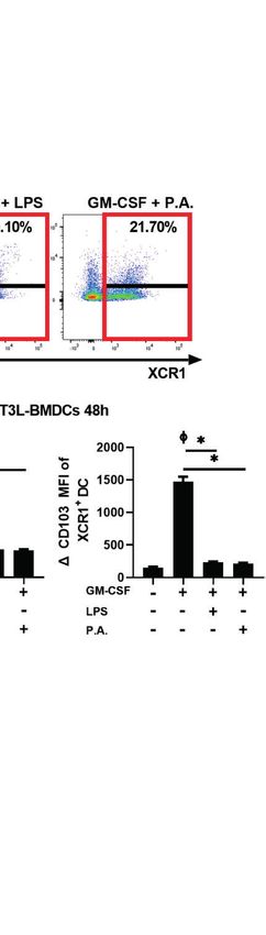
Brassard et al.                                                                                                         Bacterial Exposure and Lung Dendritic Cells

P. aeruginosa, a shift towards the cDC2 fate could support the                      propensity of pre-DCs to differentiate towards the cDC2 lineage
accumulation of cDC2s in the lung. cDC1 and cDC2-committed                          and a strong exodus of pre-DCs in response to P. aeruginosa.
precursors both express IRF8 initially, but the further                                To verify whether this impact was lung-specific, we tested
commitment to the cDC2 lineage results in a decrease in IRF8                        cDC subsets in tissues that are not in direct contact with P.
expression in time. In contrast, only cDC2-committed                                aeruginosa, such as the spleen. Total splenic cDCs were identified
precursors express IRF4 at the later stage of development                           with the same gating strategy used in the lung, but, as spleen is a
(Sichien et al., 2016). To determine whether lung exposure to                       lymphoid organ, the CD8a surface marker was evaluated on
P. aeruginosa alters the cDC1 vs cDC2 commitment, the                               cDC1s (Edelson et al., 2010; Guilliams et al., 2014). Thus,
expression of these two transcription factors was analyzed in                       CD8a + CD11b- cDCs were characterized as cDC1s, while
bone marrow pre-DCs (Guilliams et al., 2014; Grajales-Reyes                         CD11b+CD8a- cDCs were classified as cDC2s (Figure 2D,
et al., 2015). Pre-DCs were identified as lineage-, MHC II-,                         Supplementary Figure 3) (Guilliams et al., 2014; Tavernier
CD11c + , Sirpa - / l o , CD135 (FLT3) + (Figure 2A and                             et al., 2015; Guilliams et al., 2016). Total splenic cells were
Supplementary Figure 2) (Schlitzer et al., 2015; Grajales-Reyes                     decreased following i.n. instillation with P. aeruginosa, while
et al., 2015). We report that the percentage of pre–DCs from total                  cDC number remained unchanged (Figure 2E). The splenic
bone marrow cells and lineage- cells was significantly decreased                     proportion of CD8a+CD11b- cDC1s was decreased and in
in mice exposed to P. aeruginosa (Figure 2B). However, this was                     contrast the CD11b+CD8a- cDC2 proportion was increased,
accompanied by an increase in IRF4 expression following lung                        suggesting a systemic impact of P. aeruginosa on cDC
instillation with P. aeruginosa (Figure 2C). This result suggests a                 signatures in various tissues (Figure 2F).

                            A

                            B                                            C

                            D

                            E                                            F

 FIGURE 2 | Intranasal instillation with P. aeruginosa induced a systemic effect on bone marrow cDC precursors and spleen cDC populations. WT mice were
 euthanized 18 h following i.n. instillation of 5 x 10⁵ CFU of P. aeruginosa (P.A.) and bone marrow and spleen cells were analyzed by flow cytometry. (A) Bone
 marrow pre-DCs sequential gating strategy (lineage-, MHC II-, CD11c+, Sirpalo/-, CD135+) and representative flow cytometry profile of IRF4 and IRF8 expression in
 pre-DCs from naive and P. aeruginosa treated mice. (B) Percentage of bone marrow pre-DCs in total bone marrow cells and lineage- cells. (C) Bone marrow pre-
 DCs: percentage and D MFI of IRF8 and IRF4. (D) Spleen cDCs sequential gating strategy (auto-fluorescence-, CD19-, CD90.2-, NK1.1-, MHC IIHi and CD11c+) and
 representative flow cytometry profile of CD8a and CD11b expression on cDCs from naive and P. aeruginosa treated mice. (E) Total spleen cells and spleen cDC
 number. (F) Percentage of CD8a+CD11b- cDC1 and CD11b+CD8a- cDC2 from spleen total cDCs. Data are presented as mean ± SEM; n = 8-10 mice per group
 combined from two separate experiments except for panel (C), IRF4; n = 5 representative of three separate experiments. *p < 0.05 using unpaired t-test.

Frontiers in Cellular and Infection Microbiology | www.frontiersin.org          5                                          July 2021 | Volume 11 | Article 617481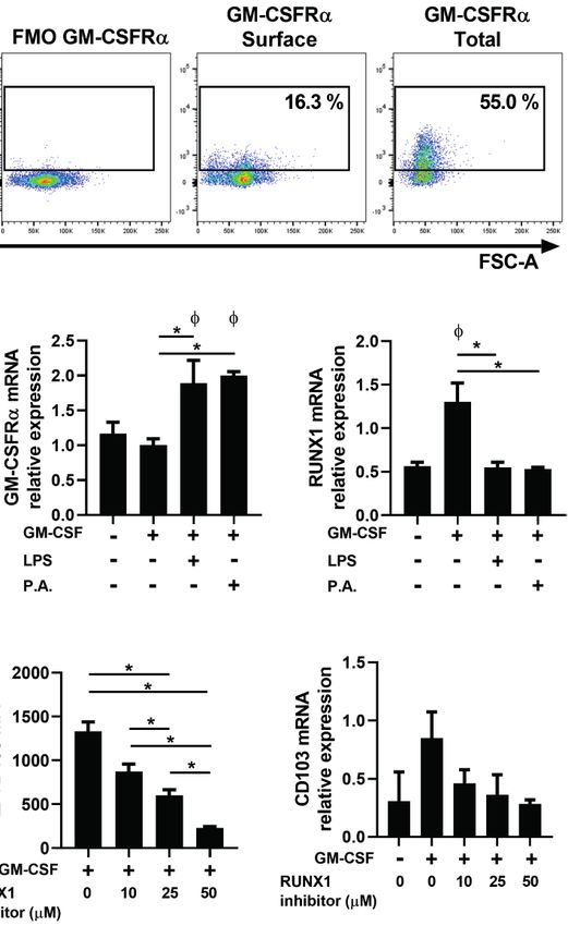
Brassard et al.                                                                                                            Bacterial Exposure and Lung Dendritic Cells

Pseudomonas aeruginosa Interferes With                                                (BMDCs) (Gating strategy, Figure 3B and Supplementary
the Capacity of cDCs to Express CD103                                                 Figure 4). Without any stimulation, the percentage of XCR1+
We previously demonstrated that LPS and inflammatory factors                           BMDCs expressing CD103 was low, with a mean of 18% positive
directly abrogate the induction of CD103 expression on cDC1s                          cells. As expected, a 12 h stimulation with GM-CSF increased
(Brassard et al., 2019). To verify whether exposure to whole                          CD103 expression on cDC1s to approximately 55% (Figure 3C).
bacteria also influences CD103 expression on cDC1s, the                                However, the presence of P. aeruginosa during the GM-CSF
proportion of XCR1+ cDC1s expressing CD103 was analyzed                               stimulation prevented the maximal induction of CD103 (Figure
18 h following i.n. instillation with P. aeruginosa. We observed a                    3C) to a level similar to that of LPS. This incapacity of XCR1+
significant decrease in the percentage of CD103+ cDCs within the                       BMDCs to fully express CD103 worsened in time, as CD103
cDC1 population in response to bacterial exposure (Figure 3A).                        expression on XCR1+ cDCs was almost entirely abrogated in
   Furthermore, we verified whether P. aeruginosa directly                             response to LPS and P. aeruginosa at 48 h (Figures 3B, C).
interferes with the capacity of cDC1s to express CD103 in                             Although few freshly isolated splenic cDC1s express CD103, its
response to GM-CSF using FLT3L-derived bone marrow DCs                                expression can be induced on splenic cDCs by GM-CSF

                           A

                           B

                           C

                           D

 FIGURE 3 | P. aeruginosa stimulation prevents GM-CSF-induced CD103 expression on cDCs. (A) Mice were euthanized 18 h after a single i.n. instillation of 5 x 10⁵ CFU
 of P. aeruginosa (P.A.) and the percentage of CD103+ cells and CD103 MFI in XCR1+ cDC1 gated lung cells were analyzed by flow cytometry. (B, C) FLT3L-BMDCs or
 (D) spleen-isolated cDCs were stimulated with GM-CSF ± LPS or P. aeruginosa (P.A.) for 12 h, 18 h or 48 h and CD103 and XCR1 expression were analyzed by flow
 cytometry. (B) Gating strategy used to identify MHC IIHiCD11c+ cDCs and representative flow cytometry profile of CD103 expression on XCR1+ FLT3L-BMDCs stimulated
 for 48 h. (C, D) Percentage of CD103+ cells and CD103 MFI from previously gated XCR1+ cDCs. Data are presented as mean ± SEM; (A % CD103) n = 10-11 pooled
 from two separate experiments, (A CD103 MFI) n = 5 representative of three separate experiments. (C, D % CD103) n = 5-6 samples of cDCs per group pooled from two
 separate experiments. (C, D CD103 MFI) n = 3 representative of two separate experiments. * = p < 0.05 compared between conditions stimulated with GM-CSF. F = p <
 0.05 compared to unstimulated condition. P-values were analyzed using unpaired t-test (A) or paired-one-way ANOVA (C, D).

Frontiers in Cellular and Infection Microbiology | www.frontiersin.org            6                                            July 2021 | Volume 11 | Article 617481
Brassard et al.                                                                                                        Bacterial Exposure and Lung Dendritic Cells

stimulation (Sathe et al., 2011; Brassard et al., 2019). We report                  receptor–ligand complex, prior to GM-CSF stimulations (Macia
that the induction of CD103 expression by GM-CSF is also                            et al., 2006; Zsiros et al., 2019). Pre-treatment with Dynasore
reduced on spleen–isolated cDCs following exposure to P.                            blocked CD103 expression on FLT3L-BMDCs suggesting that
aeruginosa, and to a level that is similar to LPS exposure                          GM-CSFR internalization is required for its expression on cDCs
(Figure 3D). Therefore, the blockade of CD103 expression on                         (Figure 4A).
cDCs by these stimuli seems to be independent of the method                            To better understand whether GM-CSFR internalization is
used to generate cDCs. Of note, neither LPS nor P. aeruginosa                       impacted by LPS or P. aeruginosa, extracellular and intracellular
altered cDCs viability (data not shown). These results also                         protein levels of the GM-CSFRa subunit were analyzed
suggested that the presence of P. aeruginosa in the lung can                        (Figures 4B, C). The combination of LPS or P. aeruginosa
directly influence the capacity of newly-recruited cDC1s to                          with GM-CSF did not alter total GM-CSFR expression
express CD103 in response to local GM–CSF.                                          (Figure 4C). However, when they were combined with GM-
                                                                                    CSF, a higher ratio of surface to total GM-CSFRa expression was
Pseudomonas aeruginosa Influences the                                                noted, indicating a reduced intracellular localization of the GM–
Localization of the GM-CSF Receptor                                                 CSFRa subunit (Figure 4C). This was not due to alterations in
The exact mechanisms by which GM-CSF induces CD103                                  new receptor synthesis, as csf2ra (GM–CSFRa) mRNA
expression remain unknown, but binding of GM-CSF to its                             expression was significantly increased in BMDCs exposed to
receptor (GM-CSFR) leads to a signaling cascade that is                             P. aeruginosa (Figure 4D).
mediated in part by GM-CSFR internalization (Broughton                                 Additionally, RUNX1 mRNA expression, a member of the
et al., 2012; Zsiros et al., 2019). To first test whether GM-CSFR                    RUNX transcription factor family, was increased following GM-
internalization is indeed involved in GM-CSF-induced cDC                            CSF stimulation, and abrogated in the presence of LPS or P.
CD103 expression, FLT3L–BMDCs were pre-treated with                                 aeruginosa (Figure 4E). Following the same pattern as RUNX1,
Dynasore, a dynamin inhibitor that blocks internalization of                        CD103 mRNA synthesis was also altered in the presence of LPS

                      A                                                         B

                      C                                                               D                            E

                      F                                      G                                                   H

 FIGURE 4 | P. aeruginosa influences GM-CSFR localization on BMDCs. FLT3L–BMDCs were stimulated for (A–G) 48 h or (B–F, H) 12 h with GM-CSF ± (A)
 Dynasore, (B–F) GM-CSF ± LPS or P. aeruginosa (P.A.) or (G, H) a RUNX1 inhibitor. (A–G) Percentage of CD103+ and D CD103 MFI on cDCs. (B) Representative
 flow cytometry profile of surface and total GM–CSFRa expression on FLT3L-BMDCs. (C) Surface (extracellular) and total (extracellular + intracellular) D MFI (first
 panel) of GM-CSFRa, and ratio of surface to total expression of GM-CSFRa (second panel). (D) mRNA relative expression of GM-CSFRa (Csf2ra). (E) mRNA relative
 expression of RUNX1. (F, H) mRNA relative expression of CD103 (Itgae). Data are presented as mean ± SEM; (A) n = 3 representative of two separate experiments.
 (C–H) n = 4-6 samples of cDCs per group pooled from two separate experiments. *p < 0.05 compared between conditions stimulated with GM–CSF. F = p < 0.05
 compared to unstimulated condition. P-values were analyzed using paired-one-way and two-way ANOVA.

Frontiers in Cellular and Infection Microbiology | www.frontiersin.org          7                                         July 2021 | Volume 11 | Article 617481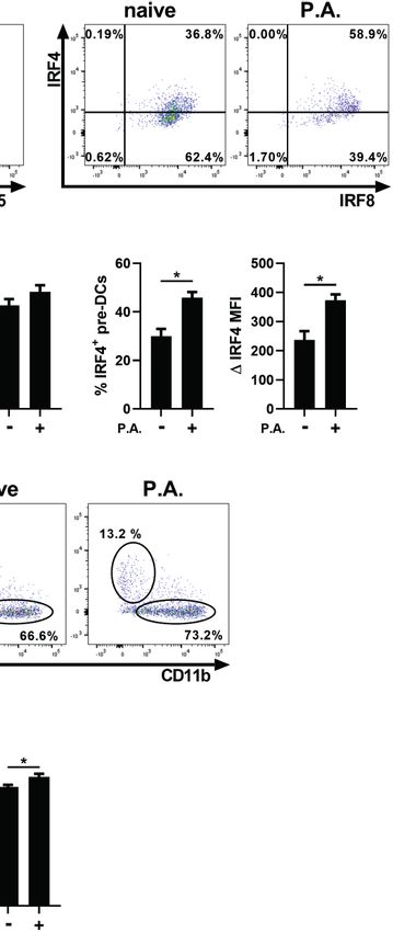
Brassard et al. Bacterial Exposure and Lung Dendritic Cells
or P. aeruginosa (Figure 4F). To confirm the involvement of and lung homogenates of the two mouse strains, (Figure 5F) and
RUNX1 in the induction of CD103 expression in cDCs, FLT3L- this was independent of the concentration used (Figure 5G).
BMDCs were pre-treated with a RUNX1 inhibitor prior to GM- This likely indicates that the bacterial clearance kinetics of this
CSF stimulation. We observed that the suppression of the model/P. aeruginosa strain may be too quick to verify whether
transactivation activity of RUNX1 and its cofactor CBF in the increase in neutrophils observed in the absence of cDC
BMDCs leads to a reduction in both CD103 protein and CD103 expression leads to better bacterial clearance.
mRNA synthesis, linking RUNX1 to CD103 expression in
cDCs (Figures 4G, H). Therefore, our results suggest that the
presence of P. aeruginosa impacts CD103 expression on cDC1s
by interfering with the intracellular localization of the GM-CSFR,
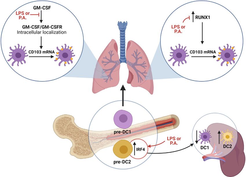
DISCUSSION
and by preventing RUNX1 mRNA expression. DCs take part in the induction of innate and adaptive immune
responses, and the efficacy of these responses is influenced by the
Lack of CD103 Expression Enhanced the nature of local cDC subsets (Macri et al., 2018). Until now, few
Lung Innate Immune Response to studies focused on the influence of bacterial infection on cDC
Pseudomonas aeruginosa populations. In this report, we demonstrated that lung exposure
Lung exposure to P. aeruginosa resulted in an increase in XCR1+ to live P. aeruginosa bacteria decreases the proportions of
cDC1s that lack CD103 expression. To determine the impact of CD103+ cDC1s in favor of CD11b+ cDC2s/monocyte-derived
an incapacity to express CD103 by cDCs on the immune DCs. We determined that this was in part modulated by
response, we sought to verify whether the absence of CD103 modifications in bone marrow pre-DC populations and an
expression on cDC1s modulates the early innate immune altered CD103 expression on XCR1+ cDC1s (Figure 6).
response to LPS and P. aeruginosa. LPS or P. aeruginosa were Furthermore, we demonstrated that the absence of CD103
injected intranasally into WT and Cd103-/- mice and cells from expression increases neutrophils recruitment in the lung in
the bronchoalveolar lavage (BAL) were analyzed 2 h (LPS) and response to LPS and P. aeruginosa.
6 h (P. aeruginosa) later. These times were chosen since CD103 is First, we realize that our gating strategy to identify cDC2s
still expressed on cDCs at that time following LPS exposure does not discard monocyte-derived DCs, so this population must
(Brassard et al., 2019), and because the increase in total cells in be considered as potentially upregulated in response to P.
response to P. aeruginosa is slower than following LPS aeruginosa. Independently of this, the altered proportions of
administration (data not shown). Of note, although CD103 is cDC populations are not restricted to lung exposure to LPS or P.
also present on T cells, they do not play a role in the rapid innate aeruginosa. Indeed, similar results were reported in a mouse
immune response to these agents (Andrew et al., 1996; model of lung infection with Chlamydia muridarum, an LPS+
Bernatchez et al., 2017). Following the i.n. administration of bacteria (Shekhar et al., 2018; Brassard et al., 2019). Interestingly,
LPS, total cells and neutrophil numbers were increased in the in the C. muridarum model, a decrease in CD103+ cDC
BAL of Cd103-/- mice compared to WT mice (Figure 5A). This proportions was observed up to 7 days post-infection,
was also observed in Cd103-/- mice exposed to P. aeruginosa suggesting that this phenomenon is not limited to acute
compared to WT (Figure 5B). These results suggest that the immune responses (Shekhar et al., 2018). This is also
absence of CD103 expression facilitates neutrophil recruitment supported by similar alterations observed by our group in a
in response to Gram–negative bacteria. 21-day mouse model of chronic allergic inflammation to
We then assessed whether this was caused by a modulation Saccharopolyspora rectivirgula antigen (Bernatchez et al., 2017).
in cDC or cDC precursors populations. The lung and spleen Our results suggest that lung exposure to P. aeruginosa altered
total cell numbers, cDC numbers, (data not shown) and the bone marrow cDC precursors towards pre-DCs that are
percentage of cDC1s and cDC2s were similar between WT and committed to the cDC2 lineage, which likely contributes to the
Cd103-/- mice in naive and P. aeruginosa exposed mice (Figures accumulation of lung cDC2s. A modification of bone marrow
5C, D). Moreover, bone marrow total cells, pre–DC numbers pre-DCs towards the cDC1 or cDC2 lineage was observed in
and IRF8 MFI were also similar between strains (Figure 5E). other immunological contexts including cancer, viral infections
The IRF4 MFI and the percentage of IRF4+ pre-DCs were and lung exposure to LPS (Beshara et al., 2018; Meyer et al., 2018
significantly lower in Cd103-/- naive mice compared to WT Brassard et al., 2019), suggesting that this mechanism is
naive mice, but similar in both strains following the exposure to conserved across various diseases and types of inflammatory
P. aeruginosa (Figure 5E). Therefore, the higher recruitment of responses. We previously observed that lung exposure to LPS
lung neutrophils in Cd103-/- mice is not caused by a difference leads to a decrease in IRF8 expression in pre-DCs, whereas an
in number or proportions in cDC or pre-DC populations. increase of IRF4 expression is reported here in response to P.
To determine whether this affected bacterial clearance, we aeruginosa (Brassard et al., 2019). Some studies suggest a
first studied the optimal time to study P. aeruginosa clearance. sequential order between IRF8 and IRF4 expression in which
Almost all bacteria were cleared at 12 h following i.n. instillation the decrease in IRF8 expression precedes the increase in IRF4
of P. aeruginosa (data not shown), thus the 6 h timepoint was expression in pre-DCs committed to the cDC2 lineage (Bajaña
selected to compare CFU number in WT and Cd103-/- mice. We et al., 2016; Sichien et al., 2016). Therefore, the differences
report similar numbers of P. aeruginosa CFU counts in the BAL reported in both studies could be explained by a difference in
Frontiers in Cellular and Infection Microbiology | www.frontiersin.org 8 July 2021 | Volume 11 | Article 617481Brassard et al. Bacterial Exposure and Lung Dendritic Cells
A
B
C D
E
F G
FIGURE 5 | Lack of CD103 expression favors the recruitment of bronchoalveolar neutrophils. (A, B) Total number (first panel), macrophages and neutrophils (second
panel) of bronchoalveolar lavage (BAL) cells were compared between WT and Cd103-/- mice (A) 2 h following LPS i.n. instillation or (B) 6 h following i.n. instillation of 5 x
105 CFU of P. aeruginosa (P.A.). (C–E) Lung, spleen and bone marrow cells were analyzed by flow cytometry 18 h after i.n. instillation with 5 x 10⁵ CFU of P. aeruginosa.
(C) Percentage of XCR1+ cDC1s and percentage of CD11b+Sirpa+ cDC2s in lung. (D) Percentage of CD8a+CD11b+ cDC1s and percentage of CD11b+CD8a- cDC2s in
spleen. (E) Total cell number, pre-DC number, DIRF8 MFI, DIRF4 MFI and percentage of IRF4+ pre-DCs in bone marrow. (F, G) P. aeruginosa CFU number present in the
lung and BAL 6 h after i.n. instillation of (F) 5 x 10⁵ CFU or (G) 5 x 10⁶ CFU of P. aeruginosa. Data are presented as mean ± SEM; (A) n = 2-5 (naive-P.A.) representative
of two independent experiments. (B) n = 3-10 (naive-P.A.) pooled from two separate experiments. (C–E) (total and pre-DC number) (F, G) n = 9-10 pooled from two
separate experiments. (E) (IRF4-IRF8) n = 5 representative of two independent experiments. *p < 0.05 using two-way ANOVA.
the timing of induction for these transcription factors in these of P. aeruginosa. Thus, our results suggest that bone marrow pre-
models. Despite this difference, both results report an imbalance DCs are biased towards the cDC2 lineage, which in turns
towards the cDC2 lineage differentiation. influences the subsets of cDC precursors recruited in the
Our results also suggest that splenic cDC populations are spleen, resulting in a modulation of splenic cDC1 and cDC2
affected by lung P. aeruginosa exposure. As the cDC turnover in proportions (Figure 6). As pre-DCs are precursors for most
the spleen is fast, even at steady state, an absence of total cDC tissue cDCs, this modification in bone marrow pre–DCs could
number increase (such as observed here) does not indicate an also influence cDC populations in other organs, and ultimately
absence of new cDC recruitment (Kamath et al., 2002), and the impact the efficacy of immune responses in case of a systemic
altered cDC populations is likely a reflection of a systemic impact infection (Ginhoux et al., 2009). We are aware that other
Frontiers in Cellular and Infection Microbiology | www.frontiersin.org 9 July 2021 | Volume 11 | Article 617481Brassard et al. Bacterial Exposure and Lung Dendritic Cells FIGURE 6 | Schematic representation of proposed mechanisms supporting the modulation of lung cDC populations in response to P. aeruginosa. LPS and P. aeruginosa directly impact the capacity of cDCs to internally localize the GM-CSFR, which is a crucial step in GM-CSF signaling leading to CD103 mRNA synthesis and CD103 expression on cDC1s. Additionally, LPS and P. aeruginosa interfere with RUNX1 expression, whose activity is needed for GM-CSF-dependent CD103 expression. Finally, exposure to LPS and P. aeruginosa alters the pre-DC transcriptional signature, favoring cDC2 precursor differentiation. Altogether, the exposure to LPS and P. aeruginosa impacts the capacity of cDCs to express CD103, as well as the cDC precursor signature. Figure 6 was Created with BioRender.com. mechanisms besides alterations in cDC precursors could be 2016; Zsiros et al., 2019), Dynasore could also exert other effects. involved in explaining the differences in the cDC population For instance, the presence of Dynasore could also block the signature in our models. As our gating strategy also included mo- internalization of other receptors, such as TLR4, express by cDCs DCs as CD11b +, this population could contribute to the (Kagan et al., 2008). However, as our assays with Dynasore were imbalance reported here (Guilliams et al., 2016). Also, a higher performed using only GM-CSF stimulation, it is very likely that migratory rate of cDC1s from the lung to the draining lymph in this case, the inhibition of CD103 expression was linked to a nodes could take part in the observed decrease percentage of this reduced blockade of GM-CSFR internalization. The exact steps population following P. aeruginosa exposure (Ho et al., 2011; linking the activation of GM-CSFR signaling to the induction of Nakano et al., 2013). CD103 mRNA expression on cDCs remained unknown. For the Supported by previously-published results (Brassard et al., first time, we demonstrate that RUNX1 expression is crucial for 2019) and present data, we propose that under homeostatic CD103 expression in cDCs. Additionally, we show that LPS and conditions, newly-recruited lung pre-DCs acquire CD103 P. aeruginosa alter RUNX1 expression, likely contributing to expression in response to GM-CSF stimulation through their reduced CD103 expression in response to these stimuli. Of final differentiation into cDCs. However, in the context of Gram- course, other cellular signaling pathways besides GM-CSF are negative bacterial exposure, the interaction with the bacteria or involved in regulating CD103 expression on cDCs. For example, LPS prevents CD103 mRNA synthesis resulting in lower GM- TGFb and retinoic acid also reportedly induced CD103 CSF-induced CD103 expression on cDC1s. Importantly, our expression (Iliev et al., 2009; Roe et al., 2017; Roe et al., 2020). results do not demonstrate that LPS nor P. aeruginosa reduce Whether LPS and P. aeruginosa alter CD103 expression in the expression of CD103 once it has been expressed, but rather response to these stimuli remains unknown, but definitely block the induction upon contact with GM-CSF. Furthermore, of interest. our observations suggest that a reduced internal localization of Despite a wealth of knowledge on CD103 modulation on the GM-CSFR could be one of the mechanisms contributing to cDCs in response to various stimuli (Nakano et al., 2013; the reduction in CD103 expression. Our results linking the GM- Bernatchez et al., 2017; Brassard et al., 2019), the physiological CSFR internalization to CD103 expression were obtained using function of this phenomenon on the host immune response Dynasore a GTPase inhibitor that inhibits dynamin activity, remained undefined. We propose here that the absence of CD103 which prevents endocytosis. Although this inhibitor is expression supports the intensity of the initial host innate frequently used to block GM-CSFR internalization (Katz et al., immune response. Anecdotally, in a model of skin bacterial Frontiers in Cellular and Infection Microbiology | www.frontiersin.org 10 July 2021 | Volume 11 | Article 617481
Brassard et al.                                                                                             Bacterial Exposure and Lung Dendritic Cells

infection, cDC1s were crucial for neutrophils recruitment to the              presence of LPS, TNF or P. aeruginosa can prevent the induction
site of infection via the secretion of VEGF-a (Janela et al., 2019),          of CD103 expression on cDC1 (Brassard et al., 2019). Currently,
which suggested that cDC1s influence neutrophil migration.                     in the lung, CD103 remains one of the main markers used to
CD103 signaling in cDC1s could be involved in promoting                       identify cDC1s (Ng et al., 2018; Shekhar et al., 2018; Monaghan
homeostasis by restraining the production of chemokines or                    et al., 2020). However, our results suggest that CD103 is not an
pro-inflammatory cytokines. In contrast, during bacterial                      ideal marker for this population in context of lung inflammation
infections, the absence of CD103 signaling on cDC1s could                     as its expression is highly modulated. Furthermore, in a few other
increase their capacity to secrete neutrophils-attracting                     particular immunological contexts, some lung CD11b+ DCs can
chemokines or induce neutrophil migration via an indirect                     also express CD103, which could lead to misinterpretations if
effect on surrounding cells. All in all, our study suggests that              used as a specific marker of cDC1s (Shane et al., 2018; Tweedle
the reduced CD103 expression on cDC1s may be a step in                        and Deepe, 2018). Finally, another disadvantage is that CD103 is
stimulating the innate immune response to Gram-                               not a marker of human lung cDC1s (Guilliams et al., 2014; Amon
negative bacteria.                                                            et al., 2020). Thus, the ideal marker to properly identify cDC1s
    The higher recruitment of lung neutrophils in Cd103-/- mice               should be constitutively expressed by cDC1s independently of
in response to P. aeruginosa was not caused by a difference in the            their differentiation stage or activation status, and similarly
number or proportions of cDC or pre-DC populations. However,                  expressed by human cDC1s.
a reduced IRF4 expression in bone marrow pre-DCs was                              In summary, we demonstrated that i.n. exposure to the Gram-
observed in Cd103-/- naive mice, suggesting a lower propensity                negative bacteria P. aeruginosa alters the proportions of CD103+
of cDC2 differentiation at steady state in these mice. As pre-DCs             cDC1 and CD11b+ cDC2 populations in favor of cDC2s, via the
don’t express CD103, an indirect mechanism must be involved to                modulation of bone marrow cDC precursors and an impact on
explain this (Brassard et al., 2019). Also, we cannot exclude the             CD103 mRNA production by cDC1s. Furthermore, we
possibility that, as WT and Cd103-/- strains were not co-housed               demonstrated that the absence of CD103 expression favors the
in our experiments, differences in microbiota could be involved               recruitment of lung neutrophils, suggesting that this
in impacting IRF4 expression.                                                 phenomenon could be an important step in the early innate
    Neutrophils are crucial to eradicate P. aeruginosa infection              immune response to P. aeruginosa.
(Koh et al., 2009). The fact that the increased neutrophil count in
Cd103-/- mice did not in turn change bacterial clearance indicates
that the number of neutrophils in WT mice was sufficient to
quickly clear bacteria. This is also supported by the fact that most
                                                                              DATA AVAILABILITY STATEMENT
of this mucoid strain of P. aeruginosa was cleared from the lung              The raw data supporting the conclusions of this article will be
past 6 h (data not shown) in WT mice. Furthermore, several                    made available by the authors, without undue reservation.
other studies observed that a higher number of lung neutrophils
do not necessarily lead to a better P. aeruginosa clearance
(Ballinger et al., 2006; Sen-Kilic et al., 2019). As CD103
expression could restrain chemokine production by cDC1s,                      ETHICS STATEMENT
mice in which cDC1s constitutively express CD103 would be a
                                                                              The animal study was reviewed and approved by Comité de
great tool to address the importance of cDC-specific CD103
                                                                              protection des animaux de l’Université Laval.
expression on neutrophil recruitment and bacterial clearance
with WT mice. Based on current knowledge, it is hard to
determine whether the alterations of cDC populations induced
by P. aeruginosa promote or impair host immune responses.                     AUTHOR CONTRIBUTIONS
First, the combination of the important lung recruitment of
cDC2s, known to secrete large amount of MIP-2 and KC                          JB and M-RB conceived the study. JB, JR, M-JB, and EB
neutrophil-attractant chemokines, and the possible higher                     performed experiments. JB and M-RB drafted and revised the
capacity of cDC1s to recruits neutrophils in absence of CD103                 paper. AML and M-JB critically revised the manuscript. MV and
expression suggest a positive impact on immune response. On                   CD contributed to the final revision and supported microbiology
the other hand, current knowledge on adaptive immune                          experiments. M-RB supervised the study. All authors contributed
responses against bacterial infection endorse the idea that                   to the article and approved the submitted version.
cDC1s support the efficacy of the adaptive immune response
compared to cDC2s. To date, no studies have established a
specific role for cDC1s or cDC2s in innate or adaptive immune                  FUNDING
responses to P. aeruginosa, but this report takes a first important
step towards better understanding the modulation of local cDC                 This work was supported by the Fonds sur les Maladies
populations in the context of Gram-negative bacterial clearance.              Respiratoires Bégin/Lavoie de l’Université Laval and by the
    An important conclusion rising from this data and our                     Fondation de l’Institut Universitaire de Cardiologie et de
previously published research is that several factors such as the             Pneumologie de l’Université Laval.

Frontiers in Cellular and Infection Microbiology | www.frontiersin.org   11                                    July 2021 | Volume 11 | Article 617481
Brassard et al. Bacterial Exposure and Lung Dendritic Cells
ACKNOWLEDGMENTS SUPPLEMENTARY MATERIAL
The authors would like to thank the CRIUCPQ animal unit as The Supplementary Material for this article can be found online
well as the CRIUCPQ animal unit personnel for their precious at: https://www.frontiersin.org/articles/10.3389/fcimb.2021.
collaboration and support. 617481/full#supplementary-material
REFERENCES Ginhoux, F., Liu, K., Helft, J., Bogunovic, M., Greter, M., Hashimoto, D., et al.
(2009). The Origin and Development of Nonlymphoid Tissue CD103+ Dcs.
Amon, L., Lehmann, C. H. K., Heger, L., Heidkamp, G. F., and Dudziak, D. (2020). J. Exp. Med. 206, 3115–3130. doi: 10.1084/jem.20091756
The Ontogenetic Path of Human Dendritic Cells. Mol. Immunol. 120, 122–129. Grajales-Reyes, G. E., Iwata, A., Albring, J., Wu, X., Tussiwand, R., Kc, W., et al.
doi: 10.1016/j.molimm.2020.02.010 (2015). Batf3 Maintains Autoactivation of Irf8 for Commitment of a CD8a(+)
Andrew, D. P., Rott, L. S., Kilshaw, P. J., and Butcher, E. C. (1996). Distribution of Conventional DC Clonogenic Progenitor. Nat. Immunol. 16, 708–717.
Alpha 4 Beta 7 and Alpha E Beta 7 Integrins on Thymocytes, Intestinal doi: 10.1038/ni.3197
Epithelial Lymphocytes and Peripheral Lymphocytes. Eur. J. Immunol. 26, Green, S. K., Schroth, M. N., Cho, J. J., Kominos, S. K., and Vitanza-jack, V. B.
897–905. doi: 10.1002/eji.1830260427 (1974). Agricultural Plants and Soil as a Reservoir for Pseudomonas
Bajaña, S., Turner, S., Paul, J., Ainsua-Enrich, E., and Kovats, S. (2016). IRF4 Aeruginosa. Appl. Microbiol. 28, 987–991. doi: 10.1128/am.28.6.987-991.1974
and IRF8 Act in CD11c+ Cells To Regulate Terminal Differentiation of Greter, M., Helft, J., Chow, A., Hashimoto, D., Mortha, A., Agudo-Cantero, J.,
Lung Tissue Dendritic Cells. J. Immunol. 196, 1666–1677. doi: 10.4049/ et al. (2012). Gm-CSF Controls Nonlymphoid Tissue Dendritic Cell
jimmunol.1501870 Homeostasis But is Dispensable for the Differentiation of Inflammatory
Ballinger, M. N., Paine, R.3rd, Serezani, C. H., Aronoff, D. M., Choi, E. S., Dendritic Cells. Immunity 36, 1031–1046. doi: 10.1016/j.immuni.2012.03.027
Standiford, T. J., et al. (2006). Role of Granulocyte Macrophage Colony- Grueter, B., Petter, M., Egawa, T., Laule-Kilian, K., Aldrian, C. J., Wuerch, A., et al.
Stimulating Factor During Gram-Negative Lung Infection With Pseudomonas (2005). Runx3 Regulates Integrin Alpha E/CD103 and CD4 Expression During
Aeruginosa. Am. J. Respir. Cell Mol. Biol. 34, 766–774. doi: 10.1165/rcmb.2005- Development of CD4-/CD8+ T Cells. J. Immunol. 175, 1694–1705.
0246OC doi: 10.4049/jimmunol.175.3.1694
Beaty, S. R., Rose, C. E.Jr., and Sung, S. S. (2007). Diverse and Potent Chemokine Guilliams, M., Dutertre, C. A., Scott, C. L., McGovern, N., Sichien, D., Chakarov,
Production by Lung CD11bhigh Dendritic Cells in Homeostasis and in Allergic S., et al. (2016). Unsupervised High-Dimensional Analysis Aligns Dendritic
Lung Inflammation. J. Immunol. 178, 1882–1895. doi: 10.4049/ Cells Across Tissues and Species. Immunity 45, 669–684. doi: 10.1016/
jimmunol.178.3.1882 j.immuni.2016.08.015
Bernatchez, E., Langlois, A., Brassard, J., Flamand, N., Marsolais, D., and Blanchet, Guilliams, M., Ginhoux, F., Jakubzick, C., Naik, S. H., Onai, N., Schraml, B. U.,
M. R. (2017). Hypersensitivity Pneumonitis Onset and Severity is Regulated by et al. (2014). Dendritic Cells, Monocytes and Macrophages: A Unified
CD103 Dendritic Cell Expression. PloS One 12, e0179678. doi: 10.1371/ Nomenclature Based on Ontogeny. Nat. Rev. Immunol. 14, 571–578.
journal.pone.0179678 doi: 10.1038/nri3712
Beshara, R., Sencio, V., Soulard, D., Barthé lé my, A., Fontaine, J., Pinteau, T., et al. Gurka, S., Hartung, E., Becker, M., and Kroczek, R. A. (2015). Mouse
(2018). Alteration of Flt3-Ligand-dependent De Novo Generation of Conventional Dendritic Cells can be Universally Classified Based on the
Conventional Dendritic Cells During Influenza Infection Contributes to Mutually Exclusive Expression of XCR1 and Sirpa. Front. Immunol. 6, 35.
Respiratory Bacterial Superinfection. PloS Pathog. 14, e1007360. doi: 10.3389/fimmu.2015.00035
doi: 10.1371/journal.ppat.1007360 Hildner, K., Edelson, B. T., Purtha, W. E., Diamond, M., Matsushita, H., Kohyama,
Brassard, J., Maheux, C., Langlois, A., Bernatchez, E., Marsolais, D., Flamand, N., M., et al. (2008). Batf3 Deficiency Reveals a Critical Role for CD8alpha+
et al. (2019). Lipopolysaccharide Impacts Murine CD103(+) DC Dendritic Cells in Cytotoxic T Cell Immunity. Science 322, 1097–1100.
Differentiation, Altering the Lung DC Population Balance. Eur. J. Immunol. doi: 10.1126/science.1164206
49, 638–652. doi: 10.1002/eji.201847910 Ho, A. W., Prabhu, N., Betts, R. J., Ge, M. Q., Dai, X., Hutchinson, P. E., et al.
Broughton, S. E., Dhagat, U., Hercus, T. R., Nero, T. L., Grimbaldeston, M. A., (2011). Lung CD103+ Dendritic Cells Efficiently Transport Influenza Virus to
Bonder, C. S., et al. (2012). The GM-CSF/IL-3/IL-5 Cytokine Receptor Family: the Lymph Node and Load Viral Antigen Onto MHC Class I for Presentation
From Ligand Recognition to Initiation of Signaling. Immunol. Rev. 250, 277– to CD8 T Cells. J. Immunol. 187, 6011–6021. doi: 10.4049/jimmunol.1100987
302. doi: 10.1111/j.1600-065X.2012.01164.x Iliev, I. D., Spadoni, I., Mileti, E., Matteoli, G., Sonzogni, A., Sampietro, G. M., et al.
Corps, E., Carter, C., Karecla, P., Ahrens, T., Evans, P., and Kilshaw, P. (2001). (2009). Human Intestinal Epithelial Cells Promote the Differentiation of
Recognition of E-cadherin by Integrin Alpha(E)Beta(7): Requirement for Tolerogenic Dendritic Cells. Gut 58, 1481–1489. doi: 10.1136/gut.2008.175166
Cadherin Dimerization and Implications for Cadherin and Integrin Janela, B., Patel, A. A., Lau, M. C., Goh, C. C., Msallam, R., Kong, W. T., et al.
Function. J. Biol. Chem. 276, 30862–30870. doi: 10.1074/jbc.M101712200 (2019). A Subset of Type I Conventional Dendritic Cells Controls Cutaneous
Crozat, K., Tamoutounour, S., Vu Manh, T. P., Fossum, E., Luche, H., Ardouin, L., Bacterial Infections Through Vegfa-Mediated Recruitment of Neutrophils.
et al. (2011). Cutting Edge: Expression of XCR1 Defines Mouse Lymphoid- Immunity 50, 1069–1083.e8. doi: 10.1016/j.immuni.2019.03.001
Tissue Resident and Migratory Dendritic Cells of the CD8a+ Type. Kagan, J. C., Su, T., Horng, T., Chow, A., Akira, S., and Medzhitov, R. (2008).
J. Immunol. 187, 4411–4415. doi: 10.4049/jimmunol.1101717 TRAM Couples Endocytosis of Toll-like Receptor 4 to the Induction of
Edelson, B. T., Kc, W., Juang, R., Kohyama, M., Benoit, L. A., Klekotka, P. A., et al. Interferon-Beta. Nat. Immunol. 9, 361–368. doi: 10.1038/ni1569
(2010). Peripheral CD103+ Dendritic Cells Form a Unified Subset Kamath, A. T., Henri, S., Battye, F., Tough, D. F., and Shortman, K. (2002).
Developmentally Related to CD8alpha+ Conventional Dendritic Cells. Developmental Kinetics and Lifespan of Dendritic Cells in Mouse Lymphoid
J. Exp. Med. 207, 823–836. doi: 10.1084/jem.20091627 Organs. Blood 100, 1734–1741. doi: 10.1182/blood.V100.5. 1734.
Franciszkiewicz, K., Le Floc’h, A., Boutet, M., Vergnon, I., Schmitt, A., and Mami- h81702001734_1734_1741
Chouaib, F. (2013). CD103 or LFA-1 Engagement at the Immune Synapse Katz, S., Zsiros, V., Dó czi, N., Szabó , A., Biczó , Á ., and Kiss, A. L. (2016). Gm-CSF
Between Cytotoxic T Cells and Tumor Cells Promotes Maturation and and GM-CSF Receptor Have Regulatory Role in Transforming Rat Mesenteric
Regulates T-cell Effector Functions. Cancer Res. 73, 617–628. doi: 10.1158/ Mesothelial Cells Into Macrophage-Like Cells. Inflammation Res. 65, 827–836.
0008-5472.Can-12-2569 doi: 10.1007/s00011-016-0967-5
Gao, Y., Nish, S. A., Jiang, R., Hou, L., Licona-Limó n, P., Weinstein, J. S., et al. King, I. L., Kroenke, M. A., and Segal, B. M. (2010). GM-CSF-Dependent, CD103+
(2013). Control of T Helper 2 Responses by Transcription Factor IRF4- Dermal Dendritic Cells Play a Critical Role in Th Effector Cell Differentiation
dependent Dendritic Cells. Immunity 39, 722–732. doi: 10.1016/ After Subcutaneous Immunization. J. Exp. Med. 207, 953–961. doi: 10.1084/
j.immuni.2013.08.028 jem.20091844
Frontiers in Cellular and Infection Microbiology | www.frontiersin.org 12 July 2021 | Volume 11 | Article 617481Brassard et al. Bacterial Exposure and Lung Dendritic Cells Koh, A. Y., Priebe, G. P., Ray, C., Van Rooijen, N., and Pier, G. B. (2009). Derived CD11b(+) Dendritic Cells Initiate and Maintain T Helper 2 Cell- Inescapable Need for Neutrophils as Mediators of Cellular Innate Immunity to Mediated Immunity to House Dust Mite Allergen. Immunity 38, 322–335. Acute Pseudomonas Aeruginosa Pneumonia. Infect. Immun. 77, 5300–5310. doi: 10.1016/j.immuni.2012.10.016 doi: 10.1128/iai.00501-09 Roe, M. M., Hashimi, M., Swain, S., Woo, K. M., and Bimczok, D. (2020). P38 Liu, K., Victora, G. D., Schwickert, T. A., Guermonprez, P., Meredith, M. M., Yao, MAPK Signaling Mediates Retinoic Acid-Induced CD103 Expression in K., et al. (2009). In Vivo Analysis of Dendritic Cell Development and Human Dendritic Cells. Immunology 161, 230–244. doi: 10.1111/imm.13246 Homeostasis. Science 324, 392–397. doi: 10.1126/science.1170540 Roe, M. M., Swain, S., Sebrell, T. A., Sewell, M. A., Collins, M. M., Perrino, B. A., Lyczak, J. B., Cannon, C. L., and Pier, G. B. (2000). Establishment of Pseudomonas et al. (2017). Differential Regulation of CD103 (ae Integrin) Expression in Aeruginosa Infection: Lessons From a Versatile Opportunist. Microbes Infect. Human Dendritic Cells by Retinoic Acid and Toll-like Receptor Ligands. 2, 1051–1060. doi: 10.1016/s1286-4579(00)01259-4 J. Leukoc. Biol. 101, 1169–1180. doi: 10.1189/jlb.1MA0316-131R Macia, E., Ehrlich, M., Massol, R., Boucrot, E., Brunner, C., and Kirchhausen, T. Sathe, P., Pooley, J., Vremec, D., Mintern, J., Jin, J. O., Wu, L., et al. (2011). The (2006). Dynasore, a Cell-Permeable Inhibitor of Dynamin. Dev. Cell 10, 839– Acquisition of Antigen Cross-Presentation Function by Newly Formed 850. doi: 10.1016/j.devcel.2006.04.002 Dendritic Cells. J. Immunol. 186, 5184–5192. doi: 10.4049/jimmunol. Macri, C., Pang, E. S., Patton, T., and O’Keeffe, M. (2018). Dendritic Cell Subsets. 1002683 Semin. Cell Dev. Biol. 84, 11–21. doi: 10.1016/j.semcdb.2017.12.009 Schlitzer, A., McGovern, N., and Ginhoux, F. (2015). Dendritic Cells and Martı́nez-Ló pez, M., Iborra, S., Conde-Garrosa, R., and Sancho, D. (2015). Batf3- Monocyte-Derived Cells: Two Complementary and Integrated Functional Dependent CD103+ Dendritic Cells are Major Producers of IL-12 That Drive Systems. Semin. Cell Dev. Biol. 41, 9–22. doi: 10.1016/j.semcdb.2015.03.011 Local Th1 Immunity Against Leishmania Major Infection in Mice. Eur. J. Schlitzer, A., Sivakamasundari, V., Chen, J., Sumatoh, H. R., Schreuder, J., Lum, J., Immunol. 45, 119–129. doi: 10.1002/eji.201444651 et al. (2015). Identification of cDC1- and Cdc2-Committed DC Progenitors Mashayekhi, M., Sandau, M. M., Dunay, I. R., Frickel, E. M., Khan, A., Goldszmid, Reveals Early Lineage Priming at the Common DC Progenitor Stage in the R. S., et al. (2011). Cd8a(+) Dendritic Cells are the Critical Source of Bone Marrow. Nat. Immunol. 16, 718–728. doi: 10.1038/ni.3200 interleukin-12 That Controls Acute Infection by Toxoplasma Gondii Sen-Kilic, E., Blackwood, C. B., Boehm, D. T., Witt, W. T., Malkowski, A. C., Tachyzoites. Immunity 35, 249–259. doi: 10.1016/j.immuni.2011.08.008 Bevere, J. R., et al. (2019). Intranasal Peptide-Based Fpva-KLH Conjugate Mayer, C. T., Ghorbani, P., Nandan, A., Dudek, M., Arnold-Schrauf, C., Hesse, C., Vaccine Protects Mice From Pseudomonas Aeruginosa Acute Murine et al. (2014). Selective and Efficient Generation of Functional Batf3-Dependent Pneumonia. Front. Immunol. 10, 2497. doi: 10.3389/fimmu.2019.02497 CD103+ Dendritic Cells From Mouse Bone Marrow. Blood 124, 3081–3091. Shane, H. L., Reagin, K. L., and Klonowski, K. D. (2018). The Respiratory doi: 10.1182/blood-2013-12-545772 Environment Diverts the Development of Antiviral Memory Cd8 T Cells. Meyer, M. A., Baer, J. M., Knolhoff, B. L., Nywening, T. M., Panni, R. Z., Su, X., J. Immunol. 200, 3752–3761. doi: 10.4049/jimmunol.1701268 et al. (2018). Breast and Pancreatic Cancer Interrupt IRF8-Dependent Shekhar, S., Peng, Y., Wang, S., and Yang, X. (2018). CD103+ Lung Dendritic Cells Dendritic Cell Development to Overcome Immune Surveillance. Nat. (Ldcs) Induce Stronger Th1/Th17 Immunity to a Bacterial Lung Infection Commun. 9, 1250. doi: 10.1038/s41467-018-03600-6 Than CD11b(hi) Ldcs. Cell Mol. Immunol. 15, 377–387. doi: 10.1038/ Monaghan, K. L., Farris, B. Y., Zheng, W., and Wan, E. C. K. (2020). cmi.2016.68 Characterization of Immune Cells and Proinflammatory Mediators in the Sichien, D., Scott, C. L., Martens, L., Vanderkerken, M., Van Gassen, S., Pulmonary Environment. J. Vis. Exp. doi: 10.3791/61359 Plantinga, M., et al. (2016). Irf8 Transcription Factor Controls Survival Moradali, M. F., Ghods, S., and Rehm, B. H. (2017). Pseudomonas Aeruginosa and Function of Terminally Differentiated Conventional and Plasmacytoid Lifestyle: A Paradigm for Adaptation, Survival, and Persistence. Front. Cell Dendritic Cells, Respectively. Immunity 45, 626–640. doi: 10.1016/ Infect. Microbiol. 7, 39. doi: 10.3389/fcimb.2017.00039 j.immuni.2016.08.013 Moser, C., Jensen, P. O., Kobayashi, O., Hougen, H. P., Song, Z., Rygaard, J., et al. Tavernier, S. J., Osorio, F., Janssens, S., and Lambrecht, B. N. (2015). Isolation of (2002). Improved Outcome of Chronic Pseudomonas Aeruginosa Lung Infection Splenic Dendritic Cells Using Fluorescence-activated Cell Sorting. Bio Protoc. is Associated With Induction of a Th1-dominated Cytokine Response. Clin. Exp. 5. doi: 10.21769/bioprotoc.1415 Immunol. 127, 206–213. doi: 10.1046/j.1365-2249.2002.01731.x Tweedle, J. L., and Deepe, G. S. Jr. (2018). Tumor Necrosis Factor Alpha Antagonism Moser, C., Johansen, H. K., Song, Z., Hougen, H. P., Rygaard, J., and Høiby, N. Reveals a Gut/Lung Axis That Amplifies Regulatory T Cells in a Pulmonary (1997). Chronic Pseudomonas Aeruginosa Lung Infection is More Severe in Fungal Infection. Infect. Immun. 86, e00109–18. doi: 10.1128/iai.00109-18 Th2 Responding BALB/c Mice Compared to Th1 Responding C3H/HeN Mice. Zsiros, V., Katz, S., Doczi, N., and Kiss, A. L. (2019). Endocytosis of GM-CSF Apmis 105, 838–842. doi: 10.1111/j.1699-0463.1997.tb05092.x Receptor b is Essential for Signal Transduction Regulating Mesothelial- Nakano, H., Burgents, J. E., Nakano, K., Whitehead, G. S., Cheong, C., Bortner, C. D., Macrophage Transition. Biochim. Biophys. Acta Mol. Cell Res. 1866, 1450– et al. (2013). Migratory Properties of Pulmonary Dendritic Cells are Determined 1462. doi: 10.1016/j.bbamcr.2019.06.005 by Their Developmental Lineage. Mucosal Immunol. 6, 678–691. doi: 10.1038/ mi.2012.106 Conflict of Interest: The authors declare that the research was conducted in the Ng, S. L., Teo, Y. J., Setiagani, Y. A., Karjalainen, K., and Ruedl, C. (2018). Type 1 absence of any commercial or financial relationships that could be construed as a Conventional Cd103(+) Dendritic Cells Control Effector Cd8(+) T Cell potential conflict of interest. Migration, Survival, and Memory Responses During Influenza Infection. Front. Immunol. 9, 3043. doi: 10.3389/fimmu.2018.03043 Copyright © 2021 Brassard, Roy, Lemay, Beaulieu, Bernatchez, Veillette, Duchaine Pauls, K., Schön, M., Kubitza, R. C., Homey, B., Wiesenborn, A., Lehmann, P., and Blanchet. This is an open-access article distributed under the terms of the Creative et al. (2001). Role of Integrin alphaE(CD103)beta7 for Tissue-Specific Commons Attribution License (CC BY). The use, distribution or reproduction in other Epidermal Localization of CD8+ T Lymphocytes. J. Invest. Dermatol. 117, forums is permitted, provided the original author(s) and the copyright owner(s) are 569–575. doi: 10.1046/j.0022-202x.2001.01481.x credited and that the original publication in this journal is cited, in accordance with Plantinga, M., Guilliams, M., Vanheerswynghels, M., Deswarte, K., Branco- accepted academic practice. No use, distribution or reproduction is permitted which Madeira, F., Toussaint, W., et al. (2013). Conventional and Monocyte- does not comply with these terms. Frontiers in Cellular and Infection Microbiology | www.frontiersin.org 13 July 2021 | Volume 11 | Article 617481
You can also read