Exploring the Relationship between Gut Microbiota and Major Depressive Disorders
←
→
Page content transcription
If your browser does not render page correctly, please read the page content below
E3S Web of Conferences 271, 03055 (2021) https://doi.org/10.1051/e3sconf/202127103055
ICEPE 2021
Exploring the Relationship between Gut Microbiota and Major
Depressive Disorders
Catherine Tian1
1Shanghai American School, Shanghai, China
Abstract. Major Depressive Disorder (MDD) is a psychiatric disorder accompanied with a high rate of
suicide, morbidity and mortality. With the symptom of an increasing or decreasing appetite, there is a
possibility that MDD may have certain connections with gut microbiota, the colonies of microbes which
reside in the human digestive system. In recent years, more and more studies started to demonstrate the
links between MDD and gut microbiota from animal disease models and human metabolism studies.
However, this relationship is still largely understudied, but it is very innovative since functional dissection
of this relationship would furnish a new train of thought for more effective treatment of MDD. In this study,
by using multiple genetic analytic tools including Allen Brain Atlas, genetic function analytical tools, and
MicrobiomeAnalyst, I explored the genes that shows both expression in the brain and the digestive system
to affirm that there is a connection between gut microbiota and the MDD. My approach finally identified 7
MDD genes likely to be associated with gut microbiota, implicating 3 molecular pathways: (1) Wnt
Signaling, (2) citric acid cycle in the aerobic respiration, and (3) extracellular exosome signaling. These
findings may shed light on new directions to understand the mechanism of MDD, potentially facilitating the
development of probiotics for better psychiatric disorder treatment.
1 Introduction
1.1 Major Depressive Disorder
Major Depressive Disorder (MDD) is a mood disorder
that will affect the mood, behavior and other physical
parts. MDD will be accompanied by depression,
decreased interest and fun in activities, weight loss and
decreased or increased appetite, frequent insomnia,
psychomotor agitation or mental retardation, suicidal
awareness but no specific plan, and so forth [1]. The
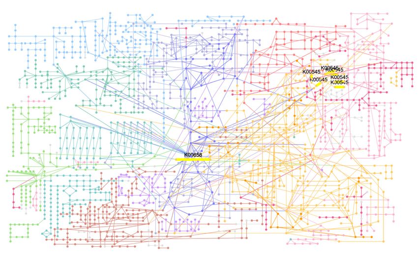
prevalence of depressive disorder occurs all over the
world (Figure 1, 2), and there is no definite medicine or
treatments that can completely cure depressive disorder.
In 2017, the number given by the WHO shows that there
are 17.3 million adults had at least one Major Depressive
episode, which is 7.1% of the US adult [2]. Some of the
risk factors for MDD in previous studies show that
females have a higher prevalence and risk of MDD [3].
Fig. 1. Crude prevalence of depressive disorders in India, 2017
Also, genetic influence and age is also a risk factor of the [5].
morbidity of MDD. Study shows that the onset of MDD
is low until the early adolescence [4].
© The Authors, published by EDP Sciences. This is an open access article distributed under the terms of the Creative Commons Attribution
License 4.0 (http://creativecommons.org/licenses/by/4.0/).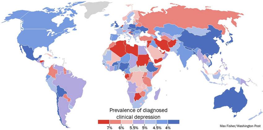
E3S Web of Conferences 271, 03055 (2021)                                                https://doi.org/10.1051/e3sconf/202127103055
ICEPE 2021

                                Fig. 2. Worldwide crude prevalence of depressive disorders, 2013 [6].

                                                                     appetite [14]. Thus, I hypothesize that MDD might be
   1.2 Major Depressive Disorder                                     linked to the enteric microorganisms, or the so-called
                                                                     “gut microbiota”, which is a component that has already
   Currently available treatments for MDD include
                                                                     been shown to associated with many brain disorders.
   antidepressant medications such as selective serotonin
                                                                         The gut microbiota contains over 3 million genes [15]
   reuptake      inhibitors   (SSRIs),       serotonin    and
                                                                     and 100 trillion microorganisms which can influence our
   norepinephrine reuptake inhibitors (SNRIs), tricyclic
                                                                     well-being in many significant aspects [16]. Recent
   antidepressants (TCAs) and psychological therapy [7].
                                                                     studies have suggested a stunning connection between
   However, these antidepressant medicines also have some
                                                                     Parkinson’s Disease (PD) and gut microbiota (such as
   severe side effects such as for SSRI, it will reduce the
                                                                     Proteus sp. and Bilophila sp.) [17]. The so-called “brain-
   concentration of the neurotransmitter serotonin in
                                                                     gut axis” is reported to directly contribute to the
   platelets, it will lead to a decrease in blood clotting
                                                                     aggregation of PD causing α-synuclein proteins in the
   ability [8]. Besides, when someone had experienced
                                                                     midbrain, by dysfunction in microglia activation.
   severely lost such as death or job loss, the antidepressant
                                                                     Progressively preclinical literature indicates that there is
   drugs may not work [9]. Another disorder that can also
                                                                     bidirectional signal transduction between the brain and
   produce distressing psychotic symptoms like MDD is
                                                                     gut microbiome, which involves a variety of
   bipolar disorders; these two disorders can lead to highly
                                                                     neuroendocrine and endocrine signal transduction
   disabling and unpleasant depressive episodes [10].
                                                                     mechanisms [18]. Research shows that probiotic
       The key to finding more effective treatments for
                                                                     administration not only affects the fMRI in the brain but
   MDD and bipolar disorder is the exploration of their
                                                                     also causes a change in the urine metabolic profile,
   molecular mechanisms. Previous genetic studies have
                                                                     further proves the hypothesis that there is bidirectional
   identified several genes whose mutation can lead to
                                                                     signal transduction between the brain and the gut
   MDD. For example, glucocorticoid receptor gene
                                                                     microbiome [19].
   NR3C1 and glycogen synthase kinase 3β is associated
                                                                         Another important aspect to link MDD and gut
   with a higher risk of MDD, implicating its potential
                                                                     microbiota is the immune system. In the past few
   connection with hormone regulation and metabolism
                                                                     decades, several published studies have already
   [11]. In other published studies, it has been shown that
                                                                     demonstrated that MDD is usually associated with
   the patients who suffer in MDD has a decreased serum
                                                                     changes in the immune system. For instance, MDD
   level of BDNF [12]. Although BDNF plays multiple
                                                                     patients are often characterized by elevated levels of pro-
   roles in the function of central nervous system, its
                                                                     inflammatory cytokines in the circulation and activation
   detailed molecular mechanisms of action on MDD have
                                                                     of microglia in the brain [20]. On the other hand, the gut
   not been further explored.
                                                                     microbiota is a part of the immune system and it is also
                                                                     one of the symptoms of MDD [21]. Therefore, I
   1.3 Gut Microbiota and its Potential Connection                   speculate that gut microbiota may be related to
   to MDD                                                            depression.
   It has long been appreciated that diet and food intake are
   associated with the onset of Major Depressive                     1.4 Rationale and Aim
       Depression. For instance, individuals who are lack
                                                                     Based on the above rationale, I aim to learn about the
   many nutrients such as B-complex vitamins and other
                                                                     relationship between MDD and gut microbiota. The
   acids such as Lithium often develop mental illness [13].
                                                                     relationship could be a very interesting exploration
   Recent psychiatric studies have shown that some of the
                                                                     strategy and may shed new insight on both the molecular
   MDD symptoms include increasing or decreasing
                                                                     mechanisms of MDD and aid on better treatments of

                                                                 2
E3S Web of Conferences 271, 03055 (2021)                                                https://doi.org/10.1051/e3sconf/202127103055
ICEPE 2021

  MDD. Based on multiple datasets including genomic,                 2.1.3 GeneCards
  metabolic, and microbiological information, I first
  conducted a computational screen to filtrate these genes           GeneCards is a database that provides human genes that
  that might have a relationship between MDD and the gut             were predicted and annotated. The knowledge base
  microbiota. Then, I further present a gene network                 contains more than 190 datasets to provide sources or
  analysis to further analyze the relationship between these         different genes and parts that can be influenced by that
  genes that has been filtrated already. Lastly, I carried out       gene. It provides a different section in the genes such as
  a systematic microbiome analysis on the candidate MDD              aliases, disorders, domains, drug, expression, function,
  gene list to explore their relationship with gut microbiota.       genomics, localization, pathway, product, protein,
                                                                     publication, and many others in order to provide more
                                                                     detail and comprehensive understanding to researchers.
  2 Materials and Methods                                                The GeneCards database is accessible from the
                                                                     following link: https://www.genecards.org/
  2.1 Data Sources
                                                                     2.1.4 Computational Tools and Algorithms
  Several publicly accessible databases of neurobiology
  and microbiology were employed in this paper. They are             1)STRING
  summarized in the following sections.                                  STRING is a well-known online genetic analytic web
                                                                     resource and database, which can predict protein-protein
  2.1.1 Allen Brain Atlas                                            interaction and depict the interaction network.
                                                                     Experimental data, computational prediction methods
  Allen Brain Atlas is a comprehensive and                           and public text collections are all collected in the
  multidisciplinary database combing the genomic,                    STRING database.
  transcriptomic, neuroanatomy, and connectome data of                   The STRING database is accessible from the
  the mouse and human brains [22]. It contains 16                    following link: https://string-db.org/
  databases about the mouse and human cell types
                                                                         2) Database for Annotation, Visualization and
  database and brain atlas. Allen Brain Atlas also provides
                                                                     Integrated Discovery (DAVID)
  data visualizing tools such as Brain Explorer and Planner
  View for researchers to grasp a comprehensive                          DAVID is a comprehensive biological knowledge
  understanding of the nervous under physiological and               base and analysis tool designed to systematically extract
  disease circumstances.                                             biological meaning from large gene lists [23]. Using
      The Allen Brain Atlas database is accessible from the          high-throughput        genomics,      proteomics,       and
  following link: https://portal.brain-map.org/                      bioinformatics scanning methods to perform functional
                                                                     analysis of large gene lists is still a difficult task [24].
  2.1.2 NIH Human Microbiome Project                                 DAVID allows researchers to gain insight into the
                                                                     biological topics in the gene list in genome-scale
  1) Human Microbiota Project (HMP) & Integrative                    research [23].
  Human Microbiota Project (iHMP)                                        DAVID is accessible from the following link:
      HMP and iHMP were created under the project NIH                https://david.ncifcrf.gov/tools.jsp
  Human Microbiome Project. iHMP is the second phase                    3) R programming language and R Studio
  of HMP. HMP generates resources to promote the
  characterization of the human microbiome to further                    R is a language and environment for statistical
  understand how the microbiome affects human health                 calculations and graphics and provides various statistical
  and disease. iHMP is engaging in providing new                     information and graphics techniques. R can easily
  calculation tools and comprehensive molecular                      produce well-designed publication-quality charts,
  perspectives on microbial activity during metabolic                including the required mathematical symbols and
  abnormalities. The database HMP and iHMP contain                   formulas. When using R, g: Profiler is also involved. g:
  31,596 samples, and a total of 161,256 files provided to           Profiler tool set can be used to find different biological
  all researchers. It is characterized by a microbial                categories in the gene list, conversion between gene
  community from 300 healthy individuals spread across               identifiers, and so on [25].
  several different parts of the human body: nasal passages,            4) MicrobiomeAnalyst
  mouth, skin, gastrointestinal tract, and urogenital tract.
  HMP contains the following parts:                                     MicrobiomeAnalyst contains 4 sections in visual and
      The HMP database is accessible from the following              meta-analysis of microbiome data, it includes: Marker
  link: https://www.hmpdacc.org/hmp/                                 Data Profiling (MDP), Shoutgun Data Profiling (SDP),
      The iHMP database is accessible from the following             Projection with Public Data (PPD), Taxon Set
  link: https://www.hmpdacc.org/ihmp/                                Enrichment Analysis (TSEA) [26].
                                                                        The MicrobiomeAnalst database is accessible from
                                                                     the following link: https://www.microbiomeanalyst.ca/
                                                                     MicrobiomeAnalyst/home.xhtml

                                                                 3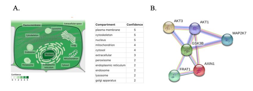
E3S Web of Conferences 271, 03055 (2021)                                                https://doi.org/10.1051/e3sconf/202127103055
ICEPE 2021

   2.1.5 Project Workflow                                           The key question to reveal the relationship between
                                                                    MDD and gut microbiota is to ensure there are genes that
                                                                    are both related to MDD and gut microbiota. In order to
                                                                    address these questions, I decided to conduct a
                                                                    computational screen of MDD related genes that might
                                                                    be involved in the regulation of gut microbiota or the
                                                                    digestive system. Conducting a computational screen at
                                                                    the first step helps to integrate the information of the
                                                                    genes which helps to narrow down the genes that shows
                                                                    both high expression in MDD and gut microbiota. By
                                                                    using a computational screen, it helps me to filtrate
                                                                    genes and finally find out the genes that is related to
                                                                    MDD and gut microbiota.
                                                                        First, I examined all relatively well-characterized
                                                                    MDD genes to confirm their RNA and protein
                                                                    expressions in the brain and to explore whether they may
                                                                    also be expressed in the digestive system. This will help
                                                                    us demonstrate if any of these genes might be involved
                                                                    in the brain-gut axis. Secondly, I employed a similar
                                                                    computational approach to search novel genetic
                                                                    candidates based on a more recent genome-wide
                                                                    association study (GWAS) that has identified 44 risk
                                                                    variants [27]. Lastly, I examine the molecular functions
                                                                    of these candidates through another computational
            Fig. 3. Screening Process in GWAS genes.                screen.

   This is a flowchart showing the screening process for
                                                                    3.1.1 Conventional MDD related genes
   GWAS genes. At first, I filtrate all the non-protein-
   coding genes, then I rate them through Genecards data.           To start with, I perform a literature search on the
   Last, I set a threshold for the genes, if the scores are         Pubmed publication database, examining Systematic
   above the threshold, I will have detailed research on that       Reviews mentioning “gut microbiota” and “Major
   gene to see if it really has a relationship between the          Depressive Disorder”. The search results suggest that
   brain and gut microbiota.                                        there are approximately 12 genes that have been well
                                                                    appreciated in the psychiatric research community [28].
   3 Results                                                        To better visualize the categories and functions of these
                                                                    genes, I then summarize these genes in Table 1.

   3.1 Computational screen for potential gut
   microbiota related MDD genes
                                           Table 1. Summary of well-known genes in MDD

               Gene Name           Category                                  Known Functions
                 GRIK4          Glutamate-gated    By activating ligand-gated ion channels and G protein-coupled
                                  ion channel      membrane receptors, it will excite the neurotransmitter in the central
                                                   nervous system.
                 BDNF            Neurotrophic      Binding of this protein to its cognate receptor promotes neuronal
                                     factor        survival in the adult brain
                 FKBP5          Chaperone and      Immunoregulation and basic cellular processes involving protein
                                stress response    folding and trafficking
                                    protein
                 COMT          Methyltransferase   Transfer of a methyl group from S-adenosylmethionine to
                                                   catecholamines
                GSK3β                Kinase        Identified as phosphorylation and inactivator of glycogen synthase
                SLC6A4              Integral       Transports the neurotransmitter serotonin from synaptic spaces into
                               membrane protein    presynaptic neurons
                 TREK1         Lipid-gated ion     Negative regulator of glucose homeostasis and is involved in energy
                               channel             metabolism, inflammation, ER-stress, mitochondrial dysfunction, and
                                                   apoptotic pathways
                CRHBP           Glutamate-gated    The major excitatory neurotransmitter in the central nervous system
                                 ionic channel
                 IGF-1           Growth factor     A member of a family of proteins involved in mediating growth and
                                                   development

                                                                4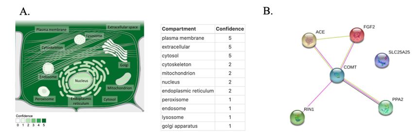
E3S Web of Conferences 271, 03055 (2021)                                                     https://doi.org/10.1051/e3sconf/202127103055
ICEPE 2021

                  IL1β              Cytokine         Involved in a variety of cellular activities, including cell proliferation,
                                                     differentiation, and apoptosis.
                  IL6               Cytokine         Implicated in a wide variety of inflammation-associated disease
                                                     states, including susceptibility to diabetes mellitus and systemic
                                                     juvenile rheumatoid arthritis
                 TNFα             Inflammatory       Implicated in a variety of diseases, including autoimmune diseases,
                                      factor         insulin resistance, and cancer.

      In order to better explore the genes whether might be             considered (small intestine, colon, stomach, and
  associated with the brain-gut axis, I recorded each gene              esophagus). Then, for the protein expression metrics,
  expression in the area of the brain and the organs of the             there are 7 levels: -2~-1 (score 1); -1~0 (score 2); 0~1
  digestive system. To reduce bias and better visualize the             (score 3); 1~2 (score 4); 2~3 (score 5); 3~4 (score 6);
  results, I introduced a gene expression metric system to              4~5 (score 7). Each tissue expression score adds up then
  analyze these data. In the RNA expression metrics, I                  divided by the total number of tissues (four tissue types
  subdivided the range of gene expression to four levels,               considered: small intestine, colon, stomach, and
  which are: 0~1 (score 1); 1~10 (score 2); 10~100 (score               esophagus). The symbol “/” represents that there is no
  3); 100~1000 (score 4). For the final RNA expression                  expression data currently available in the GeneCard
  score by each method (RNA-seq, microarray or SAGE),                   database (Table 2).
  it is the averaged expression score across all tissues
                              Table 2. RNA and protein expression levels in the human digestive system

                                            RNA expression metrics
                                                                                             Protein          Overall
                                  RNA-seq
                                                Microarray score SAGE score             expression score   Average score
                                   score
              SLC6A4                2.25                 2                   0                  1               1.38
               GRIK4                 2                   2                   1                  1                1.5
               BDNF                  2                   2                   0                  1               1.25
               FKBP5                  3                  2                   3                3.67              2.92
               COMT                  2                   2                   1                  4               2.25
           TREK1/KCNK2               3                  2.5                  1                  1               1.88
              GSK3B                   3                  2                   1                3.33              2.33
              CRHBP                  2                   2                   1                  1                1.5
                IGF-1                3                   2                   0                  1                1.5
                IL-1β               2.75                 2                   2                  1               1.94
                 IL-6                3                   2                   1                  1               1.75
                TNFα                2.25                 2                   1                  1               1.56
  Note. In RNA expression metrics, I mainly focused on the small intestine, colon, stomach, and esophagus. In Protein expression, I
  mainly focused on the colon, stomach, and esophagus. The overall score is defined as the summation of RNA score and protein score.

                                    Table 3. RNA and protein expression levels in the human brain

                                                  RNA expression metrics
                                                                                            Protein             Overall
                                       RNA-seq                           SAGE
                                                       Microarray score               expression score       Average score
                                         score                            score
                 SLC6A4                    2                   2            /                   1                  1.67
                  GRIK4                    3                   2            1                   1                  1.75
                  BDNF                     3                   2            /                   1                    2
                  FKBP5                    3                   2            2                   2                  2.25
                  COMT                     3                   3            3                   3                    3
                 KCNK2                     3                   2            3                   /                  2.67
                 GSK3B                     3                   2            2                  2.5                 2.38
                 CRHBP                     3                   2            1                  1.5                 1.88
                   IGF-1                   2                   2            /                 1.25                 1.75
                 IL1beta                   3                   2            2                   1                    2
                    IL6                    3                   2            2                   1                   2
               TNF Alpha                   2                   2            2                   1                  1.75
  Note. Expression in the Brain. In RNA expression metrics, I mainly focused on the brain. In Protein expression, I mainly focused on
  the brain, brain fetal, frontal cortex, and cerebral cortex.

      In all genes, these well-known genes have some                    has an average of 1.81, and the expression in the brain
  extent of cerebral expressions, and they all show                     has an average of 2.1. We can conclude that the
  expression in the digestive system (Table 2). Comparing               expression in the brain has a higher expression
  the average of the expression shown in the brain and the              comparing the digestive system.
  digestive system, the expression in the digestive system

                                                                    5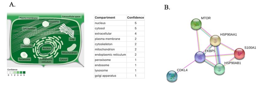
E3S Web of Conferences 271, 03055 (2021)                                              https://doi.org/10.1051/e3sconf/202127103055
ICEPE 2021

       Based on the relative expression level of all 12                 FKBP5
   known MDD related genes (Table 2 and 3), I realized                  FKBP Prolyl Isomerase 5(FKBP5) is a member from
   that FKBP5, COMT, and GSK3B are most likely to be                the immunophilin protein family and it plays a role in
   associated with brain-gut axis based on the expression           immune regulation and basic cellular processes involved
   analysis, since their expression levels are always among         in protein folding and transport. Some related diseases
   the highest in the brain and in the digestive system. I          that are related to FKBP5 are MDD and Asthma. FKBP5
   then searched the molecular and functional information           plays a role in the risk of Dajor Depressive Disorder
   of these 3 genes in the GeneCards database.                      (MDD), response to treatment, and changes in brain
                                                                    regions [29].

                                              Fig. 4. Human Microarray for FKBP5.

       It shows the Human expression in FKBP5. The red              which tissue shows high expression all the time since
   represents high expression and green represents low              there are many gene symbols and the expression is
   expression. The color shown in the first row of this             different for different donors.
   figure represents different donors. It is hard to identify

                                            Fig. 5. Human RNA expression in FKBP5.

      From the data, we can determine that the brain is                From the data, the brain did not show a very high
   expressing at an average expression for all three                expression, however, it is still maintaining at the average.
   expressions.

                                                                6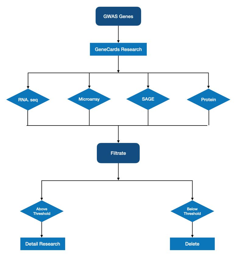
E3S Web of Conferences 271, 03055 (2021)                                                  https://doi.org/10.1051/e3sconf/202127103055
ICEPE 2021

                            Fig. 6. Information about the localization and interacting proteins for FKBP5.
    A. Localization for FKBP5. From this screenshot, we can explore that FKBP5 is most confident living in the nucleus and cytosol.
    B. Pathway for FKBP5. The screenshot presents some associated genes with FKBP5(CDKL4, HSP90AA1, HSP90AB1, MTOR,
                                                              S100A1).

      COMT                                                             bound form (MB-COMT). Diseases that are related to
      COMT (Catechol-O-Methyltransferase) transfers                    COMT are schizophrenia and panic disorder 1. COMT
  methyl groups from S-adenosylmethionine to                           will influence the level of dopamine, and dopamine
  catecholamines. COMT is also an important term in                    plays a very important role in psychiatric disorder, which
  curing for hypertension, hypertension and Parkinson’s                indirectly proves that COMT is related to psychiatric
  disease. There was two types found in the COMT tissues,              disorders [30].
  one is soluble (S-COMT), and the other is membrane

                                                Fig. 7. Human Microarray for COMT.

      It shows the human expression in COMT. The red                   which tissue shows high expression all the time since
  represents high expression and green represents low                  there are many gene symbols and the expression is
  expression. The colour shown in the first row of this                different for different donors.
  figure represents different donors. It is hard to identify

                                              Fig. 8. Human RNA expression in COMT.

                                                                   7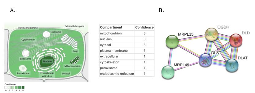
E3S Web of Conferences 271, 03055 (2021)                                               https://doi.org/10.1051/e3sconf/202127103055
ICEPE 2021

       The data shows that the Nervous area was expressing             In this data, the brain section also shows an
   at a very average rate in the RNA expression.                    expression at an average rate.

                               Fig. 9. Information about the localization and interacting proteins for COMT.
      A. Localization for COMT. The screenshot shows that COMT is most confident staying at plasma membrane, extracellular and
                                                                  cytosol.
                           B. Pathway for COMT. Some pathways for COMT are ACE, FGF2, RIN1 and PPA2.

      GSK3B                                                         stress, inflammation and is glucose homeostasis negative
      GSK3B (Glycogen Synthase Kinase 3 Beta) is                    regulator. Some related diseases are Bipolar Disorder
   involved in energy metabolism, endoplasmic reticulum             and Alzheimer Disease.

                                              Fig. 10. Human Microarray for GSK3B.

       It shows the human expression in GSK3B. The red              which tissue shows high expression all the time since
   represents high expression and green represents low              there are many gene symbols and the expression is
   expression. The colour shown in the first row of this            different for different donors.
   figure represents different donors. It is hard to identify

                                                Fig. 11. RNA expression in GSK3B.

                                                                8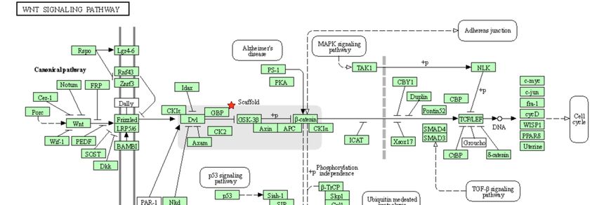
E3S Web of Conferences 271, 03055 (2021)                                                https://doi.org/10.1051/e3sconf/202127103055
ICEPE 2021

      The data shows that the Nervous area was expressing                The brain fetal and frontal cortex shows a high
  at a very average rate in the RNA expression, and it is             expression in the protein expression, but the brain and
  also expressed in SAGE where not a lot of tissues shows             cerebral cortex expression is very low.
  high expression.

                           Fig. 12. Information about the localization and interacting proteins for GSK3B.
   A. Localization for GSK3B. The screenshot shows that plasma membrane, cytoskeleton and nucleus are all the confidence area for
                                                           GSK3B to stay.
  B. Pathway for GSK3B. From the screenshot, we can explore that AKT1, AKT3, AXIN1, FRAT1 and MAP2K7re all associated with
                                                               GSK3B.

                                                                      genes, to 53 genes by only including the protein-coding
                                                                      genes (Table 4). To explore the possibility of these 53
  3.1.2 Genes identified in GWAS of MDD
                                                                      genes in brain-gut axis, I further researched through
  In section 1, I already find several genes that might be            Genecards about their expression levels in Small
  linked to gut microbiota, however, the 12 genes I                   Intestine, Colon, Stomach, and Esophagus. When
  screened from only represent a very tiny proportion of              measuring, I employed the same metrics of RNA and
  the human genome. Therefore, in order to find more                  protein expression as section 1 (Table 2).
  genes that might be related to MDD and gut microbiota,                  Next, I set a threshold for each expression score to
  I employed the GWAS data from a recent published                    reduce the genes that show high expression. For the
  article which identified 44 genetic loci whose mutations            RNA-seq score, I set the threshold to be 12; for the
  are associated with higher risks of MDD [27]. These                 Microarray score, I set the threshold to be 4; for the
  GWAS genes has helped me to narrow down the                         SAGE score, I set the threshold to be 1, and for Protein
  candidate gene list by filtering out genes unrelated to             expression score, I set the threshold to be 6. Only the
  MDD from genome wide data. Starting from these data,                gene that has all scores above the threshold for each data
  I can further discover the genes that are also related to           will be involved. These genes will be presented with its
  gut microbiota.                                                     average of RNA-seq, Microarray, SAGE, and Protein
      First, in order to simplify my computational screen, I          expression score (Table 5).
  narrowed down the list of 44 genetic loci, which has 68
                                           Table 4. Protein Coding Genes existed in GWAS

        Gene Context     Chromosome          Region [27]               SNP             Location (bp)          P value
                             No.
           RERE                1             8.390-8.895             rs159963            8,504,421          3.2 × 10⁻⁸
         SLC45A1               1             8.390-8.895             rs159963            8,504,421          3.2 × 10⁻⁸
          NEGR1                1            72.511-73.059            rs1432639          72.813.218          4.6 × 10⁻¹⁵

         DENND1B               1           197.343-197.864           rs9427672          197,754,741         3.1 × 10⁻⁸
           VRK2                2            57.765-58.485         rs11682175            57,987,593          4.7 × 10⁻⁹

           NR4A2               2           157.978-157.464           rs1226412          157,111,313         2.4 × 10⁻⁸
           GPD2                2           157.978-157.464           rs1226412          157,111,313         2.4 × 10⁻⁸
          TOPAZ1               3            44.222-44.997      chr3_44287760_l          44,287,760          4.6 × 10⁻⁸

          TCAIM                3            44.222-44.997      chr3_44287760_l          44,287,760          4.6 × 10⁻⁸

                                                                 9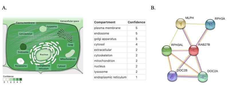
E3S Web of Conferences 271, 03055 (2021)                                        https://doi.org/10.1051/e3sconf/202127103055
ICEPE 2021

           ZNF445              3            44.222-44.997    chr3_44287760_l     44,287,760          4.6 × 10⁻⁸

           RSRC1               3           157.616-158.354        rs7430565     158,107,180          2.9 × 10⁻⁹

            MLF1               3           157.616-158.354        rs7430565     158,107,180          2.9 × 10⁻⁹

          SLC30A9              4            41.880-42.189         rs34215985     42,047,778          3.1 × 10⁻⁹

          DCAF4L1              4            41.880-42.189         rs34215985     42,047,778          3.1 × 10⁻⁹

           MEF2C               5            87.443-88.244    chr5_87992715_l     87,992,715          7.9 × 10⁻¹¹

           TENM2               5           166.977-167.056        rs4869056     166,992,078          6.8 × 10⁻⁹

           FBXL4               6            99.335-99.662         rs9402472      99,566,521          2.8 × 10⁻⁸

         TMEM106B              7            12.154-12.381         rs10950398     12,264,871          2.6 × 10⁻⁸

            VWDE               7            12.154-12.381         rs10950398     12,264,871          2.6 × 10⁻⁸

            PUM3               9             2.919-3.009          rs1354115      2,983,774           2.4 × 10⁻⁸

           ASTN2               9           119.675-119.767        rs7856424     119,733,595          8.5 × 10⁻⁹

          DENND1A              9           126.292-126.735        rs7029033     126,682,068          2.7 × 10⁻⁸

            LHX2               9           126.292-126.735        rs7029033     126,682,068          2.7 × 10⁻⁸

           SORCS3             10           106.397-106.904        rs61867293    106,563,924          7.0 × 10⁻¹⁰

            ELP4              11            31.121-31.859         rs1806153      31,850,105          1.2 × 10⁻⁹

            PAX6              11            31.121-31.859         rs1806153      31,850,105          1.2 × 10⁻⁹

            SOX5              12            23.924-24.052         rs4074723      23,947,747          3.1 × 10⁻⁸

           ENOX1              13            44.237-44.545         rs4143229      44,327,799          2.5 × 10⁻⁸

           LACC1              13            44.237-44.545         rs4143229      44,327,799          2.5 × 10⁻⁸

          CCDC122             13            44.237-44.545         rs4143229      44,327,799          2.5 × 10⁻⁸

           OLFM4              13            53.605-54.057          rs12552       53,625,781          6.1 × 10⁻¹⁹

           LRFN5              14            41.941-42.320         rs4904738      42,179,732          2.6 × 10⁻⁹

           SYNE2              14            64.613-64.878          rs915057      64.686,207          7.6 × 10⁻¹⁰

            ESR2              14            64.613-64.878          rs915057      64.686,207          7.6 × 10⁻¹⁰

            DLST              14            75.063-75.398    chr14_75356855_l    75,356,855          3.8 × 10⁻⁹

           PROX2              14            75.063-75.398    chr14_75356855_l    75,356,855          3.8 × 10⁻⁹

          RPS6KL1             14            75.063-75.398    chr14_75356855_l    75,356,855          3.8 × 10⁻⁹

            BAG5              14           103.828-104.174        rs10149470    104,017,953          3.1 × 10⁻⁹

           RBFOX1             16             6.288-6.347          rs8063603      6,310,645           6.9 × 10⁻⁹

           RBFOX1             16             7.642-7.676          rs7198928      7,666,402           1.0 × 10⁻⁸

           SHISA9             16            13.022-13.119         rs7200826      13,066,833          2.4 × 10⁻⁸

           CPPED1             16            13.022-13.119         rs7200826      13,066,833          2.4 × 10⁻⁸

           PMFBP1             16            71.631-72.849         rs11643192     72,214,276          3.4 × 10⁻⁸

           DHX38              16            71.631-72.849         rs11643192     72,214,276          3.4 × 10⁻⁸

          CRYBA1              17            27.345-28.419         rs17727765     27,576,962          8.5 × 10⁻⁹

          MYO18A              17            27.345-28.419         rs17727765     27,576,962          8.5 × 10⁻⁹

           NUFIP2             17            27.345-28.419         rs17727765     27,576,962          8.5 × 10⁻⁹

                                                             10
E3S Web of Conferences 271, 03055 (2021) https://doi.org/10.1051/e3sconf/202127103055
ICEPE 2021
DCC 18 50.358-50.958 rs11663393 50,614,732 1.6 × 10⁻⁸
RAB27B 18 51.974-52.552 rs183288 52,517,906 2.6 × 10⁻⁸
CCDC68 18 51.974-52.552 rs183288 52,517,906 2.6 × 10⁻⁸
TCF4 18 52.860-53.268 rs12958048 53,101,598 3.6 × 10⁻¹¹
L3MBTL2 22 40.818-42.216 rs5758265 41,617,897 7.6 × 10⁻⁹
CHADL 22 40.818-42.216 rs5758265 41,617,897 7.6 × 10⁻⁹
Table 5. RNA and protein expression levels in the human digestive system
RNA expression metrics Protein expression Overall
RNA-seq score Microarray score SAGE score score Average score
GPD2 3 2 2 4 2.75
SLC30A9 3 3 1 2.33 2.33
OLFM4 3 2 1 2 2
DLST 3.25 4 2 3.67 3.23
BAG5 3 2 2 4.67 2.92
CPPED1 3 3 1 3.33 2.58
DHX38 3 2 1 4 2.5
RAB27B 3 2 3 2 2.5
Note. Table 5 shows the average expressions of the genes that are above threshold in the field of Small Intestine, Colon, Stomach,
and Esophagus.
Table 6. RNA and protein expression levels in the human brain
RNA expression metrics Protein expression Overall
RNA-seq score Microarray score SAGE score score Average score
GPD2 3 2 2 4.25 6
SLC30A9 3 3 3 1.5 3.75
OLFM4 3 2 1 1 2.5
DLST 2 2 1 1.25 1.56
BAG5 3 2 2 2 2.25
CPPED1 3 2 2 3 2.5
DHX38 3 2 2 1.5 2.13
RAB27B 3 2 2 3 2.5
Note. Table 6 shows the average expression for the genes that are above threshold genes expression in the brain.
Then, in order to narrow down the genes, I collected antiapoptotic. The gene is selectively highly expressed in
the expression for the genes showed in the brain after colonic epithelium and moderately expressed in several
screening out the above threshold genes. Base on the nervous tissue (including cerebral cortex, cerebellum,
Overall protein score showed in the data Expressions of retina, and spinal cord) (Figure 13). Some disease that is
the genes that are above threshold in the field of Small related to OLFM4 are Pancreatic Cancer and Ovary
Intestine, Colon, Stomach and Esophagus, I found out 4 Serous Adenocarcinoma. OLFM4 is related to many
most interesting genes for further research: OLFM4, digestive diseases. In recent study about Helicobacter
DLST, CPPED1, RAB27B. pylori (H. pylori) infection of gastric mucosa, it shows
OLFM4 an increase in the expression of OLFM4. OLFM4 plays
Olfactomedin 4 (OLFM4) gene encodes an an important role in the mucosal defense of
extracellular matrix protein that is believed to be inflammatory bowel disease [31].
Fig. 13. Human microarray data of OLFM4 from Allen Brain Atlas.
This screenshot is a picture showing the gene all donors, amygdalohippocampal transition zone (ATZ)
OLFM4. In the screenshot, the red symbolized as a and tail of the caudate nucleus (TCd) all shows red on
higher activity in genes of interest. The colour shown in the picture, which means in these parts OLFM4 shows
the first row of this figure represents different donors. In active.
11E3S Web of Conferences 271, 03055 (2021) https://doi.org/10.1051/e3sconf/202127103055
ICEPE 2021
Fig. 14. Human RNA expression of OLFM4.
From the graph, we can identify that the internal From the graph, we can explore that internal part
section is showing a very high expression, representing didn’t show a very high expression, but its expression is
that OLMF4 did affect some parts in gut microbiota. still staying at the average.
Fig. 15. Information about the localization and interacting proteins for OLFM4.
A. Different compartment and the confidence rate for OLFM4 to live.
B. The top 5 STRING interaction network for OLFM4 (FLRT3, NOD1, NOD2)
RAB27B which displays that gut-immune system-brain axis might
RAB27B is in the RAS Oncogene Family. RAB27B play a role in RAB27B. In order to further understand
can be related to diseases including Griscelli Syndrome that which part of the brain has high expression, I did a
and Piebald Trait. Some superpathways includes further analysis through Allen Brain Atlas, and it
Response to elevated platelet cytosolic Ca2+, Vesicle- demonstrates that the frontal cortex, amygdala,
mediated transport and Metabolism of protein. diencephalon shows a high activeness. Frontal cortex
The RNA of RAB27B shows multiple major tissues and amygdala are all the brain parts that will show high
active, however, in the protein expression data, Brain activity in MDD, which further confirm that RAB27B
and stomach are the highest expression in their lineages, might be related to MDD.
12E3S Web of Conferences 271, 03055 (2021) https://doi.org/10.1051/e3sconf/202127103055
ICEPE 2021
Fig. 16. Human microarray data of RAB27B.
The microarray data shows the activity in genes of different donors. After comparing all donors with all
interest indifferent parts of the brain. The red represents activeness, Globus pallidus, internal segment in the basal
high activeness, and the green shows lower activeness. ganglia, cingulum bundle in the white matter shows high
The color shown in the first row of this figure represents activeness.
Fig. 17. RNA expression level of RAB27B.
Although brain and stomach both demonstrates high The data shows that Stomach and Brain are the
activity, mostly all the major tissues are active, therefore highest in their lineage, which displays that the gut
we cannot assure that this gene might contain the gut immune system brain axis might be involve in this gene.
immune system brain axis.
Fig. 18. T the localization and interacting proteins for RAB27B.
A. Different compartment and the confidence rate for RAB27B to live.
B. The top 5 STRING interaction network for RAB27B (DOC2A, DOC2B, MLPH, RPH3A, RPH3AL)
13E3S Web of Conferences 271, 03055 (2021) https://doi.org/10.1051/e3sconf/202127103055
ICEPE 2021
DLST of a decreasing of cell viability and induction of
Dihydrolipoamide S-Succinyltransferase (DLST) is a apoptosis was caused by the RNAi inhibition of DLST,
member of the 2-oxoacid dehydrogenase family. Some which also disrupted the TCA cycle [32]. The TCA cycle
associated diseases with DLST are Paragangliomas 7 is affected by the young and mid-age MDD patients,
and Hereditary Paraganglioma-Pheochromocytoma which indirectly proved that DLST might be associated
Syndromes. In the Human T-ALL cell line, the symptom with MDD.
Fig. 19. Human microarray data of DLST.
The red represents high expression and the green IV), Crus I (Cb-Crus I) and Crus II (Cb-Crus II) all
represents low expression. The color shown in the first shows high expression, which might show that these four
row of this figure represents different donors. In all structures play a very important role in DLST.
donors, VI(Cb-VI), VIIA(Cb-VIIA), V(Cb-V), IV(Cb-
Fig. 20. RNA expression level of DLST.
In Figure 20, the expression for brain and the Although all the tissues show expression, but we can
digestive system are mostly at the same level as other still conclude that stomach, brain, and nasal respiratory
major tissues, but the expression appears for all three epithelium shows a higher expression then other tissue.
data, which might show that it is still significant in
DLST.
14E3S Web of Conferences 271, 03055 (2021) https://doi.org/10.1051/e3sconf/202127103055
ICEPE 2021
Fig. 21. T the localization and interacting proteins for DLST.
A. Different compartment and the confidence rate for DLST to live.
B. The top 5 STRING interaction network for DLST (DLAT, DLD, MRPL15, MRPL49, OGDH).
CPPED1 promote the apoptosis of the cell. Some related diseases
Calcineurin Like Phosphoesterase Domain Contaning with CPPED1 are Trichostrongyloidiasis and
1 (CPPED1) blocks the cell cycle progression and Trichostrongylosis.
Fig. 22. Human microarray data of CPPED1.
The red represents high expression and the green thalamus (VT) and corpus callosum(cc) all shows red,
represents low expression. The color shown in the first which represents high expression, which might show that
row of this figure represents different donors. In all these four structures play a very important role in DLST.
donors, globus pallidus, internal segment (GPi), ventral
Fig. 23. RNA expression level of CPPED1.
In figure 23, both brain and digestive system was at
an average expression.
15E3S Web of Conferences 271, 03055 (2021) https://doi.org/10.1051/e3sconf/202127103055
ICEPE 2021
In Figure 24, the brain didn’t show a very high expression in the protein expression data.
expression, but the digestive system shows a very high
Fig. 24. T the localization and interacting proteins for CPPED1.
A. Different compartment and the confidence rate for CPPED1 to live.
B. The top 5 STRING interaction network for CPPED1 (PFKM, PICALM, SEPSECS).
In Figure 26, it only shows the connection between
the seven genes. And the only related genes are FKBP5
3.2 Expression and Pathway Analysis of
and COMT. These seven genes have no direct
Identified Candidate Genes
connections between each other. However, research
After exploring the 7 genes that might be highly related methods for MDD are limited, some intermediate
with MDD and gut Microbiota, I am going to further components may be missed. Therefore, I adjusted the
explore the pathways and their relations with gut settings by the STRING algorithm and expanded the
microbiota. molecular network to find more “node” genes. The
expanded STRING network has demonstrated that (1)
OLFM4 was connected through AXIN2, (2) DLST still
3.2.1 STRING Pathway Analysis of the Candidate did not show any connections, and (3) RAB27B has no
Genes connections with all 6 other genes all the time. Clearly, I
found 2 central molecular pathways which are Wnt
Signaling and citric acid cycle.
Wnt Signaling Pathway
The Wnt Signaling Pathway conducts the cell’s
migration, polarity, neural patterning, and other body
parts in the embryo. When related to some
antidepressant treatment, the content of wnt will increase
[12]. Several evidences from animal models of
depression, it shows that glycogen synthase kinase 3 beta
(GSK3β) plays a very important role in MDD [33].
Through a further research, it proves the hypothesis that
Fig. 25. Relationships between the 7 genes related to brain-gut
axis. the polymorphism in GSK3β plays a role in the
susceptibility or expression of MDD, partly because it
acts through the classic Wnt signaling pathway and
related substrates [34].
Also, Wnt Signaling Pathway is also linked to gut
microbiota. Recent studies showed Wnt signaling
pathway is tightly linked to cancer; in colorectal cancer,
it has most prominently been described [35]. We can
infer that wnt signaling pathway might have some
relationships with the digestive system through the
secretion of the Wnt signaling. By all means, GSK3B
shows a high expression in both the brain and the
digestive system, and it is also linked to Wnt Signaling
Pathway. It can be inferred that Through Wnt Signaling.
Citric Acid cycle
Citric Acid cycle is a central biochemical process in
aerobic respiration which is conserved from bacteria to
Fig. 26. Expansion of the relationships between the 7 genes human beings. It is not surprising that citric acid cycle
related to brain-gut axis. may link the metabolism of gut microbiota and their host.
16E3S Web of Conferences 271, 03055 (2021) https://doi.org/10.1051/e3sconf/202127103055
ICEPE 2021
Some evidences from an article that focused on In the graph, GO:MF stands for Gene Ontology:
leukocyte tricarboxylic acid cycle and Bipolar Disorder Molecular Function; GO:CC stands for Gene Ontology:
show that mitochondrial dysfunction plays a role in the Cellular Component; GO:BP stands for Gene Ontology:
pathophysiology of bipolar disorder [27,36]. Genetic Biological Process; KEGG stands for Kyoto
mutations on α-ketoglutarate-dependent dioxygenase Encyclopedia of Genes and Genomes; REAC stands for
(FTO) leads to a higher average body mass index (BMI) Restriction endonuclease analysis of chromosomal DNA;
higher risk of MDD [37]. TF stands for Transcription Factor; MIRNA stands for
MicroRNA; HPA stands for Human Protein Atlas;
CORUM stands for the Comprehensive Resource of
3.2.2 Gene Ontology (GO) analysis
Mammalian Protein Complexes; HP stands for
To further understand the molecular functions of the 7 haptoglobin.
candidate genes associated with both MDD and gut
microbiota, I conducted the GO analysis. To avoid bias, I 3.2.3 DAVID
chose two different GO analysis tools: g:profiler and
DAVID [23]. DAVID provides the ability to perform functional
analysis of large gene lists about biological meanings.
1) GO analysis by gprofiler (R studio based)
By using DAVID, it enables a detailed gene list for
The g:profiler algorithm first provide a Mahattan-like functional analysis for 7 genes.
plot which illustrates which GO categories the
significant GO terms fall into. The Mahattan-like plot Table 7. DAVID Functional Annotation Chart
generated based on my 7 input candidate genes indicates
one important GO term: catalytic activity (GO: 0003824) GO Category GO term P value Benjamin
(Figure 27). The number of annotated GO terms in this
gene list is relatively few, since only 7 genes have been GOTERM_CC_ extracellular
2.4E-2 2.9E-1
discovered in my computational screen. Catalytic DIRECT exosome
activity can be also named as enzyme activity in GO
Note. Within these seven genes, there is only one GO category
annotation. This result is consistent with the finding shown in DAVID, and its term is extracellular exosome.
from STRING pathway analysis in which several
enzymes of citric acid cycle are involved. After importing the 7 genes, DAVID presented a
graph that shows the related GO Category, which is
extracellular exosome. Exosome has the ability to
processes synaptic plasticity, neuronal stress response,
intercellular communication and neurogenesis in the
brain, which is connected to the brain [38]. Also, studies
confirmed by delivering antibacterial products, the
secretion of exosomes to the top of the lumen will
regulate the function of distant cells along the
gastrointestinal tract or regulate the homeostasis of the
gut microbiota [39].
Fig. 27. Manhattan-like Plot from R-studio.
17E3S Web of Conferences 271, 03055 (2021) https://doi.org/10.1051/e3sconf/202127103055
ICEPE 2021
Fig. 28. Signaling Pathways identified by DAVID algorithm.
After giving the information about the 7 genes, it The 7 genes that was chosen has the highest expression
gives the WNT Signaling Pathway. In the pathway, the shown in both the brain and the digestive system among
gene that appears is GSK3B. These results provide all genes. However, the analysis presented previously
evidence in section 2.1, further acknowledged that the has not yet shown any relationship with the gut
Wnt Signaling pathway is a pathway in these 7 genes. microbiota. Therefore, I am going to further explore the
relationship between gut microbiota through a gut
microbiome analysis.
3.3 Gut Microbiome Analysis Based on the
Screened Genes
Fig. 29. Pathway for 7 chosen genes on MicrobiomeAnalyst.
Throughout the 7 genes chosen from 1.1 and 1.2, Betalain 1 0.0027
only two genes showed pathways on Microbiomeanalyst biosynthesis
pathways which are DLST (K00658) and COMT Lysine degradation 1 0.0268
(K00545). Steroid hormone 1 0.0295
biosymthesis
Table 8. MicrobiomeAnalyst Pathways chart Citrate cycle (TCA 1 0.0471
cycle)
Name Hits P-Value Tyrosine 1 0.048
18E3S Web of Conferences 271, 03055 (2021) https://doi.org/10.1051/e3sconf/202127103055
ICEPE 2021
metabolism Acknowledgements
Carbon metabolism 1 0.212
Note. Names for the pathways found in Microbiome Analyst Catherine Tian did the analysis of the data and the
description for this research paper. Gregory Rose, the
There is a total of 6 pathways found in mentor, suggested the research topic, theoretical
MicrobiomeAnalyst. Within these 6 pathways, there are directions, and general guidance.
5 pathways that are significant. This pathway further
confirms that Citric Acid Cycle is linked to the gut
microbiota. References
The first paragraph after a section or subsection
should not be indented; subsequent paragraphs should be 1. American Psychiatric Association, A.P.A. (2013).
indented by 5 mm. Diagnostic and statistical manual of mental
disorders (5th ed.) (American Psychiatric
Association).
4 Discussion 2. The National Institute of Mental Health,
T.N.I.o.M.H. (2017). Major Depression (National
In this research, I found 7 genes that are likely to be
Institute of Mental Health, U.S. : Department of
involved in the brain-gut axis to influence MDD, which
Health and Human Services).
are: OLFM4, RAB27B, DLST, CPPED1, FKBP5,
COMT and GSK3B (section A of Results). Through 3. Luo, Z., Li, Y., Hou, Y., Liu, X., Jiang, J., Wang, Y.,
pathway and network analyses of these candidate genes, Liu, X., Qiao, D., Dong, X., Li, R., et al. (2019).
I further identified three potentially new mechanisms Gender-specific prevalence and associated factors of
that might be involved in MDD (section B of Results). major depressive disorder and generalized anxiety
Although these mechanisms still need to be disorder in a Chinese rural population: the Henan
characterized with wet lab experiments, they have rural cohort study. BMC Public Health 19, 1744.
implicated promising directions of basic MDD research. 4. Wilson, S., Vaidyanathan, U., Miller, M.B., McGue,
Through GO analysis and gut microbiota analysis, I M., and Iacono, W.G. (2014). Premorbid risk factors
further identified the terms that are related to the 7 genes for major depressive disorder: are they associated
and classified the connection between gut microbiota with early onset and recurrent course? Dev
(section C of Results). Psychopathol 26, 1477-1493.
After exploring a new mechanism that might connect 5. India State-Level Disease Burden Initiative Mental
the gut microbiota and the brain, there could be new Disorders, C. (2020). The burden of mental
antidepressant medicines that might have the function of disorders across the states of India: the Global
increasing or decreasing the rate of Alpha-ketoglutarate, Burden of Disease Study 1990-2017. Lancet
a molecule that determines the rate of the citric acid Psychiatry 7, 148-161.
cycle. Alpha-ketoglutarate plays a very important role of
6. Dewey, C. (2013). A stunning map of depression
maintains intestinal homeostasis, which we might infer
rates around the world.
that it might be linked to MDD by a decrease of Alpha-
ketoglutarate [40]. However, this treatment solution still 7. Kerr, M. (2018). Major Depressive Disorder:
needs a verification in order to carry this antidepressant Symptoms, Causes, and Treatment (Healthline:
medicine out. Healthline Media).
The limitation of this study is that I could not suggest 8. Harvard Heath Publishing, H.H.P. (2014). What are
specific gut bacterial species based on my study with the real risks of antidepressants? (Harvard Health
solid evidence. This is due to the currently available Publishing).
microbiome dataset. 9. Neuman, F. (2018). The Problem with
Antidepressant Drugs (Psychology Today: Sussex
5 Equations and mathematics Publishers).
10. BrightQuest Treatment Centers, B.T.C. Major
In this research, I focused on a new perspective about Depression and Bipolar Disorder. In BrightQuest
some perspectives about MDD which is the connection Treatment Centers.
between MDD and gut Microbiota. By using certain 11. David, J.K., Ellen, F., and Mary, L.P. (2011). Major
database to confirm the stability of the results. depressive disorder: new clinical, neurobiological,
Based on the above research, I think it is possible to and treatment perspectives.
conduct animal model experiments on 7 genes
systematically, record the expression of the brain, 12. Varidaki, A., Mohammad, H., and Coffey, E.T.
digestive system, and the reaction of the animal when the (2016). Molecular Mechanisms of Depression.
gene was taken, and further identify if the gene is related 13. Rao, T.S., Asha, M.R., Ramesh, B.N., and Rao, K.S.
with both gut microbiota and MDD. (2008). Understanding nutrition, depression and
mental illnesses. Indian J Psychiatry 50, 77-82.
14. Mills, J.G., Larkin, T.A., Deng, C., and Thomas, S.J.
(2019). Weight gain in Major Depressive Disorder:
19E3S Web of Conferences 271, 03055 (2021) https://doi.org/10.1051/e3sconf/202127103055
ICEPE 2021
Linking appetite and disordered eating to leptin and Agerbo, E., Air, T.M., Andlauer, T.M.F., et al.
ghrelin. Psychiatry Res 279, 244-251. (2018). Genome-wide association analyses identify
15. Markus, M. (2018). Gut Microbiota: Definition, 44 risk variants and refine the genetic architecture of
Importance, and Medical Uses. (Medical News major depression. Nat Genet 50, 668-681.
Today: MediLexicon International). 28. Cheung, S.G., Goldenthal, A.R., Uhlemann, A.C.,
16. Viome (2018). Anxiety? Depression? Is Your Gut Mann, J.J., Miller, J.M., and Sublette, M.E. (2019).
Microbiome to Blame? (Viome). Systematic Review of Gut Microbiota and Major
Depression. Front Psychiatry 10, 34.
17. Sampson, T.R., Debelius, J.W., Thron, T., Janssen,
S., Shastri, G.G., Ilhan, Z.E., Challis, C., Schretter, 29. Tozzi, L., Farrell, C., Booij, L., Doolin, K., Nemoda,
C.E., Rocha, S., Gradinaru, V., et al. (2016). Gut Z., Szyf, M., Pomares, F.B., Chiarella, J., O'Keane,
Microbiota Regulate Motor Deficits and V., and Frodl, T. (2018). Epigenetic Changes of
Neuroinflammation in a Model of Parkinson's FKBP5 as a Link Connecting Genetic and
Disease. Cell 167, 1469-1480 e1412. Environmental Risk Factors with Structural and
Functional Brain Changes in Major Depression.
18. Winter, G., Hart, R.A., Charlesworth, R.P.G., and
Neuropsychopharmacology 43, 1138-1145.
Sharpley, C.F. (2018). Gut microbiome and
depression: what we know and what we need to 30. Otsuka, Y., Kakeda, S., Sugimoto, K., Katsuki, A.,
know. Rev Neurosci 29, 629-643. Nguyen, L.H., Igata, R., Watanabe, K., Ueda, I.,
Kishi, T., Iwata, N., et al. (2019). COMT
19. Pinto-Sanchez, M.I., Hall, G.B., Ghajar, K., Nardelli,
polymorphism regulates the hippocampal subfield
A., Bolino, C., Lau, J.T., Martin, F.P., Cominetti, O.,
volumes in first-episode, drug-naive patients with
Welsh, C., Rieder, A., et al. (2017). Probiotic
major depressive disorder. Neuropsychiatr Dis Treat
Bifidobacterium longum NCC3001 Reduces
15, 1537-1545.
Depression Scores and Alters Brain Activity: A
Pilot Study in Patients With Irritable Bowel 31. Wang, X.Y., Chen, S.H., Zhang, Y.N., and Xu, C.F.
Syndrome. Gastroenterology 153, 448-459 e448. (2018). Olfactomedin-4 in digestive diseases: A
mini-review. World J Gastroenterol 24, 1881-1887.
20. Carsten, C., Susanne, M., Stefanie, S., Volker, A.,
Udo, D., and Judith, A. (2018). Mitochondria, 32. Anderson, N.M., Li, D., Peng, H.L., Laroche, F.J.,
Microglia, and the Immune System—How Are They Mansour, M.R., Gjini, E., Aioub, M., Helman, D.J.,
Linked in Affective Disorders? (Frontiers: Frontiers). Roderick, J.E., Cheng, T., et al. (2016). The TCA
cycle transferase DLST is important for MYC-
21. D'Amelio, P., and Sassi, F. (2018). Gut Microbiota,
mediated leukemogenesis. Leukemia 30, 1365-1374.
Immune System, and Bone. Calcif Tissue Int 102,
415-425. 33. Duda, P., Hajka, D., Wojcicka, O., Rakus, D., and
Gizak, A. (2020). GSK3beta: A Master Player in
22. Sunkin, S.M., Ng, L., Lau, C., Dolbeare, T., Gilbert,
Depressive Disorder Pathogenesis and Treatment
T.L., Thompson, C.L., Hawrylycz, M., and Dang, C.
Responsiveness. Cells 9.
(2013). Allen Brain Atlas: an integrated spatio-
temporal portal for exploring the central nervous 34. Inkster, B., Nichols, T.E., Saemann, P.G., Auer,
system. Nucleic Acids Res 41, D996-D1008. D.P., Holsboer, F., Muglia, P., and Matthews, P.M.
(2010). Pathway-based approaches to imaging
23. Huang, D.W., Sherman, B.T., Tan, Q., Kir, J., Liu,
genetics association studies: Wnt signaling,
D., Bryant, D., Guo, Y., Stephens, R., Baseler,
GSK3beta substrates and major depression.
M.W., Lane, H.C., et al. (2007). DAVID
Neuroimage 53, 908-917.
Bioinformatics Resources: expanded annotation
database and novel algorithms to better extract 35. Zhan, T., Rindtorff, N., and Boutros, M. (2017).
biology from large gene lists. Nucleic Acids Res 35, Wnt signaling in cancer. Oncogene 36, 1461-1473.
W169-175. 36. de Sousa, R.T., Streck, E.L., Forlenza, O.V.,
24. Huang da, W., Sherman, B.T., and Lempicki, R.A. Brunoni, A.R., Zanetti, M.V., Ferreira, G.K., Diniz,
(2009). Bioinformatics enrichment tools: paths B.S., Portela, L.V., Carvalho, A.F., Zarate, C.A., Jr.,
toward the comprehensive functional analysis of et al. (2015). Regulation of leukocyte tricarboxylic
large gene lists. Nucleic Acids Res 37, 1-13. acid cycle in drug-naive Bipolar Disorder. Neurosci
Lett 605, 65-68.
25. Raudvere, U., Kolberg, L., Kuzmin, I., Arak, T.,
Adler, P., Peterson, H., and Vilo, J. (2019). 37. Margarita Rivera, Adam E. Locke , Tanguy Corre,
g:Profiler: a web server for functional enrichment Darina Czamara, Christiane Wolf, Ana Ching-Lopez,
analysis and conversions of gene lists (2019 update). Yuri Milaneschi, Stefan Kloiber , Sara Cohen-
Nucleic Acids Res 47, W191-W198. Woods, James Rucker, K.J.A., , et al. (2018).
Interaction between the FTO gene, body mass index
26. Chong, J., Liu, P., Zhou, G., and Xia, J. (2020).
and depression: meta-analysis of 13701 individuals.
Using MicrobiomeAnalyst for comprehensive
Cambridge University.
statistical, functional, and meta-analysis of
microbiome data. nature protocals. 38. Saeedi, S., Israel, S., Nagy, C., and Turecki, G.
(2019). The emerging role of exosomes in mental
27. Wray, N.R., Ripke, S., Mattheisen, M., Trzaskowski,
disorders. Transl Psychiatry 9, 122.
M., Byrne, E.M., Abdellaoui, A., Adams, M.J.,
20E3S Web of Conferences 271, 03055 (2021) https://doi.org/10.1051/e3sconf/202127103055
ICEPE 2021
39. Smythies, L.E., and Smythies, J.R. (2014). 43. O'Toole, P.W., and Jeffery, I.B. (2015). Gut
Exosomes in the gut. Front Immunol 5, 104. microbiota and aging. Science 350, 1214-1215.
40. Li, S., Fu, C., Zhao, Y., and He, J. (2019). 44. Staff, M.C. (2018). Bipolar Disorder (Mayo Clinic:
Intervention with alpha-Ketoglutarate Ameliorates Mayo Foundation for Medical Education and
Colitis-Related Colorectal Carcinoma via Research).
Modulation of the Gut Microbiome. Biomed Res Int 45. The National Institute of Mental Health,
2019, 8020785. T.N.I.o.M.H. Bipolar Disorder (National Institute of
41. Harvard Health Publishing, H.H.P. (2010). Ask the Mental Health, U.S: Department of Health and
Doctor: What Is Hypomania? (Harvard Health). Human Services).
42. Johns Hopkins University School of Medicine, 46. Wang, H.X., and Wang, Y.P. (2016). Gut
J.H.U.S.o.M. (2019). New research shows Microbiota-brain Axis. Chin Med J (Engl) 129,
Parkinson's disease origins in the gut. 2373-2380.
21You can also read