MiR-377-3p-Mediated EGR1 Downregulation Promotes B a P- Induced Lung Tumorigenesis by Wnt/Beta-Catenin Transduction
←
→
Page content transcription
If your browser does not render page correctly, please read the page content below
ORIGINAL RESEARCH
                                                                                                                                                   published: 23 August 2021
                                                                                                                                              doi: 10.3389/fonc.2021.699004

                                              miR-377-3p-Mediated EGR1
                                              Downregulation Promotes B[a]P-
                                              Induced Lung Tumorigenesis by
                                              Wnt/Beta-Catenin Transduction
                                              Xinxin Ke 1, Lulu He 1, Runan Wang 1, Jing Shen 2, Zhengyang Wang 3, Yifei Shen 4,
                                              Longjiang Fan 4, Jimin Shao 1,5* and Hongyan Qi 1*
                                              1 Department of Pathology and Pathophysiology, and Department of Radiation Oncology of the Second Affiliated Hospital,
                          Edited by:
                           Zhong Liu,         School of Medicine, Zhejiang University, Hangzhou, China, 2 Department of Pathology and Pathophysiology, and Department
               Jinan University, China        of Medical Oncology of the Second Affiliated Hospital, School of Medicine, Zhejiang University, Hangzhou, China,
                                              3 Department of Pulmonary and Critical Care Medicine, Sir Run Run Shaw Hospital, School of Medicine, Zhejiang University,
                      Reviewed by:
                                              Hangzhou, China, 4 Institute of Crop Science and Institute of Bioinformatics, Zhejiang University, Hangzhou, China, 5 Key
                      Junjian Wang,
                                              Laboratory of Disease Proteomics of Zhejiang Province, Key Laboratory of Cancer Prevention and Intervention of China National
       Sun Yat-Sen University, China
                                              Ministry of Education, and Research Center for Air Pollution and Health, School of Medicine, Zhejiang University, Hangzhou, China
                    Srinivas Patnaik,
                KIIT University, India
                          Yao Wang,           Polycyclic aromatic hydrocarbons (PAHs), particularly benzo[a]pyrene (B[a]P), found in
              Wuyi University, China
                                              cigarette smoke and air pollution, is an important carcinogen. Nevertheless, early
                 *Correspondence:
                         Hongyan Qi
                                              molecular events and related regulatory effects of B[a]P-mediated cell transformation
               qihongyan@zju.edu.cn           and tumor initiation remain unclear. This study found that EGR1 was significantly
                         Jimin Shao
                                              downregulated during human bronchial epithelial cell transformation and mice lung
                shaojimin@zju.edu.cn
                                              carcinogenesis upon exposure to B[a]P and its active form BPDE, respectively. In
                 Specialty section:           contrast, overexpression of EGR1 inhibited the BPDE-induced cell malignant
       This article was submitted to          transformation. Moreover, miR-377-3p was strongly enhanced by BPDE/B[a]P
    Molecular and Cellular Oncology,
              a section of the journal        exposure and crucial for the inhibition of EGR1 expression by targeting the 3’UTR of
               Frontiers in Oncology          EGR1. MiR-377-3p antagomir reversed the effect of EGR1 downregulation in cell
           Received: 27 May 2021              malignant transformation and tumor initiation models. Furthermore, the B[a]P-induced
           Accepted: 26 July 2021
         Published: 23 August 2021
                                              molecular changes were evaluated by IHC in clinical lung cancer tissues and examined
                             Citation:
                                              with a clinic database. Mechanistically, EGR1 inhibition was also involved in the regulation
Ke X, He L, Wang R, Shen J, Wang Z,           of Wnt/b-catenin transduction, promoting lung tumorigenesis following B[a]P/BPDE
Shen Y, Fan L, Shao J and Qi H (2021)         exposure. Taken together, the results demonstrated that bBenzo[a]pyrene exposure
         miR-377-3p-Mediated EGR1
     Downregulation Promotes B[a]P-           might induce lung tumorigenesis through miR-377-3p-mediated reduction of EGR1
Induced Lung Tumorigenesis by Wnt/            expression, suggesting an important role of EGR1 in PAHs-induced lung carcinogenesis.
           Beta-Catenin Transduction.
             Front. Oncol. 11:699004.         Keywords: early growth response protein 1, miR-377-3p, malignant transformation, lung tumorigenesis, Wnt/b-
      doi: 10.3389/fonc.2021.699004           catenin pathway

Frontiers in Oncology | www.frontiersin.org                                            1                                          August 2021 | Volume 11 | Article 699004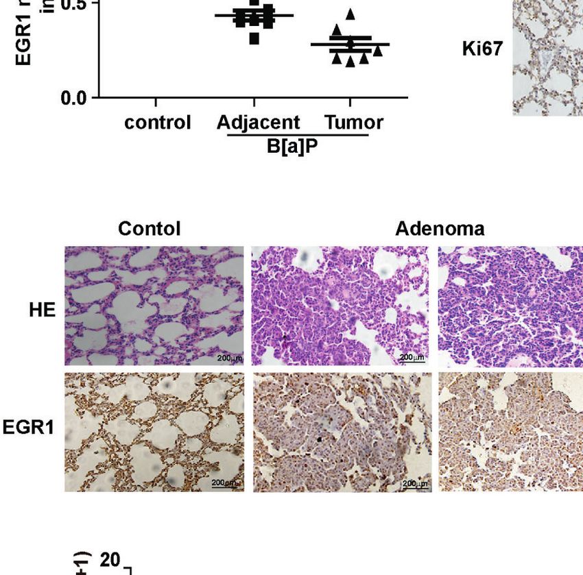
Ke et al.                                                                                         EGR1-Downregulation Promotes Lung Tumorigenesis

INTRODUCTION                                                                 mediated EGR1 downregulation facilitates cell malignant
                                                                             transformation and tumor formation by regulating the Wnt/b-
Lung cancer has the highest morbidity and mortality worldwide.               catenin pathway, suggesting an important role of the miR-377-3p/
Late diagnosis and poor prognosis are the main causes of cancer-             EGR1 axis in the malignant transformation of lung tumorigenesis
related death (1, 2), and smoking is a common risk factor. Yet,              induced by environmental carcinogen.
over the years, the increased non-smoking-related risk associated
with ambient air pollution has been frequently reported (3).
Polycyclic aromatic hydrocarbons (PAHs) are widespread
                                                                             MATERIALS AND METHODS
environmental pollutants that have been associated with
carcinogenicity (in gas or particle phase) (4). The most widely              Patient Samples
studied PAH is Benzo[a]pyrene (B[a]P), which is frequently chosen            A total of 114 non-small-cell lung cancer (NSCLC) clinical
as a substitute for evaluating the carcinogenic PAHs (5).                    samples of the Second Affiliated Hospital of Zhejiang University
    B[a]P is a human group 1 carcinogen capable of initiating and            were used in this study. The study was approved by the ethics
promoting lung tumorigenesis (6). BPDE is the main biologically              committee of the hospital. The clinical characteristics of these
active metabolite of B[a]P that can form DNA adducts of guanine              samples are shown in Table 1. The cancer tissues were formalin-
N2, thus exerting its carcinogenic effect (7). In cell-based models, B       fixed and paraffin-embedded for immunohistochemistry (IHC).
[a]P or its metabolite BPDE induce cell malignant transformation,
while in mice models, it can reduce lung tumors. Recent studies have         Cells and Reagents
shown that B[a]P-induced tumorigenesis involves DNA                          Human bronchial normal epithelium cell BEAS-2B (Cell Bank of
methylation, oxidative stress, cell cycle, inflammation, apoptosis,           the Chinese Academy of Science, Xiangya, China) and 293T cells
and other biological processes (7–9). Yet, the exact molecular               (ATCC, Manassas, VA, USA) were cultured in DMEM (Gibco,
mechanism behind this remains unclear.                                       Grand Island, NY, USA.) supplemented with 10% FBS (Gibco),
    Transient activation and regulation of immediate-early genes             streptomycin (100 g/mL), and penicillin (100 U/mL) in a
are considered primary cellular responses to an external signal in           humidified atmosphere containing 5%CO2/95% air at 37°C.
cancer development (10). Early growth response 1 (EGR1) is an                The authenticity of the cell lines used in this study has been
immediate-early gene that can be directly activated by growth                verified by STR profiling.
factors, hypoxia, ischemia, tissue injury, and apoptotic signals in             BPDE was purchased from the National Cancer Institute
different cells (11). Different roles of EGR1 have been observed in          Chemical Carcinogen Reference Standard Repository (Kansas
different tumors. EGFR1 can have double-edged effects in tumor               City, MO, USA), dissolved in DMSO, and stocked in -80°C.
development. For example, EGR1 has an oncogenic function in
prostate cancer by promoting cell proliferation and survival, but            Cell Transformation Assays
it can also act as a tumor suppressor in various cancers such as             Cells were exposed to 0.2 µM or 0.5 µM BPDE for 2 hours in a
glioma, lung, and bladder cancer by directly upregulating PTEN,              serum-free medium. Then, the treated medium was removed,
P53, and fibronectin (12–16).                                                 and cells were recovered in a fresh medium at 37°C. BPDE
    MicroRNAs (miRNAs), an endogenous short non-coding                       exposure was repeated once a week for 12 weeks. After 12 weeks
RNA, have important functions in many developmental                          of treatment, the malignant phenotype was analyzed and DMSO
systems (17). miRNAs regulate gene expression in multicellular               was used as solvent control.
organisms by affecting both the stability and translation of
mRNAs. They can target the 3’-UTR of mRNA transcripts via                    QRT-PCR
complementary sequences and repress the gene expression by                   Total RNA was extracted from cell lines or tumor and normal
post-transcriptional level (18). Their deregulation has been                 tissue samples with TRIzol reagent (Invitrogen, Carlsbad, CA,
closely related to cancer initiation and progression (19).                   USA). For gene expression, RNA was reverse transcribed using a
    miR-377-3p is a novel tumor regulatory miRNA whose biological            Prime-Script RT reagent Kit (TaKaRa). QRT-PCR was carried
functions are wildly unknown. MiR-377-3p has been shown to                   out with an SYBR Premix Ex Taq (TaKaRa). For miRNA
possess tumor-inhibiting effects in clear cell renal cell carcinoma          expression, RNA was reverse transcribed using an SYBR®
and hepatocellular carcinoma (20, 21). Contrary, previous studies            Premix Ex Taq II (TliRnaseH Plus) (TaKaRa, Dalian, China).
have shown that miR-377 promotes the proliferation and EMT                   QRT-PCR was performed using a Mir-X miRNAFirst-Strand
process in colon cancer, while the low level of miR-377 was                  Synthesis kit (Clontech, Madison, WI, USA). Experiments were
associated with a good prognosis of periampullary adenocarcinoma             performed in triplicate, and the values were normalized to
(22, 23). Moreover, recent studies demonstrated that miRNAs are              GAPDH or RNU6B using the 2(−DDCt) method for gene and
also involved in B[a]P-induced carcinogenicity (24, 25). However, the        miRNA expression analysis, respectively.
potential contribution of miRNAs in environmental carcinogens-
induced lung tumorigenesis is still not clear.                               Immunoblot Analysis
    In the present study, we found that EGR1 expression was                  Cell lysates (50 mg) were separated on a 10% SDS-PAGE gel and
strongly reduced in the malignant transformation of human lung               then transferred onto a nitrocellulose membrane (Whatman,
bronchial epithelial cells and lung tumorigenicity following B[a]P           Maidstone, UK). The membrane was blocked with 5% skim milk
and its active metabolite BPDE exposure. Moreover, miR-377-3p                solution for 2 hours and incubated overnight at 4°C with the

Frontiers in Oncology | www.frontiersin.org                              2                                August 2021 | Volume 11 | Article 699004
Ke et al.                                                                                      EGR1-Downregulation Promotes Lung Tumorigenesis

following diluted primary antibody: rabbit monoclonal anti-              400 ml of complete DMEM medium was added under the
human EGR1 (ab194357, Abcam, Hanghzou, China), and the                   chambers, whereas cells (2 × 10⁴) were added above the
mouse monoclonal anti-human GADPH (sc-47724) and anti-                   chambers in a serum-free medium. After 48 hours of incubation
human b-catenin (sc-7963) both purchased from Santa Cruz                 at 37°C, the migrated cells were fixed with 4% paraformaldehyde
Biotechnology (Santa Cruz, CA, USA). Then, the membrane was              and stained with 0.5% crystal violet. Then, the filter membrane
incubated in IRDye ® 800CW- or IRDye 680-conjugated                      was examined and photographed under a microscope.
secondary antibody (LI-COR Biosciences, Lincoln, NE, USA)
and detected by an Odyssey® infrared imaging system.                     Immunohistochemistry
                                                                         The IHC was performed using an Envision Detection System
Animal Models                                                            (DAKO, Carpinteria, CA) according to the instructions of the
A/J mice (4 weeks) and Balb/c nude mice (4 weeks) were                   manufacturer. Rabbit monoclonal anti-mouse Ki67 (ab194357)
obtained from Model Animal Research Center, Nanjing, China               was purchased from Abcam; rabbit polyclonal anti-mouse EGR1
and SLAC Laboratory Animal, Shanghai, China. All the animals             (sc-110) was purchased from Santa Cruz Biotechnology. The
were housed in an environment with a temperature of 22 ± 1°C,            IHC staining results were assessed and confirmed by two
relative humidity of 50 ± 1%, and a light/dark cycle of 12/12 h.         independent investigators blinded to the clinical data.
All animal studies (including the mice euthanasia procedure)
were done in compliance with Zhejiang University institutional           Cell Transfection
animal care regulations and guidelines, and conducted according          For lentiviral-mediated transfection, 293T cells were co-
to the AAALAC and the IACUC guidelines.                                  transfected with the lentiviral and packaging vectors. After 72 h,
    A/J mice (4 weeks) were randomly divided into two groups (12         the supernatant was collected. Supernatants were then collected
mice/group). B[a]P group was intraperitoneally injected with B[a]P       and centrifuged at 1,000 × g for 15 min at 4°C to pellet debris.
(25 mg/kg, in tricaprylin solvent) (Sigma), and the control group        Before performing the infection, the lentiviruses were recovered
was intraperitoneally injected with the tricaprylin solvent. B[a]P       and re-suspended in a fresh medium with 6 g/ml of polybrene.
treatment was given on a weekly basis for 8 weeks. The control           Stable cells with EGR1 knockdown or EGR1 overexpression were
group was treated as the same. After 4 months of restoration             selected following transduction with 0.5 mg/ml of puromycin for 2
following the treatment period, mice were sacrificed, and the lung        weeks. Transfection efficiency of EGR1 knockdown or EGR1
tissues were obtained and histologically examined. The tricaprylin       overexpression was examined by Western blot.
solvent-treated group was used as a control group.                           For miR-377-3p mimic, inhibitor, antagomir, miRNA control
    Balb/c nude mice (4 weeks) were subcutaneously injected              (GenePharma, Shanghai, China) transfection, cells were
with 5 × 10⁶ transformed cells in 100 µl volume mixed with               transfected using Lipofectamine ® RNAiMAX (Invitrogen)
Matrigel (1:1). Three days after injection, miR-377-3p antagomir         following the instructions of the manufacturer. After 72 h of
(5 nmol/mouse) or scramble control was performed by                      transfection, the cells were collected for further experiments.
intratumor injection twice a week. The long diameter (a) and
short diameter (b) of the tumors were measured; after which, the         Dual-Luciferase Reporter Assay
volume (V) was calculated using the formula V = 1/2 × a × b2.            The full-length and mutated miR-377-3p recognition elements of
Mice were sacrificed, and the tumor tissues were obtained                 3’UTR-EGR1 were synthesized and constructed into a pGL3-
and weighed.                                                             Basic vector (Promega, Madison, WI, USA). After seeding the
                                                                         cells for 24 h, the mimic or inhibitor of miR-377-3p
Soft Agar Assay                                                          (GenePharma) was co-transfected with either pGL3-EGR1-
The cells (1,000 cells/well) were suspended in a culture medium          3’UTR wild-type or mutant into BEAS-2B and 293T cells.
containing 0.4% agarose (Sigma, St Louis, MO, USA) and seeded            Dual-Luciferase Reporter Assay System was used for testing
onto a base layer of 0.7% agar bed in 12-well plates. After 2            the relative luciferase activity (Promega).
weeks, colonies were stained with crystal violet and
photographed. Colonies ≥ 0.05 mm in diameter were counted.               Immunofluorescence
                                                                         The BPDE-transformed cells were plated in culture. After
Scratch Test                                                             overexpression of EGR1, the cells were fixed for 15 min in 4%
Cells (1 x 10⁵ cells/ml) were plated in 6-well plates. The               formaldehyde solution. Then, the cells were washed with PBS
monolayer was scratched by a 10 ml sterile pipette tip. The              and treated with 0.1% Triton X-100 in PBS for 10 min. After
cells were gently rinsed twice with PBS to remove floating cells          permeabilizing the cells, we blocked the cells for 1 h in an
and incubated in 2 ml of serum free medium in 37°C, 5% CO2 air           antibody blocking buffer (10% normal goat serum, 1% BSA in
environment. Images of the scratches were taken by using an              PBS). Then, the cells were washed with PBS and incubated with
inverted microscope at 0, 24, and 48 hours of incubation. ImageJ         anti-human b-catenin primary antibody. The presented IF
software was used to analyze the percentage of wound closure.            staining pictures are the overlaid images of b-catenin staining
                                                                         in green fluorescence with nuclear 4’6-diamidino-2-
Transwell Assay                                                          phenylindole (DAPI) staining in blue fluorescence. The IF
We performed a cell migration assay with an 8 µm-pore in 24-             staining images were taken and overlaid using the Nikon NIS-
well transwell plates (Costar, Cambridge, MA, USA). Briefly,              Elements software.

Frontiers in Oncology | www.frontiersin.org                          3                                 August 2021 | Volume 11 | Article 699004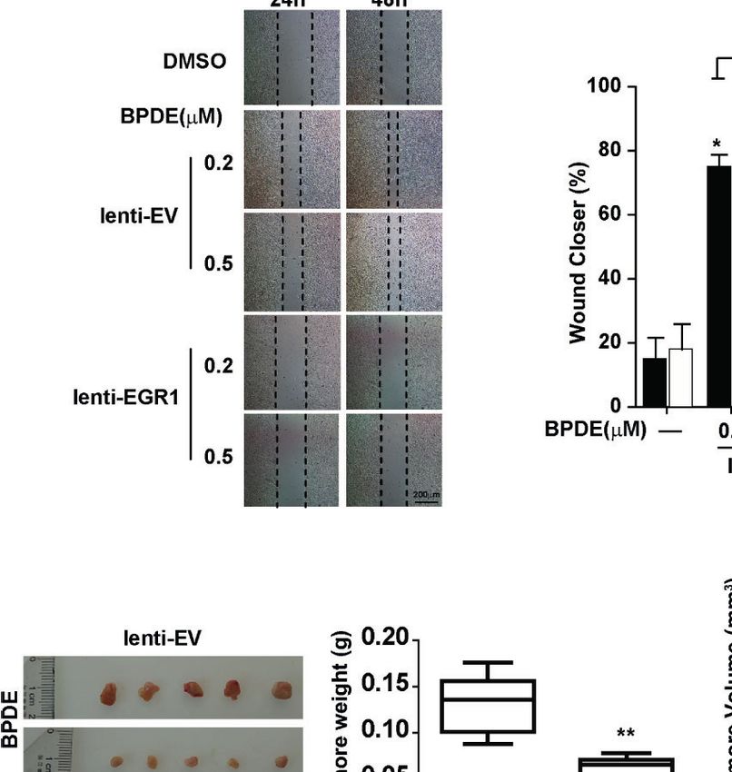
Ke et al.                                                                                                 EGR1-Downregulation Promotes Lung Tumorigenesis

Statistical Analysis                                                               proliferation assay and soft agar assay revealed that BPDE treatment
The two-tailed Student’s t-test and one-way analysis of variance                   enhanced the reproductive capacity of cells and the anchorage-
were used for statistical data analysis. The data was expressed of                 independent growth capability, respectively (Figures 1B, C). We
three separate experiments, as mean ± standard deviation (SD).                     also observed that the cell migration was enhanced upon BPDE
P ≤ 0.05 was considered to be statistically significant.                            treatment (Figures S1A, B). Xenograft assay further confirmed the
                                                                                   malignant phenotype of BPDE-induced BEAS-2B cells (Figure 1D).
                                                                                   In addition, we also confirmed the above tumorigenic effects with
RESULTS                                                                            the BPDE-induced HBE malignant transformation cell model by
                                                                                   malignant phenotype analysis (data not shown).
BPDE/B[a]P Downregulates the                                                           To investigate the genes implicated in the BPDE-induced
Expression of EGR1 In Vitro and In Vivo                                            malignant transformation process, we performed RNA-
B[a]P and its ultimate carcinogenic metabolite, BPDE, are the                      sequencing analysis. Our results showed that EGR1 was the
strong lung carcinogens found in tobacco smoke and air pollution                   most obviously downregulated gene in the transformed cells
(26). However, the molecular mechanisms underlying PAH-                            (Figure S1C). The downregulation of EGR1 expression was
induced lung tumorigenesis, particularly in the early stage, remain                confirmed in both BEAS-2B and HBE BPDE-induced cell
unclear. To indicate the critical genes involved in this process,                  transformed models (Figures 1E, F). Moreover, the EGR1
human lung epithelial cells and A/J mice were exposed to BPDE/B                    protein content was also reduced in different lung cancer cells
[a]P, respectively. Malignant transformation of BEAS-2B cells was                  contrasted with normal cells (Figure 1G).
identified upon 12 weeks of BPDE exposure (Figures 1 and S1).                           To further evaluate the effect of B[a]P on EGR1 expression
Figure 1A shows a schematic map of the strategy used to generate                   in vivo, we established a B[a]P-treated A/J mice model (Figure
the BPDE-induced malignant transformation of BEAS-2B cells. Cell                   S2A). Most mice treated with B[a]P developed primary lung
                                                                                   tumors within 6 months; this was observed by PET-CT detection
                                                                                   and histopathological analysis (Figures 2A and S2B, C). Our
TABLE 1 | Association of immunohistochemical staining for EGR1 with the            results also showed that EGR1 mRNA expression and protein
tumor clinic pathological characteristic.                                          level were decreased in the lung tumor tissues compared to the
Clinicopathological features        Case N. (%)    Egr1 expression   p-value
                                                                                   adjacent normal tissues (Figures 2B, C). Ki67 was extensively
                                                                                   assessed and reported as a predictive proliferative marker of
                                                   High      Low                   cancer cells. Moreover, the downregulation of EGR1 was not
Gender
                                                                                   only observed in adenocarcinoma but also B[a]P-treated mice
  Male                                  63 (55)     27        36                   adenoma (Figure 2D), indicating that EGR1 reduction could be
  Female                                51 (45)     20        31      0.296        the early event in B[a]P-induced tumorigenesis.
Age                                                                                    To further determine whether EGR1 downregulation was
  Mean (Range)                        62 (37-85)
                                                                                   involved in human lung carcinoma development, we expanded
  60                                  49 (43)      21        28      0.476        our study by investigating the expression of EGR1 in clinical
Tumor sizea                                                                        cancer tissues. In eight pairs of fresh cancer and adjacent normal
  2.5cm                                45 (49)     19        26      0.565        EGR1 expression in cancer tissues (Figure S3A). TCGA (The
Depth of invasion
                                                                                   Cancer Genome Atlas) database and the other two datasets
  T1                                    46 (40)     38        16
  T2                                    53 (46)     28        25                   supported in Lung Cancer Explorer confirmed that EGR1 was
  T3                                    11 (10)     1         10                   downregulated in NSCLC patient tissues compared to normal
  T4                                     4 (4)      2         2       0.013*       tissues (Figures 2E and S3B, C). Collectively, the results indicated
Lymph node metastasis                                                              that the inhibition of EGR1 was involved in cell malignant
  N0                                    50 (44)     31        19
  N1                                    29 (25)     7         22
                                                                                   transformation and mice lung tumorigenesis induced by BPDE/
  N2                                    35 (31)     9         26     0.000**       B[a]P exposure. The EGR1 reduction was also observed in clinical
Distant metastasis                                                                 cancer tissues. The above data suggested that EGR1 could have a
  M0                                   107 (94)     44        63                   tumor-suppressive role in the lung cancer process.
  M1                                    7 (6)       3         4       0.750
TNM stage
  I                                     44 (39)     29        15
                                                                                   EGR1 Reduction Mediates BPDE-Induced
  II                                    29 (25)     8         21                   Malignant Transformation
  2III                                  36 (32)     8         28                   To investigate the potential role of EGR1 downregulation in lung
  IV                                     5 (4)      2         3      0.000**       tumorigenic effects upon BPDE exposure, we established stable
Histological gradeb
                                                                                   EGR1 overexpression models in BPDE-induced transformed cells
  High                                  31 (28)     19        12
  Moderate                              63 (58)     25        38
                                                                                   with lenti-EGR1 lentivirus (Figure S4A). The ectopic expression
  Poor                                  15 (14)     3         12      0.003*       of EGR1 led to a reduced malignancy in BPDE-induced
a
                                                                                   transformed cells (Figure 3). Moreover, EGR1 overexpression
 23 cases without tumor size.
b
 5 cases without tumor histological grade.                                         reduced the cell migration ability (Figures 3A–C) and xenograft
*P < 0.05 and **P < 0.01.                                                          tumor growth (Figures 3D, E). The suppressive effect of EGR1 on

Frontiers in Oncology | www.frontiersin.org                                    4                                  August 2021 | Volume 11 | Article 699004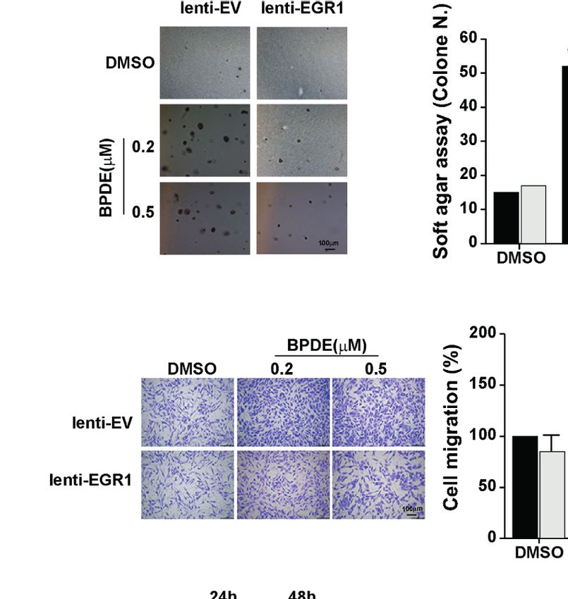
Ke et al.                                                                                                         EGR1-Downregulation Promotes Lung Tumorigenesis

                        A                                                                   E

                        B

                        C                                                                   F

                        D

                                                                                   G

 FIGURE 1 | EGR1 was downregulated in BPDE-induced malignant transformation of normal human lung epithelial cells. (A) Schematic map of BPDE-induced
 malignant transformation model. (B) Cell proliferation ability of BPDE-treated and control cells. (C) Cell anchorage-independent growth in soft agar. Top,
 representative images; bottom, quantitative results of cell colony per field. (D) Top, Tumor growth curve of the transformed cells induced by BPDE and control cells
 injected subcutaneously in nude mice (n = 12 tumors per group); bottom, representative photos of the HE staining and Ki67 immunohistochemical staining of the
 tumor mass. (E, F) EGR1 expression analyzed by qRT-PCR and Western Blot in transformed BEAS-2B and HBE cells. (G) Egr-1 expression in different lung
 epithelial and lung cancer cells. Normal bronchial epithelial cell: NL-20, BEAS-2B; lung cancer cell: H446, H520, H1299, A549. The analyses were repeated three
 times, and the results were expressed as mean ± SD. *P < 0.05, **P < 0.01 and ***P < 0.001.

Frontiers in Oncology | www.frontiersin.org                                       5                                        August 2021 | Volume 11 | Article 699004
Ke et al. EGR1-Downregulation Promotes Lung Tumorigenesis
A
B C
D
E
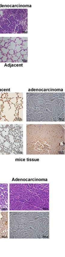
FIGURE 2 | B[a]P downregulated EGR1 expression in vivo. (A) B[a]P-induced A/J mice lung tumorigenesis. Left, representative images of primary lung tumor in
mice with or without B[a]P treatment (25mg/kg); right, representative images of HE staining. (B) EGR1 mRNA expression in B[a]P-induced murine lung tumor and
adjacent normal tissues, vehicle lung tissues. (C, D) Representative images of EGR1, Ki67 immuno-histochemical staining of B[a]P-treated murine lung tumor tissues
and adjacent tissues, vehicle lung tissues. (E) Analysis of the TCGA database of EGR1 mRNA expression in paired lung cancer and normal tissues. The analyses
were repeated three times, and the results were expressed as mean ± SD. #P < 0.05, **P < 0.01, and ***P < 0.001.
Frontiers in Oncology | www.frontiersin.org 6 August 2021 | Volume 11 | Article 699004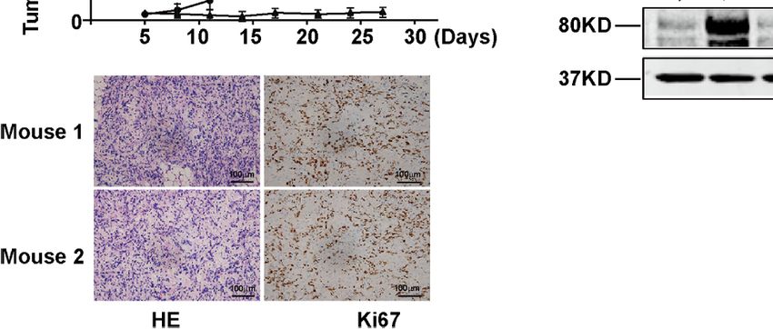
Ke et al.                                                                                                              EGR1-Downregulation Promotes Lung Tumorigenesis

cell malignant phenotypes was further confirmed by EGR1                                    downregulation was critical for promoting BPDE-induced cell
knockdown. EGR1 shRNAs introduction through lentiviral                                    malignant transformation.
vectors resulted in an increased malignancy of BEAS-2B cells
(Figures S4C–E). Moreover, the rescue of EGR1 also reversed the                           mir-377-3p Targets EGR1 and Induces its
effect of EGR1-knockdown in promoting cell transformation                                 Inhibition Following BPDE Exposure
(Figures S4F, G). The knockdown efficiency of EGR1 was                                     Notably, we found that the expression of EGR1 was inhibited
supported in Figure S4B. Our results suggested that EGR1                                  during the BPDE-induced cell malignant transformation (data

                                  A

                                  B

                                  C

                           D                                                                   E

 FIGURE 3 | EGR1 downregulation is crucial for BPDE-induced cell malignant transformation. (A) Soft-agar colony formation assays of BPDE-induced transformed
 cells infected with EGR1 overexpression lentivirus. Left, representative images; right, quantitative results of cell colony per field. (B, C) Left, representative images of
 transwell migration and wound healing assays; right, relative numbers of migration cell and percentage wound closure after treatment. (D) Representative images of
 xenograft tumors in nude mice at 4 weeks after inoculation (Left) and the tumor weight of BPDE-induced transformed cells infected with EGR1 overexpression.
 (E) Tumor growth curve of xenograft assays (n = 10 tumors per group). The analyses were repeated three times, and the results were expressed as mean ± SD.
 #
  ,*P < 0.05 and **P < 0.01.

Frontiers in Oncology | www.frontiersin.org                                           7                                          August 2021 | Volume 11 | Article 699004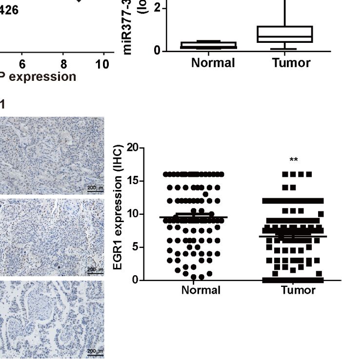
Ke et al.                                                                                        EGR1-Downregulation Promotes Lung Tumorigenesis

not shown). To investigate the molecular mechanism underlying            associated with tumor aggressiveness in lung cancer. Taken
EGR1 reduction upon BPDE treatment, we first evaluated EGR1               together, our results suggest that the upregulation of miR-377-
promoter DNA methylation level by bisulfite sequencing PCR.               3p inhibits EGR1 transcription, which is implicated in BPDE/B[a]
The DNA methylation level of the EGR1 promoter sequence did not          P-induced cell malignant transformation and lung tumorigenesis.
change after BPDE exposure (Figure S3D). Over the past decade, it
has been widely reported that miRNAs regulate gene expression by
recognizing the 3’UTR sequence. Using the microRNA database              EGR1 Inhibition is Involved in the
and target prediction tools (miRanda, PicTar, and TargetScan), we        Regulation of Wnt/b-Catenin Transduction
predicted the potential microRNAs that could target EGR1 and             in PAHs-Induced Tumorigenesis
regulate its mRNA transcription. qRT-PCR revealed that miR-              EGR1 is an important transcription factor for regulating the cell
377-3p levels were markedly increased in BPDE-treated cells              cycle, differentiation, apoptosis, and stress. To identify the potential
(Figure 4A). Transient transfection with mimics and inhibitor of         EGR1-downstream genes involved in the BPDE/B[a]P-induced
miR-377-3p showed that miR-377-3p regulates EGR1 expression              tumorigenesis, we performed the RNA-sequencing by knocking
(Figures 4B, C).                                                         down EGR1 expression. As expected, Kyoto Encyclopedia of Genes
   To further identify the effect of miR-377-3p on EGR1                  and Genomes (KEGG) analysis indicated that the Wnt/b-catenin
expression regulation, we constructed the luciferase reporter            pathway is one of the most significantly altered gene set concepts in
containing wild-type regulatory sequence with or without                 EGR1 knockdown cells, and gene set enrichment analysis (GSEA)
EGR1 binding site mutation (Figure 4D). The result showed                revealed a large fraction of Wnt/b-catenin downstream genes that
that miR-377-3p mimic reduced the reporter activity of the full-         displayed significant alterations (Figures 6A, B). Moreover, we also
length EGR1 3’UTR-containing luciferase construct, and the               observed the upregulation of b-catenin in BPDE-induced
inhibitor of miR-377-3p augmented the reporter activity in               malignant transformed cells and mice primary lung cancer tissue
BEAS-2B and 293T cells. The effect of miR-377-3p on the                  (Figures 6C–E). By transient transfection, EGR1 overexpressing
reporter activity was abrogated with the mutant-type EGR1                led to a reduction of CTNNB1 gene expression. Moreover, EGR1
3’UTR-containing luciferase construct (Figures 4E, F). These             knockdown upregulated the CTNNB1 gene expression
results indicated that miR-377-3p mediated the downregulation            (Figure 6F). Also, the rescue of EGR1 expression abrogated the
of EGR1 in BPDE-induced malignant transformed cells by                   upregulation and nuclear localization of b-catenin induced by
directly targeting its 3’UTR sequence.                                   BPDE exposure (Figures 6G, H). These data suggested that the
                                                                         Wnt/b-catenin pathway is the potential downstream signal in
mir-377-3p Antagomir Rescued the Effect                                  EGR1-mediated cell malignant transformation.
of EGR1 Downregulation in Cell Malignant                                     Furthermore, we detected the most altered genes of RNA-
Transformation and Lung Carcinogenesis                                   sequencing by knocking down EGR1 in the transformed cells. Our
To detect whether the inhibition of miR-377-3p allows for the re-        result revealed that ATF3 and ANKRD1 were downregulated in
expression of EGR1 and reduces the malignancy of BPDE-                   malignant cells and mice primary lung cancer tissues induced by
induced transformed cells, we transfected the cells with miR-            PAHs (Figures S5C, D). Ectopic expression of EGR1 resulted in the
377-3p antagomir. Our results revealed that the antagomir of             upregulation of ATF3 and ANKRD1. Moreover, siRNA of EGR1
miR377-3p reduced the malignancy phenotypes of the                       reduced the expression of ATF3 and ANKRD1 (Figure S5E). Besides,
transformed cells induced by BPDE exposure (Figures 5A–D).               the rescue of EGR1 expression in BPDE-induced transformed cells
Moreover, miR-377-3p upregulation was identified in mice lung             abrogated the inhibition of ATF3 and ANKRD1 expression induced
tumor tissues induced by B[a]P, concomitantly with EGR1                  by BPDE (Figure S5F). To sum up, our data indicated that the
downregulation (Figure 5E). Furthermore, we observed a                   downregulation of EGR1 could alter the downstream cell signals and
negative relevance between EGR1 and miR-377-3p in mice                   the expression of its target genes to contribute to the cell malignant
lung tumor tissues by the correlation analysis (Figure 5F).              transformation and lung carcinogenesis.
Consistent with our findings, the TCGA database analysis
revealed the increase of miR-377-3p and the decrease of EGR1
in human lung adenocarcinoma tissues (Figures 5G, H). Besides,           DISCUSSION
the expression of EGR1 and miR-377-3p in fresh lung cancer
tissues also showed a negative relevance (Figure S5A).                   B[a]P can directly induce lung carcinogenesis by inducing DNA
    To confirm the tumor repressive effect of EGR1 in NSCLC, we           damage and activating the signaling pathways (26–28). This
performed IHC staining to evaluate the clinical relevance of EGR1        study further investigated early events and the molecular
expression. Our results showed a high EGR1 immunoreactivity in           mechanisms of gene dysregulation that lead to cell malignant
the nuclei of adjacent normal cells compared with cancer cells. In       transformation and lung tumorigenesis following B[a]P/BPDE
114 paired cases, EGR1 was significantly inhibited in tumor tissues       exposure. We discovered that B[a]P/BPDE treatment led to miR-
(Figures 5I, J and S5B). EGR1 expression was negatively                  377-3p induction, which targeted EGR1-3’UTR and inhibited its
associated with tumor invasion, lymph node status, histological          expression, subsequently resulting in the activation of Wnt/b-
grade, and TNM stage (Table 1). Our results suggested that EGR1          catenin signal and promotion of cell malignant transformation,
functions as an onco-suppressor, and the inhibition of EGR1 was          thus further contributing to lung tumorigenesis. Consequently,

Frontiers in Oncology | www.frontiersin.org                          8                                    August 2021 | Volume 11 | Article 699004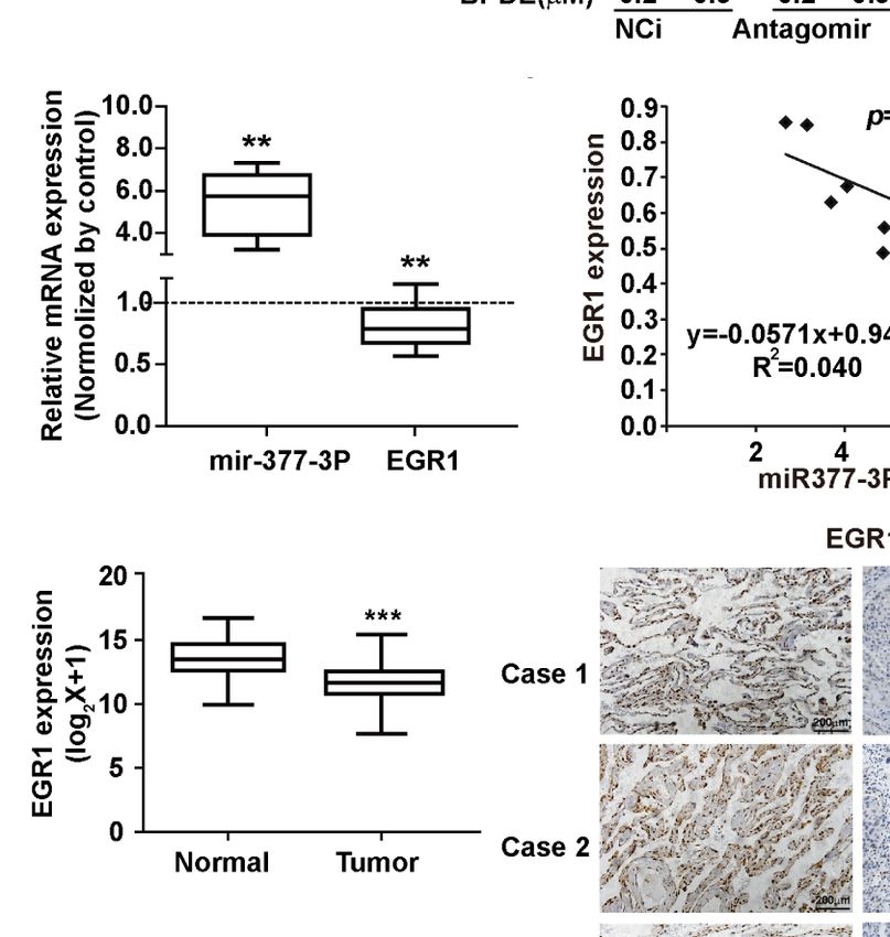
Ke et al.                                                                                                     EGR1-Downregulation Promotes Lung Tumorigenesis

                                A

                                B

                                                                      C

                                D

                            E                                                 F

 FIGURE 4 | MiR-377-3p activation mediated EGR1 inhibition following BPDE exposure. (A) Expression profile of predicted miRNAs in BPDE-treated BEAS-2B cells
 by qRT-PCR. (B, C) EGR1 expression after transfection of miR-377-3p mimic and inhibitor in BEAS-2B cells was analyzed by QRT-PCR and Western blot.
 (D) The putative target site of miR-377-3p in the 3’UTR of EGR1 (upper panel); red letters indicate the mutant luciferase reporter gene sequence (lower panel).
 (E, F) EGR1 3’UTR luciferase reporter assays in BEAS-2B cells and 293T cells. The analyses were repeated three times, and the results were expressed as
 mean ± SD. *P < 0.05 and **P < 0.01.

EGR1 could be considered a potential target for B[a]P initiation                      due to its tumor-suppressing role in the occurrence and
of lung carcinogenic actions.                                                         development of tumors. The expression of EGR1 decreases or
    As a transcription factor, EGR1 has a crucial role in human                       even disappears in a variety of human malignancies, and its
cancers. EGR1 has been increasingly attracting research attention                     expression level is associated with tumor sensitivity to

Frontiers in Oncology | www.frontiersin.org                                       9                                     August 2021 | Volume 11 | Article 699004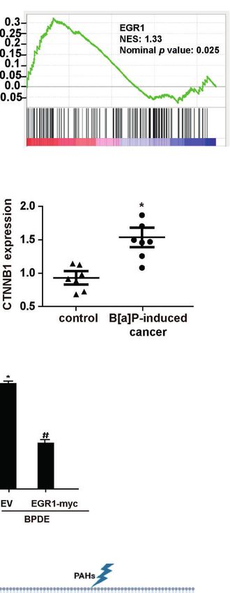
Ke et al.                                                                                                          EGR1-Downregulation Promotes Lung Tumorigenesis

                      A                                                                       C

                      B                                                                       D

                      E                                          F                                          G

                      H                                          I                                          J

 FIGURE 5 | MiR-377-3p antagomir rescued the effect of EGR1 downregulation in cell malignant transformation and lung carcinogenesis. (A) Soft-agar colony
 formation assays. Left, representative images; right, quantitative results of cell colony per field. (B) Transwell migration assays. Left, representative images; right,
 quantitative results of migratory cells per field. (C) Representative xenograft tumor images at 4 weeks after inoculation (top) and tumor weight (bottom) of BPDE-
 induced transformed cells treated with miRNA-377-3p antagomir or control. (D) The tumor growth curve of xenograft. (E) MiR-377-3p expression and EGR1 mRNA
 expression in B[a]P-induced murine lung cancer tissues by qRT-PCR. The analyses were repeated three times, and the results were expressed as mean ± SD.
 (F) The correlation analysis of EGR1 and miR-377-3p mRNA expression in mice carcinogenesis model. Y-axis showed EGR1 mRNA expression; X-axis showed miR-
 377-3p mRNA expression. (G, H) TCGA database analysis of miR-377-3p expression and EGR1 mRNA expression in human lung adenocarcinoma tissues.
 (I) Representative image of IHC staining of EGR1 in human lung cancer tissues. (J) Quantification of EGR1 staining in the adjacent normal lung epithelial cells and
 lung cancer cells in 114 paired patient tissues. #,*P < 0.05, **P < 0.01, and ***P < 0.001.

chemotherapy (29). EGR1 depletion has been associated with                              Mechanistically, EGR1 can directly transactivate P53 and PTEN,
tumor anti-apoptotic and invasion events, whereas its                                   implicated in the proliferation inhibition of lung tumor cells (31,
overexpression may depress the tumorigenicity and metastasis                            32). It can also suppress the EMT transition and cell migration in
in different cancer cells, including lung cancer (30).                                  lung cancer by regulating TGFb activity (33). Recent studies have

Frontiers in Oncology | www.frontiersin.org                                        10                                        August 2021 | Volume 11 | Article 699004
Ke et al. EGR1-Downregulation Promotes Lung Tumorigenesis
A B
C D E
F G
H I
FIGURE 6 | EGR1 inhibition was involved in the regulation of Wnt/b-catenin transduction in PAH-induced tumorigenesis. (A, B) KEGG analysis of differentially
expressed genes and Gene set enrichment plots of differentially expressed genes belonging to the Wnt pathway in EGR1 knockdown cells. (C, D) b-catenin protein
content and mRNA gene expression of CTNNB1 in BPDE-induced transformed cells. (E) mRNA level of CTNNB1 gene in mice primary lung cancer tissues.
(F, G) CTNNB1 gene expression in BEAS-2B cells upon EGR1 overexpression and knockdown and in BPDE-induced transformed cells upon EGR1 overexpression.
(H) Immunostaining of b-catenin in malignant transformed cells with or without EGR1 overexpression. (I) Schematic diagram of the regulatory mechanism of miRNA-
377-3p/EGR1 axis in tumorigenesis. The analyses were repeated three times, and the results were expressed as mean ± SD. #,*P < 0.05.
shown that EGR1 can directly and negatively regulate cell growth the early stage of malignant cell transformation upon BPDE
in different epithelial tumor cell lines (34). It can also regulate exposure. The inhibition of EGR1 promoted the progression of
KRT18 expression to inhibit the malignancy of human NSCLC BPDE-induced tumorigenicity. Moreover, the downregulation of
cells (35). Our data showed that EGR1 was strongly decreased in EGR1 was also confirmed in B[a]P-induced lung tumors in vivo.
Frontiers in Oncology | www.frontiersin.org 11 August 2021 | Volume 11 | Article 699004Ke et al. EGR1-Downregulation Promotes Lung Tumorigenesis
The results indicated that EGR1 has a tumor repressive effect in with a poor prognosis in patients with lung cancer (49). Previous
cell malignant transformation and lung tumorigenesis upon B[a] studies also reported that the Wnt/b-catenin pathway contributes to
P/BPDE treatment. the induction of EMT by transactivating several EMT-related
DNA methylation and miRNA dysregulation are important transcriptional factors, such as Snail, Slug, Twist, ZEB1, and ZEB2
molecular mechanisms of gene expression, which are critical for in lung adenocarcinoma (50). Moreover, we also observed that
epigenetic regulation in tumor formation and development by ANKRD1 and ATF3, as the target genes of EGR1, were significantly
negatively regulating targeting downstream genes (36, 37). downregulated in malignant transformed cells and mice lung cancer
Recent studies reported that miR-301b, miR-191, and miR- tissues. ATF3, a highly conserved transcription factor, was described
146a could target EGR1 mRNA and inhibit its expression, thus as a principal target of EGR1 and discussed as a tumor suppressor
contributing to oncogenesis (16, 38, 39). In this study, EGR1 was and promoter (51–53). A recent study also reported that ATF3 and
persistently decreased after BPDE exposure but without variation EGR1 are involved at the beginning of the inflammatory processes
of its promoter DNA methylation level. miRNA screening related to cancer (54). ANKRD1 is a tumor-suppressive
analysis demonstrated that miR-377-3p is a new regulator of downstream gene of the Hippo pathway, downregulated in
EGR1 by directly binding to its 3’UTR. miR-377-3p was different human cancers (55, 56). A previous study demonstrated
significantly increased in BPDE-induced malignant transformed that ANKRD1 could be inhibited by lncRNA, resulting in the
cells, as well as in the lung tumor tissues of B[a]P-treated A/J mice. promotion of pancreatic cancer proliferation and metastasis (57).
Antagonized miR-377-3p reversed the effect of EGR1 in cell Therefore, EGR1 could regulate its downstream signals and target
malignant transformation, thus supporting the critical role of genes, thus having a tumor-suppressive role in human lung cancer.
miR-377-3p in regulating EGR1 expression to promote cell In summary, our current study demonstrated the regulation
transformation and tumor formation. Recent studies reported mechanism of EGR1 inhibition induced by miR-377-3p
that miR-377 displays an ambiguous role in different cancers. activation following exposure to environmental carcinogen B
miR-377-3p can drive malignancy characteristics by upregulating [a]P/BPDE. We also discovered that EGR1 has a repressive effect
GSK-3b expression and activating the NF-kB pathway in CRC cells on lung tumorigenesis by regulating the Wnt/b-catenin signaling
(22). It can also target the pro-oncogenic genes, like E2F3, VEGF, pathway (Figure 6I). Our findings provided a novel molecular
and CDK6, or negatively regulate the Wnt/b-catenin signaling to regulatory mechanism through which the miR-377-3p/EGR1
suppress the proliferation of cancer cells (40–43). However, the dual axis was implicated in cell malignant transformation and
effect of miR-377 in tumor inhibition and promotion needs to be tumorigenesis induced by PAH.
further explored.
Clinical studies reported that the depletion of EGR1 sensitizes
the chemotherapy of cisplatin in ovarian tumors (44). The low
levels of EGR1, associated with the expression of PTEN, can DATA AVAILABILITY STATEMENT
predict poor outcomes after surgical resection of NSCLC (45). In
The datasets presented in this study can be found in online
this study, clinic tissue analysis showed a downregulation of
repositories. The names of the repository/repositories and accession
EGR1 expression in cancer tissues compared with the normal
number(s) can be found in the article/Supplementary Material.
tissue. The repression of EGR1 was associated with the local
invasion depth, lymph nodes, and TNM stages. It was also
negatively associated with histological grade (Table 1). Our
result confirmed that the deactivation of EGR1 was associated ETHICS STATEMENT
with cancer aggressiveness.
Furthermore, it is reported that EGR1 can be increased by The studies involving human participants were reviewed and
chemotherapy, and negatively regulate the Wnt/b−catenin approved by the ethics committee of the Second Affiliated
signaling pathway in CML cells (46). In this study, we observed Hospital of Zhejiang University. The patients/participants
an enrichment of Wnt/b−catenin downstream genes after EGR1 provided their written informed consent to participate in this
knockdown. Besides, b−catenin, the key effector of canonical Wnt study. The animal study was reviewed and approved by The
signaling, was activated in malignant transformed cells and lung Committee on the Use of Animals of Zhejiang University.
cancer tissues following EGR1 inhibition. The rescue of EGR1
expression reversed the upregulation and nuclear staining of
b−catenin after BPDE exposure. The Wnt/b−catenin pathway is a AUTHOR CONTRIBUTIONS
cell signaling that promotes cancer initiation and development. It
has an important role in crucial cellular processes, including cell fate XK: Conceptualization, Investigation, Resources, and Writing -
determination, embryonic development, homeostasis, motility, original draft. LH and RW: Investigation, Methodology, and
polarity, and stem cell renewal (47). It has also been reported that Validation. YS and LF: Software, Validation. ZW: Methodology,
the activation of canonical Wnt/b-catenin signaling is critical for the Validation. JShe: Review and Editing, Supervision, and Project
initiation and progression of NSCLC (48). In patient-derived administration. JSha: Review and Editing, Resources, and
xenograft models of lung cancer, the activation of WNT/b- Supervision. HQ: Writing - Review and Editing, Investigation,
catenin signaling and nuclear b-catenin staining was associated Methodology, Formal analysis, Project administration, and Funding
Frontiers in Oncology | www.frontiersin.org 12 August 2021 | Volume 11 | Article 699004Ke et al. EGR1-Downregulation Promotes Lung Tumorigenesis
acquisition. All authors contributed to the article and approved the (2016YFC1303401), and the Zhejiang Medical and Health Science
submitted version. and Technology Foundation (2018KY119).
FUNDING
SUPPLEMENTARY MATERIAL
This work was supported by the Zhejiang Provincial Natural Science
Foundation of China (No. LY18H160024, LY20H160040), the The Supplementary Material for this article can be found online
National Natural Science Foundation of China (No. 81472543, at: https://www.frontiersin.org/articles/10.3389/fonc.2021.
No. 81772919), the National Key R&D Program of China 699004/full#supplementary-material
REFERENCES 16. Yan L, Wang Y, Liang J, Liu Z, Sun X, Cai K. MiR-301b Promotes the
Proliferation, Mobility, and Epithelial-to-Mesenchymal Transition of Bladder
1. McGuire S. World Cancer Report 2014. Geneva, Switzerland World Health Cancer Cells by Targeting EGR1. Biochem Cell Biol (2017) 95(5):571–7. doi:
Organization, International Agency for Research on Cancer, WHO Press, 10.1139/bcb-2016-0232
2015. Adv Nutr (2016) 7(2):418–9. 17. O’Connell RM, Rao DS, Chaudhuri AA, Baltimore D. Physiological and
2. Bray F, Ferlay J, Soerjomataram I, Siegel RL, Torre LA, Jemal A. Global Pathological Roles for microRNAs in the Immune System. Nat Rev Immunol
Cancer Statistics 2018: GLOBOCAN Estimates of Incidence and Mortality (2010) 10(2):111–22. doi: 10.1038/nri2708
Worldwide for 36 Cancers in 185 Countries. CA: A Cancer J Clin (2018) 0:1– 18. Ventura A, Jacks T. MicroRNAs and Cancer: Short RNAs Go a Long Way.
31. doi: 10.3322/caac.21492 Cell (2009) 136(4):586–91. doi: 10.1016/j.cell.2009.02.005
3. IARC Working Group on the Evaluation of Carcinogenic Risks to Humans. 19. Hanahan D, Weinberg RA. Hallmarks of Cancer: The Next Generation. Cell
Outdoor Air Pollution. IARC Monogr Eval Carcinog Risks Hum (2016) 109:9–444. (2011) 144(5):646–74. doi: 10.1016/j.cell.2011.02.013
4. Chen H, Ma S, Yu Y, Liu R, Li G, Huang H, et al. Seasonal Profiles of 20. Chen G, Lu L, Liu C, Shan L, Yuan D. MicroRNA-377 Suppresses Cell
Atmospheric PAHs in an E-Waste Dismantling Area and Their Associated Proliferation and Invasion by Inhibiting TIAM1 Expression in Hepatocellular
Health Risk Considering Bioaccessible PAHs in the Human Lung. Sci Total Carcinoma. PloS One (2015) 10(3):e0117714. doi: 10.1371/journal.pone.0117714
Environ (2019) 683:371–9. doi: 10.1016/j.scitotenv.2019.04.385 21. Wang R, Ma Y, Yu D, Zhao J, Ma P. MiR-377 Functions as a Tumor
5. Hong WJ, Jia H, Ma W, Sinha RK, Moon H, Nakata H, et al. Distribution, Suppressor in Human Clear Cell Renal Cell Carcinoma by Targeting ETS1.
Fate, Inhalation Exposure and Lung Cancer Risk of Atmospheric Polycyclic BioMed Pharmacother (2015) 70:64–71. doi: 10.1016/j.biopha.2015.01.012
Aromatic Hydrocarbons in Some Asian Countries. Environ Sci Technol 22. Liu WY, Yang Z, Sun Q, Yang X, Hu Y, Xie H, et al. miR-377-3p Drives
(2016) 50(13):7163–74. doi: 10.1021/acs.est.6b01090 Malignancy Characteristics via Upregulating GSK-3b Expression and
6. Kasala ER, Bodduluru LN, Barua CC, Sriram CS, Gogoi R. Benzo(a)pyrene Activating NF-kb Pathway in hCRC Cells. J Cell Biochem (2018) 119:2124–
Induced Lung Cancer: Role of Dietary Phytochemicals in Chemoprevention. 34. doi: 10.1002/jcb.26374
Pharmacol Rep (2015) 67(5):996–1009. doi: 10.1016/j.pharep.2015.03.004 23. Sandhu V, Bowitz Lothe IM, Labori KJ, Lingjaerde OC, Buanes T, Dalsgaard AM,
7. He Z, Li D, Ma J, Chen L, Duan H, Zhang B, et al. TRIM36 Hypermethylation et al. Molecular Signatures of mRNAs and miRNAs as Prognostic Biomarkers in
Is Involved in Polycyclic Aromatic Hydrocarbons-Induced Cell Pancreatobiliary and Intestinal Types of Periampullary Adenocarcinomas. Mol
Transformation. Environ Pollut (2017) 225:93–103. doi: 10.1016/ Oncol (2015) 9(4):758–71. doi: 10.1016/j.molonc.2014.12.002
j.envpol.2017.03.001 24. Caiment F, Gaj S, Claessen S, Kleinjans J. High-Throughput Data Integration
8. Shahid A, Ali R, Ali N, Hasan SK, Bernwal P, Afzal SM, et al. Modulatory of RNA-miRNA-circRNA Reveals Novel Insights Into Mechanisms of Benzo
Effects of Catechin Hydrate Against Genotoxicity, Oxidative Stress, [a]Pyrene-Induced Carcinogenicity. Nucleic Acids Res (2015) 43(5):2525–34.
Inflammation and Apoptosis Induced by Benzo(a)Pyrene in Mice. Food doi: 10.1093/nar/gkv115
Chem Toxicol (2016) 92:64–74. doi: 10.1016/j.fct.2016.03.021 25. Marrone AK, Tryndyak V, Beland FA, Pogribny IP. MicroRNA Responses to
9. Li E, Xu Z, Zhao H, Sun Z, Wang L, Guo Z, et al. Macrophages Promote the Genotoxic Carcinogens Aflatoxin B1 and Benzo[a]pyrene in Human
Benzopyrene-Induced Tumor Transformation of Human Bronchial Epithelial HepaRG Cells. Toxicol Sci (2016) 149(2):496–502. doi: 10.1093/toxsci/kfv253
Cells by Activation of NF-kb and STAT3 Signaling in a Bionic Airway Chip 26. Wang GZ, Cheng X, Zhou B, Wen Z, Huang Y, Chen H, et al. CXCL13 in
Culture and in Animal Models. Oncotarget (2015) 6(11):8900–13. doi: Lung Cancers Associated With Environmental Polycyclic Aromatic
10.18632/oncotarget.3561 Hydrocarbons Pollution. Elife (2015) pii:e09419. doi: 10.7554/eLife.09419
10. Bahrami S, Drabløs F. Gene Regulation in the Immediate-Early Response 27. Li J, Tang MS, Liu B, Shi X, Huang C. A Critical Role of PI-3k/Akt/JNKs
Process. Adv Biol Regul (2016) 62:37–49. doi: 10.1016/j.jbior.2016.05.001 Pathway in Benzo[a]Pyrene Diol-Epoxide (B[a]PDE)-Induced AP-1
11. Gitenay D, Baron VT. Is EGR1 a Potential Target for Prostate Cancer Transactivation in Mouse Epidermal Cl41 Cells. Oncogene (2004) 23
Therapy? Future Oncol (2009) 5:993–1003. doi: 10.2217/fon.09.67 (22):3932–44. doi: 10.1038/sj.onc.1207501
12. Calogero A, Arcella A, De Gregorio G, Porcellini A, Mercola D, Liu C, et al. 28. Li W, Hu J, Adebali O, Adar S, Yang Y, Chiou Y, et al. Human Genome-Wide
The Early Growth Response Gene EGR-1 Behaves as a Suppressor Gene That Repair Map of DNA Damage Caused by the Cigarette Smoke Carcinogen
is Down-Regulated Independent of ARF/Mdm2 But Not P53 Alterations in Benzo[a]Pyrene. Proc Natl Acad Sci U S A (2017) 114(26):6752–7. doi:
Fresh Human Gliomas. Clin Cancer Res (2001) 7(9):2788–96. 10.1073/pnas.1706021114
13. Baron V, De Gregorio G, Krones-Herzig A, Virolle T, Calogero A, Urcis R, 29. Calogero A, Porcellini A, Lombari V, Fabbiano C, Arcella A, Miscusi M, et al.
et al. Inhibition of Egr-1 Expression Reverses Transformation of Prostate Sensitivity to Cisplatin in Primary Cell Lines Derived From Human Glioma
Cancer Cells In Vitro and In Vivo. Oncogene (2003) 22(27):4194–204. doi: Correlates With Levels of EGR−1 Expression. Cancer Cell Int (2011) 11:5. doi:
10.1038/sj.onc.1206560 10.1186/1475-2867-11-5
14. Sun M, Nie FQ, Zang C, Wang Y, Hou J, Wei C, et al. The Pseudogene 30. Hann SS, Tang Q, Zheng F, Zhao S, Chen J, Wang Z. Repression of
DUXAP8 Promotes Non-Small-Cell Lung Cancer Cell Proliferation and Phosphoinositide-Dependent Protein Kinase 1 Expression by Ciglitazone
Invasion by Epigenetically Silencing EGR1 and RHOB. Mol Ther (2017) 25 via Egr-1 Represents a New Approach for Inhibition of Lung Cancer Cell
(3):739–51. doi: 10.1016/j.ymthe.2016.12.018 Growth. Mol Cancer (2014) 13:149. doi: 10.1186/1476-4598-13-149
15. Huang RP, Fan Y, Belleet I, Niemeyer C, Gottardis MM, Mercola D, et al. 31. Fang L, Min L, Lin Y, Ping G, Rui W, Ying Z, et al. Downregulation of
Decreased Egr-1 Expression in Human, Mouse and Rat Mammary Cells and Stathmin Expression Is Mediated Directly by Egr1 and Associated With P53
Tissues Correlates With Tumor Formation. Int J Cancer (1997) 72:102–9. doi: Activity in Lung Cancer Cell Line A549. Cell Signal (2010) 22(1):166–73. doi:
10.1002/(SICI)1097-0215(19970703)72:13.0.CO;2-L 10.1016/j.cellsig.2009.09.030
Frontiers in Oncology | www.frontiersin.org 13 August 2021 | Volume 11 | Article 699004Ke et al. EGR1-Downregulation Promotes Lung Tumorigenesis
32. Yamamoto C, Basaki Y, Kawahara A, Nakashima K, Kage M, Izumi H, et al. 48. Yang J, Chen J, He J, Li J, Shi J, Cho WC, et al. Wnt Signaling as Potential
Loss of PTEN Expression by Blocking Nuclear Translocation of EGR1 in Therapeutic Target in Lung Cancer. Expert Opin Ther Targets (2016) 20:999–
Gefitinib-Resistant Lung Cancer Cells Harboring Epidermal Growth Factor 1015. doi: 10.1517/14728222.2016.1154945
Receptor-Activating Mutations. Cancer Res (2010) 70(21):8715–25. doi: 49. Ka M. Multi−layered Prevention and Treatment of Chronic Inflammation,
10.1158/0008-5472.CAN-10-0043 Organ Fibrosis and Cancer Associated With Canonical WNT/b-Catenin
33. Shan LN, Song YG, Su D, Liu YL, Shi XB, Lu SJ. Early Growth Response Protein-1 Signaling Activation. Int J Mol Med (2018) 42:713–25. doi: 10.3892/
Involves in Transforming Growth Factor-b1 Induced Epithelial-Mesenchymal ijmm.2018.3689
Transition and Inhibits Migration of Non-Small-Cell Lung Cancer Cells. Asian Pac 50. Puisieux A, Brabletz T, Caramel J. Oncogenic Roles of EMT-Inducing
J Cancer Prev (2015) 16(9):4137–42. doi: 10.7314/APJCP.2015.16.9.4137 Transcription Factors. Nat Cell Biol (2014) 16:488–94. doi: 10.1038/ncb2976
34. Kobayashi K, Sakurai K, Hiramatsu H, Inada K, Shiogama K, Nakamura S, 51. Fan F, Jin S, Amundson SA, Tong T, Fan W, Zhao H, et al. ATF3 Induction
et al. The miR-199a/Brm/EGR1 Axis Is a Determinant of Anchorage- Following DNA Damage is Regulated by Distinct Signaling Pathways and
Independent Growth in Epithelial Tumor Cell Lines. Sci Rep (2015) 5:8428. Over-Expression of ATF3 Protein Suppresses Cells Growth. Oncogene (2015)
doi: 10.1038/srep08428 21(49):7488–96. doi: 10.1038/sj.onc.1205896
35. Zhang H, Chen X, Wang J, Guang W, Han W, Zhang H, et al. EGR1 Decreases 52. Xie JJ, Xie YM, Chen B, Pan F, Guo JC, Zhao Q, et al. ATF3 Functions as a
the Malignancy of Human Non-Small Cell Lung Carcinoma by Regulating Novel Tumor Suppressor With Prognostic Significance in Esophageal
KRT18 Expression. Sci Rep (2014) 4:5416. doi: 10.1038/srep05416 Squamous Cell Carcinoma. Oncotarget (2014) 5(18):8569–82. doi: 10.18632/
36. Fabbri M, Calore F, Paone A. Epigenetic Regulation of miRNAs in Cancer. oncotarget.2322
Adv Exp Med Biol (2013) 754:137–48. doi: 10.1007/978-1-4419-9967-2_6 53. Tanaka Y, Nakamura A, Morioka MS, Inoue S, Adachi MT, Yamada K, et al.
37. Jones PA, Baylin SB. The Epigenomics of Cancer. Cell (2007) 128(4):683–92. Systems Analysis of ATF3 in Stress Response and Cancer Reveals Opposing
doi: 10.1016/j.cell.2007.01.029 Effects on Pro-Apoptotic Genes in P53 Pathway. PloS One (2011) 6(10):
38. Gao X, Xie Z, Wang Z, Chen K, Liang K, Song Z, et al. Overexpression of miR- e26848. doi: 10.1371/journal.pone.0026848
191 Predicts Poor Prognosis and Promotes Proliferation and Invasion in 54. Schoen I, Koitzsch S. ATF3-Dependent Regulation of EGR1 In Vitro and In
Esophageal Squamous Cell Carcinoma. Yonsei Med J (2017) 58(6):1101–10. Vivo. ORL J Otorhinolaryngol Relat Spec (2017) 79(5):239–25. doi: 10.1159/
doi: 10.3349/ymj.2017.58.6.1101 000478937
39. Contreras JR, Palanichamy JK, Tran TM, Fernando TR, Rodriguez-Malave NI, 55. Dethlefsen C, Hansen LS, Lillelund C, Andersen C, Gehl J, Christensen JF,
Goswami N, et al. MicroRNA-146a Modulates B-Cell Oncogenesis by Regulating et al. Exercise-Induced Catecholamines Activate the Hippo Tumor Suppressor
Egr1. Oncotarget (2015) 6(1):11023–37. doi: 10.18632/oncotarget.3433 Pathway to Reduce Risks of Breast Cancer Development. Cancer Res (2017) 77
40. Yang B, Du K, Yang C, Xiang L, Xu Y, Cao C, et al. CircPRMT5 Circular RNA (18):4894–904. doi: 10.1158/0008-5472.CAN-16-3125
Promotes Proliferation of Colorectal Cancer Through Sponging miR-377 to 56. Jimé nez AP, Traum A, Boettger T, Hackstein H, Richter AM, Dammann RH.
Induce E2F3 Expression. J Cell Mol Med (2020) 24:3431–7. doi: 10.1111/ The Tumor Suppressor RASSF1A Induces the YAP1 Target Gene ANKRD1
jcmm.15019 That is Epigenetically Inactivated in Human Cancers and Inhibits Tumor
41. Li B, Xu WW, Han L, Chan KT, Tsao SW, Lee NPY, et al. MicroRNA-377 Growth. Oncotarget (2017) 8(51):88437–52. doi: 10.18632/oncotarget.18177
Suppresses Initiation and Progression of Esophageal Cancer by Inhibiting CD133 57. Hui B, Ji H, Xu Y, Wang J, Ma Z, Zhang C, et al. RREB1-Induced
and VEGF. Oncogene (2017) 36(28):3986–4000. doi: 10.1038/onc.2017.29 Upregulation of the lncRNA AGAP2-AS1 Regulates the Proliferation and
42. Zhang J, Zhao M, Xue ZQ, Xue Y, Wang YX. miR-377 Inhibited Tumorous Migration of Pancreatic Cancer Partly Through Suppressing ANKRD1 and
Behaviors of Non-Small Cell Lung Cancer Through Directly Targeting CDK6. ANGPTL4. Cell Death Dis (2019) 10(3):207. doi: 10.1038/s41419-019-1384-9
Eur Rev Med Pharmacol Sci (2016) 20(21):4494–9.
43. Huang L, Liu Z, Hu J, Luo Z, Zhang C, Wang L, et al. MiR-377-3p Suppresses Conflict of Interest: The authors declare that the research was conducted in the
Colorectal Cancer Through Negative Regulation on Wnt/b-Catenin Signaling absence of any commercial or financial relationships that could be construed as a
by Targeting XIAP and ZEB2. Pharmacol Res (2020) 156:104774. doi: potential conflict of interest.
10.1016/j.phrs.2020.104774
44. He J, Yu JJ, Xu Q, Wang L, Zheng JZ, Liu LZ, et al. Downregulation of ATG14 Publisher’s Note: All claims expressed in this article are solely those of the authors
by EGR1-MIR152 Sensitizes Ovarian Cancer Cells to Cisplatin-Induced and do not necessarily represent those of their affiliated organizations, or those of
Apoptosis by Inhibiting Cyto-Protective Autophagy. Autophagy (2015) 11 the publisher, the editors and the reviewers. Any product that may be evaluated in
(2):373–84. doi: 10.1080/15548627.2015.1009781 this article, or claim that may be made by its manufacturer, is not guaranteed or
45. Ferraro B, Bepler G, Sharma S, Cantor A, Haura EB. EGR1 Predicts PTEN and endorsed by the publisher.
Survival in Patients With Non-Small-Cell Lung Cancer. J Clin Oncol (2005) 23
(9):1921–6. doi: 10.1200/JCO.2005.08.127 Copyright © 2021 Ke, He, Wang, Shen, Wang, Shen, Fan, Shao and Qi. This is an
46. Ma W, Liu F, Yuan L, Zhao C, Chen C. Emodin and AZT Synergistically open-access article distributed under the terms of the Creative Commons Attribution
Inhibit the Proliferation and Induce the Apoptosis of Leukemia K562 Cells License (CC BY). The use, distribution or reproduction in other forums is permitted,
Through the EGR1 and the Wnt/b−Catenin Pathway. Oncol Rep (2020) 43 provided the original author(s) and the copyright owner(s) are credited and that the
(1):260–9. doi: 10.3892/or.2019.7408 original publication in this journal is cited, in accordance with accepted academic
47. Koni M, Pinnarò V, Felice Brizzi M. The Wnt Signalling Pathway: A Tailored practice. No use, distribution or reproduction is permitted which does not comply with
Target in Cancer. Int J Mol Sci (2020) 21(20):7697. doi: 10.3390/ijms21207697 these terms.
Frontiers in Oncology | www.frontiersin.org 14 August 2021 | Volume 11 | Article 699004You can also read