Arf activation at the Golgi is modulated by feed-forward stimulation of the exchange factor GBF1
←
→
Page content transcription
If your browser does not render page correctly, please read the page content below
ß 2014. Published by The Company of Biologists Ltd | Journal of Cell Science (2014) 127, 354–364 doi:10.1242/jcs.130591

RESEARCH ARTICLE

Arf activation at the Golgi is modulated by feed-forward stimulation
of the exchange factor GBF1
Douglas Quilty1, Fraser Gray1, Nathan Summerfeldt1, Dan Cassel2 and Paul Melançon1,*

ABSTRACT                                                                           proteins (GAPs). Activation of Arf proteins occurs at the
                                                                                   membrane and requires simultaneous membrane association of
ADP-ribosylation factors (Arfs) play central roles in the regulation of
                                                                                   both substrate and the activating GEF (Cherfils and Melançon,
vesicular trafficking through the Golgi. Arfs are activated at the Golgi
                                                                                   2005). The initial association of Arfs with membranes depends
membrane by guanine-nucleotide-exchange factors (GEFs) that are
                                                                                   on its N-terminal myristoyl moiety (Haun et al., 1993;
recruited from cytosol. Here, we describe a novel mechanism for the
                                                                                   Randazzo and Kahn, 1995; Franco et al., 1996; Tsai et al.,
regulation of recruitment and activity of the ArfGEF Golgi-specific                1996), whereas stable association is triggered upon GTP-
BFA resistance factor 1 (GBF1). Conditions that alter the cellular                 induced locking of the exposed N-terminal amphipathic motif
Arf-GDP:Arf-GTP ratio result in GBF1 recruitment. This recruitment                 (Pasqualato et al., 2002). Recent work has suggested that initial
of GBF1 occurs selectively on cis-Golgi membranes in direct                        Arf association with membranes further depends on Arf
response to increased Arf-GDP. GBF1 recruitment requires Arf-                      receptors that are present in the Golgi membrane (Gommel
GDP myristoylation-dependent interactions suggesting regulation of                 et al., 2001; Honda et al., 2005).
a membrane-bound factor. Once recruited, GBF1 causes increased                        It is well established that the Golgi is sensitive to the drug
Arf-GTP production at the Golgi, consistent with a feed-forward self-              brefeldin A (BFA) (Fujiwara et al., 1988; Doms et al., 1989;
limiting mechanism of Arf activation. This mechanism is proposed to                Lippincott-Schwartz et al., 1989). This drug acts as a non-
maintain steady-state levels of Arf-GTP at the cis-Golgi during                    competitive inhibitor of a sub-family of large ArfGEFs that
cycles of Arf-dependent trafficking events.                                        includes GBF1 (Golgi-specific Brefeldin A-resistance factor 1)
                                                                                   and BIGs (BFA-inhibited GEFs) (Casanova, 2007). Whereas
KEY WORDS: Golgi, Arf, GTPase, GBF1, Brefeldin A                                   GBF1 localizes to and functions at the early Golgi, BIGs function
                                                                                   at the TGN (Kawamoto et al., 2002; Zhao et al., 2002; Zhao et al.,
                                                                                   2006; Manolea et al., 2008). Thus, GBF1 appears to be the
INTRODUCTION                                                                       primary, if not only, GEF responsible for activation of class I and
The Golgi functions as a central organizing organelle in                           class II Arfs at the ERGIC and cis-Golgi membranes.
the secretory pathway (Farquhar and Palade, 1998; Emr et al.,                         The fact that Arf activation must occur at the membrane
2009). The central Golgi stack (consisting of cis-, medial-                        surface emphasizes the importance of studying the recruitment of
and trans-cisternae) processes and facilitates the targeting                       GBF1 to Golgi membranes. Much of the information available on
of newly synthesized cargo proteins as they emerge from the                        the function of ArfGEFs such as GBF1 is derived from use of the
ER-Golgi intermediate compartment (ERGIC) and traffic through                      inhibitor BFA (Melançon et al., 2003; Casanova, 2007). Several
the trans-Golgi network (TGN). The exact mechanism through                         in vitro studies have established that the catalytic sec7 domain of
which individual compartments are created and maintained                           sensitive GEFs forms a stable, non-productive complex with BFA
remains unknown, but it is generally assumed that the                              and substrate Arf-GDP (Sata et al., 1998; Mansour et al., 1999;
recruitment of protein factors to a specific compartment                           Peyroche et al., 1999; Robineau et al., 2000; Mossessova et al.,
is required to establish and define each compartment                               2003; Renault et al., 2003). Furthermore, several groups have
(Lippincott-Schwartz, 2011).                                                       reported that BFA treatment causes accumulation of GBF1 on
   Members of the Arf family of small GTPases play central roles in                Golgi membranes in live cells (Niu et al., 2005; Szul et al., 2005;
vesicular trafficking at the Golgi. Primate cells express five                     Zhao et al., 2006). These results led to the hypothesis that release
Arf proteins (Arf1, 3, 4, 5 and 6) that are classified into three                  of GBF1 from membranes is linked to the completion of the
classes (class I: Arf1 and Arf3; class II, Arf4 and Arf5; and class III,           nucleotide exchange reaction, a process that is blocked in the
Arf6). Class I and II Arfs are found differentially distributed                    presence of BFA (Niu et al., 2005; Szul et al., 2005).
through the Golgi (Dejgaard et al., 2007; Chun et al., 2008; Manolea                  The model described above predicts co-accumulation of GBF1
                                                                                                                                                          Journal of Cell Science

et al., 2010). Arf proteins exert their regulatory effect through                  and Arf on Golgi membranes following BFA treatment. Our
cycles of GTP binding and hydrolysis, induced by Arf guanine-                      laboratory tested this prediction using live-cell imaging but failed
nucleotide-exchange factors (GEFs) and Arf GTPase-activating                       to detect accumulation of the Arf at the Golgi (Chun et al., 2008).
                                                                                   Rather, we observed rapid release of Arfs from Golgi membranes,
1
  Department of Cell Biology, University of Alberta, Edmonton, AB T6G 2H7,
                                                                                   concurrently with GBF1 accumulation. In the present study, we
Canada. 2Department of Biology, Technion-Israel Institute of Technology, Haifa     examined the recruitment of GBF1 on Golgi membranes using
32000, Israel.                                                                     several independent approaches. Our findings indicate that GBF1
*Author for correspondence (Paul.Melancon@UAlberta.ca)                             accumulation on membranes is sensitive to the Arf-GDP:Arf-
                                                                                   GTP ratio, which can explain the BFA effect. We propose that the
Received 6 March 2013; Accepted 15 October 2013                                    recruitment of GBF1 to Golgi membranes is part of a homeostatic

354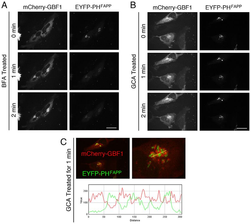
RESEARCH ARTICLE                                                                               Journal of Cell Science (2014) 127, 354–364 doi:10.1242/jcs.130591

regulatory mechanism that functions to maintain proper Arf-GTP                      Exo1 for 1 minute caused Golgi recruitment of endogenous GBF1
levels on cis-Golgi membranes.                                                      (Fig. 1B) similar to that observed for exogenous mCherry–GBF1
                                                                                    (Fig. 1A). Quantification of endogenous GBF1 recruitment,
RESULTS                                                                             shown as a ratio of mean Golgi intensity to mean cytosol
GFB1 accumulates on Golgi membranes in response to an increase in                   intensity, revealed a 2.5-fold increase in Golgi-localized GBF1
the ratio of Arf-GDP to Arf-GTP                                                     following a 1-minute Exo1 treatment (Fig. 1C).
Previous studies have revealed that treatment with BFA caused a                        To obtain independent evidence that the ratio of Arf-GDP to
dramatic accumulation of GBF1 on Golgi membranes (Niu et al.,                       Arf-GTP influences GBF1 recruitment, we examined the impact
2005; Szul et al., 2005; Zhao et al., 2006). As mentioned                           of ArfGAP1 overexpression on GBF1 distribution. Previous
above, further work from our laboratory indicated that GBF1                         studies have established that ArfGAP1 localizes to the Golgi
accumulation is not accompanied by recruitment of Arfs (Chun et                     where it promotes GTP hydrolysis on Arfs (Cukierman et al.,
al., 2008). We further observed that treatment of NRK cells with                    1995; Szafer et al., 2001; Shiba et al., 2011). Further
Exo1, a drug that reduces Arf-GTP levels but does not target the                    characterization of the N-terminal GAP domain identified a
GEF, also caused recruitment of exogenous GBF1 to Golgi and                         crucial ‘arginine finger’ residue that participates in catalysis
ERGIC membranes. To confirm and extend these results, we                            (Szafer et al., 2000; Ismail et al., 2010), and mutation of this
tested the effect of Exo1 treatment on endogenous GBF1                              arginine residue (residue 50 in human ArfGAP1) abrogated GAP
localization in HeLa cells. We first confirmed that in live HeLa                    activity in vivo (Shiba et al., 2011). To examine the potential
cells expressing mCherry–GBF1 and treated with 200 mM Exo1                          role that the ratio of Arf-GDP to Arf-GTP played in GBF1
for 1 minute, GBF1 is clearly recruited to Golgi and peripheral                     recruitment, we examined the distribution of endogenous GBF1
puncta (Fig. 1A). No change in mCherry–GBF1 localization and                        in HeLa cells transiently expressing low levels of EGFP-tagged
recruitment was observed in control cells treated with DMSO for                     wild-type (WT) or the catalytically dead R50Q point GAP1. This
1 minute. To ascertain that Exo1-dependent accumulation did not                     experiment revealed an increase in Golgi-localized GBF1 in cells
depend on overexpression of tagged GBF1, we tested its effect on                    expressing WT EGFP–GAP1. Interestingly, expression of
the endogenous protein. Treatment of HeLa cells with 200 mM                         inactive EGFP–GAP1 R50Q resulted in a clear and significant

                                                                                                                                                                     Journal of Cell Science

Fig. 1. Changes in the cellular Arf-GTP:Arf-GDP ratio cause the recruitment and accumulation of GBF1 on Golgi membranes. (A) HeLa cells expressing
mCherry–GBF1 were treated with 200 mM Exo1 or DMSO as a control and imaged by live cell spinning disc confocal microscopy as described in the Materials
and Methods. Cells from a representative experiment (n56) are displayed as focal projections of all z-slices. (B) HeLa cells were treated with 200 mM Exo1 or
DMSO as a control then stained with mouse anti-GBF1 monoclonal antibody and imaged by fixed cell spinning disc confocal microscopy. Cells from a
representative experiment (n53) are displayed as focal projections of all z-slices. (C) Quantification of GBF1 recruitment in cells treated with Exo1 or DMSO-only
was performed by measuring the ratio of mean intensity (Int) of GBF1 staining at the Golgi to mean intensity of GBF1 staining in the cytosol. A minimum of eight
cells similar to those shown in B were quantified in each of three separate experiments. (D) HeLa cells were mock transfected or transfected with plasmids
encoding EGFP–GAP1 WT or EGFP–GAP1 RQ. Cells were then fixed and stained with mouse anti-GBF1 monoclonal antibody and imaged by spinning disc
confocal microscopy. Cells from a representative experiment (n53) are displayed as focal projections of all z-slices. (E) Quantification of GBF1 recruitment in
images like those shown in D was performed as described for panel C. A minimum of eight cells were quantified in each of three separate experiments. Scale
bars: 26 mm.

                                                                                                                                                             355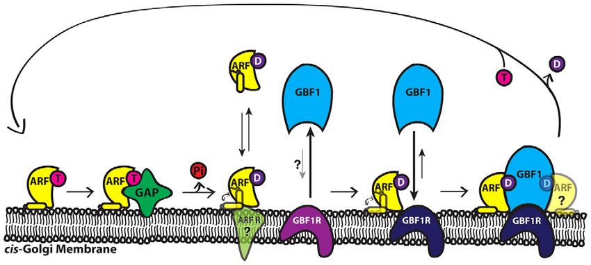
RESEARCH ARTICLE                                                                          Journal of Cell Science (2014) 127, 354–364 doi:10.1242/jcs.130591

decrease in membrane-associated GBF1 relative to mock-                         achieve low levels of Arf expression. To prevent Golgi collapse,
transfected control cells (Fig. 1D). Quantification of the ratio of            we coexpressed myc-tagged membrin, a Golgi SNARE that
Golgi-bound to free cytosolic GBF1showed that expression of                    can recruit Arf1-GDP to the Golgi. Overexpression of either
WT ArfGAP1 resulted in a 2.5-fold increase in ratio of Golgi-                  GBF1 (Claude et al., 1999) or membrin (Honda et al., 2005)
associated to free GBF1 relative to that in the mock-transfected               had previously been shown to protect from BFA-induced
control (Fig. 1E). In contrast, expression of ArfGAP1 R50Q                     Golgi collapse (Honda et al., 2005), probably as a result of
caused a 50% reduction in this ratio. This unexpected observation              sufficient Arf1-GTP production. We speculated that membrin
suggests that expression of catalytically inactive ArfGAP                      overexpression would similarly prevent Arf1-T31N-induced
decreases the Arf-GDP:Arf-GTP ratio. Altogether, our results                   Golgi collapse through enhanced production of Arf1-GTP by
suggest that GBF1 recruitment responds to re-establish steady                  more efficiently recruiting Arf1-GDP to the site of activation. The
state Arf-GTP level, and is sensitive to both increases and                    distribution of GBF1 was therefore examined in HeLa cells
decreases of the cellular ratio of Arf-GDP to Arf-GTP.                         coexpressing myc–membrin, and either the WT or T31N mutant
                                                                               forms of Arf1–EGFP. To assess relative Arf overexpression in
Arf-GDP promotes accumulation of GBF1 on Golgi membranes                       our experiment, we performed quantitative western blot analysis
Sensing a change in the ratio of Arf-GDP to Arf-GTP can result                 as described in the Materials and Methods. We observed that
from responding to either the GDP- or the GTP-bound form of                    relative band intensities of the exogenously expressed Arf–EGFP
Arf. The potential involvement of Arf-GDP could be readily                     (about 45 kDa) and endogenous Arf (about 20 kDa) were similar
tested utilizing a well-characterized the T31N point mutation that             (Fig. 2A). The anti-GFP antibody panels confirm that our Arf
interferes with GTP binding and maintains Arf in an inactive state             antibody detects the overexpressed protein. We determined that
(Dascher and Balch, 1994). In order to perform this experiment,                the fold overexpression of Arf1 WT–EGFP and Arf1 T31N–
we first had to develop a method that would allow expression of                EGFP was 1.4-fold and 0.9-fold, respectively (Fig. 2B).
dominant-inactive Arf1 T31N–EGFP while maintaining the                            We next examined GBF1 distribution in cells expressing low
integrity of the Golgi because expression of this dominant-                    levels of WT and mutant Arf1. Initial experiments focused on the
inactive mutant causes a collapse of the Golgi and a block in                  distribution of mCherry–GBF1. As predicted by Honda et al.
protein traffic (Dascher and Balch, 1994; Donaldson et al., 2005).             (Honda et al., 2005), the integrity of the Golgi was maintained in
To achieve this, we first optimized transfection conditions to                 a significant fraction of cells coexpressing myc–membrin,

                                                                                                                                                               Journal of Cell Science

Fig. 2. Increases in Arf-GDP levels cause the recruitment and accumulation of GBF1 on Golgi membranes. (A) Western blot analysis of post-nuclear
extracts prepared from HeLa cells transfected with plasmids encoding mCherry–GBF1, myc–membrin, and either WT or T31N forms of Arf1–EGFP. Different
volumes of each sample were analyzed to ensure that the signal remained within the linear range of detection. Membranes were incubated with mouse anti-Arf
1D9 and rabbit anti-GFP and were developed and analyzed. A representative western blot from four separate experiments is shown. The uncropped western
blots are shown in supplementary material Fig. S1. (B) Quantification of the fold overexpression of exogenous Arf–EGFP over endogenous Arf levels was
performed by comparing amount of signal in each band using Odyssey software. (C) HeLa cells were co-transfected with plasmids encoding mCherry–GBF1,
myc–membrin, and either WT or T31N forms of Arf1–EGFP and imaged after fixation as for Fig. 1 (n53). (D) HeLa cells were co-transfected with plasmids
encoding myc–membrin and either WT or T31N forms of Arf1–EGFP as for A. Cells were then fixed and stained with mouse anti-GBF1 monoclonal antibody and
imaged as for Fig. 1 (n53). (E) Quantification of GBF1 recruitment in cells expressing Arf1 WT and T31N was performed by measuring the percentage of total
GBF1 staining at the Golgi. Scale bars: 26 mm.

356
RESEARCH ARTICLE Journal of Cell Science (2014) 127, 354–364 doi:10.1242/jcs.130591
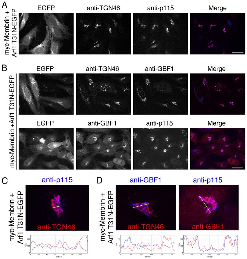
mCherry–GBF1 and low levels of Arf1 T31N (Fig. 2C). segregation of cis- and trans-markers results from continuous
Importantly, we observed a clear accumulation of GBF1 on Arf- and coat-dependent sorting events and requires recruitment
Golgi membranes and dramatic loss of cytoplasmic GBF1 in cells of GBF1 to cis-Golgi membranes (Manolea et al., 2008). We first
expressing Arf1 T31N relative to those expressing WT Arf1 confirmed that the cis-Golgi marker p115 (also known as USO1)
(Fig. 2C). Note that, although the presence of myc–membrin and trans-Golgi network marker TGN46 remained well resolved
allowed for a higher proportion of cells with intact Golgi, it was in cells expressing both inactive Arf1 T31N and myc–membrin
clearly not required for the Arf1 T31N-dependent accumulation of (Fig. 3A). Although it was clear that both markers localized
GBF1 (supplementary material Fig. S2). More importantly, Arf1 to the juxta-nuclear Golgi region, the patterns were clearly
T31N was able to induce redistribution of untagged endogenous different (Fig. 3A), as observed in cells expressing WT Arf1
GBF1 from the cytosol to the Golgi. Loss of cytosolic GBF1 did (supplementary material Fig. S3). Merging of the p115 and
not result from lower expression or degradation of GBF1 in cells TGN46 panels revealed distinct red and blue signals. A line-scan
expressing T31N Arf1 (supplementary material Fig. S1C). The analysis of a representative Golgi from the merge image in
results obtained for endogenous GBF1 were quantified as a Fig. 3A confirms the significant separation observed between
percentage of the total GBF1 signal at the Golgi and the results are TGN46 (red) and p115 (blue) signal peaks (Fig. 3C). To assess
shown in Fig. 2E. This analysis revealed a 3.5-fold increase in whether GBF1 accumulated on cis-Golgi membranes, we
membrane associated GBF1 in T31N Arf1 transfected cells repeated this analysis by comparing the distribution of
compared to WT Arf1-transfected cells (Fig. 2E). These findings endogenous GBF1 with cis-Golgi and TGN markers in cells
provide strong evidence that Arf-GDP can regulate GBF1 expressing the mutant T31N Arf1 (Fig. 3B). We observed clear
localization to Golgi membranes. colocalization of endogenous GBF1 with p115 but not with
TGN46. This was also confirmed by line-scan analysis (Fig. 3D).
GBF1 accumulates on cis-Golgi membranes in a catalytically To obtain more direct evidence that accumulated GBF1
active form remained active, we first determined whether COPI, a well-
The results presented above demonstrate that GBF1 is recruited to established effector of Arf-GTP, associated with Golgi
the Golgi in response to elevated levels of its Arf-GDP substrate. membranes in a BFA-sensitive and therefore GBF1-dependent
This suggests a potentially novel mechanism to ensure manner. HeLa cells were co-transfected with plasmids encoding
homeostatic levels of Arf-GTP on cis-Golgi membranes by mCherry–GBF1 and myc–membrin, as well as Arf1 T31N–EGFP
recruitment of GBF1. To assess whether GBF1 accumulated in an to promote recruitment of GBF1. The COPI distribution was then
active form, we first determined whether the Golgi remained examined in transfectants treated with either BFA or DMSO
polarized. Current evidence suggests that the characteristic by staining for b-COP, a subunit of the COPI coat. Results
Fig. 3. Arf-GDP-dependent GBF1 accumulation does
not alter Golgi polarity and GBF1 is recruited
specifically to the cis-Golgi. (A) HeLa cells were
co-transfected with plasmids encoding myc–membrin
and Arf1 T31N–EGFP, fixed and stained with sheep
anti-TGN46 and mouse anti-p115 and then imaged as for
Fig. 1 (n53). (B) HeLa cells were co-transfected with
plasmids encoding myc–membrin and Arf1 T31N–EGFP,
fixed and stained with either sheep anti-TGN46 and
mouse anti-GBF1 or rabbit anti-GBF1 and mouse
anti-p115 antibodies and then imaged as for Fig. 1 (n53).
(C) Line-scan analysis was performed on cells
expressing myc–membrin and Arf1 T31N–EGFP and
stained with sheep anti-TGN46 and mouse anti-p115. A
magnified image of a representative Golgi from A is
shown. (D) Line-scan analysis was performed on cells
expressing myc–membrin and Arf1 T31N–EGFP and
stained with either sheep anti-TGN46 and mouse
anti-GBF1 or rabbit anti-GBF1 and mouse anti-p115
antibodies. A magnified image of a representative Golgi
from B is shown. Scale bars: 26 mm.
Journal of Cell Science
357RESEARCH ARTICLE Journal of Cell Science (2014) 127, 354–364 doi:10.1242/jcs.130591
Fig. 4. GBF1 remains catalytically active following Arf-GDP-
dependent recruitment. (A) HeLa cells were co-transfected
with plasmids encoding myc–membrin and Arf1 T31N–EGFP
and treated with either 10 mg/ml BFA or DMSO control for
20 minutes then fixed and stained with mouse anti-b-COP and
imaged as for Fig. 1. Low expression of inactive Arf led to GBF1
accumulation in every cell analyzed (n548) and we therefore
expect all Arf–GFP-expressing cells to accumulate GBF1. COPI
recruitment in cells overexpressing the WT form of Arf1 are
shown in supplementary material Fig. S4. (B) Live HeLa cells
expressing myc–membrin, Arf1 WT–mCherry, and either the
WT or T31N form of Arf1–EGFP were imaged as for Fig. 1.
(C) Quantification of the fraction of Arf1 WT–mCherry signal at
the Golgi in cells expressing Arf1 T31N–EGFP was performed
and normalized to cells expressing Arf1 WT–EGFP. A minimum
of two cells similar to those shown in B were quantified in each
of four separate experiments. Images shown in B were extracted
from the experiments shown in supplementary material
Fig. S6B,C. Scale bars: 26 mm.
established that a large fraction of COPI associates with Arf-GDP-dependent recruitment of GBF1 does not require PtdIns4P
juxtanuclear membranes in the presence of Arf1 T31N The recent demonstration that PtdIns4P, through the activity of
(Fig. 4A). Significantly, BFA treatment completely dispersed PI4KIIIa, is required for the association of GBF1 with cis-Golgi
COPI (Fig. 4A, right panels), suggesting that accumulated GBF1 membranes suggests a readily testable mechanism in which
was active and remained sensitive to BFA. increases in Arf-GDP could result in recruitment of GBF1 by
To directly establish that recruitment of GBF1 leads to elevated elevating levels of PtdIns4P (Dumaresq-Doiron et al., 2010). To
Arf activation on Golgi membranes, we quantified the levels of test this possibility, we utilized a chimera containing the
Arf1 WT–mCherry on Golgi membranes in cells coexpressing PtdIns4P-binding PH domain of (EYFP–PHFAPP) to visually
either WT or the T31N mutant forms of Arf1–EGFP. Previous monitor intracellular PtdIns4P levels (Godi et al., 2004; De
work has established that Arf association with membranes is an Matteis and Luini, 2008). The best-characterized approach to
indirect measure of Arf activation because treatments that block rapidly increase Arf-GDP is BFA treatment, which allowed us to
Arf nucleotide exchange result in rapid redistribution of Arf to the simultaneously monitor relative changes in GBF1 and PtdIns4P
cytosol (Donaldson et al., 1992; Helms and Rothman, 1992). The levels by live-cell imaging. HeLa cells coexpressing mCherry–
experimental set up and predicted results are illustrated in GBF1 and EYFP–PHFAPP were treated with BFA and changes in
supplementary material Fig. S5. We first confirmed that GBF1 GBF1 and PtdIns4P levels at Golgi membranes were monitored.
accumulates on Golgi membranes in cells coexpressing both WT As expected, treatment with the carrier DMSO had no impact on
and mutant Arf1 (supplementary material Fig. S6A). Live-cell the distribution of either GBF1 or EYFP–PHFAPP (supplementary
imaging clearly indicated that Arf1 WT–mCherry was efficiently material Fig. S7). Treatment with BFA caused the expected
activated and associated with the Golgi whether the cells recruitment of GBF1 to Golgi membranes, but, contrary to the
coexpressed WT or mutant Arf1–EGFP (Fig. 4B). In contrast, prediction of the hypothesis above, caused a rapid loss of EYFP–
T31N Arf1–EGFP associated weakly with membranes. Most of PHFAPP from Golgi membranes (Fig. 5A).
the WT Arf1 dispersed in response to BFA treatment, leaving a BFA inhibits both the cis-Golgi-localized GBF1 and the TGN-
small fraction on Golgi membranes, similar to that observed localized BIGs (Sáenz et al., 2009). To more specifically assess a
for the T31N mutant form of Arf1 (supplementary material potential role for PtdIns4P on cis membranes where GBF1 is
Fig. S6B–D). As expected, if accumulated GBF1 is active, we localized, we turned to the more selective drug Golgicide A
observed a decrease in cytosolic WT Arf1–mCherry in cells (GCA). The bulkier GCA inhibits GBF1 activity but is excluded
Journal of Cell Science
expressing T31N Arf1–EGFP relative to those expressing WT from the BIGs’ binding site (Sáenz et al., 2009). GCA treatment
Arf1–EGFP (Fig. 4B). This observation suggests greater Arf1 of HeLa cells coexpressing mCherry–GBF1 and EYFP–PHFAPP
activation and association with membranes in cells containing caused obvious recruitment of GBF1 on Golgi membranes
accumulated GBF1. Quantification of several experiments similar (Fig. 5B). However, despite this clear increase in GBF1 signal
to that in Fig. 4B revealed that there was a 50% increase in active there was no change in the EYFP–PHFAPP intensity in all
membrane-associated Arf in cells expressing low levels of replicate experiments suggesting that PtdIns4P levels remained
inactive Arf (Fig. 4C). This result is not only conclusive constant. The dramatic decrease in PtdIns4P levels observed
evidence that GBF1 remains active following recruitment, but following BFA treatment (Fig. 5A) probably results from
also suggests that recruitment of GBF1 directly increases the inhibition of BIGs activity, as could be predicted from the
amount of Arf activation at the Golgi. well-established localization of PtdIns4P (D’Angelo et al., 2008;
358RESEARCH ARTICLE Journal of Cell Science (2014) 127, 354–364 doi:10.1242/jcs.130591
Fig. 5. Arf-GDP-dependent GBF1 recruitment
to cis-Golgi membranes is independent of
PtdIns4P levels. (A) HeLa cells expressing
mCherry–GBF1 and EYFP–PHFAPP were treated
with 5 mg/ml BFA and imaged by live cell spinning
disc confocal microscopy (n56). (B) Live HeLa
cells expressing mCherry–GBF1 and EYFP–
PHFAPP were treated with 10 mM GCA and imaged
as for Fig. 1 (n56). (C) Line-scan analysis was
performed on images of cells expressing
mCherry–GBF1 and EYFP–PHFAPP obtained
following a 1-minute treatment with 10 mM GCA.
Merge image and fourfold magnification of a
representative Golgi from B are shown. Scale
bars: 26 mm.
Bankaitis et al., 2012) and BIGs (Shinotsuka et al., 2002; substrate-driven recruitment mechanism by which GBF1 can
Zhao et al., 2002; Manolea et al., 2008) to the TGN. This maintain active levels of all Arf isoforms at the Golgi.
interpretation is further supported by our observation that
accumulated mCherry–GBF1 is well-resolved from EYFP– Arf-GDP-dependent accumulation of GBF1 requires membrane
PHFAPP (Fig. 5C). The clear separation of the GBF1 and association of Arf-GDP
PtdIns4P signals is most apparent in the magnified image and Recruitment of GBF1 to the Golgi probably involves a putative
line-scan (Fig. 5C). Taken together, these data suggest that compartment-specific membrane-bound receptor and regulation
the mechanism by which Arf-GDP recruits GBF1 to cis-Golgi of its activity should occur at the membrane. To begin addressing
membranes is independent of the PtdIns4P level and that, the mechanism by which Arf-GDP recruits GBF1 to cis-Golgi
contrary to previous reports, GBF1 recruitment does not membranes, we therefore examined whether association of
require PtdIns4P for membrane binding. Arf-GDP with membranes was required. Several studies have
demonstrated that N-terminal myristoylation of Arf proteins is
Most Golgi-associated Arfs can regulate recruitment of GBF1 required for efficient binding to membranes because mutation of
Experiments to date focused on the more abundant Arf1 isoform. the myristoylation site from glycine to alanine (G2A) abolishes
However, all Arf isoforms but Arf6 associate to some extent membrane binding (Franco et al., 1993; Haun et al., 1993; Kahn
with cis-Golgi membranes and must therefore be activated by et al., 1995). A double mutant of inactive Arf lacking the essential
GBF1 in vivo. For this reason, we predict that GBF1 recruitment myristoylation site (G2A, T31N) provided us with a tool with
should respond to all isoforms to maintain adequate levels of which to readily test our hypothesis.
each distinct Arf isoform. To test this prediction, HeLa cells HeLa cells expressing myc–membrin and low levels of either
expressing mCherry–GBF1, myc-membrin and various Arf WT, T31N, or T31N, G2A forms of Arf1–EGFP were stained
T31N–EGFP mutants were examined for accumulation of for endogenous GBF1 (Fig. 6B). As previously observed
GBF1 on Golgi membranes. The left panels in Fig. 6A show (Fig. 2A,B), expression of WT Arf1 failed to accumulate
Journal of Cell Science
the distribution of overexpressed GBF1 in cells coexpressing GBF1, whereas that of the T31N mutant led to potent
either class I (Arf1 or Arf3) or class II (Arf4 or Arf5) Arf recruitment of GBF1. As expected, the T31N, G2A double
mutants. Expression of inactive forms of both class I Arfs caused mutant did not associate to any detectable extent with Golgi
dramatic recruitment of GBF1 on a juxta-nuclear Golgi membranes. More importantly, the T31N, G2A double mutant did
(Fig. 6A). Expression of T31N Arf5 caused reproducible not cause significant accumulation of GBF1 on Golgi membranes
fragmentation of the Golgi, and, in every case examined, in any of the transfectants examined. GBF1 failed to accumulate
GBF1 accumulated on the Golgi fragments. In contrast, expression even in cells expressing significantly higher levels of the Arf1
of the other class II Arf, T31N Arf4, failed to caused GBF1 T31N, G2A double mutant (Fig. 6B, cell in the center of the
recruitment. These results indicate that most inactive Arf isoforms bottom right panel). These results indicate that the membrane
can promote GBF1 recruitment, and suggest that there is a association of Arf-GDP is required for subsequent GBF1
359RESEARCH ARTICLE Journal of Cell Science (2014) 127, 354–364 doi:10.1242/jcs.130591
Fig. 6. Arf-GDP-dependent recruitment of GBF1
to cis-Golgi membranes is not specific to an Arf
isoform and requires Arf-GDP association with
Golgi membranes. (A) Live HeLa cells were co-
transfected with plasmids encoding mCherry–GBF1,
myc–membrin, and either EGFP-tagged Arf1, Arf3,
Arf4 or Arf 5 T31N and imaged as for Fig. 1.
(B) HeLa cells were co-transfected with plasmids
encoding myc–membrin and Arf1 WT–EGFP, Arf1
T31N–EGFP or Arf1 T31N G2A–EGFP. Cells were
then fixed and stained with mouse anti-GBF1
monoclonal antibody and imaged as for Fig. 1.
(C) Quantification of GBF1 recruitment at the Golgi in
cells expressing Arf1 WT, T31N, and T31N G2A was
performed by measuring the percentage of total
GBF1 staining at the Golgi. A minimum of eight cells
similar to those shown in B were quantified in each of
three separate experiments. Scale bar: 26 mm.
recruitment. Quantification of the GBF1 signal at the Golgi and was therefore active. Accumulation of GBF1 on Golgi
from several similar experiments established that expression of membranes occurred independently of PtdIns4P levels. Arf-GDP-
T31N, G2A Arf1 did not cause a significant increase in GBF1 dependent recruitment of GBF1 depended on the myristoylation
at the Golgi. The percentage of GBF1 signal at the Golgi in of the Arf N-terminal helix that is required for membrane
cells expressing T31N, G2A Arf1 is very similar to that association. Our results suggest Arf-GDP-dependent regulation of
observed with WT Arf1, whereas expression of T31N Arf1 a membrane-associated factor for recruitment of GBF1 that is
caused a 3.5-fold increase in GBF1 level at the Golgi (Fig. 6C), located specifically on cis-Golgi membranes.
as previously observed (Fig. 2E). In summary, these results
demonstrate that the membrane association of Arf-GDP is GBF1 is recruited to Golgi membranes in response to increases
required for subsequent recruitment of GBF1 on a cis-Golgi in Arf-GDP
localized factor. It has been hypothesized (Niu et al., 2005; Szul et al., 2007) that
treatment of cells with BFA results in accumulation of GBF1
DISCUSSION on Golgi membranes due to the formation of an ‘abortive
In this study, we elucidated a novel mechanism for the regulation complex’, first observed biochemically in vitro using purified
of recruitment of GBF1 on cis-Golgi membranes. Several components (Béraud-Dufour et al., 1998; Peyroche et al., 1999;
laboratories have previously reported that treatment with BFA Renault et al., 2003). More recently, our laboratory tested
causes a rapid accumulation of GBF1 on Golgi membranes, an this hypothesis and found no coincident recruitment of GBF1
effect attributed to formation of an Arf–BFA–GEF membrane- and Arf to Golgi membranes following BFA treatment (Chun
bound complex. We extended those results and report that several et al., 2008). This result suggested that BFA inhibition in vivo
manipulations that increase Arf-GDP levels, including Exo1 induces a physiological response resulting in GBF1 recruitment
treatment and ArfGAP1 overexpression, also cause accumulation to Golgi membranes in a form that no longer binds Arf to
of endogenous GBF1 on Golgi membranes. These results are form a stable Arf–BFA–GBF1 complex. In this study, we
consistent with our previous report showing that trapping of Arf further tested the hypothesis and discovered that various
on a membrane-bound GEF is not detected in vivo (Chun et al., treatments that decrease Arf-GTP, resulting in a corresponding
Journal of Cell Science
2008), and suggest instead that it is accumulation of Arf-GDP that increase in Arf-GDP, cause GBF1 recruitment and accumulation.
promotes recruitment of GBF1. The possibility that Arf-GDP For example, as shown in Fig. 1, overexpression of active
positively regulates GBF1 association with membranes was tested ArfGAP1 led to a clear accumulation of GBF1 on Golgi
using inactive ‘GDP-arrested’ Arf T31N mutants. We found that membranes. In contrast, expression of an inactive mutant
expression of even low levels of several Arf T31N isoforms ArfGAP1 caused significant reduction of GBF1 recruitment
caused dramatic accumulation of GBF1 on membranes. Under to Golgi membranes compared to control cells, suggesting that
these conditions, the Golgi still displayed separate cis- and trans- this mutant interferes with Arf-GTP hydrolysis and thus the
Golgi markers, and the Arf-GDP-dependent enrichment of GBF1 production of Arf-GDP. This, in turn, results in a lesser
occurred selectively on cis-Golgi membranes. Moreover, the requirement for GBF1 to produce Arf-GTP, which is achieved
accumulated GBF1 resulted in increased Arf-GTP production by lower levels of recruited GBF1.
360RESEARCH ARTICLE Journal of Cell Science (2014) 127, 354–364 doi:10.1242/jcs.130591
As predicted, expression of even very low levels of the inactive and Arf activation remained BFA-sensitive in each assay (Fig. 4).
T31N mutant of several Arfs was sufficient to induce the recruitment These data establish that Arf-GDP-dependent recruitment of GBF1
and accumulation of both overexpressed and endogenous GBF1 occurs specifically on cis-Golgi membranes, results in GBF1
(Fig. 2). To further define the mechanism by which Arf-GDP activity, and demonstrate a novel and physiologically relevant
promotes GBF1 recruitment to membranes we assessed if a specific model for regulation of GBF1 recruitment.
Golgi-associated Arf isoform played a regulatory role. These
experiments established that expression of inactive mutants of Arf-GDP must be membrane-associated to regulate recruitment of
Arf1, Arf3, and Arf5 caused GBF1 accumulation on Golgi GBF1 to membranes
membranes; only Arf4 T31N failed to do so (Fig. 6). Arf4 T31N To examine the mechanism through which Arf-GDP regulates
does not disrupt the Golgi and might not properly mimic the GDP- GBF1 recruitment we first examined its potential involvement in
bound form. We expect that the true GDP-bound form of all class I modulating PtdIns4P levels using the well-characterized biosensor
and class II Arfs can regulate the recruitment of GBF1. Our results EYFP–PHFAPP (Godi et al., 2004; De Matteis and Luini, 2008).
suggest that GBF1 has evolved to respond equally to the production This hypothesis was based on a report by LeFrancois and
of most Golgi-associated Arf species. colleagues suggesting that production of PtdIns4P was required
The experiments discussed above led us to propose that Arf- for recruitment of GBF1 to Golgi membranes (Dumaresq-Doiron
GDP plays a regulatory role by activating a putative GBF1 receptor et al., 2010). However, experiments involving treatment with BFA
found specifically on membranes of the cis-Golgi and ERGIC or GCA revealed no correlation between GBF1 recruitment and
(Fig. 7). In this model, cells can monitor levels of Arf-GDP at the PtdIns4P levels (Fig. 5). Treatment with BFA, which targets both
membrane and respond by adjusting GBF1 recruitment to cis- GBF1 and BIGs, caused GBF1 accumulation and almost complete
Golgi membranes in order to maintain homeostasis by nucleotide loss of the PtdIns4P signal from the Golgi. In contrast, treatment
exchange. Several lines of evidence discussed below support the with the GBF1-specific inhibitor GCA also caused GBF1
possibility that accumulation of Arf-GDP promotes selective accumulation but had no impact on PtdIns4P levels. More
recruitment of active GBF1 on cis-Golgi membranes. importantly, GBF1 recruitment occurred on membranes that
appeared clearly distinct from membranes positive for PtdIns4P.
GBF1 enriched on membranes remains catalytically active and Whereas those experiments failed to reveal a link between GBF1
maintains Golgi polarity and function and PtdIns4P, they established a predicted but as yet
For our model to be physiological relevant, Arf-GDP-dependent uncharacterized link between the production of the TGN-
recruitment of GBF1 must result in increased GBF1 activity at the localized PtdIns4P (Odorizzi et al., 2000; De Matteis et al.,
cis-Golgi. Specifically, we would expect to observe selective 2005; D’Angelo et al., 2008) and the BFA-sensitive but GCA-
recruitment of GBF1 to membranes of the early secretory pathway, resistant BIGs, also localized at the TGN (Shinotsuka et al., 2002;
as well as subsequent Arf activation and maintenance of a Zhao et al., 2002; Manolea et al., 2008). Furthermore, these results
polarized Golgi. Under conditions that caused GBF1 enrichment, confirm that GBF1 localization occurs primarily on cis-Golgi
we indeed observed that GBF1 was recruited specifically to cis- membranes (Kawamoto et al., 2002; Zhao et al., 2002).
Golgi membranes on a Golgi that remained polarized with clearly As an alternative to the candidate approach summarized above,
resolved cis-Golgi and TGN elements (Fig. 3). The activity of we turned to the more basic question of whether Arf-GDP must
accumulated GBF1 was confirmed by the dual observations that be membrane-associated to regulate recruitment of GBF1 to
COP1 was efficiently recruited to Golgi structures and that membranes. Two plausible mechanisms appeared reasonable.
elevated GBF1 levels yielded 50% greater Arf activation relative to Arf-GDP could interact with its target in cytoplasm, possibly
control conditions (Fig. 4). As expected if COP1 and Arf GBF1 itself, to modulate its affinity for the membrane.
membrane association required GBF1 activity, COPI recruitment Alternatively, Arf-GDP could interact with its target at the
Journal of Cell Science
Fig. 7. Diagram depicting the novel ‘Arf-GDP increase’ model for regulation of GBF1 recruitment to cis-Golgi membranes. Arf-GDP acts as a trigger for
GBF1 recruitment. Regulatory Arf-GDP can arise through hydrolysis of Arf-GTP by its GAP or can be recruited directly from cytosol. Arf-GDP might be either free
or bound to an unknown receptor. GBF1 is recruited from cytosol to a no- or low-affinity receptor (magenta) that likely requires Arf-GDP for activation (dark blue).
The nature of the binding site for regulatory Arf-GDP remains unknown but must be at the membrane, possibly the GBF1 receptor itself. However, we cannot
eliminate the possibility that Arf-GDP is regulating a lipid-modifying enzyme to cause GBF1 recruitment. This self-limiting model provides a mechanism to
maintain homeostatic levels of Arf-GTP. T, GTP; D, GDP; Pi, inorganic phosphate.
361RESEARCH ARTICLE Journal of Cell Science (2014) 127, 354–364 doi:10.1242/jcs.130591
membrane, possibly a putative GBF1 receptor, to modulate membranes results in a 50% greater activation of Arf (Fig. 4B,C),
its affinity for GBF1. Such information not only helps define and therefore appears not to be compensated for by increased GAP
the regulatory mechanism but also provides avenues for activity. Together these results suggest that whereas the GEF is
identification of putative receptors. To distinguish between extremely sensitive to alterations in Arf-GTP:Arf-GDP ratio, the
these two potential mechanisms, we took advantage of a well- GAP appears to be largely unresponsive.
characterized Arf G2A mutation that abrogates myristoylation of
the N-terminal helix of Arf, which is required for its membrane Concluding remarks
association (Franco et al., 1993; Haun et al., 1993; Kahn et al., The work described here yielded novel insights into how BFA acts in
1995). As shown in Fig. 6, expression of the Arf1 T31N G2A vivo, identified a new mechanism of GBF1 recruitment regulation, and
double mutant failed to induce GBF1 recruitment, even in cells for the first time attributed a role for Arf-GDP in the cell. The work
overexpressing the putative Arf-GDP receptor, membrin. These also provides us with tools that should lead to the identification of the
results demonstrate that myristate-dependent membrane putative GBF1 receptor. The recruitment of GBF1, as proposed for
association of Arf-GDP is required to elicit recruitment of other GEFs (Richardson et al., 2012), could respond simultaneously to
GBF1, potentially to an unknown cis-Golgi-bound factor. several stimuli such as phosphoinositide level, Rab1 (Alvarez et al.,
2003) and receptor abundance and/or modification. Current work
Arf-GDP-dependent recruitment of GBF1 to cis-Golgi membranes is towards identification of a receptor will greatly add to our
likely to establish and maintain homeostatic levels of Arf-GTP understanding of GBF1 function and could potentially lead to the
ArfGEF recruitment to membranes is a crucial step in initiating discovery of therapeutics for the treatment of diseases resulting from
guanine nucleotide exchange on Arf proteins at the Golgi (Paris et al., GBF1 dysfunction and those caused by viruses that manipulate GBF1
1997). Our model for the regulation of GBF1 recruitment to cis-Golgi function and localization to ensure successful replication.
membranes centers on the response of the cell to changes in Arf-GDP
MATERIALS AND METHODS
levels (Fig. 7). This model accounts for our observation that Reagents and antibodies
increasing Arf-GDP levels promotes GBF1 membrane association, Exo1 was purchased from Calbiochem (Gibbstown, NJ) and stored in
whereas decreasing Arf-GDP levels results in reduced GBF1 DMSO as a 100 mM stock. BFA and GCA were purchased from Sigma-
association with membranes. More importantly, this self-limiting Aldrich (St. Louis, MO) and stored in DMSO at 10 mg/ml and 10 mM,
model provides a simple mechanism to maintain homeostatic levels of respectively. Monoclonal antibodies used for immunofluorescence were
Arf-GTP. Indeed, increases in the substrate Arf-GDP promote mouse anti-GBF1 (clone 25; BD Biosciences, Mississauga, ON), mouse
recruitment of GBF1 and subsequent Arf activation. Conversely, anti-b-coatomer protein I (COPI) (clone m3a5; Sigma-Aldrich), and mouse
ongoing activation eventually leads to a local reduction in Arf-GDP anti-p115 (7D1; gift from the late Dennis Shields, Albert Einstein College
levels that decreases GBF1 recruitment and establishes the desired of Medicine, NY). Polyclonal antibodies used for immunofluorescence
were sheep anti-TGN46 (AbD Serotec, Oxford, UK) and rabbit anti-GBF1
steady-state level of Arf-GTP. Stimulation of ArfGEF recruitment by
(9D4 final bleed; Manolea et al., 2008). Secondary antibodies used
Arf-GTP has been previously reported for members of the BIG were Alexa-Fluor-647-conjugated goat anti-mouse, Alexa-Fluor-555-
(Richardson and Fromme, 2012; Richardson et al., 2012; Lowery conjugated donkey anti-sheep, and Alexa-Fluor-546-conjugated goat
et al., 2013) and cytohesin families (Cohen et al., 2007; Hofmann anti-rabbit (Invitrogen, Carlsbad, CA) at 1:600. Antibodies used for
et al., 2007). The mechanism reported here for GBF1 appears new western blotting were rabbit polyclonal anti-GFP (Eusera, Edmonton, AB)
because it involves Arf-GDP. at 1:50,000 and mouse monoclonal anti-Arf (1D9; Abcam, Cambridge,
The model also takes into account our demonstration that MA) at 1:500. Secondary antibodies used for western blotting were Alexa-
association of Arf-GDP with membranes is crucial for regulation Fluor-750-conjugated goat anti-rabbit and Alexa-Fluor-680 conjugated
(Fig. 6). Such results suggest the presence of a membrane- goat anti-mouse (Invitrogen, Carlsbad, CA) at 1:10,000.
associated target, either a non-catalytic domain of GBF1 or Cell culture
possibly the GBF1 receptor itself. Whether Arf-GDP simply HeLa cells were from ECACC (Porton Down, UK). HeLa cells were
increases the affinity of GBF1 for the membrane or its receptor, cultured in DMEM containing high glucose and 2 mM L-glutamine
or is actually required for its activation remains unknown. Our and supplemented with 10% fetal bovine serum (Sigma-Aldrich) and
observation that overexpression of the GAP-dead mutant, which 100 mg/ml penicillin and streptomycin at 37 ˚C in a 5% CO2 incubator.
will reduce production of Arf-GDP, leads to near elimination of Transfections were performed using TransIT-2020 transfection reagent
GBF1 recruitment (Fig. 1D), suggests that the GBF1 receptor has or TransIT LTI transfection reagent (Mirus Bio, Madison, WI).
no or extremely low affinity for GBF1 in absence of Arf-GDP. For experiments involving co-transfection, we performed a series of
Our data allows us to also speculate on the regulation of preliminary experiments in which we compared the relative expression
levels using various ratios of plasmids. These experiments identified
ArfGAP1, whose activity opposes that of GBF1 at cis-Golgi
optimal conditions to obtain balanced expression.
membranes. Previous studies have reported that the expression of
catalytically dead mutants of ArfGAP1 did not lead to observable Construction and expression of plasmids
phenotypes on the Golgi (Liu et al., 2005). Our data can now The construction of the plasmids encoding Arf1, Arf3, Arf4, and Arf5
Journal of Cell Science
explain this unexpected result through modulation of GBF1 tagged with either GFP or mCherry has been previously described (Chun
recruitment. For example, expression of the ArfGAP1 R50Q et al., 2008). The Arf4 T31N, G2A double mutant was created by site-
mutant decreases GBF1 recruitment by roughly 50% (Fig. 1D,E), directed mutagenesis using the QuikChange kit (Stratagene, La Jolla,
thereby reducing Arf-GTP production and probably compensating CA), as per the manufacturer’s instructions. The plasmid encoding myc–
membrin (Hay et al., 1997) was a gift from J. Hay (Division of Biological
for the loss of ArfGAP activity. In other words, our results suggest
Sciences, University of Montana, Missoula, MT). The plasmid encoding
that cells adjust GBF1 recruitment to correct for decreased GAP EYFP–PHFAPP (Godi et al., 2004) was obtained through S. Grinstein (Sick
activity. Cells could establish homeostatic Arf-GTP levels by Kids, ON). Construction of a plasmid encoding ArfGAP1 tagged with EGFP
regulating GAP and/or GEF activity. Our results also suggest that at the N-terminus was as previously described (Parnis et al., 2006). A point
cells do not readily modulate endogenous GAP activity. This mutant lacking GAP activity (EGFP–GAP1 R50Q) was created by site-
conclusion is based on the fact that accumulation of GBF1 to Golgi directed mutagenesis using the QuikChange kit (Stratagene, La Jolla, CA).
362RESEARCH ARTICLE Journal of Cell Science (2014) 127, 354–364 doi:10.1242/jcs.130591
Immunofluorescence and live-cell time-lapse imaging were air dried and resuspended thoroughly in 50 ml lysis buffer and 10 ml
For live-cell imaging, cells were grown on #1D round coverslips (Fisher 66 loading buffer containing DTT (360 mM Tris-HCl pH 6.8, 60%
Scientific, Ottawa, ON). Immediately before imaging, coverslips were glycerol, 12% SDS, 0.6% Bromophenol Blue and 600 mM DTT).
transferred to an Attofluor live cell chamber (Invitrogen) and CO2- Samples were boiled for 10 minutes at 100 ˚C, and separated by Tris-
independent medium (Invitrogen) supplemented with 10% fetal bovine glycine SDS-PAGE on 12% gels using protein standards (Bio-Rad).
serum was added. Image analysis was only performed on transfectants To assess relative Arf overexpression, we performed quantitative
displaying intact Golgi and low levels of the proteins of interest. Imaging western blot analysis. After electrophoresis, proteins were transferred
was performed on the temperature controlled (37˚C) stage of an Axiovert onto nitrocellulose membranes (GE Healthcare, Chalfont St Giles, UK),
200M confocal microscope (Carl Zeis, Thornwood, NY) equipped with an immunoblotted with primary antibodies raised against Arf and GFP
UltraVIEW ERS 3E spinning disc (PerkinElmer Life and Analytical followed by Alexa-Fluor-680- and Alexa-Fluor-750-conjugated
Sciences, Waltham, MA) and a 636 objective lens (plan-Apochromat, NA (Invitrogen) secondary antibodies. Bands were subsequently detected
1.4) heated by an objective warmer set to 35˚C. Live-cell imaging was by Odyssey Li-Cor scanner and quantified using application software
performed in a room maintained at 26˚C. Images were captured with a 9100- version 3.0.21. Band intensity quantification was performed using the
50 electron multiplier charge-coupled device digital camera (Hamamatsu 10 ml-loaded lanes from four individual experiments.
Photonics, Bridgewater, NJ) and acquired using Volocity software
(PerkinElmer Life and Analytical Sciences). Live-cell experiments with Acknowledgements
dual labeling were imaged by exciting each fluorophore and detecting We thank A. Simmonds (University of Alberta, AB) for technical help with confocal
sequentially before moving to the next z-slice to avoid bleed through. Laser microscopy and secondary antibodies. We also thank P. LaPointe, R. Lehner, N.
intensity, filters, and camera gain were optimized to provide maximal signal Touret and R. Wozniak (University of Alberta, AB) for helpful discussions. We thank
without saturation or bleaching. When stated, drugs were introduced by J. Hay (University of Montana, MT), S. Grinstein (Hospital For Sick Children, ON)
for gifts of plasmids encoding myc–membrin and EYFP–PHFAPP, respectively.
addition of 250 ml of CO2-independent medium containing 66concentration
of the drug to be tested to the Attofluor chamber containing 1.25 ml
medium. Addition of drug caused a slight shift in focus at the time of Competing interests
The authors declare no competing interests.
addition, but this corrected itself for subsequent time points.
Imaging of fixed cells was performed using HeLa cells grown on square
Author contributions
coverslips (Corning Life Sciences, Tewksbury, MA). Drug treatments D.Q. performed all experiments; F.G. and N.S. generated plasmids and/or
were performed at 37 ˚C. Coverslips were then washed with phosphate- performed preliminary experiments; D.C. provided ArfGAP plasmids; P.M. and
buffered saline (PBS), and fixed with 3% paraformaldehyde in PBS at 37 ˚C D.Q. conceived the project, analysed data, designed the figures and wrote the
for 15 minutes. Cells were washed and permeabilized with 0.1% Triton X- manuscript with comments from co-authors.
100 (Sigma) in PBS and subsequently stained with antibodies. Coverslips
were mounted on slides using ProLong Gold anti-fading reagent with Funding
DAPI (Invitrogen) mounting medium and allowed to dry before imaging This study was supported by an operating grant to P.M. from the Canadian
by spinning disc confocal microscopy, as described above, without heating. Institutes of Health Research [grant numbers FRN 107519, FRN 111028], the
Israel Science Foundation [grant number 37/11 to D.C.]; and scholarships to D.Q.
from the Faculty of Medicine and Dentistry and the Faculty of Graduate Studies
Image quantification and analysis
and Research at the University of Alberta.
Quantification of ratio of Golgi signal to cytosolic signal and the
percentage signal at the Golgi was performed using Imaris 664 version Supplementary material
7.4.2 software (Bitplane AG, South Windsor, CT). Regions of interest Supplementary material available online at
were surface-wrapped in three-dimensions utilizing the surfaces option http://jcs.biologists.org/lookup/suppl/doi:10.1242/jcs.130591/-/DC1
and average pixel intensities of the Golgi and cell, and were exported to
Excel (Microsoft, Redmond, WA). Golgi and cell mean intensity values References
were corrected for cytosolic or background contributions, respectively. Alvarez, C., Garcia-Mata, R., Brandon, E. and Sztul, E. (2003). COPI
For fixed cell experiments, at least eight cells were quantified from three recruitment is modulated by a Rab1b-dependent mechanism. Mol. Biol. Cell
14, 2116-2127.
separate experiments for each condition. For live-cell experiments, at Bankaitis, V. A., Garcia-Mata, R. and Mousley, C. J. (2012). Golgi membrane
least eight cells were quantified from a total of four individual dynamics and lipid metabolism. Curr. Biol. 22, R414-R424.
experiments. All graphs were generated in Excel. Error bars represent Béraud-Dufour, S., Robineau, S., Chardin, P., Paris, S., Chabre, M., Cherfils, J.
the standard deviation in the samples quantified. Normalization in and Antonny, B. (1998). A glutamic finger in the guanine nucleotide exchange
factor ARNO displaces Mg2+ and the beta-phosphate to destabilize GDP on
Fig. 4C was performed by taking the average of the four pre-treatment ARF1. EMBO J. 17, 3651-3659.
time points and normalizing that value to 1 for each condition. In Casanova, J. E. (2007). Regulation of Arf activation: the Sec7 family of guanine
Fig. 4D, the Arf1 WT and Arf1 T31N average values were normalized to nucleotide exchange factors. Traffic 8, 1476-1485.
the average starting value of Arf1 WT and compared. Cherfils, J. and Melançon, P. (2005). On the action of Brefeldin A on Sec7-
stimulated membrane-recruitment and GDP/GTP exchange of Arf proteins.
Biochem. Soc. Trans. 33, 635-638.
Preparation of cell extracts, western blots, and analysis Chun, J., Shapovalova, Z., Dejgaard, S. Y., Presley, J. F. and Melançon, P.
(2008). Characterization of class I and II ADP-ribosylation factors (Arfs) in live
HeLa cells were grown on 10-cm plates to ,80–90% confluency. Cells cells: GDP-bound class II Arfs associate with the ER-Golgi intermediate
were transfected by TransIt-2020 reagent (Mirus Bio) with 1.4 mg compartment independently of GBF1. Mol. Biol. Cell 19, 3488-3500.
pCMV-myc-membrin and 1.75 mg pEGFP-Arf1 WT or T31N DNA for Claude, A., Zhao, B. P., Kuziemsky, C. E., Dahan, S., Berger, S. J., Yan, J. P.,
18–20 hours. Transfection efficiency of Arf plasmids was estimated to be Armold, A. D., Sullivan, E. M. and Melançon, P. (1999). GBF1: A novel Golgi-
Journal of Cell Science
associated BFA-resistant guanine nucleotide exchange factor that displays
,80–90%. Cells were released in presence of 0.25% trypsin (Invitrogen) specificity for ADP-ribosylation factor 5. J. Cell Biol. 146, 71-84.
and then collected by centrifugation. Pelleted cells were washed once in Cohen, L. A., Honda, A., Varnai, P., Brown, F. D., Balla, T. and Donaldson, J. G.
PBS and then resuspended in 1 ml 0.5% Triton X-100 (Sigma) lysis (2007). Active Arf6 recruits ARNO/cytohesin GEFs to the PM by binding their
buffer (1% Triton X-100, 150 mM NaCl, and 50 mM Tris-HCl pH 8.0) PH domains. Mol. Biol. Cell 18, 2244-2253.
Cukierman, E., Huber, I., Rotman, M. and Cassel, D. (1995). The ARF1
containing EDTA-free protease inhibitor cocktail (Roche) and incubated GTPase-activating protein: zinc finger motif and Golgi complex localization.
on a rotator for 1 hour to allow complete lysis. Nuclei and debris were Science 270, 1999-2002.
subsequently removed by centrifugation at 9300 g at 4 ˚C. Resulting D’Angelo, G., Vicinanza, M., Di Campli, A. and De Matteis, M. A. (2008). The
supernatant was then transferred to a fresh tube and precipitated by multiple roles of PtdIns(4)P — not just the precursor of PtdIns(4,5)P2. J. Cell
Sci. 121, 1955-1963.
addition of 56 volumes of 220 ˚C acetone and incubation at 220 ˚C for a Dascher, C. and Balch, W. E. (1994). Dominant inhibitory mutants of ARF1 block
minimum of 4 hours. Precipitated proteins were recovered by endoplasmic reticulum to Golgi transport and trigger disassembly of the Golgi
centrifugation at 11,200 g for 10 minutes at 4 ˚C. The resulting pellets apparatus. J. Biol. Chem. 269, 1437-1448.
363RESEARCH ARTICLE Journal of Cell Science (2014) 127, 354–364 doi:10.1242/jcs.130591
De Matteis, M. A. and Luini, A. (2008). Exiting the Golgi complex. Nat. Rev. Mol. Mansour, S. J., Skaug, J., Zhao, X. H., Giordano, J., Scherer, S. W. and
Cell Biol. 9, 273-284. Melançon, P. (1999). p200 ARF-GEP1: a Golgi-localized guanine nucleotide
De Matteis, M. A., Di Campli, A. and Godi, A. (2005). The role of exchange protein whose Sec7 domain is targeted by the drug brefeldin A. Proc.
the phosphoinositides at the Golgi complex. Biochim. Biophys. Acta 1744, 396-405. Natl. Acad. Sci. USA 96, 7968-7973.
Dejgaard, S. Y., Murshid, A., Dee, K. M. and Presley, J. F. (2007). Confocal Melançon, P., Zhao, X. and Lasell, T. K. (2003). Large Arf-GEFs of the Golgi
microscopy-based linescan methodologies for intra-Golgi localization of complex: in search of mechanisms for the cellular effects of BFA. In ARF Family
proteins. J. Histochem. Cytochem. 55, 709-719. GTPases (ed. R. A. Kahn), pp. 101-119. Dordrecht: Kluwer Academic
Doms, R. W., Russ, G. and Yewdell, J. W. (1989). Brefeldin A redistributes resident Publishers.
and itinerant Golgi proteins to the endoplasmic reticulum. J. Cell Biol. 109, 61-72. Mossessova, E., Corpina, R. A. and Goldberg, J. (2003). Crystal structure of
Donaldson, J. G., Finazzi, D. and Klausner, R. D. (1992). Brefeldin A inhibits ARF1*Sec7 complexed with Brefeldin A and its implications for the guanine
Golgi membrane-catalysed exchange of guanine nucleotide onto ARF protein. nucleotide exchange mechanism. Mol. Cell 12, 1403-1411.
Nature 360, 350-352. Niu, T. K., Pfeifer, A. C., Lippincott-Schwartz, J. and Jackson, C. L. (2005).
Donaldson, J. G., Honda, A. and Weigert, R. (2005). Multiple activities for Arf1 at Dynamics of GBF1, a Brefeldin A-sensitive Arf1 exchange factor at the Golgi.
the Golgi complex. Biochim. Biophys. Acta 1744, 364-373. Mol. Biol. Cell 16, 1213-1222.
Dumaresq-Doiron, K., Savard, M. F., Akam, S., Costantino, S. and Lefrancois, Odorizzi, G., Babst, M. and Emr, S. D. (2000). Phosphoinositide signaling
S. (2010). The phosphatidylinositol 4-kinase PI4KIIIalpha is required for the and the regulation of membrane trafficking in yeast. Trends Biochem. Sci. 25,
recruitment of GBF1 to Golgi membranes. J. Cell Sci. 123, 2273-2280. 229-235.
Emr, S., Glick, B. S., Linstedt, A. D., Lippincott-Schwartz, J., Luini, A., Malhotra, Paris, S., Béraud-Dufour, S., Robineau, S., Bigay, J., Antonny, B., Chabre, M.
V., Marsh, B. J., Nakano, A., Pfeffer, S. R., Rabouille, C. et al. (2009). Journeys and Chardin, P. (1997). Role of protein-phospholipid interactions in the
through the Golgi—taking stock in a new era. J. Cell Biol. 187, 449-453. activation of ARF1 by the guanine nucleotide exchange factor Arno. J. Biol.
Farquhar, M. G. and Palade, G. E. (1998). The Golgi apparatus: 100 years of Chem. 272, 22221-22226.
progress and controversy. Trends Cell Biol. 8, 2-10. Parnis, A., Rawet, M., Barkan, B., Rotman, M., Gaitner, M. and Cassel, D.
Franco, M., Chardin, P., Chabre, M. and Paris, S. (1993). Myristoylation is not (2006). Golgi localization determinants in ArfGAP1 and in new tissue-specific
required for GTP-dependent binding of ADP-ribosylation factor ARF1 to ArfGAP1 isoforms. J. Biol. Chem. 281, 3785-3792.
phospholipids. J. Biol. Chem. 268, 24531-24534. Pasqualato, S., Renault, L. and Cherfils, J. (2002). Arf, Arl, Arp and Sar proteins:
Franco, M., Chardin, P., Chabre, M. and Paris, S. (1996). Myristoylation-facilitated a family of GTP-binding proteins with a structural device for ‘front-back’
binding of the G protein ARF1GDP to membrane phospholipids is required for its communication. EMBO Rep. 3, 1035-1041.
activation by a soluble nucleotide exchange factor. J. Biol. Chem. 271, 1573-1578. Peyroche, A., Antonny, B., Robineau, S., Acker, J., Cherfils, J. and Jackson,
Fujiwara, T., Oda, K., Yokota, S., Takatsuki, A. and Ikehara, Y. (1988). Brefeldin C. L. (1999). Brefeldin A acts to stabilize an abortive ARF-GDP-Sec7 domain
A causes disassembly of the Golgi complex and accumulation of secretory protein complex: involvement of specific residues of the Sec7 domain. Mol. Cell
proteins in the endoplasmic reticulum. J. Biol. Chem. 263, 18545-18552. 3, 275-285.
Godi, A., Di Campli, A., Konstantakopoulos, A., Di Tullio, G., Alessi, D. R., Randazzo, P. A. and Kahn, R. A. (1995). Myristoylation and ADP-ribosylation
Kular, G. S., Daniele, T., Marra, P., Lucocq, J. M. and De Matteis, M. A. factor function. Methods Enzymol. 250, 394-405.
(2004). FAPPs control Golgi-to-cell-surface membrane traffic by binding to ARF Renault, L., Guibert, B. and Cherfils, J. (2003). Structural snapshots of the
and PtdIns(4)P. Nat. Cell Biol. 6, 393-404. mechanism and inhibition of a guanine nucleotide exchange factor. Nature 426,
Gommel, D. U., Memon, A. R., Heiss, A., Lottspeich, F., Pfannstiel, J., 525-530.
Lechner, J., Reinhard, C., Helms, J. B., Nickel, W. and Wieland, F. T. (2001). Richardson, B. C. and Fromme, J. C. (2012). Autoregulation of Sec7 Arf-GEF
Recruitment to Golgi membranes of ADP-ribosylation factor 1 is mediated by the activity and localization by positive feedback. Small GTPases 3, 240-243.
cytoplasmic domain of p23. EMBO J. 20, 6751-6760. Richardson, B. C., McDonold, C. M. and Fromme, J. C. (2012). The Sec7 Arf-
Haun, R. S., Tsai, S. C., Adamik, R., Moss, J. and Vaughan, M. (1993). Effect of GEF is recruited to the trans-Golgi network by positive feedback. Dev. Cell 22,
myristoylation on GTP-dependent binding of ADP-ribosylation factor to Golgi. 799-810.
J. Biol. Chem. 268, 7064-7068. Robineau, S., Chabre, M. and Antonny, B. (2000). Binding site of brefeldin A at
Hay, J. C., Chao, D. S., Kuo, C. S. and Scheller, R. H. (1997). Protein the interface between the small G protein ADP-ribosylation factor 1 (ARF1) and
interactions regulating vesicle transport between the endoplasmic reticulum and the nucleotide-exchange factor Sec7 domain. Proc. Natl. Acad. Sci. USA 97,
Golgi apparatus in mammalian cells. Cell 89, 149-158. 9913-9918.
Helms, J. B. and Rothman, J. E. (1992). Inhibition by brefeldin A of a Golgi Sáenz, J. B., Sun, W. J., Chang, J. W., Li, J., Bursulaya, B., Gray, N. S. and
membrane enzyme that catalyses exchange of guanine nucleotide bound to Haslam, D. B. (2009). Golgicide A reveals essential roles for GBF1 in Golgi
ARF. Nature 360, 352-354. assembly and function. Nat. Chem. Biol. 5, 157-165.
Hofmann, I., Thompson, A., Sanderson, C. M. and Munro, S. (2007). The Arl4 Sata, M., Donaldson, J. G., Moss, J. and Vaughan, M. (1998). Brefeldin
family of small G proteins can recruit the cytohesin Arf6 exchange factors to the A-inhibited guanine nucleotide-exchange activity of Sec7 domain from yeast
plasma membrane. Curr. Biol. 17, 711-716. Sec7 with yeast and mammalian ADP ribosylation factors. Proc. Natl. Acad. Sci.
Honda, A., Al-Awar, O. S., Hay, J. C. and Donaldson, J. G. (2005). Targeting of USA 95, 4204-4208.
Arf-1 to the early Golgi by membrin, an ER-Golgi SNARE. J. Cell Biol. 168, Shiba, Y., Luo, R., Hinshaw, J. E., Szul, T., Hayashi, R., Sztul, E., Nagashima,
1039-1051. K., Baxa, U. and Randazzo, P. A. (2011). ArfGAP1 promotes COPI vesicle
Ismail, S. A., Vetter, I. R., Sot, B. and Wittinghofer, A. (2010). The structure of an Arf- formation by facilitating coatomer polymerization. Cell. Logist. 1, 139-154.
ArfGAP complex reveals a Ca2+ regulatory mechanism. Cell 141, 812-821. Shinotsuka, C., Waguri, S., Wakasugi, M., Uchiyama, Y. and Nakayama, K.
Kahn, R. A., Clark, J., Rulka, C., Stearns, T., Zhang, C. J., Randazzo, P. A., (2002). Dominant-negative mutant of BIG2, an ARF-guanine nucleotide
Terui, T. and Cavenagh, M. (1995). Mutational analysis of Saccharomyces exchange factor, specifically affects membrane trafficking from the trans-Golgi
cerevisiae ARF1. J. Biol. Chem. 270, 143-150. network through inhibiting membrane association of AP-1 and GGA coat
Kawamoto, K., Yoshida, Y., Tamaki, H., Torii, S., Shinotsuka, C., Yamashina, proteins. Biochem. Biophys. Res. Commun. 294, 254-260.
S. and Nakayama, K. (2002). GBF1, a guanine nucleotide exchange factor for Szafer, E., Pick, E., Rotman, M., Zuck, S., Huber, I. and Cassel, D. (2000). Role of
ADP-ribosylation factors, is localized to the cis-Golgi and involved in membrane coatomer and phospholipids in GTPase-activating protein-dependent hydrolysis
association of the COPI coat. Traffic 3, 483-495. of GTP by ADP-ribosylation factor-1. J. Biol. Chem. 275, 23615-23619.
Lippincott-Schwartz, J. (2011). An evolving paradigm for the secretory pathway? Szafer, E., Rotman, M. and Cassel, D. (2001). Regulation of GTP hydrolysis
Mol. Biol. Cell 22, 3929-3932. on ADP-ribosylation factor-1 at the Golgi membrane. J. Biol. Chem. 276,
Lippincott-Schwartz, J., Yuan, L. C., Bonifacino, J. S. and Klausner, R. D. (1989). 47834-47839.
Rapid redistribution of Golgi proteins into the ER in cells treated with brefeldin A: Szul, T., Garcia-Mata, R., Brandon, E., Shestopal, S., Alvarez, C. and Sztul, E.
evidence for membrane cycling from Golgi to ER. Cell 56, 801-813. (2005). Dissection of membrane dynamics of the ARF-guanine nucleotide
Liu, W., Duden, R., Phair, R. D. and Lippincott-Schwartz, J. (2005). ArfGAP1 exchange factor GBF1. Traffic 6, 374-385.
dynamics and its role in COPI coat assembly on Golgi membranes of living cells. Szul, T., Grabski, R., Lyons, S., Morohashi, Y., Shestopal, S., Lowe, M.
J. Cell Biol. 168, 1053-1063. and Sztul, E. (2007). Dissecting the role of the ARF guanine nucleotide
Journal of Cell Science
Lowery, J., Szul, T., Styers, M., Holloway, Z., Oorschot, V., Klumperman, J. and exchange factor GBF1 in Golgi biogenesis and protein trafficking. J. Cell Sci.
Sztul, E. (2013). The Sec7 guanine nucleotide exchange factor GBF1 regulates 120, 3929-3940.
membrane recruitment of BIG1 and BIG2 guanine nucleotide exchange factors to Tsai, S. C., Adamik, R., Moss, J. and Vaughan, M. (1996). Purification and
the trans-Golgi network (TGN). J. Biol. Chem. 288, 11532-11545. characterization of a guanine nucleotide-exchange protein for ADP-ribosylation
Manolea, F., Claude, A., Chun, J., Rosas, J. and Melançon, P. (2008). Distinct factor from spleen cytosol. Proc. Natl. Acad. Sci. USA 93, 305-309.
functions for Arf guanine nucleotide exchange factors at the Golgi complex: Zhao, X., Lasell, T. K. and Melançon, P. (2002). Localization of large ADP-
GBF1 and BIGs are required for assembly and maintenance of the Golgi stack ribosylation factor-guanine nucleotide exchange factors to different Golgi
and trans-Golgi network, respectively. Mol. Biol. Cell 19, 523-535. compartments: evidence for distinct functions in protein traffic. Mol. Biol. Cell
Manolea, F., Chun, J., Chen, D. W., Clarke, I., Summerfeldt, N., Dacks, J. B. 13, 119-133.
and Melancon, P. (2010). Arf3 is activated uniquely at the trans-Golgi network Zhao, X., Claude, A., Chun, J., Shields, D. J., Presley, J. F. and Melançon, P.
by brefeldin A-inhibited guanine nucleotide exchange factors. Mol. Biol. Cell 21, (2006). GBF1, a cis-Golgi and VTCs-localized ARF-GEF, is implicated in ER-to-
1836-1849. Golgi protein traffic. J. Cell Sci. 119, 3743-3753.
364You can also read


