Caenorhabditis elegans HUS-1 Is a DNA Damage Checkpoint Protein Required for Genome Stability and EGL-1-Mediated Apoptosis
←
→
Page content transcription
If your browser does not render page correctly, please read the page content below
Current Biology, Vol. 12, 1908–1918, November 19, 2002, 2002 Elsevier Science Ltd. All rights reserved. PII S0960-9822(02)01262-9
Caenorhabditis elegans HUS-1 Is a DNA Damage
Checkpoint Protein Required for Genome Stability
and EGL-1-Mediated Apoptosis
E. Randal Hofmann,1,6,8 Stuart Milstein,1,2,3,8 that DNA damage increases expression of the proapo-
Simon J. Boulton,3,4 Mianjia Ye,1 ptotic gene egl-1, a response that requires hus-1 and
Jen J. Hofmann,1,6 Lilli Stergiou,6 the p53 homolog cep-1.
Anton Gartner,5 Marc Vidal,3 Conclusions: Our findings suggest that the RAD-5
and Michael O. Hengartner1,6,7 checkpoint protein is not required for HUS-1 to relocal-
1
Cold Spring Harbor Laboratory ize following DNA damage. Furthermore, our studies
1 Bungtown Road reveal a new function of HUS-1 in the prevention of
Cold Spring Harbor, New York 11724 telomere shortening and mortalization of germ cells.
2
Graduate Program in Genetics and Department of DNA damage-induced germ cell death is abrogated in
Molecular Genetics and Microbiology hus-1 mutants, in part, due to the inability of these mu-
The State University of New York at Stony Brook tants to activate egl-1 transcription in a cep-1/p53-
Stony Brook, New York 11794 dependent manner. Thus, HUS-1 is required for p53-
3
Dana-Farber Cancer Institute dependent activation of a BH3 domain protein in C.
Department of Genetics elegans.
Harvard Medical School
Boston, Massachusetts 02115
4
Cancer Research UK Introduction
Clare Hall
Clare Hall Laboratories Tumorigenesis is characterized by the accumulation of
South Mimms genetic mutations, rearrangements, amplifications, and
Hertfordshire EN6 3LD deletions — any of which can drive the progressive
United Kingdom transformation of normal cells into highly malignant de-
5
Max-Planck-Institute for Biochemistry rivatives [1]. Cancer avoidance therefore requires pre-
Department of Cell Biology cise and efficient means to maintain the integrity of the
Am Klopferspitz 18a genome. Damage to DNA triggers checkpoint controls
Martinsried 82152 that result in cell cycle arrest and repair of the lesion.
Germany In metazoans, DNA damage often results in the pro-
6
University of Zürich grammed demise of the cell, possibly due to extensive
Institute for Molecular Biology damage that is not rectifiable [2]. Alternatively, it may
Winterthurerstrasse 190 be prudent for the organism to eliminate dangerous cells
8057 Zürich in tissues that have an extensive proliferative capacity,
Switzerland even when damage has been minimal [3, 4]. Loss of
communication between the DNA lesion and the apo-
ptotic program, which allows the persistence of cells
Summary with damaged and/or unstable genomes, can lead to
tumorigenesis.
Background: The inability to efficiently repair DNA dam- Our understanding of the genetics of the DNA damage
age or remove cells with severely damaged genomes checkpoint pathway has been heavily dependent on
has been linked to several human cancers. Studies in studies done in the yeasts Saccharomyces cerevisiae
yeasts and mammals have identified several genes that and Schizosaccharomyces pombe [5]. The DNA damage
are required for proper activation of cell cycle check- checkpoint in S. pombe includes six “rad” genes: rad1⫹,
points following various types of DNA damage. How- rad3⫹, rad9⫹, rad17⫹, rad26⫹, and hus1⫹. DNA damage
ever, in metazoans, DNA damage can induce apoptosis activates Rad3, a phosphatidylinositol kinase family
as well. How DNA damage activates the apoptotic ma- member that is structurally and functionally related to
chinery is not fully understood. human ATM and ATR [6]. Rad3 is required for phosphor-
Results: We demonstrate here that the Caenorhabditis ylation of Hus1, Rad26, Chk1, and Cds1. Phosphoryla-
elegans gene hus-1 is required for DNA damage-induced tion of the kinases Chk1 and Cds1 transduces DNA
cell cycle arrest and apoptosis. Following DNA damage, damage and replication checkpoint signals to the cell
HUS-1 relocalizes and forms distinct foci that overlap cycle machinery [5]. Despite our exceptional under-
with chromatin. Relocalization does not require the standing of checkpoint arrest and repair, yeast lack an
novel checkpoint protein RAD-5; rather, relocalization apoptotic program; thus, our understanding of how a
appears more frequently in rad-5 mutants, suggesting cell decides to repair or die is lagging.
that RAD-5 plays a role in repair. HUS-1 is required for The genetics of apoptosis has been extensively stud-
genome stability, as demonstrated by increased fre- ied in the nematode Caenorhabditis elegans [7]. Al-
quency of spontaneous mutations, chromosome non- though the majority of this work has focused on develop-
disjunction, and telomere shortening. Finally, we show mental aspects of somatic apoptosis, recent research
has also focused on the germline [8]. Unlike the invari-
7
Correspondence: michael.hengartner@molbio.unizh.ch able pattern of somatic cell deaths during development,
8
These authors contributed equally to this work. germline apoptosis is not determined by lineage and can
HUS-1-Mediated Genome Stability and Apoptosis
1909
be regulated by multiple pathways. We have previously
demonstrated that C. elegans is an excellent genetic tool
that can be used to understand DNA damage-induced
cell cycle arrest and apoptosis [9, 10]. DNA damage-
mediated apoptosis is dependent on ced-3 and ced-4
and is negatively regulated by ced-9. The positive death
regulator, egl-1, is partially required, but it is not essen-
tial for radiation-induced apoptosis. Recently, three
C. elegans checkpoint mutants, op241, rad-5(mn159),
and mrt-2(e2663), have been identified that block
DNA damage-induced apoptosis and cell cycle arrest
[9, 11]. It is unclear how these checkpoint genes regulate
the apoptotic machinery. One possibility is by means of
a p53 homolog, cep-1, which is required for DNA dam-
age-induced germ cell death but not cell cycle arrest
[12, 13].
Here we show that op241 is a hypomorphic allele in the
C. elegans homolog of the S. pombe hus1⫹ checkpoint
gene. hus-1 mutants fail to induce apoptosis and prolif-
eration arrest following DNA damage and show in-
creased sensitivity to DNA damage-induced lethality.
Using a newly identified candidate null allele, we show
that hus-1 function is also required for telomere length
maintenance. HUS-1 is a nuclear protein that is ex-
pressed in early embryos and the adult germlines and
relocalizes to putative sites of DNA damage. Finally, we
demonstrate that DNA damage induces the proapo-
ptotic gene egl-1. This is dependent on hus-1 and the
p53 homolog, cep-1.
Results
Identification of Mutations in C. elegans hus-1
We have previously described mutants defective in cell
cycle arrest and apoptosis induced by DNA damage [9,
10]. Included in this group was the mutation op241,
originally identified in a strain containing the him-
7(e1480) mutation and later named dam-1(op241) (DNA
damage response) [9, 10]. We mapped op241 to a 2.1-
cM interval, between stu-4 and unc-11, on the left side
of chromosome I (Figure 1A). An in silico search for
candidate genes in this interval identified H26D21.1, a
homolog of the S. pombe gene hus1⫹. Sequence analy-
sis of H26D21.1 from op241 revealed a G to A transition
at base pair ⫹296, resulting in a G to D substitution at
amino acid 99. Because of the high sequence conserva-
tion between H26D21.1 and hus-1 homologs from other
species, we renamed this gene hus-1.
In an independent reverse genetic screen for deletions
in C. elegans homologs of known checkpoint genes, we
recovered a single hus-1 allele, op244. The op244 allele
is a 729-bp deletion that removes the last two exons
and most of the 3⬘ untranslated region of hus-1 (bp
Figure 1. Identification of hus-1 Mutants 4725–5453 on cosmid H26D21) (Figure 1A). hus-
(A) Genetic map and genomic structure of the hus-1 gene. Boxes 1(op244) mutants from homozygous parents show an
represent coding sequence. The position and nature of the hus-1
mutations are indicated.
(B) Quantification of cell proliferation arrest in wild-type, hus-
1(op241), and hus-1(op241);unc-119(ed3) strains with integrated (C) Comparison of the germline apoptotic response to irradiation
(opIs34) and nonintegrated (opEx566) transgenic constructs. The between wild-type and hus-1 mutants. Corpses were scored in the
number of cells in the proliferating region of the germline was viewed distal arm of the gonad of adult animals following indicated doses
by DIC and scored 50 m from the distal end of the gonad 12 hr of irradiation. hus-1 deletion mutants are completely defective for
following irradiation, as described in Supplementary Experimental DNA damage-induced apoptosis; however, physiological germ cell
Procedures. Each bar represents five worms ⫾ SD. death is present.Current Biology
1910
Table 1. hus-1 Deletion Mutants Display Chromosome Nondisjunction and Embryonic Lethality
F3 F3 F6
Genotype Males (%) Brood Size Survival (%) Brood Size Survival (%)
Wild-type; n ⫽ 15 0.0 ⫾ 0.0 257.4 ⫾ 13.5 99.5 ⫾ 2.6 239.1 ⫾ 21.3 99.2 ⫾ 4.3
hus-1(op241); n ⫽ 10 0.2 ⫾ 0.2 303.6 ⫾ 25.9 96.9 ⫾ 2.3 334.7 ⫾ 24.4 96.2 ⫾ 3.6
hus-1(op244); n ⫽ 15 5.7 ⫾ 10.3 222.5 ⫾ 90.0 78.5 ⫾ 15.7 60.1 ⫾ 41.8 50.0 ⫾ 17.6
incompletely penetrant maternal effect embryonic le- interacted comparably with F56D12.5 and K12H4.1 (Fig-
thality (Table 1). Complementation tests confirmed that ure 2A). These findings suggest that the op241 mutation
op241 and op244 are allelic (see the Experimental Pro- does not completely abolish the structural integrity of
cedures). An extrachromosomal array (opEx566) of a the mutant protein but specifically compromises a pro-
full-length translational fusion of HUS-1::GFP under the tein interaction domain that is important for association
control of the hus-1 promoter, which does not express with MRT-2 and PDI-2. Our findings suggest that an
in the germline, fails to rescue both cell cycle arrest and inability to form a HUS-1/MRT-2 complex in vivo com-
DNA damage-induced apoptosis in op241 (Figure 1B). promises the integrity of the DNA damage checkpoint
However, a germline-expressing transgene, opIs34, of in op241, thus providing a molecular explanation for this
the same construct fully rescues the DNA damage cell mutation.
cycle arrest and partially rescues the apoptotic defect
of op241 (4.2-fold induction of apoptotic cells in
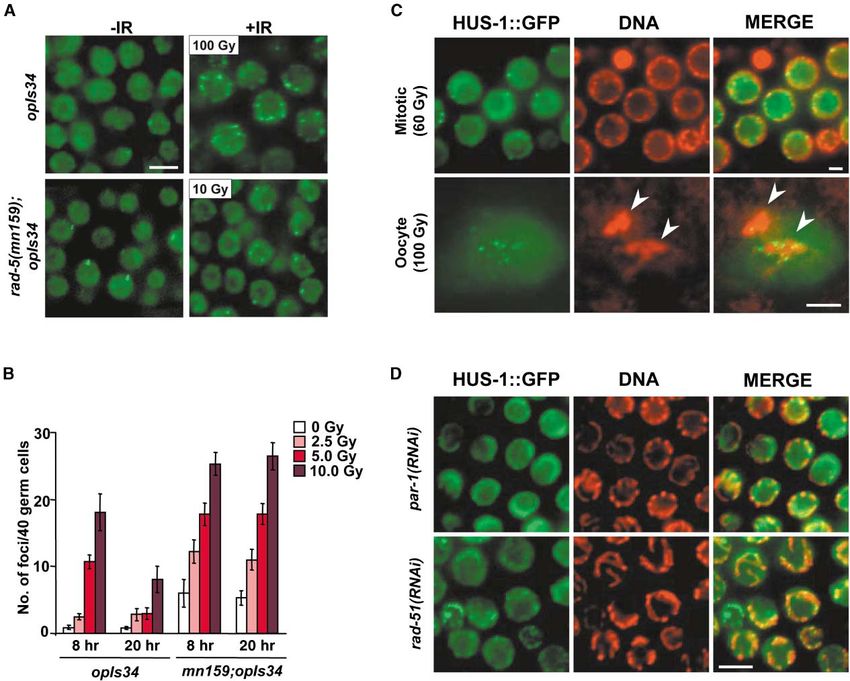
HUS-1 Is a Nuclear Protein that Requires the
op241;unc-119;opIs34 animals compared to 1.3-fold in-
Checkpoint Proteins MRT-2 and HPR-9,
duction in op241;unc-119;opEx566 and 8.6-fold induc-
but Not RAD-5, for Proper Localization
tion in wild-type) (Figure 1B).
S. pombe Hus1p has been shown to be a nuclear protein
[16]. Microscopic analysis of opIs34[hus-1::gfp] animals
DNA Damage Responses Are Defective revealed HUS-1::GFP localization in the nuclei of prolif-
in hus-1 Mutants erating germ cells, meiotic germ cells, mature oocytes,
Ionizing radiation induces several responses in C. ele- and embryos (Figure 3A). We also observed nuclear GFP
gans, including germ cell apoptosis, cell cycle arrest, expression in a subset of somatic cells, particularly pro-
and embryonic lethality [9]. We have previously shown liferating cells, in larvae (data not shown). Rad1, Rad9,
that op241 is defective for DNA damage-induced germ and Hus1 form a heterotrimer complex that structurally
cell death and cell cycle arrest [9]. We found that the resembles the proliferating cell nuclear antigen (PCNA)
op244 allele had more severe defects in both of these trimer [14, 17]. Rad17 is believed to load this complex
responses (Figures 1C, S1A, and S1B; Figure S1 is con- onto DNA at or near sites of DNA damage [18]. To deter-
tained in the Supplementary Material available with this mine the role of these interactions in C. elegans, we
article online). In addition, op244 is more sensitive to analyzed HUS-1::GFP expression in a mrt-2(e2663)
the embryonic lethal effects of ionizing radiation (Figure background. Surprisingly, the expression of HUS-
S1C). 1::GFP in the germline was greatly reduced and was
excluded from the nucleus (Figure 3A). Crossing these
hus-1(op241) Disrupts a Checkpoint worms back to wild-type worms restored proper local-
Protein Interaction ization of HUS-1::GFP to the nucleus (data not shown).
The G99D mutation in op241 affects a residue that, while This reduction of expression is likely due to degradation,
poorly conserved at the primary sequence level, borders possibly as a result of improper localization, rather than
a helix that has been proposed to interact with S. pombe transcriptional regulation, as GFP under the control of
and human Rad9 proteins (Figure 2A) [14]. Although C. the hus-1 promoter (opIs29(pRH04)) was not affected
elegans HUS-1 does not interact with HPR-9, the C. by loss of MRT-2 (data not shown).
elegans homolog of Rad9, on its own in the two-hybrid We also analyzed the expression of HUS-1::GFP in a
system [15], in vivo interaction in worms is likely (see rad-5(mn159) background. RAD-5 is homologous to S.
below). To analyze the molecular nature of the hus- cerevisiae Tel2p and is required for germ cell replication
1(op241) defect, we tested the mutant form of the protein and DNA damage checkpoints [9, 11]. HUS-1::GFP local-
for its ability to interact with four proteins (MRT-2, PDI-2, ization was not different in rad-5(mn159) than in the
K21H4.1, and F56D12.5) that interact with HUS-1 in the parental strain (Figure 3A), and this finding is consistent
yeast two-hybrid system [15]. HUS-1(⫹) interacted with with evidence that rad-5 acts independently of hus-1
these four proteins with varying degrees (Figure 2B). and mrt-2 [11]. Interestingly, HUS-1::GFP levels were
In contrast, HUS-1(G99D) is defective for its ability to significantly lower in rad-5(mn159) mutants compared
interact with the conserved checkpoint protein MRT-2, to the parental strain.
the C. elegans homolog of S. pombe Rad1, and with MRT-2 interacts with HPR-9 in a two-hybrid assay,
PDI-2, a protein disulfide isomerase homolog, in the suggesting that the Rad1/Rad9/Hus1 complex is con-
yeast-two hybrid system (Figure 2B). served in C. elegans [15]. Inhibition of hpr-9 expression
Furthermore, HUS-1(G99D) failed to interact with via RNAi was sufficient to disrupt HUS-1::GFP localiza-
MRT-2 in GST pull-down experiments from transfected tion and expression similarly to a mrt-2 RNAi-positive
cells (Figure 2C). However, HUS-1(⫹) and HUS-1(G99D) control, indicating that HPR-9 is also required for HUS-1HUS-1-Mediated Genome Stability and Apoptosis
1911
1::GFP nuclear localization is independent of the other
HUS-1-interacting proteins that we tested (PDI-2,
K21H4.1, and F56D12.5; data not shown).
HUS-1::GFP Relocalizes to Distinct Foci
that Colocalize with Chromatin
following DNA Damage
In order to determine the subcellular localization of
HUS-1 following DNA damage, we analyzed HUS-1::GFP
localization in germ cells before and after exposure to
ionizing radiation. HUS-1::GFP was diffuse in proliferat-
ing germ nuclei and weakly chromatin localized in pa-
chytene cells under normal conditions. However, follow-
ing exposure to ionizing radiation, HUS-1::GFP concen-
trates at distinct nuclear foci in all stages of germ cell
development (Figures 4A, 4C, and 4D). The presence of
these foci could be observed as early as 3 hr following
exposure to ionizing radiation. These foci overlap with
chromatin, as demonstrated by counterstaining with
DAPI (Figures 4C and 4D).
Genetic evidence of increased DNA damage sensitiv-
ity in hus-1(op241);rad-5(mn159) and rad-5(mn159) mrt-
2(e2663) double mutants suggest that rad-5 functions
in a parallel pathway to hus-1 and mrt-2 [11]. In order
to determine if relocalization of HUS-1::GFP was depen-
dent on rad-5, we irradiated worms containing the hus-
1::gfp transgene in a rad-5(mn159) background. We
found that rather than being blocked, HUS-1::GFP relo-
calization was enhanced in the absence of rad-5 func-
tion. The number of foci were present in greater numbers
in the absence of RAD-5 prior to and after exposure to
ionizing radiation (Figure 4B). Further, we found that,
while HUS-1::GFP foci numbers decreased in rad-5(⫹)
controls after 20 hr, the number of foci did not change
in rad-5 mutants (Figure 4B).
Double-strand breaks also occur under normal condi-
tions during meiotic prophase from initiation events dur-
ing recombination. These double-strand breaks fail to
“heal” in several yeast mutants, resulting in pachytene
cell cycle arrest. RNAi of the RecA-strand exchange
family member, rad-51, similarly blocks recombination
in C. elegans germ cells and results in increased germ
Figure 2. HUS-1(G99D) Is Defective for MRT-2 and PDI-2 Binding cell apoptosis [9, 19]. Under normal conditions, a limited
(A) Using the Blosum62mt2 matrix (Align-X), the alignment of the number of HUS-1::GFP foci can occasionally be ob-
putative helical Rad9 interacting region of Hus1 from human, mouse, served in the pachytene region of the germline (Figure
fly, worm, and fission yeast and Mec3 from budding yeast was 4D). In contrast, rad-51(RNAi) worms showed a dramatic
determined. The affected residue in op241 is indicated by an arrow- increase in HUS-1::GFP foci (Figure 4D).
head. See text for details.
(B) The yeast two-hybrid system was used to test for protein interac-
tions with wild-type (WT) and mutant (G99D) HUS-1-GAL4 DNA bind- The hus-1 Deletion Mutant Exhibits
ing domain (DB) fusions by scoring for LacZ expression and growth Genome Instability
on ⫺Ura plates (no selection: ⫺Leu ⫺Trp). HUS-1(WT) interacts with
Several lines of evidence suggest that loss of hus-1
GAL4 activation (AD) fusions of MRT-2, K12H4.1, F56D12.5, and
PDI-2. HUS-1(G99D) does not interact with MRT-2 and PDI-2, but function leads to genomic instability. First, hus-1 mu-
it still interacts with K12H4.1 and F56D12.5. tants show high levels of chromosomal nondisjunction,
(C) In vitro interaction of HUS-1 and MRT-2. GST-HUS-1(WT), but as evidenced by the high proportion of males (the result
not GST-HUS-1(G99D) or GST alone, interacts with Myc-epitope- of X chromosome nondisjunction) in the self-progeny of
tagged MRT-2 (lanes 1–3). GST-MRT-2 is able to interact with wild- op244 mutants. Whereas nondisjunction of the X chro-
type (WT) but not mutant Myc-epitope-tagged HUS-1(G99D) (lanes
mosomes produces less than 0.2% male progeny in the
4 and 5).
wild-type [20], early op244 generations produce about
6% males (Table 1). Second, as has been observed
localization and stabilization (Figure 3B). In contrast, for other mutants defective in genome stability, hus-1
hpr-17(RNAi)- and ced-3(RNAi)-treated worms showed mutants show abnormal levels of embryonic lethality
normal HUS-1::GFP localization (Figure 3B). HUS- [21]: early generations of op244 mutants produce aboutCurrent Biology
1912
Figure 3. HUS-1 Nuclear Localization Re-
quires MRT-2 and HPR-9
(A) Fluorescent microscopy of integrated
transgenic strains expressing fusion con-
structs in gonads and embryos of wild-type,
mrt-2(e2663), and rad-5(mn159) mutant
backgrounds. The genotype and construct
used are indicated, and expression is shown
in the indicated stage of oogenesis and early
embryos. pRH21 is a full-length translational
fusion containing 2252 bp of genomic se-
quence 5⬘ to the stop codon.
(B) Fluorescent microscopy of 4-cell-stage
embryos from F2-integrated transgenic
worms fed dsRNA produced from indicated
genes as described in the Experimental Pro-
cedures. For confirmation of ced-3(RNAi) and
hpr-17(RNAi), germ cell death and hypersen-
sitivity to radiation-induced embryonic lethal-
ity, respectively, were scored. The scale bars
represent 10 m.
78% healthy progeny that survive to adulthood (the level worms, six chromosomal bivalents can be observed by
of larval lethality was minimal) (Table 1). Embryonic sur- DAPI staining of oocytes in diakinesis. In contrast, late
vival is even lower in later generations. Embryonic sur- generations of op244 mutants often contained fewer
vival of op241 mutants was not significantly different than six bivalents. In op244 generations no longer pro-
from wild-type worms. Nondisjunction of autosomes ducing viable progeny, only 3–4 bivalents could be seen
could well explain most or all of the embryonic lethality (Figure 5B); consistent with the idea that loss of telo-
seen in op244 mutants, although other defects, such as meres led to chromosome fusions. Correspondingly, the
failure to repair endogenous damage, cannot be ex- incidence of males increases dramatically after several
cluded. Indeed, spontaneous mutations are more fre- generations and gives rise eventually to dominant Him
quent, as measured by an unc-93 reversion assay (see (high incidence of males) strains (data not shown).
below). One function of checkpoint genes is to prevent cells
Mutations in the checkpoint gene mrt-2 result not only with a damaged genome from progressing through the
in a failure to induce apoptosis in response to DNA cell cycle without correcting the DNA lesion. Mutations
damage [9], but also in progressive telomere loss, even- in genes that repair these lesions or regulate the check-
tually leading to end-to-end chromosome fusion and points that prevent cell cycle progression have been
aneuploidy [22]. As a consequence of these defects, the shown to display higher spontaneous mutation frequen-
brood sizes of mrt-2 mutants decrease over generations cies [23, 24]. Since hus-1 has a checkpoint function, we
until the animals become sterile – the Mrt (mortal germ- used the well-characterized unc-93 reversion assay to
line) phenotype from which the gene derived its name. look at spontaneous mutation frequencies in hus-1
While early generations of freshly outcrossed op244 mu- worms [25]. We found the spontaneous suppression
tants have brood sizes similar to wild-type worms, later frequency of unc-93(e1500) worms in our assay to be 1 ⫻
generations showed dramatically lower fertility (Table 1) 10⫺6 (Figure 5C). In two independent hus-1(op244);unc-
and became sterile after about 15 generations (produc- 93(e1500) strains, we found the mutation frequency to
ing very few embryos, none of which hatch). We did not be 10- to 20-fold higher than in the control strain (Figure
see a Mrt phenotype in op241 mutants kept at 20⬚C 5C). Consistent with this, the appearance of spontane-
(Table 1). However, when grown at 25⬚C, op241 mutants ous mutations has also been observed during strain
also became sterile after approximately 15 generations, maintenance (data not shown).
suggesting that op241 might be a temperature-sensitive
allele. Ionizing Radiation Induces egl-1 Transcript
To test whether the Mrt phenotype of hus-1 mutants Upregulation in a hus-1- and
was due to telomere loss, we probed Southern blots of cep-1-Dependent Manner
genomic DNA from multiple generations of op244 and In order to determine whether hus-1 is transcriptionally
op241 strains. In op244 mutants grown at 20⬚C, we saw regulated following DNA damage, we isolated mRNA
a progressive loss of telomere sequence in late genera- from adult wild-type and hus-1(op244)-irradiated worms
tions of worms (Figure 5A). As reported before, the ends and probed Northern blots with hus-1 cDNA. A single
of telomeres do not decrease in op241 mutants grown transcript of the expected size was seen in wild-type;
at 20⬚C [11]; however, worms grown at 25⬚C did display however, the transcript was truncated and levels were
telomere shortening (data not shown). In wild-type dramatically reduced in the hus-1(op244) deletion mu-HUS-1-Mediated Genome Stability and Apoptosis 1913 Figure 4. Subcellular Relocalization of HUS-1::GFP following DNA Damage (A) Fluorescent microscopy of proliferating germ cells expressing HUS-1::GFP (opIs34). Irradiated worms (opIs34 ⫽ 100 Gy; rad- 5(mn159);opIs34 ⫽ 10 Gy) were viewed 8 hr following irradiation. HUS-1::GFP is diffuse in controls. Relocalized HUS-1::GFP is seen as bright foci. The scale bar represents 5 m. (B) Quantification of HUS-1::GFP foci in wild-type and rad-5(mn159) backgrounds. Foci were scored in 40 proliferating germ cells in a single Z stack following mild doses of X-rays at indicated times. Each bar represents ten worms ⫾ SEM. (C) Colocalization of HUS-1::GFP with chromatin following exposure to ionizing radiation. The top panel shows proliferating germ cells. The bottom panel shows a single oocyte nucleus in diakinesis. Arrowheads point to two DAPI-stained bivalents from an oocyte in diakinesis. The scale bar represents 2 m. (D) Fluorescent microscopy of meiotic germ cells from worms fed dsRNA from rad-51 and a par-1 control. As in (C), HUS-1::GFP foci overlap with chromatin stained with Hoechst 33342 dye. The scale bar represents 5 m. tant. Levels of hus-1 mRNA did not change significantly construct following exposure to ionizing radiation (Fig- 60 and 180 min after exposure to IR (Figure 6A). ure 6C). Importantly, egl-1 levels did not increase in the In C. elegans, the BH3 domain protein, EGL-1, acti- hus-1(op244) deletion worms after irradiation (Figures vates the apoptotic machinery. Furthermore, egl-1 is 6A and 6B). These results are consistent with the hypoth- transcriptionally regulated in order to activate apoptosis esis that hus-1-dependent induction of egl-1 transcrip- during somatic development [26]. We subsequently hy- tion is an important element of the apoptotic DNA dam- bridized the same blot to an egl-1 probe. We found that, age response in C. elegans and that it promotes the in wild-type young adults, egl-1 is induced by 60 min increased germ cell apoptosis observed following geno- and increases by 180 min after irradiation (Figures 6A toxic stress. and 6B). Induction is significantly lower in glp-4(bn2) (a Recent work showed that the cep-1 gene, which en- temperature-sensitive mutant that lacks a germline at codes a C. elegans homolog of p53, is required for DNA the restrictive temperature) animals, suggesting that damage-induced germ cell apoptosis [12, 13]. In order egl-1 induction is largely restricted to the germline (Fig- to determine if cep-1 is also required for egl-1 induction, ure 6B). Consistent with this hypothesis, we found germ- we analyzed egl-1 expression by using real-time quanti- line induction of an egl-1::gfp transcriptional reporter tative RT-PCR (Q-RT-PCR) before and after exposure to
Current Biology
1914
Figure 5. hus-1 Deletion Mutants Show a Telomere Maintenance Defect and a High Rate of Spontaneous Mutations
(A) Southern blots were performed with a C. elegans telomere-specific probe on genomic DNA from wild-type (WT) and indicated inbred
generations of op244 after outcrossing.
(B) Oocytes in diakinesis were stained with DAPI from wild-type and late-generation hus-1(op244) mutants that no longer produced viable
progeny. Wild-type oocytes contain six easily visible bivalents, while late-generation hus-1(op244) mutants often have only 3–5 visible bivalents.
The scale bar represents 2 m.
(C) Spontaneous unc-93(e1500) reversion frequencies (log) in hus-1(⫹) and two independently generated hus(op244) strains (F1.1 and F1.2).
The unc-93(e1500) gain-of-function mutation results in severe paralysis that can be suppressed by loss of function of any one of five different
genes; this loss of function includes inactivating second site mutations within the unc-93 gene itself [47].
ionizing radiation in two different cep-1 mutant strains. ing radiation. In C. elegans, HUS-1 also mediates an
Using this technique, we confirmed that egl-1 transcripts apoptotic response to DNA damage, a pathway that is
are strongly induced in wild-type worms (Figure 6B). absent in yeast. Consistent with the observed mutant
Induction of egl-1 transcripts was reduced but not abol- phenotype, hus-1 expression is predominantly in the
ished in cep-1(w40) (Figure 6B), consistent with the ob- germline. However, we do observe expression in a sub-
servation that cep-1(w40) worms have some increased set of proliferating somatic cells. HUS-1 may have a role
germ cell apoptosis following DNA damage, albeit to a in repair and checkpoints in these cells as well. While
much lesser extent than wild-type ([12] and unpublished we have not performed a detailed mosaic analysis, the
data). In contrast, animals homozygous for the deletion fact that transgenes with no germline expression (but
allele cep-1(gk138), which completely blocks DNA dam- with high somatic cell expression) failed to rescue hus-
age-induced apoptosis (W.B. Derry and J.H. Rothman, 1(op241) suggests that HUS-1 acts cell-autonomously
personal communication), showed a total absence of to control mitotic checkpoints.
egl-1 induction (Figure 6B). This suggests that hus-1 Following DNA damage, HUS-1 is relocalized in the
and cep-1 likely act in the same pathway to mediate nucleus to distinct foci. These foci are likely sites of
DNA damage-induced apoptosis. double-strand breaks (DSBs), as RNAi suppression of
In order to learn more about the role of hus-1 in cell rad-51, a gene required for DSB repair during meiotic
cycle regulation, we determined the expression levels recombination, or elimination of the checkpoint gene
of two cyclin kinase inhibitor homologs, cki-1 and cki-2 rad-5 also resulted in increased HUS-1 relocalization.
[27, 28]. In mammalian cells, the cyclin kinase inhibitor The C. elegans checkpoint gene rad-5 was not required
p21 is induced by p53 expression following DNA dam- for HUS-1 relocalization. Rather, rad-5(mn159) mutants
age [29]. However, neither cki-1 nor cki-2 was signifi- showed an increased number of HUS-1::GFP foci in pro-
cantly induced transcriptionally by irradiation at 60 or liferating germ cells under normal conditions. Following
180 min (Figure 6A). mild insults, HUS-1::GFP foci also failed to decrease in
rad-5 mutants compared to controls. These data sug-
Discussion gest that RAD-5 is required for efficient repair of endoge-
nous and exogenous DNA damage either downstream
We have shown that HUS-1 is required for a DNA dam- or independent of HUS-1.
age checkpoint in C. elegans. As in yeast, loss of hus-1 Interestingly, the two hus-1 alleles, op241 and op244,
function abrogates DNA damage-induced cell cycle ar- have similar checkpoint defects, yet the deletion of
rest and sensitizes animals to the lethal effects of ioniz- hus-1 reveals additional functions of HUS-1 in the main-HUS-1-Mediated Genome Stability and Apoptosis
1915
tenance of genome stability. Deletion mutants show a
high frequency of meiotic nondisjunction and/or chro-
mosome loss, resulting in increased male progeny, due
to loss of the X chromosome, and high levels of embry-
onic lethality. Chromosome abnormalities have also
been reported in mouse Hus1⫺/⫺ cells [30]. One possi-
ble cause for the increase in genome instability is the
inability to recognize or repair various forms of DNA
damage. Indeed, we found that hus-1 mutants have a
mutator phenotype.
Our results also demonstrate that, unlike in yeast,
HUS-1 is required for the prevention of telomere short-
ening during replication of the genome [31]. hus-1 dele-
tion worms show a progressive shortening of telomeres,
which is associated with a progressive reduction in
brood size, reaching complete sterility by the 15th gener-
ation. In late-generation worms, outcrossing reveals a
dominant Him phenotype that is consistent with end-
to-end fusions of the X chromosome with autosomes,
due to loss of telomeric ends [22]. Fusions are also
apparent in oocytes of late-generation worms in which
only three or four bivalents, rather than the typical six,
can be detected in diakinesis. A similar mortal germline
phenotype and chromosome fusions associated with
telomere loss have previously been reported for a mu-
tant in the HUS-1-interacting protein MRT-2, and this
finding suggests that the same MRT-2/HUS-1 complex
that acts in checkpoint control might also control telo-
mere length. It is not known what role HUS-1 has in
telomere maintenance in vertebrates.
Studies of DNA damage responses in C. elegans have
revealed intriguing differences from responses in mam-
mals. DNA damage induces both a G1/S and G2/M arrest
in mammalian cells. These cell cycle arrest checkpoints
are mediated, in part, by the cyclin kinase inhibitor p21
in a p53-dependent manner [29]. However, we did not
see an increase of mRNA levels of two candidate cyclin
kinase inhibitors, cki-1 and cki-2, at the times examined.
It remains possible that one or both of these CIP homo-
logs is induced at later time points or is posttranscrip-
tionally regulated. Unlike in mammals, but similarly to
Drosophila [32], loss of CEP-1 (p53) function in C. ele-
gans does not result in a cell cycle arrest defect in
response to DNA damage [12, 13], suggesting that in-
duction of apoptosis might have been the original func-
tion of p53 family members during evolution. Mutants
of the homologs of yeast Cdc2 and Cdc25 have been
shown to disrupt normal cell cycle in the germline of C.
elegans; thus, as in yeast, these are possible candidates
for downstream effectors of HUS-1-mediated check-
Figure 6. egl-1 Is Transcriptionally Induced by Irradiation in a hus-1-
and cep-1-Dependent Manner
points [33, 34].
In mammalian cells, DNA damage induces the tran-
(A) Northern blots were performed with 2 g polyA-enriched RNA
isolated from wild-type and hus-1(op244) mutant young adult worms
at indicated times after exposure to 120 Gy ␥-irradiation. The same
blot was reprobed with a PCR-generated probe from the indicated
gene on the left. site of egl-1 fused to GFP equipped with two nuclear localization
(B) Average fold induction of egl-1 gene expression in wild-type, signals. Worms were synchronized and irradiated as L4 animals.
hus-1(op244), cep-1(w40), cep-1(gk138), and glp-4(bn2) mutants GFP expression was analyzed in dissected gonads 30 hr following
after 100 Gy X-ray irradiation as determined by real-time quantitative irradiation. Expression was seen in both proliferating and late-pa-
RT-PCR. glp-4(bn2) worms were grown at the nonpermissive tem- chytene germ cells in most (12/16) irradiated animals. Background
perature, and 90–100 worms were selected that lacked a germline. expression could be seen in the late-pachytene germ cells in few
Each bar represents the average ⫾ SD. (2/23) nonirradiated controls, but not (0/23) in the proliferating germ
(C) Induction of egl-1::gfp expression in the germline. A construct cells. The arrowhead indicates the distal end of the gonad. The
containing 2.7 kb of genomic sequence 5⬘ to the translation start scale bar represents 10 m.Current Biology
1916
ated apoptosis. In contrast, p53 status does not influ-
ence apoptosis and embryonic lethality in Hus1⫺/⫺
mice, as the Hus1⫺/⫺p53⫺/⫺ embryonic lethality is
morphologically indistinguishable from that of Hus1⫺/⫺
embryos [40]. Furthermore, induction of p53 target
genes, such as Bax, is normal in Hus1⫺/⫺ mice. How-
ever, in C. elegans, induction of egl-1 requires hus-1
and the p53 homolog, cep-1. Thus, unlike in mouse
embryos [40], hus-1 and cep-1 likely act in a common
pathway to activate the apoptotic machinery (Figure 7).
In contrast to hus-1(op244) mutants, which are highly
sensitive to IR-induced double-strand breaks (DSBs),
mouse Hus1⫺/⫺ cells are highly sensitive to hydroxy-
urea (HU) and ultraviolet (UV) radiation, but not to ioniz-
ing radiation (IR) [30]. One explanation may be that Hus1
in mice is required primarily for the replication check-
point (UV and HU), but not the DSB checkpoint (IR)
during embryogenesis. Indeed, lack of Hus1 or low lev-
els of IR fail to induce cell cycle arrest in mouse embryos;
however, apoptosis is induced [4, 30]. However, HUS1,
along with Atm and p53, may also play a primary role
in responding to DSBs after cells become more differen-
Figure 7. Model: A Pathway for DNA Damage-Induced Apoptosis tiated [41]. In several knockouts (Hus1⫺/⫺, Atm⫺/⫺,
and Cell Cycle Arrest in C. elegans
Brca⫺/⫺), apoptotic cells have been shown to have
DNA damage recruitment of the HUS-1/MRT-2/HPR-9 complex to
constitutively higher levels of Bax. Here we demonstrate
the site of the lesion, where it activates cell cycle arrest of proliferat-
ing stem cells. This complex also signals to the apoptotic machinery that, in C. elegans, loss of DNA damage sensors up-
via CEP-1-dependent upregulation of egl-1. stream of p53 results in loss of activation of the apo-
ptotic machinery and results in radioresistant cells. As
with the loss of p53, this may be a crucial step toward
scription of many genes, including the proapoptotic BH3 transformation into malignant tumors [42].
domain-containing proteins Bax, Puma, and Noxa [35– While the deletion mutant hus-1(op244) shows evi-
38]. Transcriptional activation of Bax is dependent on dence of embryonic defects due to genomic instability
p53 in a tissue-dependent manner but is independent and telomere loss, hus-1(op241) only appears to be de-
of Atm [39]. However, it is not known whether the genes fective in the DNA damage checkpoint response under
that sense damaged DNA, such as the Rad family pro- normal laboratory growth conditions. The fact that muta-
teins, are required for the induction of Bax, Puma, or tions can disrupt checkpoint function without impairing
Noxa. We demonstrate in C. elegans a dramatic increase essential genome maintenance functions during em-
in mRNA levels for the proapoptotic protein EGL-1 fol- bryogenesis (chromosome segregation) and oogenesis
lowing irradiation. We conclude that this induction is (telomere maintenance) in C. elegans might explain how
mostly in the germline, as glp-4 mutants lack strong checkpoint genes essential for viability in mammals can
egl-1 induction and a GFP reporter displayed high germ- also act as tumor suppressors. For example, even
line expression following irradiation. Induction of egl-1 though Chk1⫹/⫺ (in a Wnt-1 oncogenic background)
by ␥-irradiation is dependent on hus-1. This provides a and Atr⫹/⫺ can promote tumorigenesis, loss of hetero-
molecular explanation for how DNA damage induces zygosity is not seen in these tumors, because loss of
apoptosis in germ cells of C. elegans and suggests a either Atr or Chk1 is cell lethal [43, 44]. Thus, while
key link between a DNA damage checkpoint gene and complete loss of DNA damage checkpoint genes can
the apoptotic machinery (Figure 7). However, as in mam- induce mitotic catastrophe, more subtle mutations may
mals, DNA damage likely activates other proapoptotic contribute to oncogenic transformation by preventing
genes since egl-1(lf) mutants are only partially defective cell cycle arrest and apoptosis.
in this response.
In mice, all Hus1⫺/⫺ embryos die by 11.5 days post-
coitum (dpc) [30], and this death is at least in part due Conclusions
to a high level of chromosome abnormalities, which re- C. elegans HUS-1 is a conserved checkpoint protein
sult in increased apoptosis during embryogenesis. Simi- that is required for DNA damage-induced cell cycle ar-
larly, we showed that, while worm HUS-1 is not abso- rest and apoptosis. Following DNA damage, HUS-1 relo-
lutely required for embryonic survival, a significant calizes to putative sites of DSBs independently of the
fraction of hus-1 embryos die during embryogenesis, novel checkpoint protein RAD-5. HUS-1 is also required
likely due to genomic instability. However, we failed to for genome stability and telomere maintenance. We also
detect any increase in somatic developmental cell death demonstrate that HUS-1 checkpoint, but not essential
in hus-1 mutants (data not shown), and these findings genome maintenance, functions are dispensable for em-
are consistent with our previous report that DNA dam- bryogenesis, suggesting a possible requirement for
age does not induce somatic cell apoptosis [9]. HUS-1 in tumor suppression. DNA damage upregulates
In C. elegans, p53 function is essential for hus-1-medi- EGL-1 in a HUS-1- and CEP-1-dependent manner, pro-HUS-1-Mediated Genome Stability and Apoptosis
1917
viding a molecular link between DNA damage sensors compared to the untreated control (calibrator), normalized based
and p53-mediated activation of BH3 proteins in C. on 18S rRNA levels.
elegans.
Supplementary Material
Experimental Procedures Supplementary Material including comparative data of cell cycle
arrest and radiation sensitivities of both hus-1 alleles (Figure S1)
Isolation of op244 as well as additional methodological detail is available at http://
A deletion library was constructed by using trimethylpsoralen and images.cellpress.com/supmat/supmatin.htm.
UV mutagenesis as previously described [45]. This library was
screened for deletion mutants of H26D21.1 via nested PCR by using Acknowledgments
the following primer sequences: first round 5⬘-ATGGTCCTGCAGG
GAAATAG-3⬘ and 5⬘-ATCCGTTAACAGTGAGATACTC-3⬘ and nested We thank B. Derry and J. Rothman for the cep-1(w40) mutant and
5⬘-AGGCACATACAATAATACGGTG-3⬘ and 5⬘-ATCACGATCATGT the C. elegans Knockout Consortium for cep-1(gk138); V. Pratis
GAAGCCG-3⬘. Pooled genomic DNA from 96-well plates was and J. Austin for the unc-119 genomic rescuing plasmid and the
screened, and positive plates were screened again by rows and bombardment protocol; A. Fire for pPD117.01, pPD135.83, and
columns to get a single well address. Thawed worms from a positive pPD129.36; Y. Kohara for cDNA clone yk238e9; A. Coulson for the
address were screened by single worm PCR to confirm the presence cTel55x plasmid; and A. Brincat for help with bombardment. We
of the mutation. Homozygous worms were used to prepare genomic thank A. Hajnal and members of the Hengartner Lab for comments.
DNA for sequence analysis of the mutated H26D21.1 gene. op244 Some C. elegans strains were obtained from the Caenorhabditis
mutants were backcrossed to the wild-type eight times before phe- Genetics Center, which is funded by the National Institutes of Health
notypical analysis. (NIH) National Center for Research Resources (NCRR). This work
was supported by grants from the NIH (GM52540 to M.O.H., F32
Spontaneous Mutation Frequencies of op244 GM20801 to E.R.H., and 7 R33 CA81658-02 to S.J.B. and M.V.) and
Mutants of each genotype were singled (unc-93, n ⫽ 100; hus-1;unc- the Ernst Hadorn Foundation (to M.O.H.).
93 F1.1, n ⫽ 88; hus-1;unc-93 F1.2, n ⫽ 83) and monitored for three
generations until plates were starved. Starved animals were then Received: May 12, 2002
transferred to new plates, and the number of plates with revertants Revised: September 18, 2002
were scored as independent reversion events (unc-93, 6/100; hus- Accepted: September 23, 2002
1;unc-93 F1.1, 47/88; hus-1;unc-93 F1.2, 27/83). These events were Published: November 19, 2002
then divided by the total number of haploid genomes screened,
based on the average brood size of each genotype, to determine
References
the reversion frequency.
1. Hoeijmakers, J.H. (2001). Genome maintenance mechanisms
Transgenic Worms for preventing cancer. Nature 411, 366–374.
HUS-1::GFP lines were constructed with pRH04 and pRH21. pRH04 2. Rich, T., Allen, R.L., and Wyllie, A.H. (2000). Defying death after
is a transcriptional fusion containing 1144 bp of genomic sequence DNA damage. Nature 407, 777–783.
upstream of the start codon. pRH21 is a translational fusion con- 3. Bach, S.P., Renehan, A.G., and Potten, C.S. (2000). Stem cells:
struct containing 2252 bp including upstream regulatory sequence the intestinal stem cell as a paradigm. Carcinogenesis 21,
and all coding sequence of H26D21.1 fused in-frame to the C-ter- 469–476.
minal GFP tag. pRH21 contains GFP and let-858 sequences from 4. Heyer, B.S., MacAuley, A., Behrendtsen, O., and Werb, Z. (2000).
pPD117.01 (gift of A. Fire) and rescuing unc-119 genomic sequence Hypersensitivity to DNA damage leads to increased apoptosis
from pDPMM016 [46]. pRH25 was made by subcloning a 2.7-kb during early mouse development. Genes Dev. 14, 2072–2084.
PCR fragment of the sequence 5⬘ to the egl-1 start codon into 5. Murakami, H., and Nurse, P. (2000). DNA replication and damage
pRH20, which contains a Kpn-1 and ApaI (2xNLS::GFP::let- checkpoints and meiotic cell cycle controls in the fission and
8583⬘UTR) fragment from pPD135.83 (gift of A. Fire) and the unc- budding yeasts. Biochem. J. 349, 1–12.
119(⫹) rescuing sequence. Constructs were bombarded into unc- 6. Bentley, N.J., Holtzman, D.A., Flaggs, G., Keegan, K.S., DeMag-
119(ed3) worms as previously described [46]. Integration of each gio, A., Ford, J.C., Hoekstra, M., and Carr, A.M. (1996). The
construct was determined by loss of visible Unc-119 offspring. At Schizosaccharomyces pombe rad3 checkpoint gene. EMBO J.
least three integrated lines were generated for each construct. Go- 15, 6641–6651.
nads were dissected and were stained with Hoechst 33342 (0.5 7. Horvitz, H.R. (1999). Genetic control of programmed cell death
g/ml) or were fixed with 4% paraformaldehyde followed by cold in the nematode Caenorhabditis elegans. Cancer Res. 59,
methanol and stained with DAPI (0.2 g/ml). Images were captured 1701s–1706s.
with an ORCA-ER digital CCD camera and were processed with 8. Gumienny, T.L., Lambie, E., Hartwieg, E., Horvitz, H.R., and
OpenLab software. Hengartner, M.O. (1999). Genetic control of programmed cell
death in the Caenorhabditis elegans hermaphrodite germline.
Quantitative RT-PCR Development 126, 1011–1022.
Synchronized wild-type, cep-1(w40), cep-1(gk138), glp-4(bn2), and 9. Gartner, A., Milstein, S., Ahmed, S., Hodgkin, J., and Hengartner,
hus-1(op244) adult hermaphrodites were irradiated with 100 Gy of M.O. (2000). A conserved checkpoint pathway mediates DNA
X-rays and were left for 3 hr to recover. glp-4(bn2) worms were damage-induced apoptosis and cell cycle arrest in C. elegans.
grown at the nonpermissive temperature, and 90–100 worms were Mol. Cell 5, 435–443.
selected that lacked a germline. Total RNA was isolated with RNAzol 10. Hofmann, E.R., Milstein, S., and Hengartner, M.O. (2000). DNA
B (ams Biotechnology) according to the manufacturer’s protocol, damage-induced checkpoint pathways in the nematode Caeno-
treated with DnaseI, and further purified with the RNeasy kit (Qia- rhabditis elegans. Cold Spring Harb. Symp. Quant. Biol. 65,
gen). For cDNA synthesis, purified total RNA was reverse transcribed 467–473.
with 250 U MultiScribe Reverse Transcriptase (Applied Biosystems) 11. Ahmed, S., Alpi, A., Hengartner, M.O., and Gartner, A. (2001). C.
by using random primers. Relative amounts of egl-1 cDNA were elegans RAD-5/CLK-2 defines a new DNA damage checkpoint
subsequently estimated by real-time quantitative PCR in an ABI protein. Curr. Biol. 11, 1934–1944.
Prism 7700 Sequence detector system, by using the primers 5⬘- 12. Derry, W.B., Putzke, A.P., and Rothman, J.H. (2001). Caenorhab-
CAGGACTTCTCCTCGTGTGAAGATTC-3⬘ and 5⬘-GAAGTCATCG ditis elegans p53: role in apoptosis, meiosis, and stress resis-
CACATTGCTGCTA-3⬘, which span the single egl-1 intron. 18S rRNA tance. Science 294, 591–595.
was used for the internal standard. Average fold induction repre- 13. Schumacher, B., Hofmann, K., Boulton, S., and Gartner, A.
sents the relative expression of egl-1 following irradiation (sample) (2001). The C. elegans homolog of the p53 tumor suppressorCurrent Biology
1918
is required for DNA damage-induced apoptosis. Curr. Biol. 11, Caenorhabditis elegans gene ncc-1 encodes a cdc2-related
1722–1727. kinase required for M phase in meiotic and mitotic cell divisions,
14. Venclovas, C., and Thelen, M.P. (2000). Structure-based predic- but not for S phase. Development 126, 2227–2239.
tions of Rad1, Rad9, Hus1 and Rad17 participation in sliding 34. Ashcroft, N., and Golden, A. (2002). CDC-25.1 regulates germ-
clamp and clamp-loading complexes. Nucleic Acids Res. 28, line proliferation in Caenorhabditis elegans. Genesis 33, 1–7.
2481–2493. 35. Miyashita, T., and Reed, J.C. (1995). Tumor suppressor p53 is
15. Boulton, S.J., Gartner, A., Reboul, J., Vaglio, P., Dyson, N., Hill, a direct transcriptional activator of the human Bax gene. Cell
D.E., and Vidal, M. (2002). Combined functional genomic maps 80, 293–299.
of the C. elegans DNA damage response. Science 295, 127–131. 36. Oda, E., Ohki, R., Murasawa, H., Nemoto, J., Shibue, T., Yama-
16. Caspari, T., Dahlen, M., Kanter-Smoler, G., Lindsay, H.D., Hof- shita, T., Tokino, T., Taniguchi, T., and Tanaka, N. (2000). Noxa,
mann, K., Papadimitriou, K., Sunnerhagen, P., and Carr, A.M. a BH3-only member of the Bcl-2 family and candidate mediator
(2000). Characterization of Schizosaccharomyces pombe Hus1: of p53-induced apoptosis. Science 288, 1053–1058.
a PCNA-related protein that associates with Rad1 and Rad9. 37. Nakano, K., and Vousden, K.H. (2001). PUMA, a novel proapo-
Mol. Cell. Biol. 20, 1254–1262. ptotic gene, is induced by p53. Mol. Cell 7, 683–694.
17. Kaur, R., Kostrub, C.F., and Enoch, T. (2001). Structure-function 38. Yu, J., Zhang, L., Hwang, P.M., Kinzler, K.W., and Vogelstein, B.
analysis of fission yeast Hus1-Rad1-Rad9 checkpoint complex. (2001). PUMA induces the rapid apoptosis of colorectal cancer
Mol. Biol. Cell 12, 3744–3758. cells. Mol. Cell 7, 673–682.
18. Zou, L., Cortez, D., and Elledge, S.J. (2002). Regulation of ATR 39. Barlow, C., Brown, K.D., Deng, C.X., Tagle, D.A., and Wynshaw-
substrate selection by Rad17-dependent loading of Rad9 com- Boris, A. (1997). Atm selectively regulates distinct p53-depen-
plexes onto chromatin. Genes Dev. 16, 198–208. dent cell-cycle checkpoint and apoptotic pathways. Nat. Genet.
19. Takanami, T., Mori, A., Takahashi, H., and Higashitani, A. (2000). 17, 453–456.
Hyper-resistance of meiotic cells to radiation due to a strong 40. Weiss, R.S., Matsuoka, S., Elledge, S.J., and Leder, P. (2002).
expression of a single recA-like gene in Caenorhabditis elegans. Hus1 acts upstream of Chk1 in a mammalian DNA damage
Nucleic Acids Res. 28, 4232–4236. response pathway. Curr. Biol. 12, 73–77.
20. Hodgkin, J., Horvitz, H.R., and Brenner, S. (1979). Nondisjunc- 41. Herzog, K.H., Chong, M.J., Kapsetaki, M., Morgan, J.I., and
tion mutants of the nematode Caenorhabditis elegans. Genetics McKinnon, P.J. (1998). Requirement for Atm in ionizing radia-
91, 67–94. tion-induced cell death in the developing central nervous sys-
21. Chin, G.M., and Villeneuve, A.M. (2001). C. elegans mre-11 is tem. Science 280, 1089–1091.
required for meiotic recombination and DNA repair but is dis- 42. Symonds, H., Krall, L., Remington, L., Saenz-Robles, M., Lowe,
pensable for the meiotic G(2) DNA damage checkpoint. Genes S., Jacks, T., and Van Dyke, T. (1994). p53-dependent apoptosis
Dev. 15, 522–534. suppresses tumor growth and progression in vivo. Cell 78,
22. Ahmed, S., and Hodgkin, J. (2000). MRT-2 checkpoint protein 703–711.
is required for germline immortality and telomere replication in 43. Liu, Q., Guntuku, S., Cui, X.S., Matsuoka, S., Cortez, D., Tamai,
C. elegans. Nature 403, 159–164. K., Luo, G., Carattini-Rivera, S., DeMayo, F., Bradley, A., et al.
23. Myung, K., Datta, A., and Kolodner, R.D. (2001). Suppression (2000). Chk1 is an essential kinase that is regulated by Atr and
of spontaneous chromosomal rearrangements by S phase required for the G(2)/M DNA damage checkpoint. Genes Dev.
checkpoint functions in Saccharomyces cerevisiae. Cell 104, 14, 1448–1459.
397–408. 44. Brown, E.J., and Baltimore, D. (2000). ATR disruption leads to
24. Myung, K., Chen, C., and Kolodner, R.D. (2001). Multiple path- chromosomal fragmentation and early embryonic lethality.
ways cooperate in the suppression of genome instability in Sac- Genes Dev. 14, 397–402.
charomyces cerevisiae. Nature 411, 1073–1076. 45. Jansen, G., Hazendonk, E., Thijssen, K.L., and Plasterk, R.H.
25. De Stasio, E., Lephoto, C., Azuma, L., Holst, C., Stanislaus, (1997). Reverse genetics by chemical mutagenesis in Caeno-
D., and Uttam, J. (1997). Characterization of revertants of unc- rhabditis elegans. Nat. Genet. 17, 119–121.
93(e1500) in Caenorhabditis elegans induced by N-ethyl- 46. Praitis, V., Casey, E., Collar, D., and Austin, J. (2001). Creation of
N-nitrosourea. Genetics 147, 597–608. low-copy integrated transgenic lines in Caenorhabditis elegans.
26. Conradt, B., and Horvitz, H.R. (1999). The TRA-1A sex determi- Genetics 157, 1217–1226.
nation protein of C. elegans regulates sexually dimorphic cell 47. Greenwald, I.S., and Horvitz, H.R. (1980). unc-93(e1500): a be-
deaths by repressing the egl-1 cell death activator gene. Cell havioral mutant of Caenorhabditis elegans that defines a gene
98, 317–327. with a wild-type null phenotype. Genetics 96, 147–164.
27. Feng, H., Zhong, W., Punkosdy, G., Gu, S., Zhou, L., Seabolt,
E.K., and Kipreos, E.T. (1999). CUL-2 is required for the G1-to-
S-phase transition and mitotic chromosome condensation in
Caenorhabditis elegans. Nat. Cell Biol. 1, 486–492.
28. Hong, Y., Roy, R., and Ambros, V. (1998). Developmental regula-
tion of a cyclin-dependent kinase inhibitor controls postembry-
onic cell cycle progression in Caenorhabditis elegans. Develop-
ment 125, 3585–3597.
29. Bunz, F., Dutriaux, A., Lengauer, C., Waldman, T., Zhou, S.,
Brown, J.P., Sedivy, J.M., Kinzler, K.W., and Vogelstein, B.
(1998). Requirement for p53 and p21 to sustain G2 arrest after
DNA damage. Science 282, 1497–1501.
30. Weiss, R.S., Enoch, T., and Leder, P. (2000). Inactivation of
mouse Hus1 results in genomic instability and impaired re-
sponses to genotoxic stress. Genes Dev. 14, 1886–1898.
31. Dahlen, M., Olsson, T., Kanter-Smoler, G., Ramne, A., and Sun-
nerhagen, P. (1998). Regulation of telomere length by check-
point genes in Schizosaccharomyces pombe. Mol. Biol. Cell 9,
611–621.
32. Ollmann, M., Young, L.M., Di Como, C.J., Karim, F., Belvin,
M., Robertson, S., Whittaker, K., Demsky, M., Fisher, W.W.,
Buchman, A., et al. (2000). Drosophila p53 is a structural and
functional homolog of the tumor suppressor p53. Cell 101,
91–101.
33. Boxem, M., Srinivasan, D.G., and van den Heuvel, S. (1999). TheYou can also read