Rapid identification of methylase specificity (RIMS-seq) jointly identifies methylated motifs and generates shotgun sequencing of bacterial genomes
←
→
Page content transcription
If your browser does not render page correctly, please read the page content below
Nucleic Acids Research, 2021 1
                                                                                                              https://doi.org/10.1093/nar/gkab705

Rapid identification of methylase specificity
(RIMS-seq) jointly identifies methylated motifs and
generates shotgun sequencing of bacterial genomes
Chloé Baum1,2 , Yu-Cheng Lin1 , Alexey Fomenkov 1 , Brian P. Anton 1 , Lixin Chen1 ,
Bo Yan1 , Thomas C. Evans, Jr1 , Richard J. Roberts1 , Andrew C. Tolonen 2 and

                                                                                                                                                              Downloaded from https://academic.oup.com/nar/advance-article/doi/10.1093/nar/gkab705/6355877 by guest on 01 November 2021
Laurence Ettwiller 1,*
1
 New England Biolabs, Inc. 240 County Road Ipswich, MA 01938, USA and 2 Génomique Métabolique, Genoscope,
Institut François Jacob, CEA, CNRS, Univ Evry, Université Paris-Saclay, 91000 Évry, France

Received April 08, 2021; Revised July 29, 2021; Editorial Decision July 29, 2021; Accepted August 16, 2021

ABSTRACT                                                                        sion of foreign DNA (1). As such, profiling methylation pat-
                                                                                terns gives insight into the selective pressures driving evolu-
DNA methylation is widespread amongst eukary-                                   tion of their genomes.
otes and prokaryotes to modulate gene expres-                                      Around 90% of bacterial genomes contain at least one
sion and confer viral resistance. 5-Methylcytosine                              of the three common forms of DNA methylation: 5-
(m5C) methylation has been described in genomes                                 methylcytosine (m5C), N4-methylcytosine (m4C) and N6-
of a large fraction of bacterial species as part of                             methyladenine (m6A)) (2,3). Contrary to eukaryotes where
restriction-modification systems, each composed of                              the position of the m5C methylation is variable and subject
a methyltransferase and cognate restriction enzyme.                             to epigenetic states, bacterial methylations tend to be con-
Methylases are site-specific and target sequences                               stitutively present at specific sites across the genome. These
vary across organisms. High-throughput methods,                                 sites are defined by the methylase specificity and, in the case
such as bisulfite-sequencing can identify m5C at                                of RM systems, tend to be fully methylated to avoid cuts by
                                                                                the cognate restriction enzyme. The methylase recognition
base resolution but require specialized library prepa-
                                                                                specificities typically vary from four to eight nucleotides and
rations and single molecule, real-time (SMRT) se-                               are often, but not always, palindromic (4).
quencing usually misses m5C. Here, we present a                                    PacBio single molecule, real-time (SMRT) sequencing
new method called RIMS-seq (rapid identification of                             has been instrumental in the identification of methylase
methylase specificity) to simultaneously sequence                               specificity largely because, in addition to providing long
bacterial genomes and determine m5C methylase                                   read sequencing of bacterial genomes, m6A and m4C can
specificities using a simple experimental protocol                              easily be detected using the characteristic interpulse dura-
that closely resembles the DNA-seq protocol for Il-                             tion (IPD) of those modified bases (5). Thus, a single run
lumina. Importantly, the resulting sequencing quality                           on PacBio allows for both the sequencing and assembly
is identical to DNA-seq, enabling RIMS-seq to sub-                              of unknown bacterial genomes and the determination of
stitute standard sequencing of bacterial genomes.                               m6A and m4C methylase specificities. However, because
                                                                                the signal associated with m5C bases is weaker than for
Applied to bacteria and synthetic mixed communi-
                                                                                m6A or m4C, the IPD ratio of m5C is very similar to
ties, RIMS-seq reveals new methylase specificities,                             the IPD of unmodified cytosine. Thus, PacBio sequenc-
supporting routine study of m5C methylation while                               ing misses the m5C methylase activities (2) unless the 5-
sequencing new genomes.                                                         methylcytosine detection is enhanced by treating the library
                                                                                with Ten-eleven translocation enzyme (6). A recent study
INTRODUCTION                                                                    uses a holistic kinetic model to identify m5C using PacBio
DNA modifications catalysed by DNA methyltransferases                           reads (7). Nonetheless, methylation can only be identified in
are considered to be the most abundant form of epigenetic                       CpG context, restricting the use of this approach to organ-
modification in genomes of both prokaryotes and eukary-                         isms such as human, for which methylation is almost exclu-
otes. In prokaryotes, DNA methylation has been mainly de-                       sively in CpG sites.
scribed as part of the sequence-specific restriction modifica-                     Consequently, the identification of m5C requires special-
tion system (RM), a bacterial immune system to resist inva-                     ized methods such as bisulfite sequencing, enzyme-based

* To   whom correspondence should be addressed. Tel: +1 978 998 7910; Fax: +1 978 921 1350; Email: ettwiller@neb.com


C The Author(s) 2021. Published by Oxford University Press on behalf of Nucleic Acids Research.
This is an Open Access article distributed under the terms of the Creative Commons Attribution License (http://creativecommons.org/licenses/by/4.0/), which
permits unrestricted reuse, distribution, and reproduction in any medium, provided the original work is properly cited.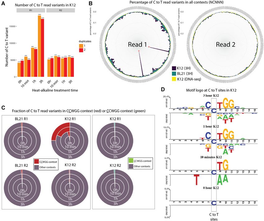
2 Nucleic Acids Research, 2021
techniques such as EM-seq (8) or hybrid techniques such and 1 M of acetic acid was added to a final concentration of
as TAPS-seq (9). Recently, MFRE-Seq has been developed 0.1 M in order to neutralize the reactions. We also tested al-
to identify m5C methylase specificities in bacteria (10). kaline concentration of 0.5M and 1M NaOH, in these cases,
MFRE-Seq uses a modification-dependent endonuclease equal amounts of acetic acid were added to the reaction to
that generates a double-stranded DNA break at methylated properly neutralize the PH. The neutralized reactions were
sites, allowing the identification of m5C for the subset of cleaned up using the Zymo oligo clean and concentrator kit
sites conforming to the recognition sites of the MFRE en- (D4060 Zymo Research) and the DNA was eluted in 20 l
zymes. Unlike PacBio sequencing, these specialized meth- of 0.1× TE.
ods do not provide the dual original sequence and methyla- PCR amplification of the samples was done following
Downloaded from https://academic.oup.com/nar/advance-article/doi/10.1093/nar/gkab705/6355877 by guest on 01 November 2021
tion readouts from a single experiment. NEBNext Ultra II library prep kit for Illumina protocol
Recently, m5C in the CpG context has been identified (ER7645, New England Biolabs) and the NEBNext® Mul-
(11) and a signal for methylation can be observed at known tiplex Oligos for Illumina® (E7337A, New England Bi-
methylated sites in bacteria using Nanopore sequencing olabs). The number of PCR cycles was tested and opti-
(12,13). So far no technique permits, from a single exper- mized for each sample following the standard procedure
iment, the dual sequencing of genomes and the de novo de- for library preparation. PCR reactions were cleaned up us-
termination of m5C methylase specificity for the non-CpG ing 0.9× NEBNext Sample purification beads (E7137AA,
contexts typically found in bacteria. New England Biolabs) and eluted in 25 l of 0.1× TE. All
Herein, we describe a novel approach called RIMS-seq the libraries were evaluated on a TapeStation High sensi-
to simultaneously sequence bacterial genomes and glob- tivity DNA1000 (Agilent Technologies) and paired-end se-
ally profile m5C methylase specificity using a protocol that quenced on Illumina.
closely resembles the standard Illumina DNA-seq with a
single, additional step. RIMS-seq shows comparable se-
Bisulfite-seq library preparation
quencing quality as DNA-seq and accurately identifies
methylase specificities. Applied to characterized strains or One percent of lambda phage gDNA (D1221, Promega)
novel isolates, RIMS-seq de novo identifies novel activities was spiked-into 300 ng gDNA to use as an unmethylated in-
without the need for a reference genome and permits the ternal control. The samples were sonicated in 1× TE buffer
assembly of the bacterial genome at metrics comparable to using the Covaris S2 (Covaris) with the standard protocol
standard shotgun sequencing. for 50 l and 200 bp insert size.
The subsequent fragmented gDNA was used as the start-
MATERIALS AND METHODS ing input for the NEBNext Ultra II library prep kit for Il-
lumina (E7645, New England Biolabs) following the manu-
Samples and genomic DNA collection facturer’s recommendations until the USER treatment step.
Skin microbiome genomic DNA (ATCC® MSA-1005) The methylated loop-shaped adapter was used for ligation.
and gut microbiome genomic DNA (ATCC® MSA-1006) After USER, a 0.6× clean-up was performed using the
were obtained from ATCC. Escherichia coli BL21 genomic NEBNext Sample purification beads (E7137AA, New Eng-
DNA was extracted from a culture of E. coli BL21 DE3 land Biolabs) and eluted in 20 l of 0.1× TE. A TapeSta-
cells (C2527, New England Biolabs) using the DNEasy tion High Sensitivity DNA1000 was used to assess the qual-
Blood and Tissue kit (69504, Qiagen). Escherichia coli K12 ity of the library before subsequent bisulfite treatment. The
MG1655 genomic DNA was extracted from a cell culture Zymo EZ DNA Methylation-Gold Kit (D5005, Zymo Re-
using the DNEasy Blood and Tissue kit (69504, Qiagen). search) was used for bisulfite treatment, following the man-
All the other gDNA from the bacteria presented in Table ufacturer’s protocol.
1 were isolated using the Monarch genomic DNA purifica- PCR amplification of the samples was done follow-
tion kit (T3010S, New England Biolabs). Xp12 phage ge- ing the suggestions from NEBNext Ultra II library prep
nomic DNA was obtained from Peter Weigele and Yian- kit for Illumina (ER7645, New England Biolabs), using
Jiun Lee at New England Biolabs. the NEBNext® Multiplex Oligos for Illumina® (E7337A,
New England Biolabs) and NEBNext® Q5U® Master Mix
(M0597, New England Biolabs).
RIMS-seq library preparation
The number of PCR cycles was tested and optimized
One hundred nanogram of gDNA was sonicated in 1× TE for each sample. The PCR reactions were cleaned up us-
buffer using the Covaris S2 (Covaris) with the standard pro- ing 0.9× NEBNext Sample purification beads (E7137AA,
tocol for 50 l and 200 bp insert size. New England Biolabs) and eluted in 25 l of 0.1× TE. All
The subsequent fragmented gDNA was used as the start- the libraries were screened on a TapeStation High sensi-
ing input for the NEBNext Ultra II library prep kit for Il- tivity DNA1000 (Agilent Technologies) and paired-end se-
lumina (E7645, New England Biolabs) following the manu- quenced on Illumina.
facturer’s recommendations until the USER treatment step.
The regular unmethylated loop-shaped adapter was used
RIMS-seq data analysis
for ligation. After the USER treatment (step included), the
samples were subjected to heat alkaline deamination: 1 M Paired-end reads were trimmed using Trim Galore 0.6.3
NaOH pH 13 was added to a final concentration of 0.1 M (option –trim1). The Acinetobacter calcoaceticus ATCC
and the reactions were placed in a thermocycler at 60◦ C for 49823 data have been trimmed using Trim Galore version
3 h. Then, the samples were immediately cooled down on ice 0.6.3 instead and downsampled to 1 million reads. Reads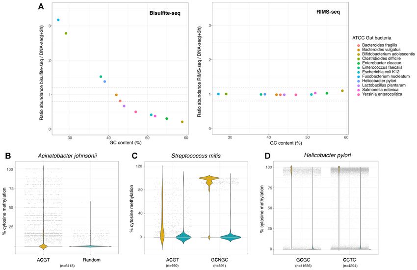
Nucleic Acids Research, 2021 3

were mapped to the appropriate genome using BWA mem              0.22.2 with default parameters. PCR duplicates were re-
with the paired-end mode (version 0.7.5a-r418 and version        moved using deduplicate bismark and methylation infor-
0.7.17-r1188 for the A. calcoaceticus). When using an as-        mation extracted using bismark methylation extractor us-
sembled genome directly from RIMS-seq data, trimmed              ing default parameters. For the microbiome, the bis-
RIMS-seq reads were assembled using SPAdes (SPAdes-              mark methylation extractor with –split by chromosome
3.13.0 (31) default parameters). Reads were split according      option was used to output one methylation report per bac-
to the read origin (Read 1 or Read 2) using samtools (ver-       terium. The motif identification was done as previously de-
sion 1.8) with -f 64 (for Read 1) and -f 128 (for Read 2) and    scribed in (10).
samtools mpileup (version 1.8) was run on the split read files

                                                                                                                                Downloaded from https://academic.oup.com/nar/advance-article/doi/10.1093/nar/gkab705/6355877 by guest on 01 November 2021
with the following parameters: -O -s -q 10 -Q 0. For Acine-      EM-seq
tobacter calcoaceticus, the unmapped reads, reads without a
mapped mate and the non-primary alignments were filtered         EM-seq was performed according to the standard protocol
out using the flags -F 12 and -F 256.                            (NEB E7120S). Motif identification was done as previously
                                                                 described in (10).

De-novo identification of motifs using RIMS-seq                  Analysis and abundance estimation in synthetic microbiomes
Programs and a detailed manual for the de-novo identifica-       RIMS-seq, DNA-seq and bisulfite-seq were performed on
tion of motifs in RIMS-seq are available on github (https:       the synthetic gut and skin microbiome as described. Reads
//github.com/Ettwiller/RIMS-seq/). Using the mpileup files,      derived from RIMS-seq, DNA-seq and bisulfite-seq were
positions and 14bp flanking genomic regions for which a          mapped as described to a ‘meta-genome’ composed of the
high quality (base quality score ≥ 35) C to T in R1 or G to      reference genomes of all the bacteria included in the cor-
A in R2 was found, were extracted for the foreground. Po-        responding synthetic community (see Supplementary Ta-
sitions and 14bp flanking regions for which a high quality       ble S3 for detailed compositions). Mapped reads were split
(base quality score ≥ 35) G to A in R1 or C to T in R2 was       according to each bacterium and RIMS-seq or bisulfite
found, were extracted for the background. C to T or G to         analysis pipelines were run on individual genomes as de-
A in the first position of reads were ignored. If the percent-   scribed above. Abundance was estimated using the number
age of C to T or G to A are above 5% for at least 5 reads        of mapped reads per bacteria and normalized to the total
at any given position, the position was ignored (to avoid        number of mapped reads. Normalized species abundances
considering positions containing true variants). Motifs that     in RIMS-seq and bisulfite-seq were compared to the nor-
are found significantly enriched (P-value < 1e−100 ) in the      malized species abundances in DNA-seq.
foreground sequences compared to background sequences
were found using mosdi pipeline mosdi-discovery with the
following parameters: ‘mosdi-discovery -v discovery -q x -i      Phylogeny of the ATCC synthetic microbiomes and visualiza-
-T 1e-100 -M 8,1,0,4 8 occ-count’ using the foreground se-       tion
quences with x being the output of the following command         The phylogenetic trees of both ATCC synthetic gut and
: ‘mosdi-utils count-qgrams -A ‘dna’’ using the background       skin microbiomes were done using OrthoFinder version
sequences. To identify additional motifs, the most signif-       2.3.11 (33) using the MSA workflow and MAFFT for
icant motif found using mosdi-discovery is removed from          the multiple sequence alignment program. The program
the foreground and background sequences using the fol-           options are available at https://github.com/davidemms/
lowing parameter: ‘mosdi-utils cut-out-motif -M X’ and the       OrthoFinder. The phylogenetic tree and abundance data
motif discovery process is repeated until no significantly en-   obtained from DNA-seq, RIMS-seq and bisulfite-seq were
riched motif can be found.                                       visualized using iTOL (34) for each synthetic community
                                                                 (see Supplementary Figure S5).
Sequence logo generation
                                                                 Quality control of the data
Using the mpileup files, positions in the genome for which
                                                                 The insert size for each downsampled filtered bam file was
a high quality (base quality score ≥ 35) C to T in R1 or a G
                                                                 calculated using Picard version 2.20.8 using the default pa-
to A in R2 was observed were extracted for the foreground
                                                                 rameters and the option CollectInsertSizeMetrics (‘Picard
using the get motif step1.pl program. Positions for which a
                                                                 Toolkit.’ 2019. Broad Institute, GitHub Repository. http:
high quality (base quality score ≥ 35) G to A in R1 or a C
                                                                 //broadinstitute.github.io/picard/; Broad Institute).
to T in R2 was observed were extracted for the background.
                                                                    The GC bias for each downsampled filtered bam file was
The ±7 bp regions flanking those positions were used to
                                                                 calculated and plotted using Picard version 2.20.8 using the
run two sample logo (32). Parameters were set as t-test, pP-
                                                                 default parameters and the option CollectGcBiasMetrics.
value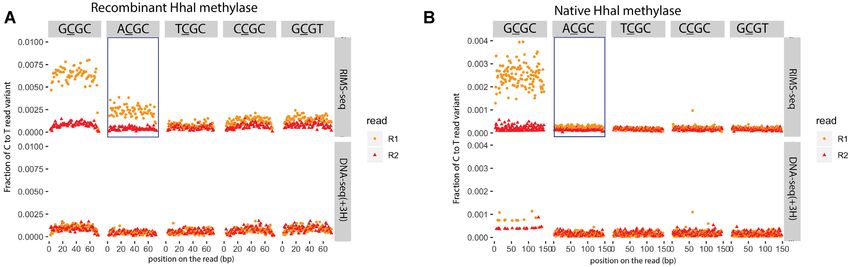
4 Nucleic Acids Research, 2021
were then mapped back to the assembly using BWA mem (R1) and reverse read (R2). Resulting from m5C deami-
0.7.17 and mapping statistics were generated using samtools nation, R1 has the C to T read variants while R2 has the
flagstat 1.10.2 reverse-complement G to A variant. This difference leads
to an overall imbalance of C to T variants between R1 and
Xp12 sequencing performance assessment R2 (17) (see also Supplementary Figure S1 for explanation).
Thus, sequence contexts for which the C to T read variants
Reads were trimmed using trimgalore 0.6.5 and mapped to are imbalanced in R1 compared to R2 correspond to m5C
the Xp12 reference genome using BWA mem 0.7.17. Insert methylase specificity(ies). Because of the limited deamina-
size and GC bias were assessed using Picard Toolkit and tion rate, RIMS-seq takes advantage of the collective signal
Downloaded from https://academic.oup.com/nar/advance-article/doi/10.1093/nar/gkab705/6355877 by guest on 01 November 2021
genome coverage using Qualimap 2.1.1. at all sites to define methylase specificity. Because C to T
imbalance can be observed at nucleotide resolution, RIMS-
Intact mass LC–MS seq identifies at base resolution which of the cytosine within
the motif is methylated.
Intact mass analysis was performed by tandem liquid The experimental steps for RIMS-seq essentially follow
chromatography–mass spectrometry (LC–MS/MS) on an the standard library preparation for Illumina sequencing
Vanquish Horizon UHPLC System equipped with a diode with an extra deamination step. Briefly, the bacterial ge-
array detector and a Thermo Q-Exactive Plus mass nomic DNA is fragmented, and adaptors are ligated to
spectrometer operating under negative electrospray ion- the ends of DNA fragments (Figure 1B and Materials
ization mode (–ESI). UHPLC was performed using a and Methods). Between the ligation step and the amplifi-
Thermo DNAPac™ RP Column (2.1 × 50 mm, 4 m) cation step, an alkaline heat treatment step is added to in-
at 70◦ C and 0.3 ml/min flow rate, with a gradient mo- crease the rate of deamination. Only un-deaminated DNA
bile phase consisting of hexafluoroisopropanol (HFIP)– or DNA containing deaminated m5C can be amplified and
N,N-diisopropylethylamine (DIEA) aqueous buffer and sequenced.
methanol. UV detection was performed at 260 nm. Intact
mass analysis was performed under Full MS mode, and
ESI-MS raw data was deconvoluted using Promass HR Validation of RIMS-seq
(Novatia Inc.). Optimization of the heat alkaline deamination step. We first
evaluated the conditions to maximize the deamination of
RESULTS m5C while minimizing other DNA damage. For this we
used bacteriophage Xp12 genomic DNA that contains ex-
Principle of RIMS-seq
clusively m5C instead of C (18) to measure the m5C deam-
Spontaneous deamination of cytosine (C) leading to uracil ination rates in various contexts.
(U) and of m5C leading to thymine (T) are examples of To estimate the overall deamination rate of m5C, we
common damage found in DNA. In-vitro, this damage is of- quantified the imbalance of C to T read variants between
ten undesirable as it can interfere with sequencing. The type R1 and R2 for 0, 10 and 30 min, 1 h, 2 h, 3 h, 5 h and 14 h
of interference during sequencing depends on whether the of heat alkaline treatment (Figure 1C). We observed an im-
deamination occurs on C or m5C. U blocks the passage of balance as early as 10 min with a 3.7-fold increase of C to T
high-fidelity polymerases typically used in library prepara- read variants in R1 compared to R2. The increase is linear
tion protocols, preventing the amplification and sequencing with time with a maximum of 212-fold increase of C to T
of U-containing DNA fragments. Conversely, DNA har- read variants in R1 compared to R2 after 14 h of heat alka-
boring T derived from m5C deamination can be normally line treatment (Figure 1D). Next, we quantified the deami-
amplified but results in C to T errors (14,15). This distinc- nation rate at all Nm5CN sequence contexts with N being
tion between blocking and mutagenic damage forms the ba- A, T, C or G and show an increase of C to T variants in
sis of the RIMS-seq method, allowing the identification of R1 in all contexts (Supplementary Figure S2A). Together,
methylase specificity based on an elevated number of reads these results show that a measurable deamination rate can
containing C to T transitions specifically at methylated sites be achieved in as soon as 10 min of heat alkaline deami-
(Figure 1A). To increase the rate of m5C deamination, the nation and that deamination efficiency is similar in all se-
DNA is subjected to a heat-alkaline treatment which has quence contexts.
been previously demonstrated to elevate the rate of both To estimate the non-specific damage to the DNA lead-
C and m5C deamination with m5C having a 1.5–3 times ing to unwanted sequencing errors, we quantified possible
higher deamination rate than for C (16). This treatment is imbalances for other variant types (Supplementary Figure
aimed at inducing a level of deamination large enough to S2B). We found that G to T variants show imbalance in
detect the m5C methylase specificity without affecting the all the conditions investigated, likely the result of oxida-
sequencing quality. For this reason, the deamination lev- tive damage resulting from sonication, a common step in
els typically obtained with RIMS-seq does not permit the library preparation between RIMS-seq and DNA-seq (17).
quantitative measurement of methylation at each genomic Interestingly, the imbalance is reduced in RIMS-seq, disap-
site but rather provides a global methylation signal charac- pearing almost completely after 14 h of heat alkaline treat-
teristic of the methylase specificity. ment (Supplementary Figure S2B). This result suggests that
Illumina paired-end sequencing allows both ends of a this treatment either converts 8-oxoG back to G or to an-
DNA fragment to be sequenced, generating a forward read other modification that ultimately blocks the polymerase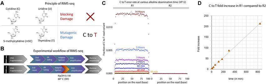
Nucleic Acids Research, 2021 5

                                                                                                                                                               Downloaded from https://academic.oup.com/nar/advance-article/doi/10.1093/nar/gkab705/6355877 by guest on 01 November 2021
Figure 1. (A) Principle of RIMS-seq. Deamination of cytidine leads to a blocking damage while deamination of m5C leads to a mutagenic C to T damage
only present on the first read (R1) of paired-end reads in standard Illumina sequencing. Thus, an increase of C to T errors in R1 in specific contexts
is indicative of m5C. (B) The workflow of RIMS-seq is equivalent to a regular library preparation for Illumina DNA-seq with an extra step of limited
alkaline deamination at 60◦ C. This step can be done immediately after adaptor ligation and does not require additional cleaning steps. (C) Fraction of C to
T variants in XP12 (m5C) at all positions in the reads for R1 and R2 after 0min (DNA-seq), 10 min, 30 min, 60 min, 2 h, 3 h, 5 h and 14 h of heat-alkaline
treatment. The C to T imbalance between R1 and R2 is indicative of deamination of m5C and increases with heat-alkaline treatment time. (D) Correlation
between the C to T fold increases in R1 compared to R2 according to time (r2 = 0.998).

from amplifying 8-oxoG-containing fragments. To prop-                            increase compared to the additional experimental time re-
erly address the disappearance of G to T variants due to                         quired.
oxidative damage in RIMS-seq, we designed an oligonu-
cleotide containing a single 8-oxoG. Using LC–MS intact                          RIMS-seq is able to distinguish methylated versus unmethy-
mass, we identified a strand break directly 5 and 3 of the 8-                  lated motifs in E. coli. To validate the application of
oxoG that is specific to oxidized G under heat alkaline treat-                   RIMS-seq to bacterial genomes, we sequenced dcm+ (K12)
ments (Supplementary text 1 and Supplementary Figure                             and dcm- (BL21) E. coli strains. In K12, the DNA cyto-
S3). Thus, the heat-alkaline treatment performed in RIMS-                        sine methyltransferase dcm methylates cytosine at CCWGG
seq induced strand breaks at oxidative damage sites, pre-                        sites (C = m5C, W = A or T) and is responsible for all
venting the amplification of 8-oxoG-containing fragments                         m5C methylation in this strain (19). E. coli BL21 has no
and de-facto decreasing the frequency of G to T in the                           known m5C methylation. Heat/alkaline treatments were
RIMS-seq libraries.                                                              performed at three time points (10 min, 1 h and 3 h). In addi-
   A slight elevation of G to C and T to C read variants can                     tion, we performed a control experiment corresponding to
be observed in RIMS-seq compared to DNA-seq but this                             the standard DNA-seq. Resulting libraries were paired-end
damage is of low frequency and therefore is not expected to                      sequenced using Illumina and mapped to their correspond-
notably affect the sequencing performance QC of RIMS-                            ing genomes (Methods).
seq.                                                                                For comparison, all datasets were downsampled to 5 mil-
   We performed QC metrics and assemblies of Xp12 for                            lion reads corresponding to 200× coverage of the E. coli
all the alkaline-heat treatment conditions, including a con-                     genome and instances of high confidence C to T variants (Q
trol DNA-seq. The overall sequencing performances were                           score > 35) on either R1 or R2 were identified. As expected,
assessed in terms of insert size, GC bias and genome cover-                      control DNA-seq experiments show comparable numbers
age. Similar results were observed between RIMS-seq and                          of C to T read variants between R1 and R2, indicating true
the DNA-seq control at all treatment times, indicating that                      C to T variants or errors during amplification and sequenc-
the RIMS-seq heat-alkaline treatment does not affect the                         ing (Figure 2A). On the other hand, the overall number of
quality of the libraries (Supplementary Figure S4).                              C to T read variants in R1 is progressively elevated for 10
   We also evaluated the quality of the assemblies compared                      min, 1 h and 3 h of heat-alkaline treatment of the E. coli
to the Xp12 reference genome and found that all conditions                       K12 samples with an overall 4-fold increase after 3 htreat-
lead to a single contig corresponding to essentially the entire                  ment compared to no treatment; heat-alkaline treatments
genome with very few mismatches (Supplementary Table                             did not increase the rate of C to T read variants in R2 (Fig-
S1). These results suggest that the heat-alkaline treatment                      ure 2A). We anticipate that the elevation of the E. coli K12
does not affect the assembly quality, raising the possibility                    C to T read variants in R1 is due to deamination of m5C.
of using RIMS-seq for simultaneous de novo genome assem-                         In this case, the elevation should be specifically found in Cs
bly and methylase specificity identification. We found that                      in the context of CCWGG (with the underlined C corre-
a 3-h treatment provides a good compromise between the                           sponding to the base under consideration). To demonstrate
deamination rate (resulting in ∼0.3% of m5C showing C to                         this, we calculated the fraction of C to T read variants in
T transition) and duration of the experiment. We found that                      CCWGG compared to other contexts. We observed a large
longer incubation times (up to 14 h) increased the deamina-                      elevation of the C to T read variants in the CCAGG and
tion rate by up to 1% and decided this is a slight sensitivity                   CCTGG contexts for K12 (Figure 2B). As expected, the
6 Nucleic Acids Research, 2021
Downloaded from https://academic.oup.com/nar/advance-article/doi/10.1093/nar/gkab705/6355877 by guest on 01 November 2021
Figure 2. (A) Bar plots representing the number of C to T read variants for K12 in R1 and R2 after different heat/alkaline treatment times. Colors represent
duplicate experiments. (B) Circular bar plots representing the percentage of C to T read variants in all NCNNN contexts (with N = A, T, C or G) for
Read 1 (R1, left) and Read 2 (R2, right) in DNA-seq performed on K12 (yellow bars), RIMS-seq (3H) performed on BL21 (green) and RIMS-seq (3H)
performed on K12 (dark blue). Red asterisks denote CCWGG contexts with W being either A or T. (C) Proportion of C to T read variants in CCWGG (red)
or CCWGG (green) contexts compared to other NCNNN or CNNNN contexts for R1 and R2 in K12 and BL21. The C to T read variants in CCWGG
and CCWGG motifs represent less than 2% of all variants except in K12 (R1 only) after 10 min, 1- and 3-h treatments where the CCWGG motifs represent
4.1%, 22.5% and 32.6% of all C to T read variants respectively. The increase of C to T read variants in the CCWGG context is therefore specific to R1 in
K12 strain. (D) Visualization of the statistically significant differences in position-specific nucleotide compositions around C to T variants in R1 compared
to R2 using Two Sample Logo (21) for the K12 sample subjected to (from top to bottom) 3 h, 1 h, 10 min and 0 min heat alkaline treatment.
C to T read variants show no elevation at CCAGG and all the other NCNNN and CNNNN contexts, respectively.
CCTGG contexts for the E. coli BL21 strain that is miss- After 3 h of heat-alkaline treatment, the fraction of C to T
ing the dcm methylase gene (Figure 2B). Thus, this C to T read variants in a CCWGG context increased, rising from
read variant elevation is specific to the E. coli K12 strain only 1.9% in regular DNA-seq to ∼25% of all the C to T
subjected to heat-alkaline treatments, consistent with deam- variants. This increase is only observable in R1 of the K12
ination detectable only on methylated sites. Taken together, strain (Figure 2C). Conversely, no increase can be observed
these results indicate that the elevated rate of C to T vari- in a CCWGG context for which the C to T variant rate at the
ants observed in R1 from E. coli K12 is the result of m5C first C is assessed (Figure 2C). Thus, RIMS-seq identified
deamination in the CCWGG context. the second C as the one bearing the methylation, consistent
Next, we assessed whether the difference in the C to T with the well described dcm methylation of E. coli K12 (20)
read variant context between R1 and R2 at the CCWGG (19), highlighting the ability of RIMS-seq to identify m5C
motif provides a strong enough signal to be discernible over methylation at base resolution within the methylated motif.
the background noise. For this, we calculated the fraction of Next, we calculated significant (P-value < 0.01) differ-
C to T read variants in CCWGG and CCWGG compared to ences in position-specific nucleotide compositions aroundNucleic Acids Research, 2021 7
C to T variants in R1 compared to R2 using Two Sam- For this, we performed RIMS-seq on A. calcoaceticus
ple Logo (21). We found a signal consistent with the dcm ATCC 49823 genomic DNA as described above as well
methylase specificity in K12 RIMS-seq samples at 1 and as a control DNA-seq experiment for which the alkaline
3 h of heat alkaline treatment (Figure 2D) demonstrating treatment was replaced by 3 h incubation in TE (DNA-
that it is possible to identify methylase specificities in ge- seq(+3H)). We compared the de novo assembly obtained
nomic sequence subject to as little as 1 h of alkaline treat- from the reads generated by the DNA-seq(+3H) and the de
ment. These results support the application of RIMS-seq novo assembly obtained from the reads generated by RIMS-
for the de novo identification of methylase specificity at base seq (see Materials and Methods). In brief, the alkaline treat-
resolution. ment did not alter the important metrics for assembly qual-
Downloaded from https://academic.oup.com/nar/advance-article/doi/10.1093/nar/gkab705/6355877 by guest on 01 November 2021
ity such as the rate of mismatches and N50 demonstrating
RIMS-seq identifies the correct methylase specificity de that the elevated C to T variant rate at methylated sites is
novo in E. coli K12. In order for RIMS-seq to iden- not high enough to cause assembly errors (Figure 3B).
tify methylase specificities de novo, we devised an analy- We then proceeded to map the RIMS-seq reads to the as-
sis pipeline based on MoSDi (22) to find sequence mo- sembly and motifs were identified using the RIMS-seq de
tif(s) that are over-represented around C to T transitions novo motif discovery pipeline. As expected, the same mo-
in R1 reads (Figure 3A, analysis pipeline available at https: tifs found when mapping to the reference genome are also
//github.com/Ettwiller/RIMS-seq). In brief, the pipeline ex- found in the A. calcoaceticus de novo assembly with similar
tracts the sequence context at each C to T read variant significance (GATC (P-value = 1.44e−1255 ) and CGCG (P-
in R1 (foreground) and R2 (background). MoSDi identi- value = 8.6e−228 ) (Figure 3C). These motifs correspond to
fies the highest over-represented motif in the foreground the methylase specificities expected in this strain indicating
sequences compared to the background sequences. To ac- that RIMS-seq can be applied for genome sequencing and
commodate the presence of multiple methylases in the same assembly of any bacterium without the need for a reference
host, the first motif is subsequently masked in both the fore- genome.
ground and background sequences and the pipeline is run
again to find the second highest over-represented motif and RIMS-seq can be complemented with SMRT sequencing
so on until no significant motifs can be found (see Ma- to obtain a comprehensive overview of methylase specifici-
terials and Methods for details). Running the pipeline us- ties. RIMS-seq performed in parallel with SMRT se-
ing the K12 strain RIMS-seq data identifies one significant quencing has the advantage of comprehensively identify-
over-represented motif corresponding to the CCWGG mo- ing all methylase specificities (m5C, m4C and m6A methy-
tif (P-value = 9.71e−77 , 4.25e−858 and 3.61e−4371 for 10, 60 lations) and results in an assembly of higher quality than
and 180 min of alkaline treatment respectively) with the cy- with short reads illumina data. We applied this hybrid
tosine at position 2 being m5C. approach to Acinetobacter calcoaceticus ATCC 49823 for
Summing up, we devised a novel sequencing strategy which a SMRT sequencing and assembly had been done
called RIMS-seq and its analysis pipeline to identify m5C previously (4). RIMS-seq was performed as described above
methylase specificity de novo. When applied to E. coli and the reads were mapped to the genome assembly ob-
K12, RIMS-seq identifies the dcm methylase specificity as tained from SMRT-sequencing. We again found the two
CCWGG with the methylated site located on the second C, m5C motifs: CGCG (P-value = 1.84e−1535 ) and GATC (P-
consistent with the reported dcm methylase specificity (Ta- value = 4.93e−6856 ) from the RIMS-seq data in addition
ble 1). to the 13 m6A motifs described previously using SMRT
sequencing (4). This result demonstrates the advantage of
RIMS-seq identifies multiple methylase specificities de novo such a hybrid approach in obtaining closed genomes with
within a single microorganism. To assess whether RIMS- comprehensive epigenetic information.
seq can identify methylase specificity in strains express-
ing multiple methylases, we repeated the same procedure XP12 can be used as a spiked-in to measure the deamination
on a strain of Acinetobacter calcoaceticus ATCC 49823 rate. To ensure the correct level of heat-alkaline deam-
expressing two m5C methylases with known specificities ination rate, XP12 can be used as spiked-in to measure
(4). RIMS-seq identifies CGCG (P-value = 2.33e–174 ) and the deamination rate at m5C. To illustrate the practicality
GATC (P-value = 3.02e–1308 ) (Table 1) both motifs have of such control, we subjected Haemophilus influenzae Rd
been confirmed by MFRE-seq (10). Thus, RIMS-seq is able ATCC 51907 (Table 1) spiked-in with XP12 DNA to various
to de novo identify methylase specificities in bacteria ex- NaOH concentrations and treatment times. We observed
pressing multiple methylases. deamination rates varying from 0.24% (0.1 M NaOH, 3 h)
to 2.72% (0.5 M NaOH, 3 h) (Supplementary Figure S2C).
RIMS-seq can be applied for genome sequencing and m5C We further investigated the error rates in both the bacteria
profiling in bacteria without a reference genome. We inves- and XP12 for substitutions other than C to T at various
tigated whether RIMS-seq can be used to identify methylase heat alkaline conditions (Supplementary Figure S2C) and
specificities of uncharacterized bacteria for which a refer- found that all substitution rates are comparable to the rates
ence genome is unavailable. More specifically, we evaluated obtained using standard DNA-seq. Taken together, these
if the reads generated using RIMS-seq can be used for both results indicate that the heat alkaline treatments in the mea-
identifying methylase specificities and generating an assem- sured ranges are not expected to notably affect the sequenc-
bly of comparable quality to DNA-seq. ing performance QC in bacteria.8 Nucleic Acids Research, 2021
Downloaded from https://academic.oup.com/nar/advance-article/doi/10.1093/nar/gkab705/6355877 by guest on 01 November 2021
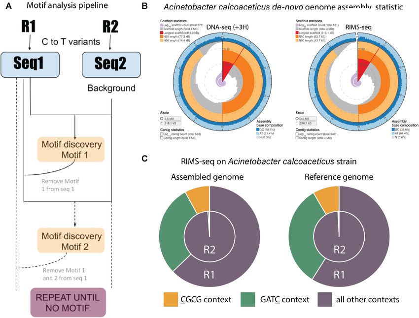
Figure 3. De novo discovery of methylase specificity using RIMS-seq. (A) Description of the RIMS-seq motif analysis pipeline. First, C to T read variants
are identified in both Read 1 and Read 2 separately. Then, the MosDI program searches for overrepresented motifs. Once a motif is found, the pipeline is
repeated until no more motifs are found, enabling identification of multiple methylase specificities in an organism. (B) Assembly statistics obtained using
the sequence from the standard DNA-seq (+3H, left) and RIMS-seq (right). Visualization using assembly-stats program (https://github.com/rjchallis/
assembly-stats). The corresponding table with the statistical values is available in the supplementary material (Supplementary Table S2). (C) Fractions of
C to T read variants in CGCG (yellow) or GATC (green) contexts compared to other contexts for R1 and R2 in Acinetobacter calcoaceticus ATCC 49823
using the assembled or the reference genome. The increase of C to T read variants in these contexts are similar when using either the assembled or reference
genomes
Table 1. Methylases specificity obtained using RIMS-seq and validated using different methods. The method is indicated by a number next to the motif. :
Evidence for the validated motifs are (1) bisulfite-seq (Materials and Methods), (2) REBASE (4), (3) EM-seq (material and method), (4) MFRE-seq (10),
(5) mTet1-enhanced SMRT sequencing (6)
Organism Accession numbers (biosample) RIMS-seq motif(s) Validated motif(s)
Escherichia coli K12 SAMN02604091 CCWGG CCWGG (1,2,4)
Acinetobacter calcoaceticus ATCC 49823 SAMN14530202 GATC GATC (4)
CGCG CGCG (2,4)
Bacillus fusiformis 1083 SAMN17843035 ACCTGC ACCTGC (2,3)
GCAGGT GCAGGT (2,3)
Bacillus amyloliquefaciens H ATCC 49763 SAMN12284742 GCWGC GCWGC (3)
Clostridium acetobutylicum ABKn8 SAMN17843114 GCNNGC GCNNGC (3)
Aeromonas hydrophila NEB724 SAMN14533640 GCCGGC GCCGGC (3)
Haemophilus influenzae Rd ATCC 51907 SAMN02603991 GRCGYC* GRCGYC (5)
ACCGCACT
AGTGCGGT
Haemophilus parahaemoltyicus ATCC 10014 SAMN11345835 GCGC GCGC (2)
M.HhaI clone (E. coli) NA RCGC GCGC (4)
CCWGG(a) CCWGG (1,2,4)(a)
(a) The E. coli strain used is Dcm+, resulting in the discovery of both the Dcm (CCWGG) and M.HhaI motifs (GCGC). RIMS-seq discovered RCGC
instead of GCGC motif (see text for explanation). * P-value = 1.0e−91 (standard detection threshold ofNucleic Acids Research, 2021 9
Downloaded from https://academic.oup.com/nar/advance-article/doi/10.1093/nar/gkab705/6355877 by guest on 01 November 2021
Figure 4. C to T error profile in GCGC (canonical recognition site), ACGC, TCGC, CCGC and GCGT. in R1 reads (orange) and R2 reads (red) for
RIMS-seq (upper panel) and DNA-seq(+3H) (lower panel) A. Recombinant HhaI methylase expressed in E. coli B. Native HhaI methylase expressed in
Haemophilus parahaemolyticus. Elevation of C to T in the R1 read variant can be observed in the context of GCGC for both the recombinant and native
HhaI genomic DNA and in the context of ACGC only for DNA from the recombinant but not the native HhaI.
RIMS-seq can be applied to a variety of RM systems consequently the star activity results in hemi-methylation
of the ACGC sites and not the GCGT motif.
Methylases targets are usually palindromic sequences be-
tween 4 nt and 8 nt, and a single bacterium often possesses
RIMS-seq can be applied to microbial communities
several, distinct MTase activities (23). Next, we tested the
general applicability of RIMS-seq and the de novo motif dis- We assessed whether RIMS-seq can be applied to mixed
covery pipeline using bacterial genomic DNA from our in- microbial communities using synthetic gut and skin micro-
house collections of strains. biomes from ATCC containing 12 and 6 bacterial species,
For some bacterial strains, the methylase recognition respectively. We also complemented the RIMS-seq exper-
specificities have been previously experimentally character- iment with the control experiment DNA-seq(+3H) and
ized. In all of those strains, RIMS-seq confirms the specifici- a bisulfite treatment to validate the RIMS-seq findings.
ties and identifies the methylated cytosine at base resolution Reads were mapped to their respective microbiome refer-
(Table 1). We have tested the identification of 4-mers motifs ence genomes (Materials and Methods). For the gut mi-
such as GATC, CGCG (Acinetobacter calcoaceticus) and crobiome we found a mapping rate (properly paired only)
GCGC (Haemophilus parahaemolyticus) up to 8-mers mo- of 95.79%, 95.77% and 66.2% for RIMS-seq, DNA-seq
tifs such as ACCGCACT and AGTGCGGT (Haemophilus and bisulfite-seq respectively. Concerning the skin micro-
influenzae). Motifs can be palindromic or non-palindromic biome, 85.89%, 85.35% and 54.9% of reads were mapped
(Table 1 and Supplementary Table S3). In the latter case, for RIMS-seq, DNA-seq and bisulfite-seq respectively. The
RIMS-seq defines non-palindromic motifs at strand res- low mapping rate for bisulfite-seq is a known challenge as
olution. For example, RIMS-seq identifies methylation at the reduction of the alphabet to A, G, T generates ambigu-
two non-palindromic motifs ACCTGC as well as its reverse ous mapping (24).
complement GCAGGT in the Bacillus fusiformis strain (Ta- To use RIMS-seq as an equivalent to DNA-seq for mixed
ble 1). community applications, RIMS-seq should produce se-
A number of RM systems have been expressed in other quencing quality metrics that are similar to standard DNA-
hosts such as E. coli for biotechnological applications. For seq, especially on the estimation of species relative abun-
the methylase M.HhaI recognizing GCGC (4), we per- dances. We therefore compared RIMS-seq sequencing per-
formed RIMS-seq and a control DNA-seq(+3H) on both formances with DNA-seq(+3H) and bisulfite sequencing.
the native strain (Haemophilus parahaemolyticus ATCC We found that bisulfite sequencing elevates abundances of
10014) and in E. coli K12 expressing the recombinant ver- AT-rich species such as Clostridioides difficile (71% AT),
sion of M.HhaI. Interestingly, we found that the de novo Enterococcus faecalis (63% AT) and Fusobacterium nuclea-
RIMS-seq analysis algorithm identifies RCGC (with R be- tum (73% AT) (Figure 5A, Supplementary Figure S5). For
ing either A or G) for the recombinant strain and GCGC for example, bisulfite sequencing over-estimated the presence
the native strain (Figure 4A). Conversely, no notable eleva- of Clostridioides difficile by a factor of 2.65 and Staphy-
tion of C to T read variants are observed at ACGC for the lococcus epidermidis by a factor of 3.9 relative to DNA-
native strain (Figure 4B), confirming the de novo motif dis- seq. This over-estimation of an AT rich genome by bisul-
covery results from the analysis pipeline. Collectively, these fite is a known bias of bisulfite sequencing and relates to
results suggest that the recombinant methylase shows star damage at cytosine bases (25). Conversely, we found that
activity, notably in the context of ACGC, that is not found the species abundances are similar between DNA-seq(+3H)
in the native strain. We hypothesize that the star activity is and RIMS-seq (abundance ratios between 0.8 and 1.2) indi-
the result of the over-expression of the methylase in E. coli cating that RIMS-seq can be used to quantitatively estimate
K12. Interestingly, ACGC is not a palindrome motif and microbial composition.10 Nucleic Acids Research, 2021
Downloaded from https://academic.oup.com/nar/advance-article/doi/10.1093/nar/gkab705/6355877 by guest on 01 November 2021
Figure 5. (A) Bacterial abundance in the ATCC gut microbiome calculated from bisulfite-seq data (left) and RIMS-seq (Right) normalized to
DNAseq(+3H). The normalized abundance is plotted relative to the GC content of each bacterium. (B) Methylation levels in Acinetobacter johnsonii
(ATCC skin microbiome).The methylation level was calculated for cytosine positions in the context of ACGT (yellow) and randomly selected positions in
other contexts (blue). These bisulfite-seq data suggest some sites are methylated in the context of ACGT, but they are not fully methylated. (C) Methylation
level in Streptococcus mitis (ATCC skin microbiome) calculated from bisulfite-seq data. The methylation level was calculated for cytosine positions in the
context of ACGT and GCNGC (yellow) as well as for randomly selected positions in other contexts (blue). (D) Methylation level in Helicobacter pylori
(ATCC gut microbiome) calculated from bisulfite-seq data. The methylation level was calculated for cytosine positions in the context of GCGC and CCTC
(yellow) as well as for randomly selected positions in other contexts (blue).
RIMS-seq identifies known and novel methylase specificities cytosines in any other context. We see a methylation level
in synthetic microbial communities. Overall, we found mo- above 90% at the cytosines in the CCTC context confirm-
tifs for 6 out of the 12 gut microbiome species and five out ing the existence of this methylated motif in Helicobacter
of the six skin microbiome species (Supplementary Table pylori (Figure 5D). Interestingly, m4C methylation in He-
S3). The motifs range from four to eight nucleotides long licobacter pylori has been shown to also occur at TCTTC
and 70% are palindromic. Interestingly, we found an un- (26), resulting in the composite motif CYTC (TCTTC and
known palindromic motif GGCSGCC (with S being either NCCTC) found in the bisulfite data. Contrary to bisulfite,
C or G) from Micrococcus luteus (NC 012803.1) in the skin RIMS-seq does not identify m4C methylation (27), hence
community. To our knowledge, this is the first time this 7nt the identification of the CCTC motif instead.
motif is identified, showing the potential of RIMS-seq to Also, interestingly, bisulfite-seq results indicate that the
identify new methylase specificities. Results obtained with ACGT motif in Acinetobacter johnsonii and Streptococcus
RIMS-seq were also validated using bisulfite sequencing. mitis from the ATCC synthetic skin microbiome are not
RIMS-seq identified two motifs in Helicobacter pylori from fully methylated (Figure 5B). Most of the sites in Acine-
the ATCC synthetic gut microbiome: GCGC as well as an tobacter johnsonii show a methylation of about 10% while
additional non-palindromic motif CCTC that was identi- in Streptococcus mitis, the average methylation per site is
fied by the bisulfite analysis pipeline as CYTC with Y being 23% (Figure 5C). These results highlight that despite the low
either C or T. The CCTC motif is very common in Heli- methylation levels, RIMS-seq is able to detect the ACGT
cobacter pyloris species, it has been described to be modified motif at high significance (P-value < 1e−100 ). We took ad-
at m5C on one strand, while modified at m6A on the other vantage of the fact that Streptococcus mitis has two methy-
strand (4). In order to confirm the RIMS-seq motif, we in- lated motifs, ACGT and GCNGC with an average methy-
vestigated the bisulfite-seq data and compared the methyla- lation per site at 23% and 91% respectively (Figure 5C) to
tion level in cytosines present in the CCTC context versus evaluate the sequencing depth required for RIMS-seq toNucleic Acids Research, 2021 11
de-novo identify both motifs. As expected, the fully methy- seq to determine their specificities. Nonetheless, bacterial
lated GCNGC motif is found using 4 times fewer sequenc- methylases can be involved in other processes such as, but
ing reads than the ACGT motif, with a required 1 million not limited to, DNA mismatch repair (28), gene regulation
and 4 million mapped reads respectively (Supplementary (29) and sporulation (30) and the recognition sites may not
Figure S6A and B). necessarily be fully methylated. Partially methylated sites
can be found using RIMS-seq but more analysis needs to be
done to evaluate how pervasive methylation needs to be to
DISCUSSION
provide a RIMS-seq signal. In other cases, methylated mo-
In this study, we developed RIMS-seq, a sequencing tifs are too specific or under purifying selection, resulting in
Downloaded from https://academic.oup.com/nar/advance-article/doi/10.1093/nar/gkab705/6355877 by guest on 01 November 2021
method to simultaneously obtain high quality genomic se- just a handful of sites in the genome. In these cases, RIMS-
quences and discover m5C methylase specificity(ies) in bac- seq signals can only be obtained with enough read cover-
teria using a single library preparation. The simplicity of the age to compensate for the scarcity of those sites. While the
procedure makes RIMS-seq a cost effective and time saving methylase specificities are of great interest in bacteria due to
method with only an additional 3 h sodium hydroxide incu- their diversity in recognition sequences, applying RIMS-seq
bation and an additional column-based cleaning step. The- to humans would lead to the identification of the already
oretically, the cleaning step can be avoided if a small volume well-described CpG context. In this case, other technolo-
of the library is used for the amplification step, but we have gies such as EM-seq or bisulfite-seq are more appropriate
not tested this procedure. By increasing the sodium hydrox- as they enable the precise genomic location to be obtained.
ide concentration to 0.5M or even 1M, the incubation time In summary, RIMS-seq is a new technology allowing the
can be reduced to 30 min. simultaneous investigation of both the genomic sequence
Due to the limited deamination rate, RIMS-seq is equiv- and the methylation in prokaryotes. Because this technique
alent to short read DNA-seq in terms of sequencing qual- is easy to implement and shows similar sequencing met-
ity. Sequencing QC metrics such as coverage, GC content rics to DNA-seq, RIMS-seq has the potential to substitute
and mapping rate are similar for RIMS-seq and DNA- DNA-seq for microbial studies.
seq. Thus, RIMS-seq can be used for applications such as,
but not limited to, shotgun sequencing, genome assembly DATA AVAILABILITY
and estimation of species composition of complex micro-
bial communities. This dual aspect of RIMS-seq is anal- The data have been deposited with links to BioProject ac-
ogous to SMRT sequencing for which methylation is in- cession number PRJNA706563 in the NCBI BioProject
ferred from the IPD ratio. We showed that both PacBio database (https://www.ncbi.nlm.nih.gov/bioproject/).
and RIMS-seq can be complementary with the ability to Custom-built bioinformatics pipelines to analyse se-
obtain a complete methylome: m6A and m4C methylase quencing reads from RIMS-seq are available at https://
specificities can be obtained from SMRT sequencing while github.com/Ettwiller/RIMS-seq/.
m5C methylase specificity can be obtained from RIMS-
seq. Combining both sequencing technologies also allows SUPPLEMENTARY DATA
for a hybrid assembly strategy resulting in closed reference
genomes of high sequencing accuracy. Supplementary Data are available at NAR Online.
We applied RIMS-seq to several bacteria and identified a
variety of methylation motifs, ranging from 4 nt to 8 nt long, ACKNOWLEDGEMENTS
palindromic and non-palindromic. Some of these motifs
We thank Peter Weigele and Yian-Jiun Lee from New Eng-
were identified for the first time, demonstrating the poten-
land Biolabs for the Xp12 genomic DNA and genomic se-
tial of the technology to discover new methylase specifici-
quence. We thank Ivan Correa and Nan Dai for their as-
ties, from known as well as from unknown genomes. We also
sistance with LC–MS and Ira Schildkraut for his help with
validated that RIMS-seq can identify multiple methylase
methylase specificities.
specificities from a synthetic microbial community and es-
timate species abundances. However, RIMS-seq has caveats
similar to metagenomics sequencing when applied to study FUNDING
natural microbial communities. Closely related species are Funding for open access charge: New England Biolabs.
likely to co-exist and assigning the motif to the correct Conflict of interest statement. C.B., Y.C.L., A.F., B.P.A.,
species can be challenging. Furthermore, single nucleotide L.C., T.C.E., R.R. and L.E. are or were employees of New
polymorphisms found in microbial communities may con- England Biolabs Inc. a manufacturer of restriction enzymes
found the identification of the C to T deamination, in- and molecular reagents.
creasing the background noise for the detection of motifs.
Finally, species in microbiomes are unevenly represented
which can cause RIMS-seq to identify motifs only in the REFERENCES
most abundant species. 1. Loenen,W.A.M., Dryden,D.T.F., Raleigh,E.A., Wilson,G.G. and
Because RIMS-seq is based on a limited deamination, it Murray,N.E. (2014) Highlights of the DNA cutters: a short history of
requires the combined signal over many reads to be large the restriction enzymes. Nucleic Acids Res., 42, 3–19.
2. Blow,M.J., Clark,T.A., Daum,C.G., Deutschbauer,A.M.,
enough to effectively identify methylase specificity. For the Fomenkov,A., Fries,R., Froula,J., Kang,D.D., Malmstrom,R.R.,
vast majority of the methylases in RM systems, methyla- Morgan,R.D. et al. (2016) The epigenomic landscape of prokaryotes.
tion is present at enough sites across the genome for RIMS- PLoS Genet., 12, e1005854.12 Nucleic Acids Research, 2021
3. Beaulaurier,J., Schadt,E.E. and Fang,G. (2019) Deciphering bacterial 18. Kuo,T.T., Huang,T.C. and Teng,M.H. (1968) 5-Methylcytosine
epigenomes using modern sequencing technologies. Nat. Rev. Genet., replacing cytosine in the deoxyribonucleic acid of a bacteriophage for
20, 157–172. Xanthomonas oryzae. J. Mol. Biol., 34, 373–375.
4. Roberts,R.J., Vincze,T., Posfai,J. and Macelis,D. (2015) REBASE––a 19. Marinus,M.G. and Morris,N.R. (1973) Isolation of deoxyribonucleic
database for DNA restriction and modification: enzymes, genes and acid methylase mutants of Escherichia coli K-12. J. Bacteriol., 114,
genomes. Nucleic Acids Res., 43, D298–D299. 1143–1150.
5. Flusberg,B.A., Webster,D.R., Lee,J.H., Travers,K.J., Olivares,E.C., 20. Palmer,B.R. and Marinus,M.G. (1994) The dam and dcm strains of
Clark,T.A., Korlach,J. and Turner,S.W. (2010) Direct detection of Escherichia coli–a review. Gene, 143, 1–12.
DNA methylation during single-molecule, real-time sequencing. Nat. 21. Crooks,G.E., Hon,G., Chandonia,J.-M. and Brenner,S.E. (2004)
Methods, 7, 461–465. WebLogo: a sequence logo generator. Genome Res., 14, 1188–1190.
6. Clark,T.A., Lu,X., Luong,K., Dai,Q., Boitano,M., Turner,S.W., 22. Marschall,T. and Rahmann,S. (2009) Efficient exact motif discovery.
Downloaded from https://academic.oup.com/nar/advance-article/doi/10.1093/nar/gkab705/6355877 by guest on 01 November 2021
He,C. and Korlach,J. (2013) Enhanced 5-methylcytosine detection in Bioinformatics, 25, i356–i364.
single-molecule, real-time sequencing via Tet1 oxidation. BMC Biol., 23. Vasu,K. and Nagaraja,V. (2013) Diverse functions of
11, 4. restriction-modification systems in addition to cellular defense.
7. Tse,O.Y.O., Jiang,P., Cheng,S.H., Peng,W., Shang,H., Wong,J., Microbiol. Mol. Biol. Rev., 77, 53–72.
Chan,S.L., Poon,L.C.Y., Leung,T.Y., Chan,K.C.A. et al. (2021) 24. Grehl,C., Wagner,M., Lemnian,I., Glaser,B. and Grosse,I. (2020)
Genome-wide detection of cytosine methylation by single molecule Performance of mapping approaches for whole-genome bisulfite
real-time sequencing. Proc. Natl. Acad. Sci. U.S.A., 118, sequencing data in crop plants. Front. Plant Sci., 11, 176.
e2019768118. 25. Olova,N., Krueger,F., Andrews,S., Oxley,D., Berrens,R.V.,
8. Sun,Z., Vaisvila,R., Hussong,L.-M., Yan,B., Baum,C., Saleh,L., Branco,M.R. and Reik,W. (2018) Comparison of whole-genome
Samaranayake,M., Guan,S., Dai,N., Corrêa,I.R. Jr et al. (2021) bisulfite sequencing library preparation strategies identifies sources of
Nondestructive enzymatic deamination enables single-molecule biases affecting DNA methylation data. Genome Biol., 19, 33.
long-read amplicon sequencing for the determination of 26. Vitkute,J., Stankevicius,K., Tamulaitiene,G., Maneliene,Z.,
5-methylcytosine and 5-hydroxymethylcytosine at single-base Timinskas,A., Berg,D.E. and Janulaitis,A. (2001) Specificities of
resolution. Genome Res., 31, 291–300. eleven different DNA methyltransferases of Helicobacter pylori strain
9. Liu,Y., Siejka-Zielińska,P., Velikova,G., Bi,Y., Yuan,F., Tomkova,M., 26695. J. Bacteriol., 183, 443–450.
Bai,C., Chen,L., Schuster-Böckler,B. and Song,C.-X. (2019) 27. Vilkaitis,G. and Klimasauskas,S. (1999) Bisulfite sequencing protocol
Bisulfite-free direct detection of 5-methylcytosine and displays both 5-methylcytosine and N4-methylcytosine. Anal.
5-hydroxymethylcytosine at base resolution. Nat. Biotechnol., 37, Biochem., 271, 116–119.
424–429. 28. Modrich,P. and Lahue,R. (1996) Mismatch repair in replication
10. Anton,B.P., Fomenkov,A., Wu,V. and Roberts,R.J. (2021) fidelity, genetic recombination, and cancer biology. Annu. Rev.
Genome-wide identification of 5-methylcytosine sites in bacterial Biochem., 65, 101–133.
genomes by high-throughput sequencing of MspJI restriction 29. Casadesús,J. and Low,D. (2006) Epigenetic gene regulation in the
fragments. PLoS One, 16, e0247541. bacterial world. Microbiol. Mol. Biol. Rev., 70, 830–856.
11. Simpson,J.T., Workman,R.E., Zuzarte,P.C., David,M., Dursi,L.J. and 30. Oliveira,P.H., Ribis,J.W., Garrett,E.M., Trzilova,D., Kim,A.,
Timp,W. (2017) Detecting DNA cytosine methylation using nanopore Sekulovic,O., Mead,E.A., Pak,T., Zhu,S., Deikus,G. et al. (2020)
sequencing. Nat. Methods, 14, 407–410. Epigenomic characterization of Clostridioides difficile finds a
12. Rand,A.C., Jain,M., Eizenga,J.M., Musselman-Brown,A., conserved DNA methyltransferase that mediates sporulation and
Olsen,H.E., Akeson,M. and Paten,B. (2017) Mapping DNA pathogenesis. Nat. Microbiol., 5, 166–180.
methylation with high-throughput nanopore sequencing. Nat. 31. Nurk,S., Bankevich,A., Antipov,D., Gurevich,A.A.,
Methods, 14, 411–413. Korobeynikov,A., Lapidus,A., Prjibelski,A.D., Pyshkin,A.,
13. Tourancheau,A., Mead,E.A., Zhang,X.-S. and Fang,G. (2021) Sirotkin,A., Sirotkin,Y. et al. (2013) Assembling single-cell genomes
Discovering multiple types of DNA methylation from bacteria and and mini-metagenomes from chimeric MDA products. J. Comput.
microbiome using nanopore sequencing. Nat. Methods, 18, 491–498. Biol., 20, 714–737.
14. Fogg,M.J., Pearl,L.H. and Connolly,B.A. (2002) Structural basis for 32. Vacic,V., Iakoucheva,L.M. and Radivojac,P. (2006) Two sample logo:
uracil recognition by archaeal family B DNA polymerases. Nat. a graphical representation of the differences between two sets of
Struct. Biol., 9, 922–927. sequence alignments. Bioinformatics, 22, 1536–1537.
15. Duncan,B.K. and Miller,J.H. (1980) Mutagenic deamination of 33. Emms,D.M. and Kelly,S. (2019) OrthoFinder: phylogenetic
cytosine residues in DNA. Nature, 287, 560–561. orthology inference for comparative genomics. Genome Biol., 20, 238.
16. Wang,R.Y., Kuo,K.C., Gehrke,C.W., Huang,L.H. and Ehrlich,M. 34. Letunic,I. and Bork,P. (2021) Interactive Tree Of Life (iTOL) v5: an
(1982) Heat- and alkali-induced deamination of 5-methylcytosine and online tool for phylogenetic tree display and annotation. Nucleic
cytosine residues in DNA. Biochim. Biophys. Acta, 697, 371–377. Acids Res., 49, W293–W296.
17. Chen,L., Liu,P., Evans,T.C. and Ettwiller,L.M. (2017) DNA damage
is a pervasive cause of sequencing errors, directly confounding variant
identification. Science, 355, 752–756.You can also read