Evolution of a highly functional circular DNA aptamer in serum
←
→
Page content transcription
If your browser does not render page correctly, please read the page content below
Nucleic Acids Research, 2020 1
                                                                                                                        doi: 10.1093/nar/gkaa800

Evolution of a highly functional circular DNA aptamer
in serum
Yu Mao1 , Jimmy Gu2 , Dingran Chang2 , Lei Wang1 , Lili Yao1 , Qihui Ma1 , Zhaofeng Luo3 ,
Hao Qu1 , Yingfu Li 2,* and Lei Zheng1,*
1
 School of Food and Biological Engineering, Hefei University of Technology, Hefei 230009, China, 2 Department of

                                                                                                                                                              Downloaded from https://academic.oup.com/nar/advance-article/doi/10.1093/nar/gkaa800/5918318 by guest on 19 October 2020
Biochemistry and Biomedical Sciences, McMaster University, Hamilton L8S4K1, Canada and 3 School of Life
Sciences, University of Science and Technology of China, Hefei 230026, China

Received March 24, 2020; Revised August 26, 2020; Editorial Decision September 11, 2020; Accepted September 15, 2020

ABSTRACT                                                                        stability in biological media. Standard nucleic acids are vul-
                                                                                nerable to nucleases in biological media, which seriously
Circular DNA aptamers are powerful candidates for                               hampers the practical applications of the aptamers derived
therapeutic applications given their dramatically en-                           from them (7). Chemical modifications are typically used
hanced biostability. Herein we report the first effort                          to make aptamers nuclease-resistant after they are created
to evolve circular DNA aptamers that bind a human                               by in vitro selection or SELEX (1–3). However, chemi-
protein directly in serum, a complex biofluid. Target-                          cal modifications often require complicated organic synthe-
ing human thrombin, this strategy has led to the dis-                           sis, can reduce the activity of aptamers and produce un-
covery of a circular aptamer, named CTBA4T-B1, that                             desired biological side effects (8–10). Circularization of a
exhibits very high binding affinity (with a dissoci-                            linear aptamer is an attractive alternative for several rea-
ation constant of 19 pM), excellent anticoagulation                             sons. First, a circular construct becomes completely resis-
activity (with the half maximal inhibitory concentra-                           tant to exonucleases, a primary source for DNA degrada-
                                                                                tion in biological fluids, especially in blood. Second, cir-
tion of 90 pM) and high stability (with a half-life of
                                                                                cularization of a linear aptamer can increase its thermal
8 h) in human serum, highlighting the advantage of                              stability. Moreover, circularization permits the use of nat-
performing aptamer selection directly in the environ-                           ural nucleotides, which could avoid potential toxicity asso-
ment where the application is intended. CTBA4T-B1                               ciated with chemical modification (11–14). Recently, Tan’s
is predicted to adopt a unique structural fold with a                           group reported circular bivalent aptamers (cb-apt) by lig-
central two-tiered guanine quadruplex capped by two                             ating two aptamer-based oligonucleotide sequences. Com-
long stem–loops. This structural arrangement dif-                               pared with the precursor aptamer, cb-apt has improved sta-
fers from all known thrombin binding linear DNA ap-                             bility and better binding activity (15). Jia’s group designed
tamers, demonstrating the added advantage of evolv-                             two double-strand circular aptamers for targeting circulat-
ing aptamers from circular DNA libraries. The method                            ing tumor cells (CTCs), resulting in enhanced ability to cap-
described here permits the derivation of circular DNA                           ture CTCs in vivo (16). Although a linear aptamer can be
                                                                                redesigned into a circular aptamer, additional nucleotides
aptamers directly in biological fluids and could po-
                                                                                will have to be carefully selected and incorporated into new
tentially be adapted to generate other types of ap-                             constructs to minimize the impact of circularization on the
tamers for therapeutic applications.                                            activity of the aptamer (17,18).
                                                                                   A large number of excellent linear DNA aptamers have
INTRODUCTION                                                                    been selected to bind important protein biomarkers. We
                                                                                believe that excellent circular DNA aptamers can be re-
Aptamers are nucleic acids with well-folded tertiary struc-                     engineered from these linear aptamers to make them more
tures to recognize a target molecule (1–3). Because of their                    desirable for applications in intended biological fluids.
excellent molecular recognition properties, biocompatibil-                      However, rational engineering of a circular DNA aptamer
ity and non-immunogenicity, aptamers hold the potential                         that is highly active in biological fluids from a known DNA
for diverse applications, including therapeutics and diag-                      aptamer represents a daunting challenge. Instead, we con-
nostics (4–6).                                                                  ceived an ‘adaptation’ strategy to achieve two tasks in one:
   A prerequisite for successful biomedical application of                      converting a known linear aptamer into a circular aptamer
aptamers is their nuclease resistance and conformational

* To
   whom correspondence should be addressed. Tel: +1 905 525 9140; Fax: +1 905 522 9033; Email: liying@mcmaster.ca
Correspondence may also be addressed to Lei Zheng. Email: lzheng@hfut.edu.cn


C The Author(s) 2020. Published by Oxford University Press on behalf of Nucleic Acids Research.
This is an Open Access article distributed under the terms of the Creative Commons Attribution License (http://creativecommons.org/licenses/by/4.0/), which
permits unrestricted reuse, distribution, and reproduction in any medium, provided the original work is properly cited.
2 Nucleic Acids Research, 2020
and making it highly functional in an intended biological (GE Healthcare) and Bio-Rad image system. The inten-
fluid. Since this is the first study of this nature, we chose to sity of each band was analyzed using Image Quant soft-
focus on TBA15 , a widely studied 15-nucleotide (nt) DNA ware (Molecular Dynamics). Thermal melting tempera-
aptamer that is known to bind human thrombin with high tures were measured by Roche LightCycler96 quantitative
affinity and specificity (19–22). Our objectives were to con- fluorescence PCR instrument. PT times were measured by
vert this linear DNA aptamer into a circular form with sim- using a semi-automatic coagulometer (Ruimai, Nanjing,
ilar or improved properties and to make it highly functional China).
in human serum.
Thrombin is a serine protease that plays a key role in
Library preparation
Downloaded from https://academic.oup.com/nar/advance-article/doi/10.1093/nar/gkaa800/5918318 by guest on 19 October 2020
blood coagulation, and its pivotal roles in coronary heart
disease and other thrombotic disorders have precipitated The DNA library, denoted Lib, contains a 15-nucleotide
the efforts toward identifying thrombin binders as anticoag- thrombin binding aptamer flanked with two 20-nucleotide
ulant agents (23–25). TBA15 , with the sequence 5 -GGTTG random domains and two constant domains at the 5 -
GTGTG GTTGG-3 , was derived by SELEX for binding end and 3 -end (note that the constant domain contains a
to human ␣-thrombin and has been shown to have attrac- recognition site for the restriction enzyme EcoRV). Circular
tive anticoagulation activity (26). A number of studies have DNA library was prepared from 5 -phosphorylated linear
been devoted to further improve the activity of this aptamer. DNA oligonucleotides through template-assisted ligation
For example, Mayer’s group used a poly-dA linker to con- with T4 DNA ligase. Each linear DNA oligonucleotide was
nect two aptamers which target distinct protein subdomains phosphorylated as follows: a reaction mixture (50 l) was
and the fusion aptamer displays 30-fold enhanced anticoag- made to contain 1 nM linear oligonucleotide, 20 U PNK
ulant activity when compared to TBA15 (27). Soh’s group (U: unit), 1× PNK buffer A (50 mM Tris–HCl, pH 7.6 at
reported a selection strategy to derive the most fitting linker 25◦ C, 10 mM MgCl2 , 5 mM DTT, 0.1 mM spermidine) and
sequence for the same aptamer pair and the derived biva- 2 mM ATP. The mixture was incubated at 37◦ C for 30 min,
lent aptamer shows 200-fold improvement in binding affin- followed by heating at 90◦ C for 5 min. The circularization
ity (28). However, linear aptamers like these are susceptible reaction was conducted in a volume of 400 l, produced by
to nuclease digestion, which seriously hinders their thera- adding 306 l of H2 O and 2 l of a DNA template (LT1
peutic applications. Placing TBA15 into a circular form and or LT2, 500 M) to the phosphorylation reaction mixture
deriving a highly functional circular DNA aptamer were ex- above. After heating at 90◦ C for 3 min and cooling down at
pected to overcome this issue. room temperature (RT) for 10 min, 40 l of 10× T4 DNA
ligase buffer (400 mM Tris–HCl, 100 mM MgCl2 , 100 mM
DTT, 5 mM ATP, pH 7.8 at 25◦ C) and 2 l of T4 DNA
MATERIALS AND METHODS ligase (5 U/l) were added. This mixture was incubated at
Oligonucleotides and other materials RT for 2 h before heating at 90◦ C for 5 min to deactivate
the ligase. The ligated circular DNA molecules were con-
All DNA oligonucleotides (Supplementary Table S1) were centrated by standard ethanol precipitation and purified by
synthesized and purified with HPLC by Sangon Biotech 10% dPAGE (Supplementary Figure S1). The concentra-
Company, Ltd (Shanghai, China). T4 polynucleotide kinase tion of the circular DNA template was determined spectro-
(PNK), T4 DNA ligase, phi29 DNA polymerase, EcoRV, scopically.
adenosine 5 -triphosphates (ATP) and deoxyribonucleoside
5 -triphosphates (dNTPs) were purchased from New Eng-
land Biolabs (NEB, Beijing, China). ␥ -[32 P]-ATP was ac- In vitro selection of circular aptamers
quired from Perkin Elmer (Woodbridge, ON, Canada). Hu- In the first round, the circular DNA library (1 nmol), which
man ␣-thrombin was purchased from Haematologic Tech- was denatured at 95◦ C for 10 min and then cooled on ice
nologies. The process of protein immobilization on mag- for 15 min, was incubated with beads (1 × 10⁸ beads / mL)
netic beads (M-270, carboxylic acid functionalized, Life with rotation for 1 h at room temperature in 100 l of bind-
Technologies) was performed according to the manufac- ing buffer (1× PBSMT, pH 7.2) containing 137 mM NaCl,
turer’s protocol for two-step coupling using ethyl (dimethy- 2.68 mM KCl, 8.1 mM Na2 HPO4 , 1.76 mM KH2 PO4 , 1
laminopropyl) carbodiimide and N-hydroxysuccinimide mM MgCl2 , and 0.025% Tween-20. The tube was then ap-
(Sigma). LightCycler® 480 high resolution melting master plied to a magnet (Qiagen), the supernatant was collected
was purchased from Roche. Human serum was purchased into a new tube. The collected circular oligonucleotides were
from XinFan Biotech (Shanghai, China). Water was puri- then incubated with bead-bound thrombin (13.5 pmol) with
fied with a Milli-Q Synthesis from a Millipore system. All rotation for 1 h at room temperature in 100 l of 1× PB-
other chemicals were purchased from Sigma-Aldrich and SMT. The tube was then applied to a magnet, the super-
used without further purification. natant was removed, and the beads were washed three times
with 1× PBSMT (100 l). Bound aptamers were eluted
with 50 l of ultrapure water at 95◦ C for 15 min with the
Instruments
help of magnet. After recovery by ethanol precipitation, the
Absorbance measurements were recorded with a microplate circular DNA denoted circular thrombin binding aptamer
reader (Tecan infinite M1000 Pro, Männedorf, Switzer- (CTBA) was amplified by two rounds of RCA, restrict di-
land). The autoradiogram and fluorescent images of gels gestion and circularization. The RCA for the first round of
were obtained using Typhoon 9200 variable mode imager selection was performed in 50 l of 1× RCA buffer (made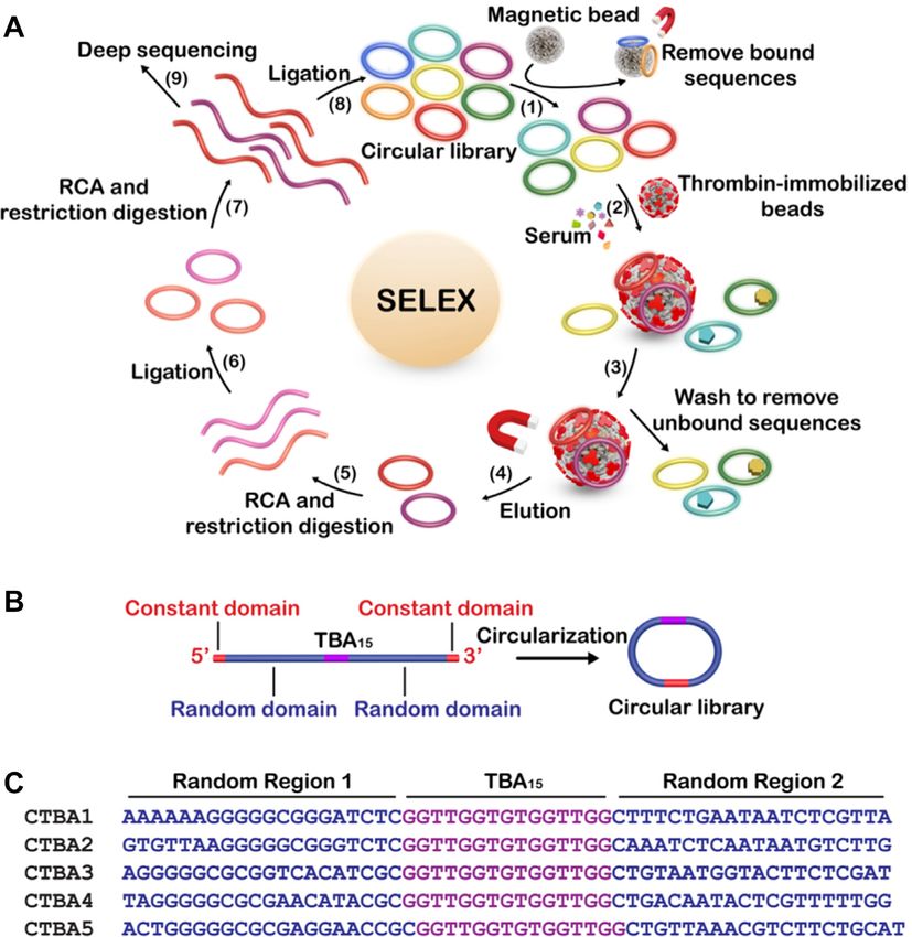
Nucleic Acids Research, 2020 3
from 10× stock, which is made of 330 mM Tris-acetate, pH FASTA format for further analysis (30). Sequences were du-
7.9 at 37◦ C, 100 mM magnesium acetate, 660 mM potas- plicated and tagged with copy number using USEARCH
sium acetate, 1% (v/v) Tween 20, 10 mM DTT) containing v7.0.1090 i86linux32 sequence analysis package (31). USE-
CTBA derived from last step, 2 M LT1 (100 pmol), 1 mM ARCH was also used for clustering of duplicated popula-
dNTP. After heating at 90◦ C for 3 min, the solution was tions using the cluster smallmem command at 0.9 identity
cooled at room temperature for 10 min. Subsequently, 0.5 l threshold. PANDAseq and USEARCH software packages
of phi29 DNA polymerase (10 U/l) was added, followed were run on Ubuntu Linux 12.04 LTS. Analysis of sequence
by incubation at 30◦ C for 30 min. Finally, the mixture was populations, rankings and base composition were done us-
heated to 65◦ C for 10 min to deactivate the polymerase. To ing Microsoft Excel 2010 running on a Windows 8 PC.
the RCA reaction mixture above, 2 l of 500 M LT2 (1
Downloaded from https://academic.oup.com/nar/advance-article/doi/10.1093/nar/gkaa800/5918318 by guest on 19 October 2020
nmol) was introduced. The mixture was heated at 90◦ C for
Thrombin inhibition assays
3 min and cooled at RT for 10 min, followed by the addi-
tion of 10 l of 10× Fast Digestion Buffer (100 mM Tris– Thrombin inhibition assays were performed by monitor-
HCl, pH 8.0, 50 mM MgCl2 , 1 M NaCl, 1 mg/ml BSA) ing the formation of insoluble fibrin by measuring the ab-
and 5 l of FastDigest EcoRV (unit size 400 reactions; the sorbance of the reaction at 350 nm on a microplate reader
total volume is 400 l). The total reaction volume was in- (Tecan) at 25◦ C. To determine the half-maximal inhibitory
creased to 100 l. The reaction mixture was then incubated concentrations (IC50 ) of the thrombin inhibitors, we pre-
at 37◦ C for 16 h (29). The restriction enzyme was inactivated pared reactions containing 0.1 nM thrombin, 2 M fibrino-
at 65◦ C for 10 min. The monomerized RCA products were gen (Sigma) and various inhibitors at a range of different
concentrated by standard ethanol precipitation and purified concentrations in 100 l of selection buffer in duplicate. For
by dPAGE. The DNA was then eluted and circularized into each thrombin inhibitor, the absorbance at steady state were
circular DNA template B (CDTB), which was used for the blank subtracted then normalized. These values were fitted
second RCA reaction. The reaction condition was identical to a four-parameter logistic model (Origin) by nonlinear re-
to the first RCA except for the replacement of LT1 with LT2. gression to calculate the IC50 . The model takes the form: y =
For the restriction digestion after RCA, LT2 was replaced D + (A – D)/(1 + (x/C)B , where y = normalized absorbance,
LT1. Serum was introduced into the binding buffer after x = the inhibitor concentration, and with the four parame-
two rounds of selection. Seven rounds of selection were con- ters, A = absorbance with no inhibition, B = the slope fac-
ducted while the amount of the serum was increased from tor, C = IC50 and D = absorbance at maximum inhibition
2% (round 3) to 5% (round 4), 10% (round 5), 20% (round (32).
6) and 50% (round 7). The DNA pool from round 7 was
used for deep sequencing.
Circular dichroism analysis
Circular dichroism (CD) studies were carried out using a
Sequencing protocol
JASCO J-600 instrument according to the manufacturer’s
CTBA in round 7 was digested into linear DNA sequences instructions. All constructs were scanned from 320 to 220
as previously described. 2 l of 0.05 M linear CTBA was nm at 20 M DNA in a 0.1-cm quartz cuvette. All DNA
amplified by PCR. There were two PCR steps. In PCR1, a samples were incubated for 30 min at room temperature in
reaction mixture (50 l) was prepared to contain the DNA the 1× selection buffer before scanning.
above, 0.4 M each of forward primer (FP) and reverse
primer (RP; their sequences are provided in Supplementary
Fluorescence measurements of G-quadruplex–ligand interac-
Table S1), 200 M each of dNTPs (dATP, dCTP, dGTP
tion
and dTTP), 1× PCR buffer (75 mM Tris–HCl, pH 9.0, 2
mM MgCl2 , 50 mM KCl, 20 mM (NH4 )2 SO4 ) and 1.5 U Thioflavin T (ThT; 1 M) was incubated with a relevant
Taq DNA polymerase. The DNA was amplified using the circular DNA (1 M) in binding buffer for 10 min. Fluo-
following thermocycling steps: 94◦ C for 3 min; 15 cycles rescence measurements were carried out in a 96-well assay
at 94◦ C (30 s), 42◦ C (45 s) and 72◦ C (45 s); 72◦ C for 1 plate using a microplate reader (Tecan infinite M1000 Pro,
min. 1 l of the PCR1 product was diluted with H2 O to Männedorf, Switzerland) under room temperature. The ex-
100 l, 2 l of which was used as the template for PCR2 citation wavelength of the solution was set at 425 nm, and
using the same primers (their sequences are provided in the emission spectra were collected from 450 to 600 nm with
Supplementary Table S1) while following the same pro- a step of 2 nm.
tocol above for PCR1 except that the annealing temper-
ature increased to 48◦ C. The DNA product generated in
Electrophoretic mobility shift assays (EMSA)
PCR2 was analyzed and purified by 2% agarose gel elec-
trophoresis and sent out for deep sequencing. Paired-end Binding reactions were performed in 20 l of 1× binding
next generation sequencing (NGS) was done using an Illu- buffer containing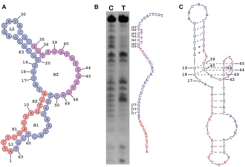
4 Nucleic Acids Research, 2020
by storage phosphor screen and imaged on a GE Typhoon Bio-Rad image system and the bands were quantified with
Biomolecular Imager and the resulting bands were quanti- ImageQuant software.
fied using ImageJ software.
Prothrombin time assay
Methylation interference assay
Prothrombin time (PT) assay on human plasma samples
A solution of 10 M FAM-labeled DNA (500 pmol) in was measured by using a semi-automatic coagulometer
1× PBSMT was heated to 90◦ C for 1 min and cooled to with a specific kit for blood coagulation test (Sun Biotech,
room temperature. For control samples, the same amount of Shanghai, China). Briefly, this method relies on the high
FAM-labeled DNA in ddH2 O was heated to 90◦ C for 1 min sensitivity of thromboplastin reagent based on recombinant
Downloaded from https://academic.oup.com/nar/advance-article/doi/10.1093/nar/gkaa800/5918318 by guest on 19 October 2020
and cooled to room temperature quickly to disrupt the fold- human tissue factors. The addition of recombiplastin to the
ing. The methylation reaction was initiated by adding an plasma, in presence of calcium ions, initiates the activation
equal volume of 0.4% (v/v) DMS (freshly made) to each of of extrinsic pathway converting the fibrinogen into fibrin,
the above reaction mixture, followed by incubation at room with a formation of solid gel. The procedure was performed
temperature for 40 min. Methylated DNA was recovered according to the manufacturer’s instructions. In our exper-
by ethanol precipitation, followed by two washes in 70% imental conditions, each thrombin inhibitor (200 M, 1.7
ethanol, and subjected to methylation-dependent cleavage l) was incubated with 100 l of plasma at 37◦ C for 3 min,
in 50 l 10% (v/v) piperidine for 30 min at 90◦ C. The resul- after that 200 l of the kit solution containing recombiplas-
tant cleavage products were dried under vacuum and ana- tin was added with consequent activation of extrinsic path-
lyzed by 20% denaturing PAGE. way. The PT measurement, for each incubation time, was
produced in triplicate and the average and its standard error
Competitive binding assay values were calculated and expressed as seconds. The basal
clotting time was determined by measuring the clotting time
Binding reactions were prepared with 32 P-labeled CTBA4T in absence of any thrombin inhibitor.
at 500 pM, 50 pM thrombin and a titration of unlabeled
competitor aptamer (Published thrombin aptamers TBA15
RESULTS
(Bock et al.) and ‘TBA29 ’ (Tasset et al.) targeting fibrino-
gen and heparin epitopes respectively were used as com- Directed evolution of circular aptamers in serum
petitors of CTBA4T for thrombin binding). Competition
The adaption strategy is illustrated in Figure 1A. The cir-
reactions were performed in 1× binding buffer and incu-
cular DNA library was prepared by T4 DNA ligase medi-
bated at room temperature for 1 h. Samples were prepared
ated circularization of Lib, a linear DNA library containing
for electrophoresis by addition of loading dye to 1× final
TBA15 flanked with a 20-nt random region and a constant
concentration.10% 0.5× TBE native PAGE was prerun at
region on each side (see Figure 1B for its construction, and
200 V for 1 h at 4◦ C. Samples were loaded and gel was
Supplementary Table S1 in the Supporting Information for
run at 200 V for 1 h. Storage phosphor screens were ex-
the sequences of all DNA molecules used in this work). The
posed for 1 h. Visualization of DNA bands was done us-
yield of circularization was calculated to be 86% (Supple-
ing a GE Typhoon Biomolecular Imager and the result-
mentary Figure S1). The use of two random regions to flank
ing bands were quantified with ImageJ software. Non-linear
TBA15 was intended to provide TBA15 the best chance to
regression performed in GraphPad Prism using a one-site
recruit additional functional nucleotides from both ends as
competitive binding model to estimate IC50 . Y = (top –
well as minimize the impact of cyclization on the binding ac-
bottom)/(1+10(X – log IC50) ) + bottom.
tivity of TBA15 . The circular DNA pool was first mixed with
bare carboxylic acid magnetic beads (CAMB) in a counter-
Thermal melting temperature analysis selection step to remove bead-binding sequences (step 1).
The high resolution melting experiments were carried out The unbound DNA was incubated with the CAMB coated
with Roche LightCycler96 quantitative fluorescence PCR with thrombin (step 2). This step was conducted directly
instrument. Samples were prepared at a concentration of 10 in serum. The unbound DNA molecules were removed by
M in LightCycler® 480 high resolution melting master, washing (step 3) and the bound species were eluted by heat-
which contained ResoLight Dye, a double-stranded DNA ing (step 4). The DNA recovered was then amplified by
binding Dye. The high resolution melting curves were ob- a circle-to-circle amplification strategy we reported before
tained by following the fluorescence change in the 37–90◦ C (29). Briefly, the bound circular DNA from step 4 was am-
range using a scan rate of 0.05◦ C/s and 20 readings/◦ C. plified by rolling circle amplification (RCA) (33–37). Fol-
lowing RCA and restriction digestion of long RCA products
into monomers (step 5), circularization was performed to
Stability assay
produce a new circular DNA (step 6), which was used as the
10 M FAM-labeled anti-thrombin aptamers (1000 pmol) template for another RCA and restriction digestion (step 7).
were incubated in 50% human serum at 37◦ C. At established Circularization was again performed to produce the circu-
times, ranging from 0 to 24 h, 10 l of samples were col- lar DNA pool for the next round of selection (step 8). It is
lected, heated at 90◦ C for 10 min and stored at –20◦ C for noteworthy that the circle-to-circle amplification has never
further analysis. The samples were then mixed with 10 l been attempted in previous aptamer selection experiments.
2× denaturing gel loading buffer and analyzed for 10% de- It represents a simpler method over polymerase chain reac-
naturing PAGE. Visualization of the gel was done using a tion (PCR) because it is isothermal and equipment-free.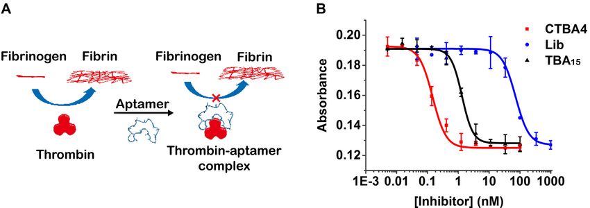
Nucleic Acids Research, 2020 5

                                                                                                                                                     Downloaded from https://academic.oup.com/nar/advance-article/doi/10.1093/nar/gkaa800/5918318 by guest on 19 October 2020
Figure 1. (A) Strategy for adapting TBA15 into a circular DNA aptamer in serum. (B) Circular DNA library design. (C) The sequence of top 5 aptamer
candidates. Each circular aptamer also contains the following constant sequence of 5 -TGTCT CGGAT ATCTC GACTA GTCA-3 .

Selected circular aptamer effectively inhibits thrombin                    Aptamer characterization and optimization
∼10 distinct circular DNA molecules were used as the ini-
     14
                                                                           The 79-nt sequence of CTBA4 can be divided into 5 se-
tial DNA library, with which seven rounds of selection were                quence regions as shown in Figure 3: CR1 (constant region
carried out under increasing stringency to favor isolation of              1, A1 –A14 ), RR1 (random region 1, T15 –G34 ), CR2 (G35 –
circular aptamers with the best activity in serum (2%, 5%,                 G49 ), RR2 (C50 –G69 ), CR3 (T70 –T79 ). A mutant aptamer of
10%, 20% and 50% serum in rounds 3, 4, 5, 6 and 7, re-                     CTBA4, named CTBA4MT, in which all the nucleotides in
spectively). The DNA pool from round 7 was subjected to                    the selected RR1 and RR2 sequence elements were mutated
high-throughput DNA sequencing (Supplementary Table                        to dT-nucleotides, was made and tested using TIFP. The
S2). 4 015 918 sequence reads were obtained; the top five se-              IC50 value of CTBA4MT increases by 22-fold, suggesting
quences account for ∼30%, indicating effective enrichment                  that some or all of the nucleotides within the RR1 and RR2
(the top fifty sequences from deep sequencing result were                  elements in CTBA4 were indeed functionally important. We
shown in Supplementary Figure S2).                                         then carried out T-tract walking experiment to determine
   We assessed the top five sequences for their ability to                 the importance of a group of consecutive nucleotides within
inhibit thrombin-induced fibrin polymerization (TIFP), a                   each region of CTBA4; in this experiment, each chosen nu-
process closely associated with blood-clotting (Figure 2A).                cleotide group was substituted with the same number of T
Each circular aptamer was incubated with thrombin and                      residues. Substitutions within CR2 led to the complete loss
fibrinogen. By monitoring A350 (absorbance at 350 nm;                      of activity (CTBA4M6 and CTBA4M7), this was not sur-
Supplementary Figure S3), we can determine the amount                      prising because CR2 was the seeding aptamer. Interestingly,
of insoluble fibrin produced and calculate the half maxi-                  substitutions within RR1 resulted in significantly increased
mal inhibitory concentration, IC50 (Supplementary Table                    IC50 (IC50 of CTBA4M3, CTBA4M4 and CTBA4M5 in-
S3). CTBA4 showed the best activity (Figure 2B), with an                   creased by 494-, 190- and 1390-fold, respectively), indicat-
IC50 of 0.15 nM. As controls, TBA15 and the original cir-                  ing nucleotides in this region were evolved to play important
cular DNA library had the IC50 values of 1.31 and 72.14                    roles in thrombin binding. Substitutions elsewhere had no
nM, respectively. The TIFP inhibition efficiency of CTBA4                  or less significant impact on IC50 , suggesting most residues
is 480-fold better than the circular library and 9-fold better             within CR1, RR2 and CR3 are unimportant for throm-
than TBA15 . These results are consistent with our specu-                  bin recognition. Among these mutants, CTBA4M10 and
lation that in vitro selection can produce highly functional               CTBA4M11 exhibited nearly identical IC50 to the mother
circular aptamers from a linear aptamer.                                   sequence. Based on these observations, we synthesized a
6 Nucleic Acids Research, 2020

                                                                                                                                                    Downloaded from https://academic.oup.com/nar/advance-article/doi/10.1093/nar/gkaa800/5918318 by guest on 19 October 2020
Figure 2. (A) Inhibition of thrombin-induced fibrin polymerization (TIFP) by an aptamer. (B) Inhibition of TIFP by CTBA4, TBA15 and Lib, measured
as absorbance at 350 nm as the function of inhibitor concentration. The data were fitted to a four-parameter logistic model.

Figure 3. T-substitution study of CTBA4.

shortened aptamer, CTBA4T, in which T65 -T79 was deleted.                  single-stranded element of B2, we were wondering if a new
This molecule had an IC50 of 90 pM.                                        G4 structure is formed that is now responsive for thrombin
  It was worth noting that there are five consecutive G                    binding.
residues in RR1 of CTBA4. In fact, the existence of ∼5                        The technique of circular dichroism (CD) was employed
consecutive guanines in either RR1 or RR2 is a common                      to provide evidence for the existence of G4 in the structure
feature of all top 50 selected sequences (green Gs in Supple-              of CTBA4T (Supplementary Figure S4). The CD spectrum
mentary Figure S2; 45 5Gs, 3 6Gs, 1 7Gs and 1 4Gs), sug-               indeed contains a negative peak close to 260 nm and a pos-
gesting an important structural or functional role for this                itive peak close to 290 nm, features that are indicative of
new evolved element (more to follow).                                      an antiparallel G-quadruplex (38). The slight shifting of the
                                                                           two peaks toward wavelengths lower than those usually seen
CTBA4 forms a unique G-quadruplex structure                                with pure antiparallel G4 structures may be caused by the
                                                                           influence of the flanking duplex structures (39,40).
To understand the structural basis of CTBA4T for signif-                      Methylation interference was performed to identify gua-
icantly increased TIFP inhibition efficiency over TBA15 ,                  nine residues involved in the G4 formation; this technique
we used mfold to predict Watson-Crick elements within                      can detect the sensitivity of N7 atoms of guanines to methy-
CTBA4T. The structure with the lowest free energy is shown                 lation and is widely used for G4 structure probing (41,42).
in Figure 4A. It contains two loops (L1, L2), two bulges (B1,              Because it is difficult to determine methylation interference
B2) and three stems (S1, S2, S3). One interesting prediction               directly with a circular DNA molecule, we sought to use
is the placement of G35 G36 T37 in S3, as these 3 nucleotides              the linear counterpart (LTBA4T). However, a comparison
were supposed to fold into the G-quadruplex (G4) structure                 of the predicted secondary structures for LTBA4T (Sup-
for thrombin binding in the original library design. Since                 plementary Figure S5b) and CTBA4T reveals significant
the remaining nucleotides in the original seeding aptamer                  disparities. However, a slightly modified version of the ap-
are still predicted to be a single-stranded element as part of             tamer, CTBA4T-B1 was predicted to form a structure (Sup-
B2 and there are also consecutive G residues in the other                  plementary Figure S5c) comparable to CTBA4T; for this
Nucleic Acids Research, 2020 7
Downloaded from https://academic.oup.com/nar/advance-article/doi/10.1093/nar/gkaa800/5918318 by guest on 19 October 2020
Figure 4. (A) Predicted secondary structure of CTBA4T. (B) Methylation interference of LTBA4T-B1: 20% dPAGE of LTBA4T-B1 after methylation
interference and piperidine cleavage. Four pairs of guanines showing significant methylation interference are highlighted along with their base number. C
refers to the control sample (without thrombin) and T refers to the test sample (with thrombin). (C) The proposed structure for CTBA4T.
reason, its linear form, LTBA4T-B1 (Supplementary Figure to create the G4 pairing is shown in Supplementary Figure
S5d) was used for the methylation interference experiment. S6.
Ten guanines within B2 exhibited significant methylation The proposed structure is very unique in that it con-
interference (Figure 4B), and they can be grouped as four tains both G-quadruplex and duplex elements. This struc-
pairs of two consecutive G residues: G18 G19 (or G17 G18 ), ture differs from all known G4 structures in two major
G39 G40 , G44 G45 , G48 G49 . This points to a possibility that ways (43–46). First, it has a circular topology as common
these guanines form the two-tiered G4 as illustrated in Fig- G4 structures are made of linear nucleic acids. Second, it
ure 4C. G17 (or G19 ) and G42 also showed strong methy- has two large and structured loop elements. This may be
lation interference. It is possible that these two guanine also linked to its circular topology, because, as a circular
residues participate in some important tertiary interactions molecule, CTBA4T may require more nucleotides to build
that involve their N7 atoms. Alternatively, being located inter-strand connecting loops to stabilize its core G4 ele-
very close to the G4 element may place them in tight spatial ment. Given its better performance over TBA15 , the isola-
arrangements, preventing them from tolerating the bulky tion of CTBA4T points to the added advantage of evolving
methyl group on N7. It is noteworthy that G35 and G36 did aptamers from circular DNA libraries since this approach
not exhibit substantial methylation interference, consistent can generate aptamers with structural folds that are unique
with the structural model predicted with mfold in which to the circular topology.
these two G nucleotides are arranged as part of the S3 stem There are also many unpaired nucleotides near and away
(the N7 atoms of guanine in DNA duplex does not produce from the G-quadruplex element, which may further partic-
strong methylation interference). ipate in some very important tertiary interactions that ulti-
The methylation experiment strongly suggests that mately contribute to the significantly increased TIFP inhi-
G18 G19 or (G17 G18 ) within RR1 was evolved to partner bition efficiency and affinity over TBA15 .
with G39 G40 , G44 G45 , and G48 G49 within CR2 (i.e. the seed- We next designed 14 mutants of CTBA4T with mutations
ing aptamer region) to create a new G4 structure. In the targeting B1, L2, B2, S3 and examined their relative TIFP
process, the G35 G36 pair that are an integral part of the inhibition efficiencies in an attempt to gather information
G4 structure of TBA15 were re-arranged into a stem that on the functionality of these structural elements (Figure 5).
is next to the G4 element (Figure 4C). Since G17 G18 G19 all We found that B1 could be eliminated altogether and the
showed methylation interference and they can be arranged resultant mutant CTBA4T-B1 retained the full TIFP inhi-
as G18 G19 pair or as G17 G18 pair, for the formation of a G4 bition activity. Similarly, the full activity was maintained
structure, an alternative structure in which G17 G18 was used when the sequence of L2 was scrambled (CTBA4T-M1).8 Nucleic Acids Research, 2020
Downloaded from https://academic.oup.com/nar/advance-article/doi/10.1093/nar/gkaa800/5918318 by guest on 19 October 2020
Figure 5. Permutations of B1, B2, S3 and L2. The putative secondary structure of CTBA4T is shown on the left, and mutant aptamers are shown on the
right. The name of each mutant aptamer is given underneath each altered motif. Nucleotides shown in green are the actual altered nucleotides in each
construct in comparison to CTBA4T.
These two observations indicate that B1 and L2 do not play bition activities observed with the other mutants made in
any role in target recognition. S3 largely support the existence of the proposed S3 stem el-
Several mutants (CTBA4T-M2-8) were designed to ex- ement, but also suggest that nucleotides within this element
amine functionalities of the nucleotides in S3. The most may play important, but not absolutely essential, roles in
interesting observation is that the G-toT mutation of G36 the structural formation and/or target recognition of the
(CTBAT-M2), which is known to participate in the forma- new evolved aptamer.
tion of G4 in the seed aptamer TBA15 , did not affect the Six mutants (CTBA4T-M9-14) targeting B2 were also
TIFP inhibition efficiency. This finding is consistent with tested. The G-toT mutation of G18 (CTBA4T-M9) and G19
our proposed structural model of CTBA4T that does not (CTBA4T-M10) led to complete loss of TIFP inhibition
involve G36 in the G4 structure formation. The TIFP inhi- activity, consistent with the proposed structural model ofNucleic Acids Research, 2020 9
Nuclease resistance in human serum and the anticoagulant
activity in blood
The nuclease resistance of CTBA4T, CTBA4T-B1 and their
linear forms in biological media was investigated next. Fig-
ure 6A shows that the half-life of CTBA4T, CTBA4T-B1
and their linear forms in 50% human serum was circa 8 h,
while the half-life of TBA15 was less than 5 min, illustrat-
ing the superb capability of the selected aptamers against
nuclease degradation. To further evaluate the anticoagu-
Downloaded from https://academic.oup.com/nar/advance-article/doi/10.1093/nar/gkaa800/5918318 by guest on 19 October 2020
lant activity in blood, CTBA4T, CTBA4T-B1, LTBA4T,
Figure 6. (A) Stability of TBA15 , LTBA4T, CTBA4T, LTBA4T-B1 and
CTBA4T-B1 in serum. (B) PT values of Argatroban, TBA15 , LTBA4T, LTBA4T-B1, TBA15 and Argatroban (a small molecule in-
LTBA4T-B1, CTBA4T and CTBA4T-B1. ***P < 0.001 versus vehicle, hibitor of thrombin) were subjected to Prothrombin Time
◦◦◦ P < 0.001 versus TBA (n = 3).
15 (PT) assay (49,50). CTBA4T and CTBA4T-B1 showed sig-
nificantly prolonged PT over LTBA4T, LTBA4T-B1, TBA15
and Argatroban (Agtb), while CTBA4T-B1 exhibited the
CTBA4T that places G18 and G19 as part of the G4 element. highest PT value, indicating a much higher anticoagulant
The G-toT mutant of G17 (CTBA4T-M11) resulted in sig- activity in human plasma (Figure 6B). Interestingly, despite
nitifcant loss of TIFP inhibition activity while the mutants that LTBA4T and LTBA4T-B1 are as stable as CTBA4T
carrying T15C, A16T, or C50T mutation (CTBA4T-M12- and CTBA4T-B1 in human serum, they produced much
14) exhibited substantial activity losses. These observations poorer PT values. Given the observation that each circular
suggest that these four unpaired nucleotides next to the pro- form exhibits a higher melting temperature than their lin-
posed G4 structure may participate in target recognition or ear counterpart, we believe the high PT value of each circu-
play important structural roles. lar construct reflects their high structural stability and thus
The existence of a G4 structure in CTBA4 was further their ability to resist the interference from other DNA bind-
confirmed with the experiment with Thioflavin T, a fluores- ing molecules in biological media. The result clearly under-
cent dye that specifically binds G4 and produce enhanced scores the potential therapeutic value of CTBA4T-B1 for
fluorescence (Supplementary Figure S7) (47). blood coagulation.
DISCUSSION
High binding affinity and thermal stability In summary, we report the first effort to derive a protein-
binding circular DNA aptamer directly in serum, resulting
CTBA4T and CTBA4T-B1 were then radioactively labeled in the isolation of a highly functional circular DNA ap-
and tested for thrombin binding by electrophoretic mobility tamer that specifically and strongly binds thrombin, and
shift assays (EMSA; Supplementary Figure S8). The frac- shows potent activity to inhibit thrombin-induced fibrin
tion of bound aptamer (top band) was plotted as a func- polymerization. This condition-driven strategy to evolve
tion of thrombin concentration, resulting in a Kd of 23 pM a functional circular aptamer from a precursor linear ap-
and 19 pM for CTBA4T and CTBA4T-B1, respectively. tamer requires no structural information for the target
Strikingly, their binding affinity was much better than the protein, and thus can be expanded to any protein target
circular control, CTBA4MT, and their linear forms (Sup- with and without determined structures. The isolation of
plementary Figure S9), showing a huge improvement from CTBA4T-B1 also highlights the advantage of selecting cir-
their mother sequence TBA15 (∼800-fold for CTBA4T and cular aptamers directly in biological media, because this ap-
∼1000-fold for CTBA4T-B1). proach can produce aptamers with improved physical sta-
It is noteworthy that CTBA4T recognizes the same bility and functionality in the intended biological media.
epitope in thrombin, exosite I, that is recognized by The use of RCA as an amplification tool has also signifi-
TBA15 . This was clear from the competitive binding as- cantly simplified the enrichment of circular aptamers. Taken
say shown Supplementary Figure S10: CTBA4T in the together, we envision that the demonstrated strategy can be
aptamer/thrombin complex can be replaced by TBA15 exploited for creating a wide variety of high-quality circular
at high concentrations. This is not surprising as TBA15 DNA aptamers that are both stable and highly functional in
was used as the seeding aptamer. As a control, thrombin- real biological samples, thus boosting the practical utilities
complexed CTBA4T could not be displaced by TBA29 , a 29- of DNA aptamers for biomedical applications.
nt DNA aptamer that is known to bind exosite II of throm-
bin (Supplementary Figure S9) (48).
Thermal melting experiments were carried out to de- SUPPLEMENTARY DATA
termine the relative thermal stabilities for the circular ap- Supplementary Data are available at NAR Online.
tamers. The thermal melting temperatures (Tm ) obtained
from high resolution melting curves were shown in Supple-
FUNDING
mentary Table S4. Inspection of Supplementary Table S4
reveals a remarkable enhancement (∼+10◦ C) of the thermal National Key Research and Development Program of
stability for CTBA4T and CTBA4T-B1 versus their linear China [2017YFC1600603]; Key Science & Technology Spe-
form (LTBA4T and LTBA4T-B1). cific Projects of Anhui Province [15czz03117, 16030701078];10 Nucleic Acids Research, 2020
China Postdoctoral Science Foundation Funded Project complexes between thrombin and thrombin-binding aptamer shed
[113-471038]; Natural Sciences and Engineering Research light on the role of cations in the aptamer inhibitory activity. Nucleic
Acids Res., 40, 8119–8128.
Council of Canada (NSERC). Funding for open access 23. Davie,E.W. and Kulman,J.D. (2006) An overview of the structure and
charge: National Key Research and Development Program function of thrombin. Semin. Thromb. Hemostasis, 32, 3–5.
of China. 24. Crawley,J.T., Zanardelli,S., Chion,C.K. and Lane,D.A.J. (2007) The
Conflict of interest statement. None declared. central role of thrombin in hemostasis. Thromb. Hemostasis, 5,
95–101.
25. Franchini,M. and Mannucci,P.M. (2012) Thrombin and cancer: from
molecular basis to therapeutic implications. Semin. Thromb.
REFERENCES Hemostasis, 38, 95–101.
Downloaded from https://academic.oup.com/nar/advance-article/doi/10.1093/nar/gkaa800/5918318 by guest on 19 October 2020
1. Ellington,A.D. and Szostak,J.W. (1990) In vitro selection of RNA 26. Griffin,L.C., Tidmarsh,G.F., Bock,L.C., Toole,J.J. and Leung,L.L.K.
molecules that bind specific ligands. Nature, 346, 818–822. (1993) In vivo anticoagulant properties of a novel nucleotide-based
2. Tuerk,C. and Gold,L. (1990) Systematic evolution of ligands by thrombin inhibitor and demonstration of regional anticoagulation in
exponential enrichment: RNA ligands to bacteriophage T4 DNA extracorporeal circuits. Blood, 81, 3271–3276.
polymerase. Science, 249, 505–510. 27. Müller,J., Wulffen,B., Pçtzsch,B. and Mayer,G. (2007) Multidomain
3. Robertson,D.L. and Joyce,G.F. (1990) Selection in vitro of an RNA targeting generates a high-affinity thrombin-Inhibiting bivalent
enzyme that specifically cleaves single-stranded DNA. Nature, 344, aptamer. ChemBioChem., 8, 2223–2226.
467–468. 28. Ahmad,K.M., Xiao,Y. and Soh,H.T. (2012) Selection is more
4. Liu,J., Cao,Z. and Lu,Y. (2009) Functional nucleic acid sensors. intelligent than design: improving the affinity of a bivalent ligand
Chem. Rev., 109, 1948–1998. through directed evolution. Nucleic Acids Res., 40, 11777–11783.
5. Keefe,A.D., Pai,S. and Ellington,A.D. (2010) Aptamers as 29. Mao,Y., Liu,M., Tram,K., Gu,J., Salena,B.J., Jiang,Y. and Li,Y.
therapeutics. Nat. Rev. Drug Discov., 9, 537–550. (2015) Optimal DNA templates for rolling circle amplification
6. Tan,W., Donovan,M.J. and Jiang,J. (2013) Aptamers from cell-based revealed by in vitro selection. Chem. Eur. J., 21, 2420–2424.
selection for bioanalytical applications. Chem. Rev., 113, 2842–2862. 30. Masella,A.P., Bartram,A.K., Truszkowski,J.M., Brown,D.G. and
7. Osborne,S.E. and Ellington,A.D. (1997) Nucleic acid selection and Neufeld,J.D. (2012) PANDAseq: paired-end assembler for illumina
the challenge of combinatorial chemistry. Chem. Rev., 97, 349–370. sequences. BMC Bioinformatics, 13, 31.
8. Kusser,W. (2000) Chemically modified nucleic acid aptamers for in 31. Edgar,R.C. (2010) Search and clustering orders of magnitude faster
vitro selections: evolving evolution. Rev. Mol. Biotechnol., 74, 27–38. than BLAST. Bioinformatics, 26, 2460–2461.
9. Lee,J.H., Canny,M.D., De Erkenez,A., Krilleke,D., Ng,Y.S., 32. Findlay,J.W. and Dillard,R.F. (2007) Appropriate calibration curve
Shima,D.T., Pardi,A. and Jucker,F. (2005) A therapeutic aptamer fitting in ligand binding assays. AAPS J., 9, E260–E267.
inhibits angiogenesis by specifically targeting the heparin binding 33. Fire,A. and Xu,S.Q. (1995) Rolling replication of short DNA circles.
domain of VEGF165. Proc. Natl. Acad. Sci. U.S.A., 102, Proc. Natl. Acad. Sci. U.S.A., 92, 4641–4645.
18902–18907. 34. Liu,D., Daubendiek,S.L., Zillman,M.A., Ryan,K. and Kool,E.T.
10. Dellafiore,M.A., Montserrat,J.M. and Iribarren,A.M. (2016) (1996) Rolling circle DNA synthesis:? small circular oligonucleotides
Modified nucleoside triphosphates for in-vitro selection techniques. as efficient templates for DNA polymerases. J. Am. Chem. Soc., 118,
Front. Chem., 4, 18. 1587–1594.
11. Bohjanen,P.R., Colvin,R.A., Puttaraju,M., Been,M.D. and 35. Liu,M., Zhang,W., Zhang,Q., Brennan,J.D. and Li,Y. (2015)
Garcia-Blanco,M.A. (1996) A small circular TAR RNA decoy Biosensing by tandem reactions of structure switching, nucleolytic
specifically inhibits Tat-activated HIV-1 transcription. Nucleic Acids digestion, and DNA amplification of a DNA assembly. Angew.
Res., 24, 3733–3738. Chem. Int. Ed., 54, 9637–9641.
12. Di Giusto,D.A. and King,G.C. (2004) Construction, stability, and 36. Liu,M., Zhang,Q., Li,Z., Gu,J., Brennan,J.D. and Li,Y. (2016)
activity of multivalent circular anticoagulant aptamers. J. Biol. Programming a topologically constrained DNA nanostructure into a
Chem., 279, 46483–46489. sensor. Nat. Commun., 7, 12704.
13. Tang,X., Su,M., Yu,L., Lv,C., Wang,J. and Li,Z. (2010) 37. Liu,M., Zhang,Q., Chang,D., Gu,J., Brennan,J.D. and Li,Y. (2017) A
Photomodulating RNA cleavage using photolabile circular antisense DNAzyme feedback amplification strategy for biosensing. Angew.
oligodeoxynucleotides. Nucleic Acids Res., 38, 3848–3855. Chem. Int. Ed., 56, 6142–6146.
14. Di Giusto,D.A., Knox,S.M., Lai,Y., Tyrelle,G.D., Aung,M.T. and 38. Dolinnaya,N.G., Yuminova,A.V., Spiridonova,V.A.,
King,G.C. (2006) Multitasking by multivalent circular DNA Arutyunyan,A.M. and Kopylova,A.M. (2012) Coexistence of
aptamers. ChemBioChem, 7, 535–544. G-quadruplex and duplex domains within the secondary structure of
15. Kuai,H., Zhao,Z., Mo,L., Liu,H., Hu,X., Fu,T., Zhang,X. and 31-mer DNA thrombin-binding aptamer. J. Biomol. Struct. Dyn., 30,
Tan,W. (2017) Circular bivalent aptamers enable in vivo stability and 524–531.
recognition. J. Am. Chem. Soc., 139, 9128–9131. 39. Lim,K.W., Khong,Z.J. and Phan,A.T. (2014) Thermal stability of
16. Dong,H., Han,L., Wu,Z.S., Zhang,T., Xie,J., Ma,J., Wang,J., Li,T., DNA quadruplex–duplex hybrids. Biochemistry, 53, 247–257.
Gao,Y., Shao,J., Sinko,P.J. and Jia,L. (2017) Biostable aptamer rings 40. Lim,K.W., Nguyen,T.Q.N. and Phan,A.T. (2014) Joining of multiple
conjugated for targeting two biomarkers on circulating tumor cells in duplex stems at a single quadruplex loop. J. Am. Chem. Soc., 136,
vivo with great precision. Chem. Mater., 29, 10312–10325. 17969–17973.
17. Di Giusto,D.A., Wlassoff,W.A., Gooding,J.J., Messerle,B.A. and 41. Shen,Y., Brennan,J.D. and Li,Y. (2005) Characterizing the secondary
King,G.C. (2005) Proximity extension of circular DNA aptamers structure and identifying functionally essential nucleotides of
with real-time protein detection. Nucleic Acids Res., 33, e64. pH6DZ1, a fluorescence-signaling and RNA-cleaving
18. Wang,L., Tram,K., Ali,M.M., Salena,B.J., Li,J. and Li,Y. (2014) deoxyribozyme. Biochemistry, 44, 12066–12076.
Arrest of rolling circle amplification by protein-binding DNA 42. McManus,S.A. and Li,Y. (2008) A deoxyribozyme with a novel
aptamers. Chem. Eur. J., 20, 2420–2424. guanine quartet-helix pseudoknot structure. J. Mol. Bol., 375,
19. Bock,L.C., Griffin,L.C., Latham,J.A., Vermaas,E.H. and Toole,J.J. 960–968.
(1992) Selection of single-stranded DNA molecules that bind and 43. Tucker,W.O., Shum,K.T. and Tanner,J.A. (2012) G-quadruplex DNA
inhibit human thrombin. Nature, 355, 564–566. aptamers and their ligands: structure, function and application. Curr.
20. Tasset,D.M., Kubik,M.F. and Steiner,W.J. (1997) Oligonucleotide Pharm. Des., 18, 2014–2026.
inhibitors of human thrombin that bind distinct epitopes. Mol. Biol., 44. Neidle,S. (2016) Quadruplex nucleic acids as novel therapeutic
272, 688–698. targets. J. Med. Chem., 59, 5987–6011.
21. Paborsky,L.R., McCurdy,S.N., Griffin,L.C., Toole,J.J. and 45. Platella,C., Riccardi,C., Montesarchio,D., Roviello,G.N. and
Leung,L.L.K. (1993) The single-stranded DNA aptamer-binding site Musumeci,D. (2017) G-quadruplex-based aptamers against protein
of human thrombin. J. Biol. Chem., 268, 20808–20811. targets in therapy and diagnostics. Biochim. Biophys. Acta, Gen.
22. Russo Krauss,I., Merlino,A., Randazzo,A., Novellino,E., Subj., 1861, 1427–1447.
Mazzarella,L. and Sica,F. (2012) High-resolution structures of twoNucleic Acids Research, 2020 11
46. Kwok,C.K. and Merrick,C.J. (2017) G-guadruplexes: prediction, 49. Scuotto,M., Rivieccio,E., Varone,A., Corda,D., Bucci,M.,
characterization, and biological application. Trends Biotechnol., 12, Vellecco,V., Cirino,G., Virgilio,A., Esposito,V., Galeone,A. et al.
997–1013. (2015) Site specific replacements of a single loop nucleoside with a
47. Mohanty,J., Barooah,N., Dhamodharan,V., Harikrishna,S., dibenzyl linker may switch the activity of TBA from anticoagulant to
Pradeepkumar,P.I. and Bhasikuttan,A.C. (2013) Thioflavin T as an antiproliferative. Nucleic Acids Res., 43, 7702–7716.
efficient inducer and selective fluorescent sensor for the human 50. Virgilio,A., Petraccone,L., Vellecco,V., Bucci,M., Varra,M., Irace,C.,
telomeric G-quadruplex DNA. J. Am. Chem. Soc., 135, 367–376. Santamaria,R., Pepe,A., Mayol,L., Esposito,V. et al. (2015)
48. Tasset,D.M., Kubik,M.F. and Steiner,W.J. (1997) Oligonucleotide Site-specific replacement of the thymine methyl group by fluorine in
inhibitors of human thrombin that bind distinct epitopes. Mol. Biol., thrombin binding aptamer significantly improves structural stability
272, 688–698. and anticoagulant activity. Nucleic Acids Res., 43, 10602–10611.
Downloaded from https://academic.oup.com/nar/advance-article/doi/10.1093/nar/gkaa800/5918318 by guest on 19 October 2020You can also read