CRISPR-Cas9-Based Discovery of the Verrucosidin Biosynthesis Gene Cluster in Penicillium polonicum - Frontiers
←
→
Page content transcription
If your browser does not render page correctly, please read the page content below
ORIGINAL RESEARCH
                                                                                                                                                   published: 21 May 2021
                                                                                                                                          doi: 10.3389/fmicb.2021.660871

                                             CRISPR-Cas9-Based Discovery of
                                             the Verrucosidin Biosynthesis Gene
                                             Cluster in Penicillium polonicum
                                             Silvia Valente 1,2 , Edoardo Piombo 1,2 , Volker Schroeckh 3 , Giovanna Roberta Meloni 1,2 ,
                                             Thorsten Heinekamp 3 , Axel A. Brakhage 3,4* and Davide Spadaro 1,2*
                                             1
                                               AGROINNOVA – Centre of Competence for the Innovation in the Agro-Environmental Sector, Grugliasco, Italy,
                                             2
                                               Department of Agricultural, Forest and Food Sciences, Università degli Studi di Torino, Grugliasco, Italy, 3 Department
                                             of Molecular and Applied Microbiology, Leibniz Institute for Natural Product Research and Infection Biology – Hans Knöll
                                             Institute, Jena, Germany, 4 Department of Microbiology and Molecular Biology, Institute for Microbiology, Friedrich Schiller
                                             University, Jena, Germany

                            Edited by:       Penicillium polonicum, commonly found on food matrices, is a mycotoxigenic species
                   Esther Garcia-Cela,
            University of Hertfordshire,
                                             able to produce a neurotoxin called verrucosidin. This methylated α-pyrone polyketide
                       United Kingdom        inhibits oxidative phosphorylation in mitochondria and thereby causes neurological
                      Reviewed by:           diseases. Despite the importance of verrucosidin as a toxin, its biosynthetic genes
                  Jéssica Gil-Serna,
                                             have not been characterized yet. By similarity analysis with the polyketide synthase
   Complutense University of Madrid,
                                Spain        (PKS) genes for the α-pyrones aurovertin (AurA) and citreoviridin (CtvA), 16 PKS
               Alessandra Lanubile,          genes for putative α-pyrones were identified in the P. polonicum genome. A single
    Catholic University of the Sacred
                           Heart, Italy
                                             PKS gene, verA, was found to be transcribed under verrucosidin-producing growth
                  *Correspondence:
                                             conditions. The annotated functions of the genes neighboring verA correspond to those
                    Axel A. Brakhage         required for verrucosidin biosynthesis. To prove the involvement of verA in verrucosidin
           Axel.Brakhage@hki-jena.de
                                             biosynthesis, the clustered regularly interspaced short palindrome repeats (CRISPR)
                     Davide Spadaro
             davide.spadaro@unito.it         technology was applied to P. polonicum. In vitro reconstituted CRISPR-Cas9 was used
                                             to induce targeted gene deletions in P. polonicum. This approach allowed identifying and
                   Specialty section:
                                             characterizing the verrucosidin biosynthetic gene cluster. VerA deletion mutants were
         This article was submitted to
                    Food Microbiology,       no longer able to produce verrucosidin, whereas they were displaying morphological
               a section of the journal      characteristics comparable with the wild-type strain. The available CRISPR-Cas9
             Frontiers in Microbiology
                                             technology allows characterizing the biosynthetic potential of P. polonicum as a valuable
         Received: 29 January 2021
           Accepted: 15 April 2021
                                             source of novel compounds.
           Published: 21 May 2021
                                             Keywords: secondary metabolites, Penicillium, mycotoxins, CRISPR-Cas, alpha-pyrone polyketides
                              Citation:
                  Valente S, Piombo E,
              Schroeckh V, Meloni GR,       INTRODUCTION
       Heinekamp T, Brakhage AA and
                     Spadaro D (2021)
                                            Penicillium polonicum is a ubiquitous fungus, found as a contaminant of food matrices, such as
      CRISPR-Cas9-Based Discovery
of the Verrucosidin Biosynthesis Gene
                                            meat (Sunesen and Stahnke, 2003; Wigmann et al., 2018), nuts (Prencipe et al., 2018), and fresh
      Cluster in Penicillium polonicum.     fruit, like grape berries and strawberries (Jensen et al., 2013; Santini et al., 2014). It is also reported
          Front. Microbiol. 12:660871.      as a postharvest pathogen on apple (Ouhibi et al., 2018), pear (Scholtz and Korsten, 2016), onion
     doi: 10.3389/fmicb.2021.660871         (Duduk et al., 2014), chestnut (Prencipe et al., 2018), and cactus pear (Faedda et al., 2015). The

Frontiers in Microbiology | www.frontiersin.org                                      1                                              May 2021 | Volume 12 | Article 660871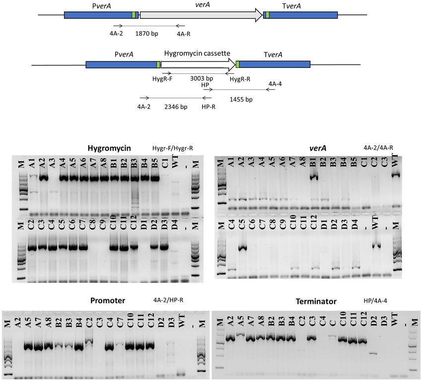
Valente et al.                                                                                                  CRISPR-Cas9 for Verrucosidin Cluster

fungus can grow as saprophyte in diverse environments (Sonjak               and resistance proteins. Thousands of gene clusters have been
et al., 2006). P. polonicum is able to produce a variety of secondary       described in fungal genomes, a number that is higher compared
metabolites (SMs), including mycotoxins, which exhibit toxic                to the chemically identified compounds (Yu et al., 2015). Most
effects on animals and humans. Characteristic compounds                     of these uncharacterized BGCs are often “silent,” meaning their
produced by P. polonicum are penicillic acid, aspterric acid,               genes are not transcribed under laboratory conditions and a
verrucofortine, cyclopenins, nephrotoxic glycopeptides, and                 number of strategies exist for their activation (Bergmann et al.,
verrucosidin (Frisvad et al., 2004), a neurotoxic compound                  2007, 2010; Fischer et al., 2016). On the other hand, gene
initially isolated from Penicillium verrucosum var. cyclopium,              deletion on active BGCs can be used to characterize the function
and later from other Penicillium species, such as Penicillium               of the tailoring genes and to assign a BGC to a compound
expansum (Burka et al., 1983; Frisvad et al., 2004; Kim et al.,             (Valente et al., 2020).
2016).                                                                          The clustered regularly interspaced short palindrome repeats
    Penicillium polonicum strain X6 was previously identified,              (CRISPR) technology is by now the method of choice in genome
by adopting a polyphasic approach based on molecular,                       studies of filamentous fungi (Nødvig et al., 2015; Fang and Tyler,
morphological, and chemical characterization (Prencipe et al.,              2016; Pohl et al., 2016; Deng et al., 2017; Krappmann, 2017; Nagy
2018). A multilocus phylogenetic analysis was performed using               et al., 2017; Nielsen et al., 2017; Weber et al., 2017; Song et al.,
internal transcribed spacer (ITS) region, calmodulin, and                   2019; Tong et al., 2019; Wang and Coleman, 2019). It allows
β-tubulin partial genes. Morphological analysis was conducted               marker-free genome editing by generating mutations at specific
on three different media: malt extract agar (MEA), czapek yeast             sites in the genome. CRISPR is based on the endonuclease Cas9,
agar (CYA), and yeast extract sucrose (YES). Finally, the SMs               that, when associated with small sequences of crRNA (CRISPR-
produced in vivo were analyzed, confirming the ability of this              RNA) and tracrRNA (Trans-activating crRNA), usually referred
strain to produce verrucosidin.                                             to as guide RNA (gRNA), recognizes genomic protospacer
    From the chemical point of view, verrucosidin is a highly               sequences and site-specifically cuts the double strand of DNA
reduced polyketide, composed of a methylated α-pyrone, a                    (Tong et al., 2019).
polyene linker, and an epoxidated tetrahydrofuran ring. Many                    In the present manuscript, we described the bioinformatic
polyketides share the α-pyrone structure, such as citreoviridin             identification of a number of α-pyrone polyketide synthases
and aurovertins, and have been extensively studied because of               (PKSs) in P. polonicum. One BGC was selected based on
their ability to inhibit mitochondrial oxidative phosphorylation,           the putative function of the biosynthetic genes of the cluster,
resulting in cytotoxicity and potential antitumor activity (Li              and the verrucosidin-encoding PKS was confirmed to be
et al., 2018). Verrucosidin acts on the central nervous system,             expressed in verrucosidin-producing Penicillium strains. Besides,
causing neurological diseases, first diagnosed in cattle and                we established the CRISPR-Cas9 technology in P. polonicum
experimentally confirmed in mice (Hodge et al., 1988; Fink-                 X6, which helped us characterize the verrucosidin BGC in
Gremmels et al., 1991). Verrucosidin is the most cytotoxic and              P. polonicum.
genotoxic compound among tremorgenic mycotoxins (Núñez
et al., 2000; Sabater-Vilar et al., 2003). Based on its cytotoxicity,
verrucosidin has been evaluated for its effect against cancer cells         MATERIALS AND METHODS
(Park et al., 2007; Thomas et al., 2013), making this compound
interesting for medical purposes. Despite the importance of                 Fungal Strains
this metabolite for human health and its potential benefit in               Penicillium polonicum strain X6 and P. crustosum strain CAL64
medicine, only few studies have been conducted to elucidate its             were isolated from chestnut production chain (Prencipe et al.,
biosynthesis. Núñez et al. (2000) determined favorable growth               2018). Penicillium aurantiogriseum CBS 112021 was obtained
conditions of P. polonicum for verrucosidin production, whereas             from the Westerdijk Fungal Biodiversity Institute and used as
Aranda et al. (2002) screened verrucosidin-producer strains to              a positive control of verrucosidin production. The strains were
develop a molecular probe to identify mycotoxigenic fungi in                grown on Potato Dextrose Agar plates (PDA, Merck KGaA,
foodstuff, which was further used by Rodríguez et al. (2012) to             Darmstadt, Germany) with 50 µg/ml streptomycin (Merck
design a TaqMan probe. Nowadays, the elucidated biosyntheses                KGaA) in the dark at 25◦ C for 7–10 days. P. polonicum mutants
of the structurally related α-pyrone polyenes citreoviridin,                obtained were grown on PDA supplemented with 100 µg/ml
aurovertins, and aspernidgulenes (Lin et al., 2016, 2019; Li et al.,        of hygromycin B (Thermo Fischer Scientific, Waltham, MA,
2018), available fungal genome sequences (Weber and Kim, 2016;              United States) under the same conditions. All the strains were
Nielsen and Nielsen, 2017), and bioinformatic tools open up                 maintained in glycerol stock at –80◦ C.
the possibility to identify the biosynthetic genes responsible for
verrucosidin biosynthesis.                                                  Growth of Penicillium spp. in vitro
    Biosynthetic gene clusters (BGCs) (Brakhage, 2013; Keller,              Conidial suspensions were obtained by adding 5 ml of sterile
2019) consist of central biosynthetic genes, often PKS or/and               water with 0.01% (v/v) Tween-20 and gently scraping the
non-ribosomal peptide synthetase (NRPSs) genes, and are                     surface of fungal cultures grown on Petri dishes, as in Spadaro
associated with additional genes encoding so-called tailoring               et al. (2013). The final conidia concentration was measured
enzymes, which are required for the modification of the                     using a hemocytometer and adjusted by dilution to different
carbon structure or encode transporters, transcription factors,             concentrations depending on each assay.

Frontiers in Microbiology | www.frontiersin.org                         2                                      May 2021 | Volume 12 | Article 660871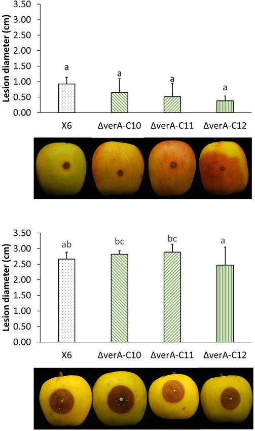
Valente et al.                                                                                               CRISPR-Cas9 for Verrucosidin Cluster

    To evaluate verrucosidin production in vitro, 1 ml of                  according to the manufacturer’s instruction. All primer sequences
conidial suspension (108 conidia/ml) was inoculated in 30 ml               used in the PCR are listed in Supplementary Table 1.
of malt extract broth (MEB, w/v: 2% malt extract, 2% glucose,
0.1% peptone) and in 30 ml of Czapek yeast broth (CYB, w/v:                RNA Extraction and RT-qPCRs
0.5% yeast extract, 3.5% Czapek broth). Flasks were kept at 26◦ C          RNA was extracted from 100 mg of fungal mycelium using
for 10 days, 55% RH with 12 h of light and 12 h of dark. After             Spectrum Plant Total RNA (Sigma-Aldrich, St. Louis, MO,
10 days of incubation, the fungal mycelium was separated from              United States), following the manufacturer’s instructions.
the liquid media through a double layer of sterile gauze and half of       Mycelium was placed in a 2-ml tube with two tungsten
the mycelium was immediately frozen in liquid nitrogen, stored             beads and tubes were immersed in liquid nitrogen for 1 min.
at –80◦ C, and used for RNA extraction. The remaining fungal               Then, samples were immediately lysed using TissueLyser II at
tissue was used for verrucosidin extraction.                               20.00 Hz for 1 min.
                                                                              DNase treatment and first-strand cDNA synthesis were
                                                                           performed according to Valente et al. (2020) using a TURBO
Bioinformatic Analyses
                                                                           DNA-freeTM Kit (Thermo Fischer Scientific) and a High Capacity
To identify BGCs in the genomes of P. polonicum strain
                                                                           cDNA Reverse Transcription Kit (Thermo Fischer Scientific).
IBT4502 (GCA_002072265.1) and P. polonicum strain hy4
                                                                              RT-qPCR was performed with StepOneTM and
(GCA_003344595.1), antiSMASH (Weber et al., 2015) was used
                                                                           StepOnePlusTM Real-Time PCR System with Power SYBRTM
and only clusters containing a putative PKS similar to both CtvA
                                                                           Green PCR Master Mix (Thermo Fischer Scientific); cycling
protein (Q0C9L7.1) and AurA (A0A0M4L8I7.1) were further
                                                                           conditions were 5 min at 95◦ C, followed by 45 cycles of 10 s
considered (query coverage ≥50% and e-value > e−5 ). All genes
                                                                           at 95◦ C, 30 s at 58◦ C, and 30 s at 72◦ C. In order to determine
found to be highly similar to the putative PKS genes were
                                                                           relative gene expression (RGE), the 211cq method (Pfaffl, 2001)
considered as part of the gene cluster whereas proteins with
                                                                           was used with cDNA of samples, by comparing the amplification
unknown function encoded by genes found far away from the
                                                                           of β-tubulin gene with the amplification of the target gene.
core gene were omitted. The genes showing 70% query coverage
                                                                           P. aurantiogriseum was used as reference strain for verrucosidin
and identity with differentially expressed genes (DEGs) of Kim
                                                                           production, as it is reported to be a verrucosidin producer
et al. (2016) were also reported. The proteins in these clusters
                                                                           (Fink-Gremmels et al., 1991). All primer sequences used in
were additionally blasted against P. expansum (ALJY00000000.1)
                                                                           RT-qPCR reactions are listed in Supplementary Table 1.
to verify their presence. BLAST using Non-Redundant database
(Madden, 2013) and Interproscan (Quevillon et al., 2005) was
                                                                           Verrucosidin Extraction
used to find functional domains and predict a putative function.
                                                                           Verrucosidin produced in vitro was extracted from
                                                                           approximately 0.5 g of fungal mycelium. The mycelium
DNA Extraction and PCR                                                     was placed in 2-ml tubes, and 1.5 ml of MeOH:chloroform (1:2,
Fungal DNA was extracted from mycelium using E.Z.N.A.                      v/v) was added to the samples, which were then subjected to
R
 Fungal DNA Mini Kit (Omega Bio-tek, Norcross, GA,                         ultrasound for 30 min. After centrifugation at 4452 × g for
United States) with brief modifications. The mycelium was lysed            5 min, the liquid phase was transferred into a new tube. The
by adding the required amount of lysis buffer and two tungsten             mycelium pellet was subjected again to two further extractions:
beads in a TissueLyser II (Qiagen, Hilden, Germany) at 20.00 Hz            the first with ethyl acetate and the second with isopropanol. The
speed for 20 min. DNA concentration and purity were checked                extracts were combined and concentrated at 45◦ C (Eppendorf
by a spectrophotometer (Nanodrop 2000, Thermo Scientific,                  concentrator 5301, Hamburg, Germany). The dry extracts were
Wilmington, DE, United States).                                            then resuspended in 500 µl of H2 O:acetonitrile (1:1, v/v) and
   To amplify the genomic DNA (gDNA) of Penicillium spp.,                  transferred into a HPLC vial for HPLC-MS/MS analysis.
Taq DNA Polymerase (Qiagen) was used with PCR mixture                         Verrucosidin produced on apples was extracted from 5 g
containing 1 × PCR buffer, 0.2 mM dNTPs, 0.4 µM of each                    of decayed tissue around the inoculation site, samples were
primer, 0.5 U of polymerase, and 10 ng of gDNA. PCR parameters             homogenized with 5 ml of water, and the protocol described
to amplify fragments between 100 and 400 bp were as follows:               in Valente et al. (2020) was adopted to extract verrucosidin
3 min at 95◦ C; 35 cycles of 30 s at 95◦ C, 30 s at 58◦ C and 30 s         from clear juice.
at 72◦ C; 5 min at 72◦ C. PCR parameters to amplify fragments
between 2 and 3 kbp were as follows: 5 min at 95◦ C; 35 cycles of          Chemical Analyses
30 s at 95◦ C, 45 s at 65◦ C and 3 min at 72◦ C; 5 min at 72◦ C.           The HPLC-MS/MS system consisted of a binary pump and
   To amplify DNA repair template from plasmid pUChph1                     a vacuum degasser (1260 Agilent Technologies, Santa Clara,
(Liebmann et al., 2004), the Phusion Flash High-Fidelity PCR               CA, United States) connected to a Varian auto-sampler Model
Master Mix (Thermo Fischer Scientific) was used according to               410 Prostar (Varian, Palo Alto, CA, United States), equipped
the manufacturer’s instructions. PCR parameters were as follows:           with a 20-µl loop and coupled with a Varian 310-MS TQ
10 s at 98◦ C; 30 cycles of 10 s at 98◦ C, 30 s at 60◦ C and 20 s          Mass Spectrometer.
at 72◦ C; 1 min at 72◦ C. The amplified PCR product was loaded                To characterize the metabolic profile of wild-type and
onto an agarose gel, cut out, and extracted using Zymoclean Gel            knockout strains, 10 µl of each extract was analyzed using
DNA Recovery Kit (Zymo Research, Irvine, CA, United States),               a C18 analytical column (Luna 3 µm, 150 × 2 mm,

Frontiers in Microbiology | www.frontiersin.org                        3                                     May 2021 | Volume 12 | Article 660871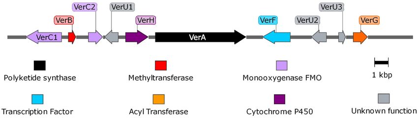
Valente et al.                                                                                                    CRISPR-Cas9 for Verrucosidin Cluster

100 Å, Phenomenex, Torrance, CA, United States) and the                 were resuspended in 1 ml of STC Buffer and kept on ice until
chromatographic separation was achieved by gradient conditions          transformation with PEG.
for 45 min at a flow rate of 300 µl/min. Solvent A was water and           CRISPR RNAs were designed using Alt-R Custom Cas9
solvent B was acetonitrile both containing 0.1% formic acid. The        crRNA (CRISPR RNA) Design Tool1 and are listed in
gradient was programmed as follows: 0–3 min isocratic 5% B,             Supplementary Table 1. The off-target analysis was performed
followed by a linear gradient to 100% B, ending at 40 min and           using Blast. Alt-R CRISPR-Cas9 crRNA, Alt-R CRISPR-
                                                                                                 R                                      R

from 40 to 45 min isocratic 100% B. Full-scan mass spectra were         Cas9 tracrRNA (trans-activating crRNA), and Alt-R S.p.                R

acquired in the positive-ion mode over the m/z range from 100 to        Cas9 nuclease V3 were purchased from IDT (Integrated
700 using the TQ mass analyzer.                                         DNA Technologies, Inc., Coralville, IA, United States) and
    To perform the verrucosidin qualitative analysis, the same          were combined to obtain a ribonucleoprotein (RNP) complex
HPLC-MS/MS system was used. HPLC was equipped with a                    according to the manufacturer’s instructions. Briefly, crRNA
Pursuit XRs ULTRA 2.8 µm C18 (100 × 2 mm, Varian) column                (2 nM) and tracrRNA (2 nM) were heated at 95◦ C for 5 min and
and a binary mixture as a mobile phase: solvents A and B were           cooled at room temperature to obtain gRNA. Three microliters
composed of 40 of 0.05% formic acid and 60% of acetonitrile,            of each gRNA was mixed with 4 µl of Cas9 at room temperature
respectively. The isocratic mode was used at a flow rate of             for 20 min. The entire mixture of RNP together with 10 µl
0.2 L/min for 5 min. A mass spectrometer was equipped with              of repair template (3 µg) and 40 µl of PEG20 solution (20%
an electrospray ionization (ESI) source operating in positive ion       PEG6000) were gently mixed with 80 µl of protoplasts (107
mode, whereas Product Ion Scan (PS) mode was used for triple-           protoplasts/ml). As a control, instead of DNA, 10 µl of water
quadrupole: m/z 417→100–427. The collision gas (Ar) pressure            was used. The mixtures were kept on ice for 30 min, and then
was set at 2 mbar.                                                      900 µl of PEG60 solution (60% of PEG6000) was added. After
                                                                        30 min of incubation on ice, protoplasts were gently spread on
                                                                        Petri dishes with regeneration media (RM; w/v: Malt extract 2%,
Protoplasts Preparation and                                             peptone 1%, glucose 2%; sucrose 0.8 M) supplemented with or
CRISPR-Cas9 Procedure                                                   without hygromycin.
Protocols used to obtain protoplasts from Aspergillus fumigatus
(Weidner et al., 1998), Ophiostoma piceae (Wang et al.,                 Characterization of Mutants in vitro
1999), Penicillium nalgiovense (Fierro et al., 2004), Penicillium       Mutants were selected on PDA supplemented with hygromycin
paxilli (Young et al., 1998; McMillan et al., 2003), Penicillium        B. Conidia were obtained in order to assure uniformity in the
chrysogenum (Pohl et al., 2016), Penicillium crustosum, and             genetic material. The deletion event was confirmed by PCR,
Penicillium janthinellium (Nicholson et al., 2015) were used as         sequencing PCR products, and Southern blot. Southern blot
the basis to design a protocol for protoplast preparation of            analysis was performed according to Stroe et al. (2020), and the
P. polonicum. Fungal mycelium was obtained by inoculating               probes used are listed in Supplementary Table 1.
2 ml of conidial suspension (1.25 × 10⁸ conidia/ml) in 50 ml               Deletion mutants were compared with wild-type P. polonicum
of YGG (8 g/L KCl, 16 g/L glucose, 6.6 g/L yeast nitrogen base,         X6 by inoculating 5 µl of spore suspension (1 × 10⁶
1.5 g/L citric acid, 6 g/L KH2 PO4 , and 2 g/L yeast extract).          conidia/ml) on PDA and incubating the plates at 25◦ C in the
Flasks were shaken on a rotary shaker (180 rpm) at 26◦ C for            dark. Colony diameter (cm) and number of asexual spores
20 h. Fungal mycelium was filtered through Miracloth (Merck             (conidia/plate) were measured up to 7 days post inoculation
KGaA) and washed with MgSO4 0.6 M. A pre-treatment was                  (dpi). Additionally, deletion mutants were inoculated in liquid
conducted by mixing 5 g of mycelium in 50-ml tubes with                 media (CYB and MEB) as previously described in order to
5 mM Na2 EDTA and 25 mM 2-mercaptoethanol; tubes were                   examine verrucosidin production.
kept in horizontal position in a rotary shaker (80 rpm) at
30◦ C for 20 min. Fungal mycelium was washed with 0.6 M
                                                                        Characterization of Mutants in vivo
MgSO4 and then digested with 2 g of Vinotaste Pro (Lamothe-
                                                  R

                                                                        Apples cv. Ambrosia and cv. Opal were surface disinfected and
Abiet, Canéjan, France) and 0.1 g of Lysing Enzymes from
                                                                        wounded as described in Zhang et al. (2012). Ten microliters
Trichoderma harzianum (Sigma-Aldrich) suspended in 15 ml of
                                                                        of conidial suspension (1 × 10⁶ conidia/ml) of each strain was
Osmo Solution (1.2 M MgSO4 , 10 mM sodium phosphate buffer).
                                                                        pipetted into each wound, whereas controls were inoculated
The lytic solution was kept at 30◦ C, 80 rpm. Every 30 min, the
                                                                        with deionized Ringer solution. Inoculated apples were placed
mycelium-lysis solution was mixed using a serological pipette
                                                                        in plastic trays, covered with a transparent polyethylene film,
and visually checked under a microscope; about 3–4 h were
                                                                        and stored for 14 days at room temperature. To verify the
necessary to digest the cell wall and obtain protoplasts. The
                                                                        pathogenicity of P. polonicum X6, apples cv. Gala were used.
suspension was filtered through Miracloth in order to separate
and remove undigested mycelia. Protoplasts were then recovered
using Trapping Buffer (0.6 M sorbitol, 0.1 M Tris–HCl, pH               Statistical Analyses and Software
7) and by centrifuging for 25 min. The intermediate layer was           All statistical analyses were performed with one-way ANOVA
separated and washed twice with STC Buffer (2.4 M sorbitol,             followed by Tukey’s b multiple comparison test using IBM
10 mM CaCl2 , and 10 mM Tris–HCl, pH 7.5). All centrifugation
steps were performed at 3,000 × g at 4◦ C. Finally, protoplasts         1
                                                                            https://eu.idtdna.com/site/order/designtool/index/CRISPR_CUSTOM

Frontiers in Microbiology | www.frontiersin.org                     4                                            May 2021 | Volume 12 | Article 660871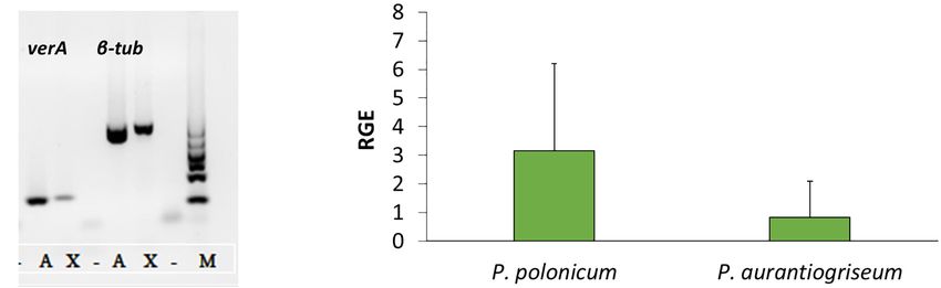
Valente et al.                                                                                                                   CRISPR-Cas9 for Verrucosidin Cluster

SPSS statistics software version 24 (SPSS Inc., Chicago, IL,                          X6, P. aurantiogriseum CBS 112021, and P. crustosum CAL64
United States); p < 0.05 was considered significant.                                  were grown in induction media. As shown in Supplementary
   SnapGene software (from Insightful Science; available at                           Figure 2, P. polonicum and P. aurantiogriseum were able to
snapgene.com) was used to draw gene clusters and visualize                            produce verrucosidin, whereas P. crustosum failed to produce
annotated DNA sequences.                                                              this compound. Consistently, we could verify the corresponding
                                                                                      PKS gene sequence in the genomes of P. polonicum X6 and
                                                                                      P. aurantiogriseum CBS 112021 by PCR, but not in the
                                                                                      P. crustosum genome (Figure 2A). Moreover, the expression of
RESULTS                                                                               the verA gene was confirmed both in P. polonicum X6 and in
                                                                                      P. aurantiogriseum CBS 112021 by qPCR (Figure 2B).
Putative Verrucosidin Gene Cluster
Using the PKS genes for aurovertin (AurA) and citreoviridin
(CtvA) biosyntheses as matrix, 16 putative BGCs                                       Deletion of PKS Through CRISPR-Cas9
(Supplementary Figure 1) were found in the genome of                                  To investigate the role of verA in verrucosidin biosynthesis, the
P. polonicum. Of them, 10 clusters were present in both                               gene was deleted using CRISPR-Cas9. For this purpose, a method
available P. polonicum genomes, whereas clusters 4 (later                             to obtain protoplasts for P. polonicum strain X6 was developed.
identified as verrucosidin BGC), 12, and 13 were only present                         The method allowed obtaining 1 × 10⁷ protoplasts/ml starting
in P. polonicum IBT 4502 and clusters 14, 15, and 16 in                               from 5 g of mycelium.
P. polonicum strain hy4 genome (Supplementary Table 2).                                  In order to delete target genes with the endonuclease Cas9,
Cluster 4 was further investigated due to the presence of genes                       the protospacers were designed based on the sequenced promoter
encoding putative tailoring enzymes involved in verrucosidin                          and terminator of target genes in P. polonicum X6. To obtain
biosynthesis (Figure 1): a putative methyltransferase (named                          the deletion mutants, two sets of gRNA were used, allowing to
cl4B or verB), two Flavin adenine dinucleotide (FAD)-dependent                        cut both the promoter and the terminator and to excise the
monooxygenases (cl4C1 and cl4C2 or verC1 and verC2), an                               target gene. The endonuclease Cas9 was mixed in vitro with
acyltransferase (cl4G or verG), and a cytochrome P450 (cl4H or                        gRNA and a repair DNA template encoding the hygromycin
verH) gene. To monitor verrucosidin production, P. polonicum                          resistance cassette. The repair DNA template was amplified using

  FIGURE 1 | Representation of verrucosidin gene cluster in P. polonicum. Maps were obtained with SnapGene software.

  FIGURE 2 | Presence and expression of verA gene in Penicillium spp. Amplification of verA and β-tubulin genes from gDNA (A): X, P. polonicum X6; A,
  P. aurantiogriseum CBS121001; M, GelPilot 100 bp Ladder; –, negative control (PCR mix without DNA). Relative gene expression (RGE) of verA (cl4A) gene in
  P. polonicum and P. aurantiogriseum (B) was evaluated 10 days post inoculation on CYB. The expression is relative to the expression of the β-tubulin gene, and
  P. aurantiogriseum was used as reference strain.

Frontiers in Microbiology | www.frontiersin.org                                   5                                             May 2021 | Volume 12 | Article 660871
Valente et al. CRISPR-Cas9 for Verrucosidin Cluster primers with an additional tail of 50 bp providing at both flanking positive transformants were further confirmed by sequencing regions DNA micro-homology close to the PAM site. This way, the amplified PCR products (Supplementary Figure 3) and by the correct integration of the repair DNA through homology- Southern blot analysis (Supplementary Figure 4). The results mediated end joining (MMEJ) was achieved. Obtained knockout showed that one of the strains displayed a repetition of 50 bp mutants were assessed through PCR, amplifying both the of micro-homology in the promoter region (Supplementary hygromycin resistance cassette and the target gene (Figures 3A– Figure 3), whereas 1cl4A_C10, 1cl4A_C11, and 1cl4A_C12 C). Sixteen mutants, in that the hygromycin resistance cassette mutants displayed a correct integration of the hph gene and lack was successfully amplified whereas amplification of the verA PKS of ectopic integrations (Supplementary Figures 3, 4) and were gene failed, were randomly chosen and the correct integration of therefore further studied. the repair template was confirmed using PCR with primer pairs designed inside the hph gene and in the promoter and terminator Phenotype of Mutants region of the deleted PKS gene (Figures 3A,D,E). In most cases, Three P. polonicum X6 verA mutants were phenotypically correct 1verA mutants were found (Figures 3D,E). Some of the characterized in vitro and on apples. Compared to wild-type FIGURE 3 | PCR analysis of verA mutants. Schematic presentation of the verA (cl4A) locus in the wild-type and deletion mutants; homology sequences (50 bp) are indicated in green, and primers used are marked by arrows (A). Amplification of hygromycin resistance cassette (B) and verA gene (C); confirmation of orientation of inserted repair DNA (D,E); M, GelPilot Wide Range Ladder; WT, wild-type P. polonicum X6; A–D, deletion mutants for verA; –, negative control (PCR mix without DNA). Frontiers in Microbiology | www.frontiersin.org 6 May 2021 | Volume 12 | Article 660871
Valente et al. CRISPR-Cas9 for Verrucosidin Cluster
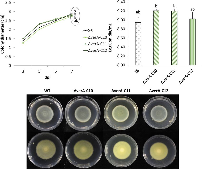
strain, the mutant strains displayed the same ability to grow DISCUSSION
(Figures 4A,C,D) and no significant differences in conidiation
(Figure 4B). Analyzing the metabolic profile of wild-type strain We report the identification of the BGC for the α-pyrone
and knockout mutants, the verrucosidin chromatographic peak polyketide verrucosidin in the genome of P. polonicum, a
was absent in deletion mutants (Supplementary Figure 5), known verrucosidin producer (Frisvad et al., 2004). Initial
independently of the used induction medium (Table 1 and similarity analyses with the PKS genes for aurovertin (AurA)
Supplementary Figure 6). By contrast, verrucosidin formation and citreoviridin (CtvA) had ended up with 16 BGCs
in P. polonicum X6 was not influenced, when the PKS gene of putatively encoding the biosynthesis of α-pyrone polyketides.
another α-pyrone BGC, cl3A, was deleted (data not shown). Besides P. polonicum, we confirmed P. aurantiogriseum to be
The pathogenicity of the wild-type strain X6 of P. polonicum able to produce verrucosidin (Fink-Gremmels et al., 1991),
was previously verified on apple (Supplementary Table 3). The whereas P. crustosum did not produce this compound, as
effect of gene deletion on the virulence of P. polonicum was expected (Frisvad et al., 2004). Under the used laboratory
assessed on apple, as it is a host species for this pathogen and conditions, meaning cultivation in different induction media,
the virulence of the fungal strain can be quantified by measuring the verA gene was expressed and verrucosidin formation
the lesion diameter, whereas on chestnut, the symptoms are was verified in both producer species P. polonicum X6 and
mild and not easily measured. The virulence was tested on P. aurantiogriseum CBS 112021.
two apple cultivars. On apples cv. Ambrosia, verA deletion To delete verA, encoding a putative highly reducing polyketide
mutants displayed a slightly reduced virulence 7 days after the synthase (HR-PKS), for the first time an in vitro method for
inoculation (Supplementary Table 4), whereas this behavior was CRISPR-Cas9 gene deletion was established in P. polonicum
not observed after 14 days of storage (Figure 5A) or on apples cv. X6. Up to the present, CRISPR-Cas9 technology was adopted
Opal (Figure 5B). only in the Penicillium species P. chrysogenum. Protoplasts of
FIGURE 4 | Effect of verA deletion on P. polonicum growth in vitro. Colony diameter after 3 to 7 days post inoculation (A) conidia production (B) and plate view (front
C and reverse D) at 7 days post inoculation (dpi) on PDA. WT, wild-type P. polonicum X6; 1verA, deletion mutants for verA (cl4A). Values followed by the same letter
are not statistically different by Tukey’s b multiple comparison test (p < 0.05).
Frontiers in Microbiology | www.frontiersin.org 7 May 2021 | Volume 12 | Article 660871Valente et al. CRISPR-Cas9 for Verrucosidin Cluster
TABLE 1 | Effect of gene deletion on verrucosidin production in vitro and in vivo.
Strain CYB MEB APPLE
P. aurantiogriseum CBS 112021* + + +
P. polonicum X6 wild-type + + +
P. polonicum X6 1cl4A_C10 − − −
P. polonicum X6 1cl4A_C11 − − −
P. polonicum X6 1cl4A_C12 − − −
Penicillium polonicum wild type, P. polonicum deletion mutants, and
P. aurantiogriseum were inoculated in CYB and MEB broth or on apples. Presence
(+) or absence (−) of verrucosidin production at 10 days post inoculation.
*Penicillium aurantiogriseum CBS 112021 was used as a qualitative standard
since the verrucosidin standard is not commercially available.
a strain harboring the Cas9 gene on an AMA1 plasmid were
transformed with a synthesized gRNA (Pohl et al., 2016). In this
work, the method of Al Abdallah et al. (2017) was followed,
who successfully transformed protoplasts of A. fumigatus with
RNP and a repair template harboring antibiotic resistance to
select mutants. This strategy does not require the generation
of plasmids or the expression of the endonuclease Cas9, but is
based on transforming protoplasts with the endonuclease protein
Cas9 together with crRNA and tracrRNA. Therefore, there is an
immediate cleavage activity when the RNP is targeted into the
cells and the RNP is degraded by the host cell within a short
period, reducing the possibility of off-target activity (Wang and
Coleman, 2019). This strategy requires both accurate design of
the crRNA and a protocol to transform fungi. A method to
obtain protoplasts in P. polonicum X6 was optimized based on
available protocols used for other filamentous fungi (Weidner
et al., 1998; Young et al., 1998; Wang et al., 1999; McMillan
et al., 2003; Fierro et al., 2004; Nicholson et al., 2015; Pohl
et al., 2016) and allows obtaining a high amount of protoplasts.
Mutants for putative PKS gene were obtained, confirming that
the CRISPR-Cas9 method is effective and could be used for
gene editing in P. polonicum. The finding of a small number
of mutants displaying incorrect insertion of the foreign DNA FIGURE 5 | Analysis of verA deletion on the virulence of P. polonicum in vivo.
necessitate screening and confirmation of mutants by molecular Lesion diameter (cm) and pictures of apples cv. Ambrosia (A) and Opal (B)
techniques such as PCR and Southern blot analysis. As an were recorded 14 days after inoculation. WT, wild-type P. polonicum X6;
1verA, deletion mutants for verA (cl4A). Values followed by the same letter are
example, when sequencing the terminator region of the deleted
not statistically different by Tukey’s b multiple comparison test (p < 0.05).
verA gene in the mutant B4, we observed an unexpected insertion,
that suggested correct cutting of the double-strand DNA by
the endonuclease, but missing MMEJ. To our knowledge, there
were no previous reports about MMEJ occurring only at one P. polonicum strain hy4, confirming high variability in secondary
excision site. Therefore, the integration of repair DNA could have metabolism between strains of the same species (Ballester
occurred by non-homologous end-joining (Krappmann, 2017). et al., 2015). The biosynthetic genes were also absent in
The efficiency of homologous recombination could be improved P. expansum strain NRRL 62431. This fungal genome was initially
by adjusting the length of the homology sequence or by changing deposited as P. aurantiogriseum and later assigned to another
the amount of donor DNA, as previously shown in A. fumigatus species (Ballester et al., 2015). Due to the lack of verrucosidin
(Al Abdallah et al., 2017). gene cluster, we can conclude that both P. expansum strains
Deletion mutants for the HR-PKS encoded by verA were no NRRL 62431 and P. polonicum strain hy4 are not able to
longer able to produce verrucosidin in vitro confirming that produce this SM.
verA is responsible for the biosynthesis of this compound. The Secondary metabolites are known to play a central role in
here identified gene cluster was bioinformatically hypothesized many biological processes such as growth development (Calvo
as verrucosidin cluster in P. polonicum by Li et al. (2018), et al., 2002) and pathogenesis (Scharf et al., 2014; Macheleidt
but this prediction was not experimentally proven. Surprisingly, et al., 2016), which were extensively reviewed (Fox and Howlett,
the verrucosidin gene cluster was absent in the genome of 2008; Keller, 2019). In this work, the deletion of the verA gene did
Frontiers in Microbiology | www.frontiersin.org 8 May 2021 | Volume 12 | Article 660871Valente et al. CRISPR-Cas9 for Verrucosidin Cluster
not affect P. polonicum in terms of growth rate and conidiation will allow characterizing the biosynthetic potential of this
on PDA. Concerning the effect of deletion on the virulence of interesting fungal species.
the strains, apples were used as preferential host, as P. polonicum
is reported as a postharvest pathogen on pome fruit (Scholtz
and Korsten, 2016; Ouhibi et al., 2018). The pathogenicity of the DATA AVAILABILITY STATEMENT
wild-type strain X6 of P. polonicum was previously verified on
apple, where the virulence is easily quantified by measuring the The datasets presented in this study can be found in
lesion diameter. Slight differences in virulence of 1verA mutants online repositories. The names of the repository/repositories
were observed on apple cv. Ambrosia, whereas this behavior and accession number(s) can be found in the article/
was not observed on apples cv. Opal. These results are not Supplementary Material.
surprising, as the apple cultivar is known to be a key factor in the
virulence, such as in the case of patulin biosynthesis, which may
be considered a cultivar-dependent aggressiveness factor (Banani AUTHOR CONTRIBUTIONS
et al., 2016; Snini et al., 2016). However, a reduced virulence of
1verA mutants was observed only during the first steps of blue SV, EP, VS, TH, AB, and DS conceived and designed the study.
mold development on apple fruit; therefore, further studies are SV performed the experiments. EP, VS, and GM performed
needed to confirm these preliminary observations. bioinformatic, Southern blot, and chemical analyses, respectively.
SV and EP retrieved the literature and drafted the manuscript. All
authors reviewed and approved the final manuscript.
CONCLUSION
The verrucosidin biosynthesis BGC was identified by
FUNDING
bioinformatic and genetic approaches. Sixteen HR-PKS gene- This work was supported by the German Academic Exchange
containing BGCs were discovered in the available genomes Service (DAAD) with a scholarship in the program Research
of P. polonicum. These BGCs are probably involved in Grants – Short-Term Grants, 2019.
the biosynthesis of α-pyrone type polyketides, which could
have many biological activities, such as cytotoxic, antitumor,
antimicrobial, and anti-germination activities and are therefore ACKNOWLEDGMENTS
a potential source of novel compounds (Schäberle, 2016; Li
et al., 2018; Stroe et al., 2020). Based on the putative function We thank Christina Täumer from the Leibniz Institute for
of the genes present in the clusters, one of these BGCs was Natural Product Research and Infection Biology for technical
further characterized through deletion, in order to investigate its assistance, and the students at the University of Torino who
involvement in verrucosidin biosynthesis. To delete the selected helped characterize the mutants.
PKS gene, CRISPR-Cas9 was adopted in P. polonicum. This
allowed obtaining mutants by replacing the target genes with
repair template sequences with 50 bp homology to chromosomal SUPPLEMENTARY MATERIAL
sequences. By applying this technology, the verrucosidin BGC
was confirmed by creating deletion mutants for verA gene The Supplementary Material for this article can be found
coding for HR-PKS known to be the key enzyme of the online at: https://www.frontiersin.org/articles/10.3389/fmicb.
biosynthesis. The availability of the CRISPR-Cas technology 2021.660871/full#supplementary-material
REFERENCES metabolism of the postharvest pathogen Penicillium griseofulvum. BMC
Genomics 17:19. doi: 10.1186/s12864-015-2347-x
Al Abdallah, Q., Ge, W., and Fortwendel, J. R. (2017). A Simple and Universal Bergmann, S., Funk, A. N., Scherlach, K., Schroeckh, V., Shelest, E., Horn, U., et al.
System for Gene Manipulation in Aspergillus fumigatus: In Vitro -Assembled (2010). Activation of a Silent Fungal Polyketide Biosynthesis Pathway through
Cas9-Guide RNA Ribonucleoproteins Coupled with Microhomology Repair Regulatory Cross Talk with a Cryptic Nonribosomal Peptide Synthetase Gene
Templates. mSphere 2, 446–417e. doi: 10.1128/msphere.00446-17 Cluster. Appl. Environ. Microbiol. 76, 8143–8149. doi: 10.1128/AEM.00683-10
Aranda, E., Rodríguez, M., Benito, M. J., Asensio, M. A., and Córdoba, Bergmann, S., Schümann, J., Scherlach, K., Lange, C., Brakhage, A. A., and
J. J. (2002). Molecular cloning of verrucosidin-producing Penicillium Hertweck, C. (2007). Genomics-driven discovery of PKS-NRPS hybrid
polonicum genes by differential screening to obtain a DNA probe. metabolites from Aspergillus nidulans. Nat. Chem. Biol. 3, 213–217. doi: 10.
Int. J. Food Microbiol. 76, 55–61. doi: 10.1016/S0168-1605(02)00 1038/nchembio869
008-9 Brakhage, A. A. (2013). Regulation of fungal secondary metabolism. Nat. Rev.
Ballester, A.-R., Marcet-Houben, M., Levin, E., Sela, N., Selma-Lázaro, C., Microbiol. 11, 21–32. doi: 10.1038/nrmicro2916
Carmona, L., et al. (2015). Genome, Transcriptome, and Functional Analyses Burka, L. T., Ganguli, M., and Wilson, B. J. (1983). Verrucosidin, a Tremorgen from
of Penicillium expansum Provide New Insights Into Secondary Metabolism and Penicillium verrucosum var. cyclopium. J. Chem. Soc. Chem. Commun. 1670,
Pathogenicity. Mol. Plant Microbe Interact. 28, 232–248. doi: 10.1094/MPMI- 544–545. doi: 10.1039/C39830000544
09-14-0261-FI Calvo, A. M., Wilson, R. A., Bok, J. W., and Keller, N. P. (2002). Relationship
Banani, H., Marcet-Houben, M., Ballester, A. R., Abbruscato, P., González- between Secondary Metabolism and Fungal Development. Microbiol. Mol. Biol.
Candelas, L., Gabaldón, T., et al. (2016). Genome sequencing and secondary Rev. 66, 447–459. doi: 10.1128/mmbr.66.3.447-459.2002
Frontiers in Microbiology | www.frontiersin.org 9 May 2021 | Volume 12 | Article 660871Valente et al. CRISPR-Cas9 for Verrucosidin Cluster
Deng, H., Gao, R., Liao, X., and Cai, Y. (2017). CRISPR system in filamentous intermediates on mammalian maxi-K ion channels. Mol. Genet. Genomics 270,
fungi: Current achievements and future directions. Gene 627, 212–221. doi: 9–23. doi: 10.1007/s00438-003-0887-2
10.1016/j.gene.2017.06.019 Nagy, G., Szebenyi, C., Csernetics, Á, Vaz, A. G., Tóth, E. J., Vágvölgyi, C., et al.
Duduk, N., Vasiæ, M., and Vico, I. (2014). First report of Penicillium polonicum (2017). Development of a plasmid free CRISPR-Cas9 system for the genetic
causing blue mold on stored onion (Allium cepa) in Serbia. Plant Dis. 98:1440. modification of Mucor circinelloides. Sci. Rep. 7, 1–10. doi: 10.1038/s41598-017-
doi: 10.1094/PDIS-05-14-0550-PDN 17118-2
Faedda, R., Pane, A., Cacciola, S. O., Granata, G., Salafia, L., and Sinatra, F. (2015). Nicholson, M. J., Eaton, C. J., Stärkel, C., Tapper, B. A., Cox, M. P., and Scott,
Penicillium polonicum causing a postharvest soft rot of cactus pear fruits. Acta B. (2015). Molecular cloning and functional analysis of gene clusters for the
Hortic. 1067, 193–197. doi: 10.17660/ActaHortic.2015.1067.26 biosynthesis of indole-diterpenes in Penicillium crustosum and P. janthinellum.
Fang, Y., and Tyler, B. M. (2016). Efficient disruption and replacement of an Toxins 7, 2701–2722. doi: 10.3390/toxins7082701
effector gene in the oomycete Phytophthora sojae using CRISPR/Cas9. Mol. Nielsen, J. C., and Nielsen, J. (2017). Development of fungal cell factories for the
Plant Pathol. 17, 127–139. doi: 10.1111/mpp.12318 production of secondary metabolites: Linking genomics and metabolism. Synth.
Fierro, F., Laich, F., García-Rico, R. O., and Martín, J. F. (2004). High efficiency Syst. Biotechnol. 2, 5–12. doi: 10.1016/j.synbio.2017.02.002
transformation of Penicillium nalgiovense with integrative and autonomously Nielsen, M. L., Isbrandt, T., Rasmussen, K. B., Thrane, U., Hoof, J. B., Larsen,
replicating plasmids. Int. J. Food Microbiol. 90, 237–248. doi: 10.1016/S0168- T. O., et al. (2017). Genes Linked to production of secondary metabolites in
1605(03)00306-4 Talaromyces atroroseus revealed using CRISPR-Cas9. PLoS One 12:1–9. doi:
Fink-Gremmels, J., Henning, J., and Leistner, A. (1991). Quantitative 10.1371/journal.pone.0169712
determination of verrucosidin producing Penicillium aurantiogriseum. Nødvig, C. S., Nielsen, J. B., Kogle, M. E., and Mortensen, U. H. (2015). A CRISPR-
Microbiologie, Aliments. Nutr. Microbiol. Foods Feed. Nutr. 5, 155–160. Cas9 system for genetic engineering of filamentous fungi. PLoS One 10:1–18.
Fischer, J., Schroeckh, V., and Brakhage, A. A. (2016). “Awakening of Fungal doi: 10.1371/journal.pone.0133085
Secondary Metabolite Gene Clusters,” in Gene Expression Systems in Fungi: Núñez, F., Díaz, M. C., Rodríguez, M., Aranda, E., Martín, A., and Asensio,
Advancements and Applications, eds M. Schmoll and C. Dattenböck (Cham: M. A. (2000). Effects of substrate, water activity, and temperature on growth
Springer), 253–273. doi: 10.1007/978-3-319-27951-0_11 and verrucosidin production by Penicillium polonicum isolated from dry-cured
Fox, E. M., and Howlett, B. J. (2008). Secondary metabolism: regulation and role in ham. J. Food Prot. 63, 231–236. doi: 10.4315/0362-028X-63.2.231
fungal biology. Curr. Opin. Microbiol. 11, 481–487. doi: 10.1016/j.mib.2008.10. Ouhibi, S., Santos, C., Ghali, R., Soares, C., Hedhili, A., Paterson, R., et al. (2018).
007 Penicillium tunisiense sp. nov., a novel species of Penicillium section Ramosa
Frisvad, J. C., Smedsgaard, J., Larsen, T. O., and Samson, R. A. (2004). Mycotoxins, discovered from Tunisian orchard apples. Int. J. Syst. Evol. Microbiol. 68,
drugs and other extrolites produced by species in Penicillium subgenus 3217–3225. doi: 10.1099/ijsem.0.002962
Penicillium. Stud. Mycol. 49, 201–241. Park, H. R., Ryoo, I. J., Choo, S. J., Hwang, J. H., Kim, J. Y., Cha, M. R., et al.
Hodge, R. P., Harris, C. M., and Harris, T. M. (1988). Verrucofortine, a major (2007). Glucose-deprived HT-29 human colon carcinoma cells are sensitive
metabolite of Penicillium verrucosum var. cyclopium, the fungus that produces to verrucosidin as a GRP78 down-regulator. Toxicology 229, 253–261. doi:
the mycotoxin verrucosidin. J. Nat. Prod. 51, 66–73. doi: 10.1021/np50055a 10.1016/j.tox.2006.11.049
008 Pfaffl, M. W. (2001). A new mathematical model for relative quantification in
Jensen, B., Knudsen, I. M. B., Andersen, B., Nielsen, K. F., Thrane, U., Jensen, D. F., real-time RT-PCR. Nucleic Acids Res. 29:e45. doi: 10.1093/nar/29.9.e45
et al. (2013). Characterization of microbial communities and fungal metabolites Pohl, C., Kiel, J. A. K. W., Driessen, A. J. M., Bovenberg, R. A. L., and Nygård, Y.
on field grown strawberries from organic and conventional production. Int. J. (2016). CRISPR/Cas9 Based Genome Editing of Penicillium chrysogenum. ACS
Food Microbiol. 160, 313–322. doi: 10.1016/j.ijfoodmicro.2012.11.005 Synth. Biol. 5, 754–764. doi: 10.1021/acssynbio.6b00082
Keller, N. P. (2019). Fungal secondary metabolism: regulation, function and drug Prencipe, S., Siciliano, I., Gatti, C., Garibaldi, A., Gullino, M. L., Botta, R., et al.
discovery. Nat. Rev. Microbiol. 17, 167–180. doi: 10.1038/s41579-018-0121-1 (2018). Several species of Penicillium isolated from chestnut flour processing
Kim, H. Y., Heo, D. Y., Park, H. M., Singh, D., and Lee, C. H. (2016). Metabolomic are pathogenic on fresh chestnuts and produce mycotoxins. Food Microbiol. 76,
and transcriptomic comparison of solid-state and submerged fermentation 396–404. doi: 10.1016/j.fm.2018.07.003
of Penicillium expansum KACC 40815. PLoS One 11:e0149012. doi: 10.1371/ Quevillon, E., Silventoinen, V., Pillai, S., Harte, N., Mulder, N., Apweiler, R.,
journal.pone.0149012 et al. (2005). InterProScan: Protein domains identifier. Nucleic Acids Res. 33,
Krappmann, S. (2017). CRISPR-Cas9, the new kid on the block of fungal molecular W116–W120. doi: 10.1093/nar/gki442
biology. Med. Mycol. 55, 16–23. doi: 10.1093/mmy/myw097 Rodríguez, A., Córdoba, J. J., Werning, M. L., Andrade, M. J., and Rodríguez, M.
Li, W., Ma, Z., Chen, L., and Yin, W. B. (2018). Synthesis and production of the (2012). Duplex real-time PCR method with internal amplification control for
antitumor polyketide aurovertins and structurally related compounds. Appl. quantification of verrucosidin producing molds in dry-ripened foods. Int. J.
Microbiol. Biotechnol. 102, 6373–6381. doi: 10.1007/s00253-018-9123-1 Food Microbiol. 153, 85–91. doi: 10.1016/j.ijfoodmicro.2011.10.020
Liebmann, B., Müller, M., Braun, A., and Brakhage, A. A. (2004). The cyclic AMP- Sabater-Vilar, M., Nijmeijer, S., and Fink-Gremmels, J. (2003). Genotoxicity
dependent protein kinase A network regulates development and virulence in assessment of five tremorgenic mycotoxins (fumitremorgen B, paxilline,
Aspergillus fumigatus. Infect. Immun. 72, 5193–5203. doi: 10.1128/IAI.72.9. penitrem A, verruculogen, and verrucosidin) produced by molds isolated from
5193-5203.2004 fermented meats. J. Food Prot. 66, 2123–2129. doi: 10.4315/0362-028X-66.11.
Lin, T. S., Chen, B., Chiang, Y. M., and Wang, C. C. C. (2019). Discovery 2123
and Elucidation of the Biosynthesis of Aspernidgulenes: Novel Polyenes from Santini, A., Mikušová, P., Sulyok, M., Krska, R., Labuda, R., and Šrobárová,
Aspergillus Nidulans by Using Serial Promoter Replacement. ChemBioChem 20, A. (2014). Penicillium strains isolated from Slovak grape berries taxonomy
329–334. doi: 10.1002/cbic.201800486 assessment by secondary metabolite profile. Mycotoxin Res. 30, 213–220. doi:
Lin, T. S., Chiang, Y. M., and Wang, C. C. C. (2016). Biosynthetic Pathway of 10.1007/s12550-014-0205-3
the Reduced Polyketide Product Citreoviridin in Aspergillus terreus var. aureus Schäberle, T. F. (2016). Biosynthesis of α-pyrones. Beilstein J. Org. Chem. 12,
Revealed by Heterologous Expression in Aspergillus nidulans. Org. Lett. 18, 571–588. doi: 10.3762/bjoc.12.56
1366–1369. doi: 10.1021/acs.orglett.6b00299 Scharf, D. H., Heinekamp, T., and Brakhage, A. A. (2014). Human and Plant Fungal
Macheleidt, J., Mattern, D. J., Fischer, J., Netzker, T., Weber, J., Schroeckh, V., Pathogens: The Role of Secondary Metabolites. PLoS Pathog. 10:e1003859. doi:
et al. (2016). Regulation and Role of Fungal Secondary Metabolites. Annu. Rev. 10.1371/JOURNAL.PPAT.1003859
Genet. 50, 371–392. doi: 10.1146/annurev-genet-120215-035203 Scholtz, I., and Korsten, L. (2016). Profile of Penicillium species in the pear supply
Madden, T. (2013). “The BLAST sequence analysis tool,” in The NCBI Handbook, chain. Plant Pathol. 65, 1126–1132. doi: 10.1111/ppa.12494
2nd Edn, (Maryland: National Center for Biotechnology Information). Snini, S. P., Tannous, J., Heuillard, P., Bailly, S., Lippi, Y., Zehraoui, E., et al. (2016).
McMillan, L. K., Carr, R. L., Young, C. A., Astin, J. W., Lowe, R. G. T., Parker, E. J., Patulin is a cultivar-dependent aggressiveness factor favouring the colonization
et al. (2003). Molecular analysis of two cytochrome P450 monooxygenase genes of apples by Penicillium expansum. Mol. Plant Pathol. 17, 920–930. doi: 10.1111/
required for paxilline biosynthesis in Penicillium paxilli, and effects of paxilline mpp.12338
Frontiers in Microbiology | www.frontiersin.org 10 May 2021 | Volume 12 | Article 660871Valente et al. CRISPR-Cas9 for Verrucosidin Cluster Song, R., Zhai, Q., Sun, L., Huang, E., Zhang, Y., Zhu, Y., et al. (2019). CRISPR/Cas9 Weber, T., and Kim, H. U. (2016). The secondary metabolite bioinformatics portal: genome editing technology in filamentous fungi: progress and perspective. Computational tools to facilitate synthetic biology of secondary metabolite Appl. Microbiol. Biotechnol. 103, 6919–6932. doi: 10.1007/s00253-019-10 production. Synth. Syst. Biotechnol. 1, 69–79. doi: 10.1016/j.synbio.2015.12.002 007-w Weber, T., Blin, K., Duddela, S., Krug, D., Kim, H. U., Bruccoleri, R., et al. Sonjak, S., Frisvad, J. C., and Gunde-Cimerman, N. (2006). Penicillium mycobiota (2015). AntiSMASH 3.0-A comprehensive resource for the genome mining of in Arctic subglacial ice. Microb. Ecol. 52, 207–216. doi: 10.1007/s00248-006- biosynthetic gene clusters. Nucleic Acids Res. 43, W237–W243. doi: 10.1093/ 9086-0 nar/gkv437 Spadaro, D., Lorè, A., Garibaldi, A., and Gullino, M. L. (2013). A new strain of Weidner, G., D’Enfert, C., Koch, A., Mol, P. C., and Brakhage, A. A. Metschnikowia fructicola for postharvest control of Penicillium expansum and (1998). Development of a homologous transformation system for the human patulin accumulation on four cultivars of apple. Postharv. Biol. Technol. 75, 1–8. pathogenic fungus Aspergillus fumigatus based on the pyrG gene encoding doi: 10.1016/j.postharvbio.2012.08.001 orotidine 5’-monophosphate decarboxylase. Curr. Genet. 33, 378–385. doi: 10. Stroe, M. C., Netzker, T., Scherlach, K., Krüger, T., Hertweck, C., Valiante, V., et al. 1007/s002940050350 (2020). Targeted induction of a silent fungal gene cluster encoding the bacteria- Wigmann, ÉF., Jahn, R. C., Scherer, C. D., Saccomori, F., Alcano-González, M., specific germination inhibitor fumigermin. Elife 9:e52541. doi: 10.7554/eLife. de, J., et al. (2018). Detection and identification of Penicillium spp. in a frozen 52541 chicken nuggets production facility. Food Microbiol. 70, 42–48. doi: 10.1016/j. Sunesen, L. O., and Stahnke, L. H. (2003). Mould starter cultures for dry sausages fm.2017.09.002 - Selection, application and effects. Meat Sci. 65, 935–948. doi: 10.1016/S0309- Young, C., Itoh, Y., Johnson, R., Garthwaite, I., Miles, C. O., Munday-Finch, S. C., 1740(02)00281-4 et al. (1998). Paxilline-negative mutants of Penicillium paxilli generated by Thomas, S., Sharma, N., Gonzalez, R., Pao, P. W., Hofman, F. M., Chen, T. C., et al. heterologous and homologous plasmid integration. Curr. Genet. 33, 368–377. (2013). Repositioning of Verrucosidin, a Purported Inhibitor of Chaperone doi: 10.1007/s002940050349 Protein GRP78, as an Inhibitor of Mitochondrial Electron Transport Chain Yu, J., Jurick, M. W., and Bennett, J. W. (2015). Current status of genomics research Complex I. PLoS One 8:1–14. doi: 10.1371/journal.pone.0065695 on mycotoxigenic fungi. Int. J. plant Biol. Res. 3:1035. Tong, Y., Weber, T., and Lee, S. Y. (2019). CRISPR/Cas-based genome engineering Zhang, D., Spadaro, D., Valente, S., Garibaldi, A., and Gullino, M. L. (2012). in natural product discovery. Nat. Prod. Rep. 36, 1225–1372. doi: 10.1039/ Cloning, characterization, expression and antifungal activity of an alkaline c8np00089a serine protease of Aureobasidium pullulans PL5 involved in the biological Valente, S., Cometto, A., Piombo, E., Meloni, G. R., Ballester, A. R., González- control of postharvest pathogens. Int. J. Food Microbiol. 153, 453–464. doi: Candelas, L., et al. (2020). Elaborated regulation of griseofulvin biosynthesis in 10.1016/j.ijfoodmicro.2011.12.016 Penicillium griseofulvum and its role on conidiation and virulence. Int. J. Food Microbiol. 328:108687. doi: 10.1016/j.ijfoodmicro.2020.108687 Conflict of Interest: The authors declare that the research was conducted in the Wang, H. L., Kim, S. H., Siu, H., and Breuil, C. (1999). Transformation of absence of any commercial or financial relationships that could be construed as a sapstaining fungi with hygromycin B resistance plasmids pAN7-1 and pCB1004. potential conflict of interest. Mycol. Res. 103, 77–80. doi: 10.1017/S0953756298006698 Wang, Q., and Coleman, J. J. (2019). Progress and Challenges: Development and Copyright © 2021 Valente, Piombo, Schroeckh, Meloni, Heinekamp, Brakhage and Implementation of CRISPR/Cas9 Technology in Filamentous Fungi. Comput. Spadaro. This is an open-access article distributed under the terms of the Creative Struct. Biotechnol. J. 17, 761–769. doi: 10.1016/j.csbj.2019.06.007 Commons Attribution License (CC BY). The use, distribution or reproduction in Weber, J., Valiante, V., Nødvig, C. S., Mattern, D. J., Slotkowski, R. A., Mortensen, other forums is permitted, provided the original author(s) and the copyright owner(s) U. H., et al. (2017). Functional reconstitution of a fungal natural product gene are credited and that the original publication in this journal is cited, in accordance cluster by advanced genome editing. ACS Synth. Biol. 6, 62–68. doi: 10.1021/ with accepted academic practice. No use, distribution or reproduction is permitted acssynbio.6b00203 which does not comply with these terms. Frontiers in Microbiology | www.frontiersin.org 11 May 2021 | Volume 12 | Article 660871
You can also read