Severe T-System Remodeling in Pediatric Viral Myocarditis
←
→
Page content transcription
If your browser does not render page correctly, please read the page content below
ORIGINAL RESEARCH
                                                                                                                                          published: 18 January 2021
                                                                                                                                     doi: 10.3389/fcvm.2020.624776

                                            Severe T-System Remodeling in
                                            Pediatric Viral Myocarditis
                                            Dominik J. Fiegle 1 , Martin Schöber 2 , Sven Dittrich 2 , Robert Cesnjevar 3 , Karin Klingel 4 ,
                                            Tilmann Volk 1,5 , Muhannad Alkassar 2*† and Thomas Seidel 1,5*†
                                            1
                                             Institute of Cellular and Molecular Physiology, Friedrich-Alexander-Universität Erlangen-Nürnberg, Erlangen, Germany,
                                            2
                                             Department of Pediatric Cardiology, Friedrich-Alexander-Universität Erlangen-Nürnberg, Erlangen, Germany, 3 Department
                                            of Pediatric Cardiac Surgery, University Hospital Erlangen, Erlangen, Germany, 4 Cardiopathology, University Hospital
                                            Tuebingen, Tübingen, Germany, 5 Muscle Research Center Erlangen (MURCE), Friedrich-Alexander-Universität
                                            Erlangen-Nürnberg, Erlangen, Germany

                                            Chronic heart failure (HF) in adults causes remodeling of the cardiomyocyte transverse
                                            tubular system (t-system), which contributes to disease progression by impairing
                                            excitation-contraction (EC) coupling. However, it is unknown if t-system remodeling
                                            occurs in pediatric heart failure. This study investigated the t-system in pediatric viral
                                            myocarditis. The t-system and integrity of EC coupling junctions (co-localization of
                           Edited by:       L-type Ca2+ channels with ryanodine receptors and junctophilin-2) were analyzed by
                         Estela Azeka,      3D confocal microscopy in left-ventricular (LV) samples from 5 children with myocarditis
        University of São Paulo, Brazil
                                            (age 14 ± 3 months), undergoing ventricular assist device (VAD) implantation, and 5
                        Reviewed by:
                   Leonardo Sacconi,
                                            children with atrioventricular septum defect (AVSD, age 17 ± 3 months), undergoing
           University of Florence, Italy    corrective surgery. LV ejection fraction (EF) was 58.4 ± 2.3% in AVSD and 12.2 ± 2.4% in
                     David Crossman,        acute myocarditis. Cardiomyocytes from myocarditis samples showed increased t-tubule
           The University of Auckland,
                         New Zealand        distance (1.27 ± 0.05 µm, n = 34 cells) and dilation of t-tubules (volume-length ratio:
                *Correspondence:            0.64 ± 0.02 µm2 ) when compared with AVSD (0.90 ± 0.02 µm, p < 0.001; 0.52 ±
                Muhannad Alkassar           0.02 µm2 , n = 61, p < 0.01). Intriguingly, 4 out of 5 myocarditis samples exhibited
  muhannad.alkassar@uk-erlangen.de
                    Thomas Seidel
                                            sheet-like t-tubules (t-sheets), a characteristic feature of adult chronic heart failure. The
              thomas.seidel@fau.de          fraction of extracellular matrix was slightly higher in myocarditis (26.6 ± 1.4%) than in
                 † Theseauthors share
                                            AVSD samples (24.4 ± 0.8%, p < 0.05). In one case of myocarditis, a second biopsy
                     senior authorship      was taken and analyzed at VAD explantation after extensive cardiac recovery (EF from
                                            7 to 56%) and clinical remission. When compared with pre-VAD, t-tubule distance and
                    Specialty section:
                                            density were unchanged, as well as volume-length ratio (0.67 ± 0.04 µm2 vs. 0.72 ±
          This article was submitted to
                   Pediatric Cardiology,    0.05 µm2 , p = 0.5), reflecting extant t-sheets. However, junctophilin-2 cluster density
                a section of the journal    was considerably higher (0.12 ± 0.02 µm−3 vs. 0.05 ± 0.01 µm−3 , n = 9/10, p <
  Frontiers in Cardiovascular Medicine
                                            0.001), approaching values of AVSD (0.13 ± 0.05 µm−3 , n = 56), and the measure
       Received: 01 November 2020
       Accepted: 22 December 2020
                                            of intact EC coupling junctions showed a distinct increase (20.2 ± 5.0% vs. 6.8 ±
        Published: 18 January 2021          2.2%, p < 0.001). Severe t-system loss and remodeling to t-sheets can occur in
                              Citation:     acute HF in young children, resembling the structural changes of chronically failing adult
     Fiegle DJ, Schöber M, Dittrich S,
                                            hearts. T-system remodeling might contribute to cardiac dysfunction in viral myocarditis.
        Cesnjevar R, Klingel K, Volk T,
       Alkassar M and Seidel T (2021)       Although t-system recovery remains elusive, recovery of EC coupling junctions may be
      Severe T-System Remodeling in         possible and deserves further investigation.
            Pediatric Viral Myocarditis.
   Front. Cardiovasc. Med. 7:624776.        Keywords: myocarditis, transverse tubular system, excitation-contracting coupling, heart failiure, remodeling,
     doi: 10.3389/fcvm.2020.624776          confocal micoscopy, ryanodine receptor (RyR)

Frontiers in Cardiovascular Medicine | www.frontiersin.org                        1                                         January 2021 | Volume 7 | Article 624776
Fiegle et al.                                                                                                        T-System Remodeling Pediatric Myocarditis

INTRODUCTION                                                              TABLE 1 | Patient data.

The cardiomyocyte transverse tubular system (t-system), a dense           No Region Sex         Age          LVEF      Diagnosis
                                                                                              [months]        [%]
network of membrane invaginations (t-tubules), is essential
for efficient Ca2+ cycling and excitation-contraction (EC)                AVSD
coupling. T-tubules guarantee the formation and maintenance               1   LVOT      M        12.9          62      AVSD
of a large number of close junctions between L-type Ca2+                  2   LVOT      M        17.3          58      AVSD and hypertrophic cardiomyopathy
channels (LTCC) and ryanodine receptors (RyR), which is                   3   LVOT      F        17.7          64      VSD
facilitated by junctophilin-2, a protein spanning the distance            4   LVOT      M        27.5          55      AVSD
between the plasma and SR membranes and interacting with                  5   LVOT      M         9.1          53      VSD
both LTCCs and RyRs (1–4). Upon electrical activation, Ca2+               Myocarditis
enters the cell through LTCC, binds to adjacent RyRs and
                                                                          1   LV        F        21.5           7      Acute myocarditis (adenovirus and HHV6)
elicits additional Ca2+ release from the sarcoplasmic reticulum.
                                                                          2   LV        F         6.3          13      Acute myocarditis (adenovirus)
The resulting increase in cytosolic Ca2+ concentration triggers
                                                                          3   LV        F        16.4          11      Acute myocarditis (HHV6/7)
cardiomyocyte contraction.
                                                                          4   LV        F        15.2          10      Acute myocarditis (PVB19 and HHV6)
    With a complex 3D structure and diameters ranging from
                                                                          5   LV        M        12.4          20      Acute myocarditis (PVB19)
0.1 to 0.5 µm, t-tubules can be visualized best by 3D confocal
microscopy and related imaging techniques (5). In conjunction             Clinical data comprising myocardial biopsy region, sex (m, male; f, female), age at time of
with computational image analysis, pathological alterations of            surgery, left ventricular ejection fraction (LVEF) at time of surgery and the diagnosis. The
                                                                          order is consistent with Figure 4. AVSD, atrioventricular septal defect; VSD, ventricular
the t-system can be quantified in cardiac disease. Remodeling             septal defect; HHV, Human herpes virus (subtype 6 or 7); PVB19, Parvovirus B19; LVOT,
and loss of the t-system are common features of chronic heart             left-ventricular outflow tract.
failure in adults, impairing EC coupling, contributing to disease
progression and limiting recovery (6–11). It was reported that a
sheet-like structure of t-tubules, resulting from one-dimensional         Echocardiography
dilation along the myocyte long axis, is a typical feature of human       Echocardiographic examinations of myocarditis patients were
heart failure (11), and similar structures were observed in animal        carried out before VAD implantation. In one patient, additional
models of cardiac disease (12, 13). However, as most studies              echocardiographic examinations were performed 1 month after
investigated either animal or adult human hearts, little is known         VAD implantation and directly before VAD explantation. In
about the t-system in young children. In particular, it is unknown        AVSD patients, echocardiography was performed few days before
if t-system remodeling does also occur in acute heart failure             or after surgery to assess ventricular function. In all patients,
in children.                                                              left ventricular ejection fraction (LVEF) was calculated according
    A common cause of acute pediatric heart failure is                    to Simpson, from end-diastolic and end-systolic optical sections
fulminant viral myocarditis (14), but the mechanisms underlying           in 2- and 4-chamber view. In some patients, regional wall-
contractile dysfunction and recovery in myocarditis remain                movements, and global longitudinal systolic strain (GLS) were
elusive (15). Cell death (16), metabolic alterations (17), and            analyzed with speckle tracking.
immune response mechanisms (18) have been described, but data
on structures of cardiac EC coupling, such as the t-system or EC
coupling junctions, are rare (19). Thus, it is unclear if they are        Histopathological Diagnostics
affected by acute myocarditis.                                            Formalin-fixed, paraffin-embedded heart tissue probes
    This study describes and displays severe t-system loss and            were examined by histopathological (Dallas criteria) and
remodeling to t-sheets in pediatric patients with acute viral             immunohistological methods for the identification of
myocarditis and explores a case of partial structural recovery of         inflammatory infiltrates. In some samples, Masson’s trichrome
EC coupling junctions during mechanical circulatory support.              stain was used to visualize collagen fibers. Samples for virus
                                                                          diagnostics were snap-frozen in liquid nitrogen and stored at
                                                                          −80◦ C. After RNA-DNA extraction, genome amplification of
                                                                          cardiotropic viruses was performed by realtime (RT) PCR or
METHODS                                                                   nested RT-PCR, according to the ESC recommendation (20).
Human Cardiac Specimens
Left-ventricular myocardial samples and endomyocardial                    Tissue Preparation
biopsies were collected during implantation and explantation              Myocardial biopsies were either used instantly for cardiomyocyte
of ventricular assist devices from pediatric hearts with viral            isolation, snap-frozen in liquid nitrogen or fixed with PFA and
myocarditis (age 17 ± 3, n = 5), and during atrioventricular              embedded in paraffin, using standard methods. Frozen tissue
septal defect (AVSD) surgeries in children of comparable age              was embedded in sectioning compound (TissueTek O.C.T, 4583)
(14 ± 3 months, n = 5). See Table 1 for details. The study was            and cut at −20◦ C with a cryotome into 60 µm thick sections.
approved by the local institutional review boards and followed            Sections were immediately fixed in paraformaldehyde (PFA, 2 %
the declaration of Helsinki principles. Legal guardians of all            in PBS) for 10 min and then used for immunostaining. Paraffin-
patients gave their written informed consent.                             embedded samples were processed to 30 µm thick sections on a

Frontiers in Cardiovascular Medicine | www.frontiersin.org            2                                                January 2021 | Volume 7 | Article 624776
Fiegle et al.                                                                                            T-System Remodeling Pediatric Myocarditis

microtome and rehydrated in a xylol/ethanol dilution series on            Confocal Imaging
microscope slides before staining.                                        For confocal imaging, cardiomyocyte suspension was transferred
                                                                          to a dish with coverslip bottom (thickness: 0.15 mm) and
                                                                          mounted on the stage of a Leica LSM780 inverted confocal
Cardiomyocyte Isolation
                                                                          microscope. Microscope slides with embedded tissue sections
Cardiomyocytes were isolated as described previously (21). In
                                                                          were directly mounted on the stage. Confocal image stacks of
brief, tissue specimens were embedded in low melting-point
                                                                          1,280 × 384 × 26 to 1,536 × 512 × 52 pixels, for single cells, and
agarose (Roth, 4 % w/v) and cut into 300 µm thick sections with
                                                                          1,280 × 1,280 × 125 pixels, for tissue sections, with a voxel size
a vibratome (Leica VT1200 S), while being submerged in cutting
                                                                          of 0.1 × 0.1 × 0.2 µm3 were acquired using a 63x oil immersion
solution [containing in mM: 138 NaCl, 0.33 NaH2 PO4, 5.4 KCl,
                                                                          lens (Zeiss Plan Apochromat, NA 1.4). The pixel dwell time was
2 MgCl2 , 0.5 CaCl2 , 10 HEPES, 10 glucose, 30 butanedione
                                                                          set to 1.26 µs and signal attenuation in depth was corrected
monoxime (BDM), pH 7.4]. The agarose was then removed, and
                                                                          by gradual laser power increase (22). Di8-ANEPPS was excited
the slices were rinsed twice with isolation solution [containing in
                                                                          at 488 nm with an argon laser and emission was recorded at
mM: 10 beta-hydroxybutyrate, 30 BDM, 10 glucose, 70 glutamic
                                                                          507-690 nm. Multilabel-stained cells, were excited sequentially in
acid, 20 KCl, 10 KH2 PO4 , 10 MgCl2 , 20 taurin and 2% bovine
                                                                          three separate tracks with [1] 405 nm, [2] 488 and 633 nm, and
serum albumin (BSA), pH 7.4], transferred into a petri dish and
                                                                          [3] 561 nm. Emission was detected at 418–478 nm, 490–515 nm
isolated by enzymatic digestion at 37◦ C with constant agitation
                                                                          and [1] 531–704 nm, [2] 496–576 nm, and [3] 638-735 nm and
on a rocker. First, 0.5 mg/ml proteinase [type XXIV, (∼3.5–
                                                                          560-665 nm.
7 U/ml), Sigma, P8038] was applied for 12 min. After two
washing steps 4 mg/ml collagenase (type I, 330 U/mg, Merck,
CAS 9001-12-1) and 5 µM CaCl2 were applied for 30 min. Next,
                                                                          Image Processing and Analysis
                                                                          Confocal image stacks were noise-filtered, deconvolved by
the Ca2+ concentration in the solution was elevated in several
                                                                          application of the Richardson-Lucy algorithm, and corrected for
steps and BDM was washed out gradually. Cardiomyocytes were
                                                                          depth-dependent signal attenuation (22). Multilabel fluorescence
immediately used for subsequent experiments.
                                                                          images of EC coupling proteins were corrected for spill-over
                                                                          between AF488 and Pacific Orange by linear unmixing according
Fluorescent Staining                                                      to published methods (23, 24). Morphological watershed
Living isolated cardiomyocytes were stained in a modified                 segments were generated and manually refined to obtain cell
Tyrode’s Solution (containing in mM: 130 NaCl, 0.4 NaH2 PO4 ,             masks of isolated cardiomyocytes and for individual cells from
5.8 NaHCO3 , 5.4 KCl, 0.5 MgCl2 , 25 HEPES, 22 glucose, 2                 whole tissue sections, in the case of AVSD samples (22, 25).
CaCl2, 30 BDM) with 8 µM of the lipophilic membrane dye Di8-              Cardiomyocyte masks were morphologically closed and used for
ANEPPS (Di8, Enzo LifeSciences, ENZ-52204). After a minimum               further analysis of intracellular components. Histogram-based
of 20 min incubation at room temperature, the cells were imaged           thresholds were applied to separate signal from background.
by confocal microscopy.                                                   T-system measures, i.e. intracellular distance of each voxel to
    Excitation-contraction (EC) coupling proteins junctophilin-2          the closest t-tubule (TT distance), t-system volume normalized
(JPH2), L-type Ca2+ channel (LTCC), and ryanodine receptor                to cell volume (TT density), and volume-to-length ratio of t-
subtype 2 (RyR) were stained by immunofluorescence in                     tubules (TT shape) were calculated as described (11, 24). EC
PFA-fixed, isolated cardiomyocytes or myocardial tissue slices.           coupling protein clusters (RyR, LTCC, JPH2) were detected by
Primary antibodies against JPH2 (Thermo Fisher, 40-5300),                 connected-component analysis. Cluster densities were calculated
LTCC (Alomone, AGP-001), and RyR (Thermo Fisher, MA3                      as the number of clusters per cell volume (clusters per µm3 ).
916) were diluted 1:200 in staining solution (phosphate buffered          Co-localization was defined as overlapping fraction between
saline, PBS, supplemented with 1% BSA, 5% normal goat serum,              clusters. The percentage of LTCC clusters co-localized with
0.25% TritonX-100) and incubated for a minimum of 16h in the              both RyR and JPH2 clusters was used as a measure of intact
dark at 4◦ C. After washing with PBS the specimen were incubated          EC coupling junctions. Three-dimensional representations were
                           ′
with the nuclear stain, 4 ,6- diamidino-2-phenylindole (DAPI,             reconstructed from segmented images, using ParaView (v5.8).
Roth, 6335.1, 1 µM) and fluorophore-conjugated secondary                  The amount of extracellular matrix as an indicator of fibrosis
antibodies, goat anti-rabbit Pacific Orange (Thermo Fisher,               was measured by calculating the volume fraction of WGA-stained
P31584), goat anti-guinea pig AF647 (Thermo Fisher, A21450),              image regions after application of a histogram-based threshold
goat anti-mouse AF488 (Thermo Fisher, A21121), at 1:200                   (image mode + standard deviation) (13, 26).
dilution for 4 h at room temperature. After two final washing
steps, the stained cardiomyocytes were imaged by confocal                 Statistics
microscopy in PBS. Tissue sections were additionally stained for          Data in the text and figures are reported as mean ± standard
3 h at room temperature with wheat germ agglutinin (WGA-                  error. One-way analysis of variance (ANOVA) was used to test if
CF555, Biotium, 29076, 40 µg/ml in PBS). The sections were                the samples stemmed from different populations. Linear mixed-
then washed twice with PBS, embedded in Fluoromount G                     effects models were applied for AVSD and myocarditis samples to
(Invitrogen, 00-4958-02) on a microscope slide, covered with              account for repeated measures (cells) per sample and for possible
a coverslip and allowed to dry for a minimum of 24 h before               variance resulting from sample preparation or cell isolation (27,
confocal microscopy.                                                      28). The intercept of each sample was included as a random

Frontiers in Cardiovascular Medicine | www.frontiersin.org            3                                    January 2021 | Volume 7 | Article 624776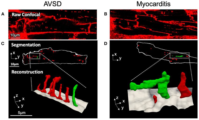
Fiegle et al.                                                                                          T-System Remodeling Pediatric Myocarditis

effect according to the following model: Y ∼ X + (1 | G), for           ratios in myocarditis (0.636 ± 0.024 µm2 ), when compared with
dependend variable Y (response), predicting variable X (fixed           AVSD cardiomyocytes (0.524 ± 0.023 µm2 , p < 0.05). Signs of
effect) and grouping variable G with a possible random effect on        cellular hypertrophy were also present in myocarditis, because
the intercept (1). As grouping variable, the sample ID was used.        cell area, measured at the optical section with the largest area,
The two-tailed unequal variances t-test was applied to test for         was nearly doubled (1,260 ± 95 vs. 606 ± 48 µm2 , p < 0.001).
significant differences between pre- and post-VAD groups, with          From one myocardits sample, isolated cardiomyocytes were
the level of significance set to p
Fiegle et al.                                                                                                                  T-System Remodeling Pediatric Myocarditis

  FIGURE 1 | Confocal microscopic images and 3D t-system reconstruction of LV tissue samples from pediatric patients with AVSD (left) or fulminant myocarditis (right).
  (A,B) Raw confocal images of myocardial tissue sections stained with WGA. (C,D) The surface membrane (white) and t-system (red, green) were discerned by image
  segmentation. Reconstructions represent three-dimensional views of the highlighted areas, with surface membranes (gray) and the t-system. Scale bars in (A,C) also
  apply to (B,D), respectively.

  FIGURE 2 | Quantification of t-system remodeling and cell size in AVSD and acute viral myocarditis. (A,B) Distance maps of the cells shown in Figure 1, with
  color-coded intracellular distance to the nearest t-tubule in µm (TT distance). (A) Example cell from AVSD, (B) Example cell from myocarditis. (C) Quantification of the
  intracellular t-tubule distance (TT distance), volume density (TT density), and shape (TT volume/length). (D) Cell area. Number of analyzed cells/patients: 61/5 (AVSD),
  34/5 (myocarditis). **p < 0.01, ***p < 0.001, linear mixed-effects model with patient group (AVSD or myocarditis) as predicting variable and intercept as random effect
  by patient (i.e., sample). Scale bar in (A) also applies to (B).

explore whether functional cardiac recovery was associated with                          comparison of the t-system of isolated cardiomyocytes from
structural recovery of the t-system and EC coupling junctions,                           pre- and post VAD did not provide evidence for t-system
we investigated these structures in more detail (Figure 7).                              recovery because t-sheets were still widely present (Figure 7B),
Tissue morphology and collagen deposition of pre- and post-                              and TT distance and density were not significantly changed
VAD biopsies were not visibly different (Figure 7A). Also,                               (Figure 7D). TT volume-length ratio remained at 0.67 ± 0.04

Frontiers in Cardiovascular Medicine | www.frontiersin.org                           5                                           January 2021 | Volume 7 | Article 624776
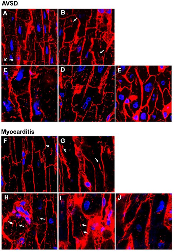
Fiegle et al.                                                                                                                  T-System Remodeling Pediatric Myocarditis

  FIGURE 3 | Confocal microscopic images of myocardial samples from AVSD and myocarditis patients. (A–E) Tissues obtained from five pediatric patients with
  atrioventricular septal defect (AVSD), stained with WGA (red), and DAPI (blue) (F–J) Tissues obtained from five pediatric patients with myocarditis. Enlarged t-system
  components (t-sheets) are indicated with white arrows. The tissue in (F) was retrieved at time of VAD explantation, (G–J) at time of implantation. Note the low t-system
  density in (F–J) when compared with (A–E) and that t-sheets appear as longitudinal components in the xy view. Scale bar in (A) also applies to (B–J).

µm2 (pre-VAD: 0.72 ± 0.05 µm2 ), which exceeded markedly                                 suggest that, although cardiac function improved markedly,
the value of AVSD samples (compare with Figure 2C) and                                   there was only marginal recovery of the overall t-system
was consistent with persisting t-sheets. In summary, these data                          structure. Thus, t-system recovery might not explain the

Frontiers in Cardiovascular Medicine | www.frontiersin.org                           6                                           January 2021 | Volume 7 | Article 624776
Fiegle et al. T-System Remodeling Pediatric Myocarditis
coupling junctions by confocal microscopy (Figure 8). From raw
confocal images shown in Figures 8A,B, the overall appearance
of clusters seemed similar between pre- and post-VAD cells.
However, inspection of Figures 8C,D revealed a larger number of
LTCC clusters in co-localization with JPH2 and RyR in post-VAD
myocytes, which was hardly visible in pre-VAD myocytes. This
indicates a greater fraction of functional EC coupling junctions.
Cluster analysis of the segmented confocal images (Figure 8E)
showed extensive recovery of the JPH2 cluster density during
VAD therapy, reaching values comparable to the AVSD control
group (pre: 0.05 ± 0.01 µm−3 , post: 0.12 ± 0.02 µm−3 , p < 0.001
n = 10/9 cells, respectively; AVSD: 0.13 ± 0.05, n = 56 cells).
Furthermore, cluster analysis confirmed a significantly higher
co-localization of LTCC with JPH2 and RyR in post- than pre-
VAD (pre: 6.8 ± 2.2%, post: 20.2 ± 5%, p < 0.05, Figure 8F).
Because LTCC staining quality was insufficient in fixed tissue
preparations, no co-localization analysis was performed in the
AVSD group (see Supplementary Figure 2). In conclusion, the
results indicate improved JPH2 densities and a higher density
of intact EC coupling junctions at time of VAD explantation,
suggesting a potential molecular basis for the observed functional
cardiac recovery.
DISCUSSION
Acute myocarditis in young children frequently causes heart
failure (15, 32), but the mechanisms leading to contractile
dysfunction are poorly understood. Also, the determinants of
sustained recovery are largely unclear. Here, we report severe t-
system loss and abnormal t-tubule structure (t-sheets) in young
children suffering from viral myocarditis. Such severe t-system
remodeling is commonly observed in chronically failing adult
hearts (7, 9), correlates with HF duration and may impair cardiac
recovery (11), but has to our knowledge not been described in
children. Thus, our results provide evidence that remodeling of
the t-system is not restricted to chronic pathologies in adult
patients, but can as well occur in young children and acute
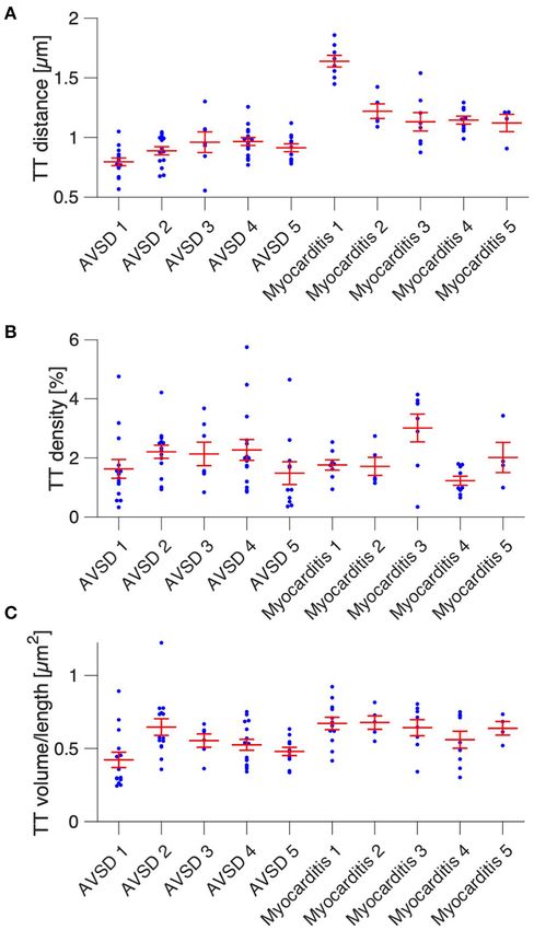
FIGURE 4 | Cardiomyocyte t-system parameters grouped by patient. (A) heart failure. We also shine light on recovery of EC coupling
Intracellular t-tubule (TT) distance of cells from five AVSD and five myocarditis junctions related to improved cardiac function after mechanical
patients (one sample per patient). (B) Cardiomyocyte TT density (TT volume circulatory support.
divided by cell volume). (C) Mean cardiomyocyte TT volume-to-length ratio, a
measure of TT enlargement. Number of analyzed cells: 13/14/6/15/11 (AVSD
1-5) and 8/5/8/9/4 (Myocarditis 1-5), respectively. One-way ANOVA (patient as T-System Remodeling in Children and
categorical variable): p < 0.01 in (A–C). Acute Heart Failure
Generally, little is known on the t-system in human infants
and young children. One study reported a lack of t-tubules in
newborns and an already dense t-system in 5- and 7-month old
prominent functional recovery of the investigated heart during
infants (33). This fits well to our finding that the t-system in the
circulatory support.
control group (age: 9 – 27 months) was dense and contained
regularly-shaped t-tubules. It is therefore likely that in humans,
Remodeling and Recovery of EC Coupling as in other mammals, the t-system develops during early infancy
Junctions (34–37) and that cardiac EC coupling relies on a dense t-system
To investigate whether other structures of EC coupling might not only in adults, but also in children. Of note, this renders t-
be responsible for functional recovery we immunostained the tubule loss a possible pathomechanism in pediatric heart disease.
junctional proteins junctophilin-2 (JPH2), L-type Ca2+ channels The pronounced loss of t-tubules in the children with viral
(LTCC), and cardiac ryanodine receptor (RyR) in myocytes myocarditis investigated here (Figure 2) may have contributed
obtained pre- and post-VAD and analyzed the integrity of EC to the acute reduction of cardiac output and is in accordance
Frontiers in Cardiovascular Medicine | www.frontiersin.org 7 January 2021 | Volume 7 | Article 624776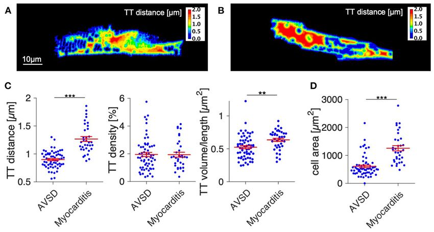
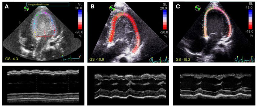
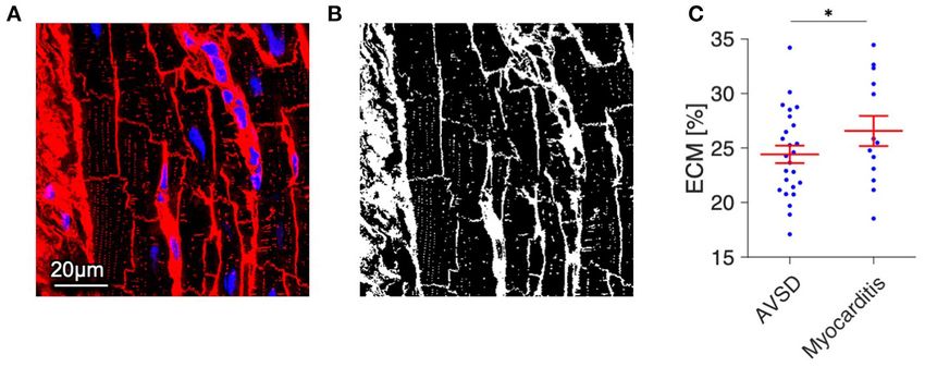
Fiegle et al.                                                                                                                T-System Remodeling Pediatric Myocarditis

  FIGURE 5 | Quantification of extracellular matrix (ECM) as a measure of fibrosis in myocardial tissue from AVSD and Myocarditis patients. (A) Example of a confocal
  image from AVSD tissue stained with WGA (red) and DAPI (blue). (B) The resulting binary image after application of a histogram-based intensity-threshold was used to
  calculate the fraction of ECM. (C) ECM fraction of myocardial samples from 5 AVSD and 5 Myocarditis patients, obtained from 25 and 13 confocal image stacks,
  respectively. *p < 0.05, linear mixed-effects model with patient group (AVSD or Myocarditis) as predicting variable and intercept as random effect by patient (i.e.,
  sample). Scale bar in (A) also applies to (B).

  FIGURE 6 | Speckle tracking and M-mode echocardiography of a 21-month old patient with fulminant viral myocarditis. Apical 4-chamber views with tracing of the
  endo- and epicardium and longitudinal strain (SL) of the left ventricle (top), and M-mode view (bottom) (A) 1 day before VAD implantation, (B) after 1 month of VAD
  therapy, (C) after 3 months of VAD therapy. Global strain (GS) is indicated in yellow. Note the different color scaling in (C).

with studies reporting diminished contractility after acute t-                          a general association of myocarditis with hypertrophy (45).
system loss in de-tubulated animal myocytes or myocardium                               However, hypertrophy is not considered a classical symptom of
(38, 39). Presuming that t-system alterations in the studied                            myocarditis. Thus, our finding might result from investigating
patients developed along with clinical symptoms, the sudden                             particularly severe cases which required VAD implantation.
onset of cardiac dysfunction suggests that t-system deterioration
in humans may arise in short periods of time, as observed in some
animal models (40, 41). This would have important implications                          Inflammation as a Possible Trigger of
when considering that so far only very limited evidence exists                          T-System Remodeling
for the possibility of structural t-system recovery (42, 43). Acute                     The triggers of t-system loss in vivo, especially in human
heart failure, for example due to myocarditis, could become                             hearts, remain elusive. Excessive strain and wall stress (46),
chronic when involving t-system remodeling. Future studies                              mechanical load (47), fibrosis (13, 29), and dysregulation of
could investigate this hypothesis by probing t-system remodeling                        several t-system associated proteins (48) have been suggested.
in acute heart failure and subsequently after clinical recovery                         We found only mildly increased amounts of extracellular matrix
or chronification.                                                                      in myocarditis samples (Figure 5), rendering it unlikely that
   We also found markedly greater sizes of cardiomyocytes in                            fibrosis was the sole trigger of t-system remodeling, because
the myocarditis group, which is in accordance with reports                              previous studies reported much higher degrees of fibrosis
of transient hypertrophy in pediatric myocarditis (44) and                              in association with t-system alterations (13, 29). Although

Frontiers in Cardiovascular Medicine | www.frontiersin.org                          8                                           January 2021 | Volume 7 | Article 624776
Fiegle et al.                                                                                                              T-System Remodeling Pediatric Myocarditis

  FIGURE 7 | Histology and t-system quantification after functional cardiac recovery. (A) Masson’s trichrome stain of myocardial tissue of a 21-month old myocarditis
  patient before VAD implantation (Pre-VAD) and at time of VAD explantation (Post-VAD) 3 months later. Cardiomyocytes appar red, collagen appears blue. (B) Pre- and
  Post-VAD confocal microscopic images of isolated and membrane-stained cardiomyocytes. In one example cell, filled arrow heads point to the surface membrane,
  empty arrow heads point to t-system components. (C) Overview of patient’s cardiac function expressed as left-ventricular ejection fraction (LVEF). Dashed red lines
  and arrows indicate times of VAD implantation and explantation. (D) Cardiomyocyte t-tubule (TT) distance, TT density, and TT volume/length ratio (n = 8/9 cells for
  Pre- and Post-VAD, respectively). n.s., not significant (p > 0.05), unpaired two-tailed t-test.

increased mechanical load is present in fulminant myocarditis,                        (53), it seems possible that downregulation of inflammatory
we suggest the hypothesis that t-system loss is facilitated                           pathways protects from t-system remodeling. However, these
or triggered by inflammatory pathways. These are highly                               concepts require further investigation, and it should be noted
active in myocarditis (14, 49), but also in chronic heart                             that although treatment with glucocorticoids seems favorable
failure (50) and after myocardial infarction (51), where t-                           especially in non-viral myocarditis (54), it may also have adverse
system remodeling is regularly observed. Excessive activation of                      effects (55).
inflammatory signaling in cardiomyocytes, for instance through
viral infection, could affect cellular processes required for t-                      Structural and Functional Recovery
system maintenance. Disturbed autophagy, for example, was                             Analysis of the t-system and junctional proteins in the sample
reported to contribute to t-system loss in isolated cardiomyocytes                    obtained after circulatory support and clinical recovery revealed
(24), while viral proteases are able to degrade proteins of the                       only marginal recovery of the t-system. Although TT density
autophagic machinery (18). Interestingly, it was recently shown                       was increased, TT distance and morphology (presence of
that glucocorticoids prevent t-system loss in cardiomyocyte                           t-sheets) were far from normal. The volume-length ratio,
culture and that glucocorticoid receptor knockout causes t-                           an indicator of t-tubule dilation to t-sheets (11), was still
system loss in vivo (24). As glucocorticoids exert anti-                              elevated and could explain why TT volume density reached
inflammatory effects, e.g., via NF-κB inhibition (52), and NF-                        nearly normal values, while TT distance—the presumably
κB inactivation has been shown to preserve calcium handling                           most relevant parameter for EC coupling (56)—remained

Frontiers in Cardiovascular Medicine | www.frontiersin.org                        9                                           January 2021 | Volume 7 | Article 624776
Fiegle et al.                                                                                                                T-System Remodeling Pediatric Myocarditis

  FIGURE 8 | Structural integrity of excitation-contraction (EC) coupling junctions before and after VAD therapy of a myocarditis patient. (A,B) Raw confocal images of
  fixed isolated cardiomyocytes from pre- and post-VAD of the patient presented in Figures 6, 7, co-stained for LTCC (red), RyR (green), and JPH2 (blue) and with DAPI
  (not shown). (C,D) Overlay of binary images for the EC coupling proteins shown in (A,B), with magnifications of boxed regions. The cell surface, obtained from
  autofluorescence, is shown white, nuclei are shown white with black asterisk. Co-localizations of LTCC, JPH2, and RyR appear cyan, magenta, yellow, or white (see
  color legend). (E) Cardiomyocyte JPH2 cluster density (JPH2 density) of AVSD as reference and the Pre- and Post-VAD sample. (F) Fraction of LTCC clusters that
  were co-localized with both RyR and JPH2, as a measure of intact EC coupling junctions (n = 10/9 cells for Pre/Post-VAD). *p < 0.05, **p < 0.01, ***p < 0.001,
  unpaired, two-tailed t-test with Holm–Bonferroni multiple-comparison correction.

increased. One should consider, however, that the biopsy at                             near the apex and microscale deposition of ECM could have
explantation was taken adjacent to the scar caused by VAD                               impeded t-system recovery particularly at the biopsy site.
implantation and might therefore differ structurally from other                         Thus, the question remains open whether t-system recovery is
regions of the ventricle. Although no major differences of                              generally possible.
collagen deposition between pre- and post-VAD biopsies were                                Regarding the integrity of EC coupling junctions,
apparent (Figure 7A), overall fibrosis, altered strain patterns                         however, cardiomyocytes showed clear improvements after

Frontiers in Cardiovascular Medicine | www.frontiersin.org                         10                                          January 2021 | Volume 7 | Article 624776
Fiegle et al.                                                                                             T-System Remodeling Pediatric Myocarditis

mechanical circulatory support, which may have contributed                  children and may contribute to contractile dysfunction in
to improvements in contractile function. JPH2 cluster density               acute viral myocarditis. Furthermore, this study provides strong
improved despite persistent t-system remodeling and reached                 evidence of severe t-tubule loss and remodeling to t-sheets
levels of the control AVSD group. This confirms other studies               in acute heart failure, challenging the idea that chronic heart
reporting that JPH2 degradation and t-system remodeling are                 failure or ischemic cardiomyopathy is required to elicit t-system
accompanied in cardiac disease (12, 57), but it also suggests               remodeling in humans. Although t-system recovery remains
that they may recover independently from each other. Because                elusive, recovery of EC coupling junctions may be possible and
JPH2 is thought to improve coupling between LTCC and RyRs                   deserves further investigation. Moreover, we suggest to further
(1–4), it is likely that the integrity of EC coupling junctions             explore the t-system as a prognostic biomarker (11, 62) not only
ameliorated secondary to increased JPH2 expression, even                    in adults but also in pediatric heart disease.
though there was no t-system recovery (Figure 7). This would
fit to observations of high plasticity of junctophilin-2 and
ryanodine receptors (4) and to the observation that Ca2+                    DATA AVAILABILITY STATEMENT
release is more rapid and more efficient in compact dyads,
                                                                            The raw data supporting the conclusions of this
i.e., EC coupling junctions of high integrity (58). Investigating
                                                                            article will be made available by the authors, without
this idea in more detail and finding ways to induce reverse
                                                                            undue reservation.
remodeling of EC coupling junctions could be promising targets
of future studies.

Limitations                                                                 ETHICS STATEMENT
The t-system was imaged in WGA-stained, fixed tissue sections               The studies involving human participants were reviewed and
in 9 out of 10 samples, while from the sample used for                      approved by Institutional Review Boards of the University
recovery analysis (Figure 6) isolated cells stained with Di-                of Tuebingen and the Friedrich-Alexander-University (FAU)
8-ANEPPS were imaged. Both WGA and Di-8-ANEPPS are                          Erlangen-Nuremberg. Written informed consent to participate in
widely used reliable markers of the t-system in humans and                  this study was provided by the participants’ legal guardian/next
animals (7, 9, 11, 24, 37), but it has been reported that t-                of kin.
tubules and caveolae can decline quickly after cell isolation
(59, 60) and that PFA fixation may alter tissue dimensions
(61). To examine if these effects may have confounded t-                    AUTHOR CONTRIBUTIONS
system measures, we stained and imaged tissue sections and
isolated myocytes from a human ventricular biopsy with                      MA, SD, KK, TS, and TV contributed to the conception and
both methods, but could not detect differences in t-system                  design. RC, DF, and MS contributed to the data acquisition and
density or morphology (Supplementary Figure 1). In addition,                experiments. MA, DF, and TS contributed to the data analysis.
statistical analysis of AVSD vs. myocarditis, excluding the isolated        MA, DF, TS, and TV contributed to the interpretation of the data.
cardiomyocytes, still yielded significantly increased TT distance           DF and TS drafted the manuscript. All authors critically revised
in myocarditis (p < 0.001).                                                 and approved the manuscript.
   Because AVSD hearts may have been subjected to increased
load due to valve defects, it is possible that the t-system in
these samples was not completely normal. However, considering               FUNDING
that increased cardiac load has been associated with a loss
rather than a gain of t-tubules (47), it is unlikely that the               This work was supported by the Interdisciplinary
detected t-system loss in myocarditis resulted from a flawed                Center for Clinical Research (IZKF) at the University
control. We also acknowledge that samples were obtained                     Hospital of the Friedrich-Alexander University of
from different cardiac regions in AVSD (LV outflow tract)                   Erlangen-Nürnberg (FAU).
and myocarditis hearts (LV apex), but although t-tubule
remodeling may vary regionally, it seems to correlate with
                                                                            ACKNOWLEDGMENTS
function rather than with specific regions of the heart (9). To
substantiate the results regarding recovery of the t-system and             We would like to thank Julia Seufert and Jasmin Raufer for their
EC coupling junctions, studies with a larger number of cases                valuable support.
are required to enable statistical analysis. The case explored
here shall generate new hypotheses and motivate studies in
this field.                                                                 SUPPLEMENTARY MATERIAL
Conclusions                                                                 The Supplementary Material for this article can be found
The results from the presented study indicate that remodeling               online at: https://www.frontiersin.org/articles/10.3389/fcvm.
of EC coupling junctions and the t-system can occur in young                2020.624776/full#supplementary-material

Frontiers in Cardiovascular Medicine | www.frontiersin.org             11                                   January 2021 | Volume 7 | Article 624776
Fiegle et al. T-System Remodeling Pediatric Myocarditis
REFERENCES 19. Goller T, Galle J, Eggers HJ, Bultmann B. Experimental reovirus myocarditis
in newborn mice. Electron microscopic observations. Virchows Arch B Cell
1. Sun XH, Protasi F, Takahashi M, Takeshima H, Ferguson DG, Franzini- Pathol Incl Mol Pathol. (1986) 50:373–86. doi: 10.1007/BF02889915
Armstrong C. Molecular architecture of membranes involved in excitation- 20. Caforio AL, Pankuweit S, Arbustini E, Basso C, Gimeno-Blanes J, Felix SB,
contraction coupling of cardiac muscle. J Cell Biol. (1995) 129:659–71. et al. Current state of knowledge on aetiology, diagnosis, management, and
doi: 10.1083/jcb.129.3.659 therapy of myocarditis: a position statement of the European society of
2. Takeshima H, Komazaki S, Nishi M, Iino M, Kangawa K. Junctophilins: a cardiology working group on myocardial and pericardial diseases. Eur Heart
novel family of junctional membrane complex proteins. Mol Cell. (2000) J. (2013) 34:2636–48, 2648a–d. doi: 10.1093/eurheartj/eht210
6:11–22. doi: 10.1016/S1097-2765(05)00005-5 21. Fiegle DJ, Volk T, Seidel T. Isolation of Human Ventricular Cardiomyocytes
3. Munro ML, Jayasinghe ID, Wang Q, Quick A, Wang W, Baddeley D, et al. from Vibratome-Cut Myocardial Slices. JoVE. (2020) (159):e61167.
Junctophilin-2 in the nanoscale organisation and functional signalling of doi: 10.3791/61167
ryanodine receptor clusters in cardiomyocytes. J Cell Sci. (2016) 129:4388–98. 22. Seidel T, Edelmann JC, Sachse FB. Analyzing remodeling of cardiac
doi: 10.1242/jcs.196873 tissue: a comprehensive approach based on confocal microscopy
4. Jones PP, MacQuaide N, Louch WE. Dyadic plasticity in cardiomyocytes. and 3D reconstructions. Ann Biomed Eng. (2016) 44:1436–48.
Front Physiol. (2018) 9:1773. doi: 10.3389/fphys.2018.01773 doi: 10.1007/s10439-015-1465-6
5. Savio-Galimberti E, Frank J, Inoue M, Goldhaber JI, Cannell MB, Bridge 23. Schwab BC, Seemann G, Lasher RA, Torres NS, Wulfers EM, Arp M, et al.
JH, et al. Novel features of the rabbit transverse tubular system revealed Quantitative analysis of cardiac tissue including fibroblasts using three-
by quantitative analysis of three-dimensional reconstructions from confocal dimensional confocal microscopy and image reconstruction: towards a basis
images. Biophys J. (2008) 95:2053–62. doi: 10.1529/biophysj.108.130617 for electrophysiological modeling. IEEE Trans Med Imaging. (2013) 32:862–
6. Louch WE, Bito V, Heinzel FR, Macianskiene R, Vanhaecke J, Flameng W, 72. doi: 10.1109/TMI.2013.2240693
et al. Reduced synchrony of Ca2+ release with loss of T-tubules-a comparison 24. Seidel T, Fiegle DJ, Baur TJ, Ritzer A, Nay S, Heim C, et al. Glucocorticoids
to Ca2+ release in human failing cardiomyocytes. Cardiovasc Res. (2004) preserve the t-tubular system in ventricular cardiomyocytes by
62:63–73. doi: 10.1016/j.cardiores.2003.12.031 upregulation of autophagic flux. Basic Res Cardiol. (2019) 114:47.
7. Lyon AR, MacLeod KT, Zhang Y, Garcia E, Kanda GK, Lab MJ, et al. Loss of doi: 10.1007/s00395-019-0758-6
T-tubules and other changes to surface topography in ventricular myocytes 25. Seidel T, Dräbing T, Seemann G, Sachse FB. A semi-automatic
from failing human and rat heart. Proc Natl Acad Sci USA. (2009) 106:6854–9. approach for segmentation of three-dimensional microscopic image
doi: 10.1073/pnas.0809777106 stacks of cardiac tissue. Lect Notes Comput Sic. (2013) 7945:300–7.
8. Sacconi L, Ferrantini C, Lotti J, Coppini R, Yan P, Loew LM, et al. doi: 10.1007/978-3-642-38899-6_36
Action potential propagation in transverse-axial tubular system is 26. Emde B, Heinen A, Godecke A, Bottermann K. Wheat germ agglutinin
impaired in heart failure. Proc Natl Acad Sci USA. (2012) 109:5815–9. staining as a suitable method for detection and quantification of fibrosis in
doi: 10.1073/pnas.1120188109 cardiac tissue after myocardial infarction. Eur J Histochem. (2014) 58:2448.
9. Crossman DJ, Young AA, Ruygrok PN, Nason GP, Baddelely D, Soeller C, et al. doi: 10.4081/ejh.2014.2448
T-tubule disease: Relationship between t-tubule organization and regional 27. Pinheiro J, Bates D. Mixed-Effects Models in S and S-PLUS. Springer Science &
contractile performance in human dilated cardiomyopathy. J Mol Cell Cardiol. Business Media (2006).
(2015) 84:170–8. doi: 10.1016/j.yjmcc.2015.04.022 28. Sikkel MB, Francis DP, Howard J, Gordon F, Rowlands C, Peters NS, et al.
10. Crocini C, Ferrantini C, Scardigli M, Coppini R, Mazzoni L, Lazzeri E, et al. Hierarchical statistical techniques are necessary to draw reliable conclusions
Novel insights on the relationship between T-tubular defects and contractile from analysis of isolated cardiomyocyte studies. Cardiovasc Res. (2017)
dysfunction in a mouse model of hypertrophic cardiomyopathy. J Mol Cell 113:1743–52. doi: 10.1093/cvr/cvx151
Cardiol. (2016) 91:42–51. doi: 10.1016/j.yjmcc.2015.12.013 29. Crossman DJ, Shen X, Jullig M, Munro M, Hou Y, Middleditch M, et al.
11. Seidel T, Navankasattusas S, Ahmad A, Diakos NA, Xu WD, Tristani- Increased collagen within the transverse tubules in human heart failure.
Firouzi M, et al. Sheet-like remodeling of the transverse tubular system Cardiovasc Res. (2017) 113:879–91. doi: 10.1093/cvr/cvx055
in human heart failure impairs excitation-contraction coupling and 30. Greiner J, Sankarankutty AC, Seemann G, Seidel T, Sachse FB. Confocal
functional recovery by mechanical unloading. Circulation. (2017) 135:1632– microscopy-based estimation of parameters for computational modeling of
45. doi: 10.1161/CIRCULATIONAHA.116.024470 electrical conduction in the normal and infarcted heart. Front Physiol. (2018)
12. Pinali C, Malik N, Davenport JB, Allan LJ, Murfitt L, Iqbal MM, 9:239. doi: 10.3389/fphys.2018.00239
et al. Post-myocardial infarction T-tubules form enlarged branched 31. Amabile N, Fraisse A, Bouvenot J, Chetaille P, Ovaert C. Outcome
structures with dysregulation of junctophilin-2 and bridging integrator of acute fulminant myocarditis in children. Heart. (2006) 92:1269–73.
1 (BIN-1). J Am Heart Assis. (2017) 6:e004834. doi: 10.1161/JAHA.116. doi: 10.1136/hrt.2005.078402
004834 32. Kindermann I, Barth C, Mahfoud F, Ukena C, Lenski M, Yilmaz A,
13. Seidel T, Sankarankutty AC, Sachse FB. Remodeling of the transverse tubular et al. Update on myocarditis. J Am Coll Cardiol. (2012) 59:779–92.
system after myocardial infarction in rabbit correlates with local fibrosis: doi: 10.1016/j.jacc.2011.09.074
a potential role of biomechanics. Prog Biophys Mol Biol. (2017) 130 (Pt. 33. Wiegerinck RF, Cojoc A, Zeidenweber CM, Ding G, Shen M, Joyner RW,
B):302–14. doi: 10.1016/j.pbiomolbio.2017.07.006 et al. Force frequency relationship of the human ventricle increases
14. Kawai C. From myocarditis to cardiomyopathy: mechanisms of inflammation during early postnatal development. Pediatr Res. (2009) 65:414–9.
and cell death: learning from the past for the future. Circulation. (1999) doi: 10.1203/PDR.0b013e318199093c
99:1091–100. doi: 10.1161/01.CIR.99.8.1091 34. Kim HD, Kim DJ, Lee IJ, Rah BJ, Sawa Y, Schaper J. Human fetal heart
15. Fung G, Luo H, Qiu Y, Yang D, McManus B. Myocarditis. Circ Res. (2016) development after mid-term: morphometry and ultrastructural study. J Mol
118:496–514. doi: 10.1161/CIRCRESAHA.115.306573 Cell Cardiol. (1992) 24:949–65. doi: 10.1016/0022-2828(92)91862-Y
16. Zhou F, Jiang X, Teng L, Yang J, Ding J, He C. Necroptosis may be a novel 35. Haddock PS, Coetzee WA, Cho E, Porter L, Katoh H, Bers DM,
mechanism for cardiomyocyte death in acute myocarditis. Mol Cell Biochem. et al. Subcellular [Ca2+]i gradients during excitation-contraction coupling
(2018) 442:11–8. doi: 10.1007/s11010-017-3188-5 in newborn rabbit ventricular myocytes. Circ Res. (1999) 85:415–27.
17. Remels AHV, Derks WJA, Cillero-Pastor B, Verhees KJP, Kelders MC, doi: 10.1161/01.RES.85.5.415
Heggermont W, et al. NF-kappaB-mediated metabolic remodelling in the 36. Munro ML, Soeller C. Early transverse tubule development begins in
inflamed heart in acute viral myocarditis. Biochim Biophys Acta Mol Basis Dis. utero in the sheep heart. J Muscle Res Cell Motil. (2016) 37:195–202.
(2018) 1864:2579–89. doi: 10.1016/j.bbadis.2018.04.022 doi: 10.1007/s10974-016-9462-4
18. Yajima T. Viral myocarditis: potential defense mechanisms within the 37. Lipsett DB, Frisk M, Aronsen JM, Norden ES, Buonarati OR, Cataliotti A, et al.
cardiomyocyte against virus infection. Future Microbial. (2011) 6:551–66. Cardiomyocyte substructure reverts to an immature phenotype during heart
doi: 10.2217/fmb.11.40 failure. J Physiol. (2019) 597:1833–53. doi: 10.1113/JP277273
Frontiers in Cardiovascular Medicine | www.frontiersin.org 12 January 2021 | Volume 7 | Article 624776Fiegle et al. T-System Remodeling Pediatric Myocarditis
38. Ferrantini C, Coppini R, Sacconi L, Tosi B, Zhang ML, Wang GL, et al. Impact 53. Zhang XQ, Tang R, Li L, Szucsik A, Javan H, Saegusa N, et al. Cardiomyocyte-
of detubulation on force and kinetics of cardiac muscle contraction. J Gen specific p65 NF-kappaB deletion protects the injured heart by preservation of
Physiol. (2014) 143:783–97. doi: 10.1085/jgp.201311125 calcium handling. Am J Physiol Heart Circ Physiol. (2013) 305:H1089–1097.
39. Bourcier A, Barthe M, Bedioune I, Lechene P, Miled HB, Vandecasteele G, doi: 10.1152/ajpheart.00067.2013
et al. Imipramine as an alternative to formamide to detubulate rat ventricular 54. Frustaci A, Chimenti C. Immunosuppressive therapy in myocarditis. Circ J.
cardiomyocytes. Exp Physiol. (2019) 104:1237–49. doi: 10.1113/EP087760 (2015) 79:4–7. doi: 10.1253/circj.CJ-14-1192
40. Louch WE, Mørk HK, Sexton J, Strømme TA, Laake P, Sjaastad I, et al. T- 55. Mason JW, O’Connell JB, Herskowitz A, Rose NR, McManus BM, Billingham
tubule disorganization and reduced synchrony of Ca2+ release in murine ME, et al. A clinical trial of immunosuppressive therapy for myocarditis. The
cardiomyocytes following myocardial infarction. J Physiol. (2006) 574:519–33. myocarditis treatment trial investigators. N Engl J Med. (1995) 333:269–75.
doi: 10.1113/jphysiol.2006.107227 doi: 10.1056/NEJM199508033330501
41. Wu CY, Jia Z, Wang W, Ballou LM, Jiang YP, Chen B, et al. PI3Ks maintain 56. Torres NS, Sachse FB, Izu LT, Goldhaber JI, Spitzer KW, Bridge JH. A modified
the structural integrity of T-tubules in cardiac myocytes. PLoS ONE. (2011) local control model for Ca2+ transients in cardiomyocytes: junctional flux
6:e24404. doi: 10.1371/journal.pone.0024404 is accompanied by release from adjacent non-junctional RyRs. J Mol Cell
42. Ibrahim M, Navaratnarajah M, Siedlecka U, Rao C, Dias P, Moshkov AV, et al. Cardiol. (2014) 68:1–11. doi: 10.1016/j.yjmcc.2013.12.019
Mechanical unloading reverses transverse tubule remodelling and normalizes 57. Zhang C, Chen B, Guo A, Zhu Y, Miller JD, Gao S, et al. Microtubule-mediated
local Ca(2+)-induced Ca(2+)release in a rodent model of heart failure. Eur J defects in junctophilin-2 trafficking contribute to myocyte transverse-tubule
Heart Fail. (2012) 14:571–80. doi: 10.1093/eurjhf/hfs038 remodeling and Ca2+ handling dysfunction in heart failure. Circulation.
43. Lawless M, Caldwell JL, Radcliffe EJ, Smith CER, Madders GWP, Hutchings (2014) 129:1742–50. doi: 10.1161/CIRCULATIONAHA.113.008452
DC, et al. Phosphodiesterase 5 inhibition improves contractile function and 58. Novotova M, Zahradnikova AJr, Nichtova Z, Kovac R, Kralova
restores transverse tubule loss and catecholamine responsiveness in heart E, Stankovicova T, et al. Structural variability of dyads relates to
failure. Sci Rep. (2019) 9:6801. doi: 10.1038/s41598-019-42592-1 calcium release in rat ventricular myocytes. Sci Rep. (2020) 10:8076.
44. Kosutic J. Severe transient left ventricular hypertrophy in an infant with doi: 10.1038/s41598-020-64840-5
acute myocarditis and heart failure. Pediatr Cardiol. (2004) 25:677–80. 59. Mitcheson JS, Hancox JC, Levi AJ. Action potentials, ion channel
doi: 10.1007/s00246-003-0617-x currents and transverse tubule density in adult rabbit ventricular myocytes
45. Pinamonti B, Alberti E, Cigalotto A, Dreas L, Salvi A, Silvestri F, et al. maintained for 6 days in cell culture. Pflugers Arch. (1996) 431:814–27.
Echocardiographic findings in myocarditis. Am J Cardiol. (1988) 62:285–91. doi: 10.1007/s004240050073
doi: 10.1016/0002-9149(88)90226-3 60. Burton RAB, Rog-Zielinska EA, Corbett AD, Peyronnet R, Bodi I, Fink
46. Frisk M, Ruud M, Espe EK, Aronsen JM, Roe AT, Zhang L, et al. Elevated M, et al. Caveolae in rabbit ventricular myocytes: distribution and
ventricular wall stress disrupts cardiomyocyte t-tubule structure and calcium dynamic diminution after cell isolation. Biophys J. (2017) 113:1047–59.
homeostasis. Cardiovasc Res. (2016) 112:443–51. doi: 10.1093/cvr/cvw111 doi: 10.1016/j.bpj.2017.07.026
47. Ibrahim M, Nader A, Yacoub MH, Terracciano C. Manipulation of 61. Holda MK, Klimek-Piotrowska W, Koziej M, Piatek K, Holda J. Influence
sarcoplasmic reticulum Ca(2+) release in heart failure through mechanical of different fixation protocols on the preservation and dimensions of cardiac
intervention. J Physiol. (2015) 593:3253–9. doi: 10.1113/JP270446 tissue. J Anat. (2016) 229:334–40. doi: 10.1111/joa.12469
48. Guo A, Zhang C, Wei S, Chen B, Song LS. Emerging mechanisms of 62. Nikolova AP, Hitzeman TC, Baum R, Caldaruse AM, Agvanian S, Xie
T-tubule remodelling in heart failure. Cardiovasc Res. (2013) 98:204–15. Y, et al. Association of a novel diagnostic biomarker, the plasma cardiac
doi: 10.1093/cvr/cvt020 bridging integrator 1 score, with heart failure with preserved ejection
49. Corsten MF, Schroen B, Heymans S. Inflammation in viral fraction and cardiovascular hospitalization. JAMA Cardiol. (2018) 3:1206–10.
myocarditis: friend or foe? Trends Mol Med. (2012) 18:426–37. doi: 10.1001/jamacardio.2018.3539
doi: 10.1016/j.molmed.2012.05.005
50. Dick SA, Epelman S. Chronic heart failure and inflammation: Conflict of Interest: The authors declare that the research was conducted in the
what do we really know? Circ Res. (2016) 119:159–76. absence of any commercial or financial relationships that could be construed as a
doi: 10.1161/CIRCRESAHA.116.308030 potential conflict of interest.
51. Lagan J, Naish JH, Simpson K, Zi M, Cartwright EJ, Foden P, et al.
Substrate for the myocardial inflammation-heart failure hypothesis identified Copyright © 2021 Fiegle, Schöber, Dittrich, Cesnjevar, Klingel, Volk, Alkassar and
using novel USPIO methodology. JACC Cardiovasc Imaging. (2020). Seidel. This is an open-access article distributed under the terms of the Creative
doi: 10.1016/j.jcmg.2020.02.001 Commons Attribution License (CC BY). The use, distribution or reproduction in
52. Auphan N, DiDonato JA, Rosette C, Helmberg A, Karin M. other forums is permitted, provided the original author(s) and the copyright owner(s)
Immunosuppression by glucocorticoids: inhibition of NF-kappa B activity are credited and that the original publication in this journal is cited, in accordance
through induction of I kappa B synthesis. Science. (1995) 270:286–90. with accepted academic practice. No use, distribution or reproduction is permitted
doi: 10.1126/science.270.5234.286 which does not comply with these terms.
Frontiers in Cardiovascular Medicine | www.frontiersin.org 13 January 2021 | Volume 7 | Article 624776You can also read