Upregulation of p53 by tannic acid treatment suppresses the proliferation of human colorectal carcinoma
←
→
Page content transcription
If your browser does not render page correctly, please read the page content below
Acta Pharm. 71 (2021) ???–??? Original research paper
https://doi.org/10.2478/acph-2021-0036
Upregulation of p53 by tannic acid treatment suppresses
the proliferation of human colorectal carcinoma
SERDAR KARAKURT1,* The present study’s objective is to clarify the molecular
SINAN KANDIR 2
mechanisms of tannic acid effects on the viability of human
ÇİĞDEM GÖKÇEK-SARAÇ3
colorectal carcinoma (CRC). Tannic acid is stable for up to
1
Selcuk University, Faculty of Science 48 h and is localized in both cytoplasm and nucleus. It
Department of Biochemistry, Konya dose-dependently inhibited the viability of CRC cell lines;
Turkey SW-620 and HT-29 with IC50 values of 7.2 ± 0.8 and 37.6 ± 1.4
µmol L–1. Besides, metastatic, invasive, and colony forma-
2
Cukurova University, Ceyhan Faculty tion properties of CRC cells were significantly inhibited
of Veterinary Medicine, Department of following the tannic acid treatment (p < 0.001). Tannic acid
Physiology, Adana, Turkey has been found to modulate enzyme, protein, and gene ex-
pressions of NQO1 in different levels and the upregulation
3
Faculty of Engineering, Department of of protein/gene expressions of p53 (p < 0.001), which leads
Biomedical Engineering, Akdeniz the cells to trigger apoptosis. In conclusion, the present in
University, Antalya, Turkey vitro study may supply a significant background for in vivo
studies in which the molecular mechanisms of antioxidant
and chemopreventive activities of tannic acid will com-
pletely clarify.
Accepted October 27, 2020 Keywords: tannic acid, colorectal carcinoma, wound healing,
Published online November 10, 2020 cell viability, NQO1, p53
After cardiovascular diseases, cancer is the second most common cause of death,
a ffecting millions worldwide (1). The uncontrolled cell cycle progression and the loss of
apoptotic mechanisms are induced by activating oncogenes or deactivating tumor sup-
pressor genes. Continually developing cancer cells have the potential to be metastatic.
Therefore, early and effective treatments are essential in cancer treatment. Colorectal car-
cinoma (CRC), with its high morbidity and mortality rates, causes significant problems (2).
A progressive change in genetic code during the development and progression of CRC
results in tumor transformation from the normal colonic mucosa (3). Increased CRC inci-
dence is related to malnutrition, smoking, intestinal inflammatory problems, polyps,
genetic factors, and aging (4). Many attempts, including surgery, chemotherapy, and radio-
therapy, were developed against CRC’s proliferation and invasion (5–7). Although the use
of chemically derived drugs, such as cisplatin-based therapy, prevents DNA repair mecha
nisms and induces apoptosis in cancer cells, it potentially causes severe side effects (8, 9).
* Correspondence; e-mail: kserdar1@yahoo.com
1
S. Karakurt et al.: Upregulation of p53 by tannic acid treatment suppresses the proliferation of human colorectal carcinoma, Acta Pharm.
71 (2021) ???–???.
The protective effects of secondary plant metabolites on human health are well known
(10). Phenolic compounds with aromatic rings containing hydroxyl groups are various
plant-derived substances with biological activities ranging from antioxidant and anticancer
properties (11, 12). One of the polyphenolic compounds is tannic acid (TA), produced
from the secondary metabolism of plants. It can be obtained from various biological sam-
ples including grapes, grass, blackberries, and dates (13–15). TA has a central core com-
posed of glucose esterified with gallic acid (Fig. 1d). TA’s aromatic hydroxyl groups effec-
tively form strong complexes with proteins and other macromolecules under particular
environmental conditions. Antimicrobial, antifungal, antiviral, and cytotoxic properties
of TA were demonstrated in many studies (16-19). TA’s possesses inhibitory potential due
to the regulatory effect of many enzyme activities (20, 21). One of these enzymes is NAD(P)H:
quinone oxidoreductase 1 (NQO1, DT-diaphorase, EC 1.6.99.2), a FAD-containing, homo
dimer protein (22). Two electron reducting properties of NQO1 make it critical against
many procarcinogens and protect cells from oxidative stress and reactive oxygen species
(ROS) (23).
The development of various cancer types is associated with a decrease in NQO1 activ-
ity by polymorphism (24, 25). Three protective strategies have been identified due to ele-
vated NQO1 expression: one-step quinone detoxification, maintenance of endogenous
antioxidants, and regulation of p53 stability (26). An important transcription factor, p53,
a) b) c)
d)
Fig. 1. Bio-imaging, structure, and stability of TA. SW-620 cells were treated with 5 µmol L–1 of TA
and incubated for 45 min. The scale is 100 µm. a) brightfield; b) fluorescence; c) merge of the images;
d) UV spectra and structures of TA and GA. l ex = 350 nm.
2
S. Karakurt et al.: Upregulation of p53 by tannic acid treatment suppresses the proliferation of human colorectal carcinoma, Acta Pharm.
71 (2021) ???–???.
has a crucial role in the suppression of carcinogenesis, which induces either growth arrest
or apoptosis (27). The upregulation of NQO1 inhibits proteasomal degradation of p53, p73
and p33 and maintains the stability of these enzymes (28). The increased level of NQO1
has been linked to the low level of CRC (29).
EXPERIMENTAL
Chemicals
Tannic acid (TA; T0200), phenylmethanesulfonylflouride (PMSF; P7626), bicinchoninic
acid (D8284), bovine serum albumin (BSA; A7511), and DCPIP were obtained from Sigma-
-Aldrich (USA). NQO1 (Anti-NQO1 antibody (ab34173, 31 kDa), p53 (ab32389, 53 kDa),
GAPDH (ab181602, 36 kDa), p53 (PAb 240, 53 kDa), and Goat Anti Rabbit IgG (ab216773)
were purchased from Abcam (UK). Bax (Proteintech (50599-2), 21 kDa), Bcl-2 (Proteintech
(12789-1), 26 kDa) were obtained from Proteintech (USA). Alamar blue was purchased from
Invitrogen Life Technologies). Non-fat dry milk (170-6404) and tetramethyl ethylene diamine
(TEMED; 161-0801) were from Bio-Rad Laboratories (USA). Primers were designed by
Iontek (Turkey). Leibovitz (L-15), EMEM, l-glutamine were obtained from ATCC (American
Type Culture Collection, USA). All purchased chemicals and solvents were of the a nalytical
standard at the highest grade of purity available.
Biology
In vitro bioimaging. – The penetration and the localization of TA in SW-620 cells were
detected via fluorescence microscopy technique. Cells were seeded into a glass-bottom
dark 24-well plate and incubated overnight at 37 °C and 5 % CO2. Cells were then washed
with 10 mmol L–1 PBS and pretreated with DAPI (1/1000) for 30 min. The excess amount of
DAPI was removed, and cells were incubated with serum-free growth medium containing
5 µmol L–1 of TA for 45 min. Following the incubation, the cells were washed twice with
PBS to remove the excess TA, and fluorescence intensity was monitored with a fluorescence
microscope (ZOE Bio-Rad, USA) at an excitation wavelength of 355 nm and an emission
wavelength of 433 nm (30).
Stability studies of TA. – To determine TA stability in in vitro conditions, 40 µg–1 of tan-
nic acid and its monomeric subunit gallic acid were dissolved in FBS free growth medium
and change in the UV-spectra monitored by spectrophotometer (Shimadzu, Japan) at 37 °C
up to 48 h.
Cell viability and proliferation studies. – Human colorectal carcinoma cell lines, SW620,
and HT-29 colon cancer cells and human healthy epithelial cell line CCD-18Co were pur-
chased from ATCC. SW620, HT-29, and CCD-18Co cells were grown in Leibovitz (L-15) and
EMEM growth mediums, respectively, supplemented with 10 % FBS (fetal bovine serum)
and 2 mM glutamine and were grown at 37 °C and 5 % CO2. The growth media used for
these cell lines were also used for the dilution of TA. 3 × 10⁴ cells were transferred to 24-
well plates and treated with various TA concentrations ranging from 0–200 µmol L–1 and
were incubated 48 h. The proliferation and viability of colon cells were determined by
3
S. Karakurt et al.: Upregulation of p53 by tannic acid treatment suppresses the proliferation of human colorectal carcinoma, Acta Pharm.
71 (2021) ???–???.
using Alamar blue (31). Cells were treated with Alamar blue (10 % of the well) for 3 h, and
the cell suspension was transferred to a 96-well plate. The change in the color was mea-
sured spectrophotometrically by reading absorbance at 570 nm and 600 nm. The IC50 values
were calculated using the sigmoidal plot of the cell viability.
Wound healing assay. – To determine the effects of TA on metastatic properties of h
uman
CRC cells, in vitro wound healing studies were performed. Before the wound healing
studies, cells were seeded into 6-well plates and treated with TA t the equivalent concen-
tration of IC50 values (7.2 µmol L–1 for SW-620 and 37.6 µmol L–1 for HT-29) for 48 h. Control
groups were supplemented with the growth medium without TA. After 48 h, 5 × 10⁴ of
SW620 and HT-29 cells were seeded into 24-well plate using CytoSelect 24-well Wound
Healing inserts. Following overnight incubation at 37 °C, and 5 % CO2, the inserts were
removed and the cells were washed with 10 mmol L–1 PBS to remove unattached cells.
Reduction in the scratch areas was monitored by a Trinocular inverted microscope (VWR,
USA). The cell movement was monitored for 24 h, and the images were taken with a digital
camera embedded in the microscope. The wound healing percentage was calculated based
on the reduction in the scratch area measured by Image J software.
Colony formation assay. – To observe the effects of TA on colony formation properties
of Human CRC cells, a soft agar colony formation assay was performed. SW620 and HT-29
cells were seeded into a 24-well plate and treated with 7.2 µmol L–1 and 37.6 µmol L–1 of TA,
respectively for 48 h at 37 °C and 5 % CO2. The cells were harvested, counted with TC20
automated cell counter, and 5 × 10³ of cells were transferred into a 6-well plate containing
1 % base agar as described in our previous study and incubated at 37 °C and 5 % CO2 for
15 days (32). The colonies were then stained with 0.1 % toluidine blue and the number of
colonies was counted and analyzed by Image J software.
Cell invasion assay. – To determine the effects of TA on cell invasive properties of SW-
620 and HT-29 cells, the cells were seeded into a 24-well plate and treated with 7.2 and 37.6
µmol L–1 of TA, respectively for 48 h at 37 °C and 5 % CO2, then harvested and transferred
into the BioCoat Matrigel Invasion Chambers. According to the manufacturer’s instruc-
tions, the invasive properties of cells were determined (BD Biosciences, USA).
Apoptosis assays. – Following the treatment of SW-620 and HT-29 cells with an equiva-
lent concentration of IC50 values for 48 h, the cells were harvested by trypsin-EDTA, and
the ratios of apoptotic and necrotic cells were calculated using Annexin V-FITC and
7-Aminoactinomycin D (7-AAD dye) (BD Biosciences), according to the manufacturer’s
instructions. Analyses of the cells were performed by The NovoCyte Flow Cytometry
Systems (Acea, USA).
Protein lysis and Western blotting assay. – Following the cell treatment with TA (concen-
tration equivalent of IC50 values for 48 h), the protein extraction from SW620 and HT-29
cells was performed using RIPA buffer as described manufactural protocols, and the BCA
method was used to determine the protein concentrations by using Bovine serum albumin
(BSA) as protein standard (33, 34). The whole-cell extracts were used for protein expression
through SDS-PAGE, followed by Western blot. 15 µg of protein were separated on precast
7.5 % SDS-PAGE gels and transferred to PVDF membrane. After blocking with 5 % non-fat dry
milk, primary antibodies (NQO1, p53, Bax, Bcl-2, and GAPDH) were added and incubated
4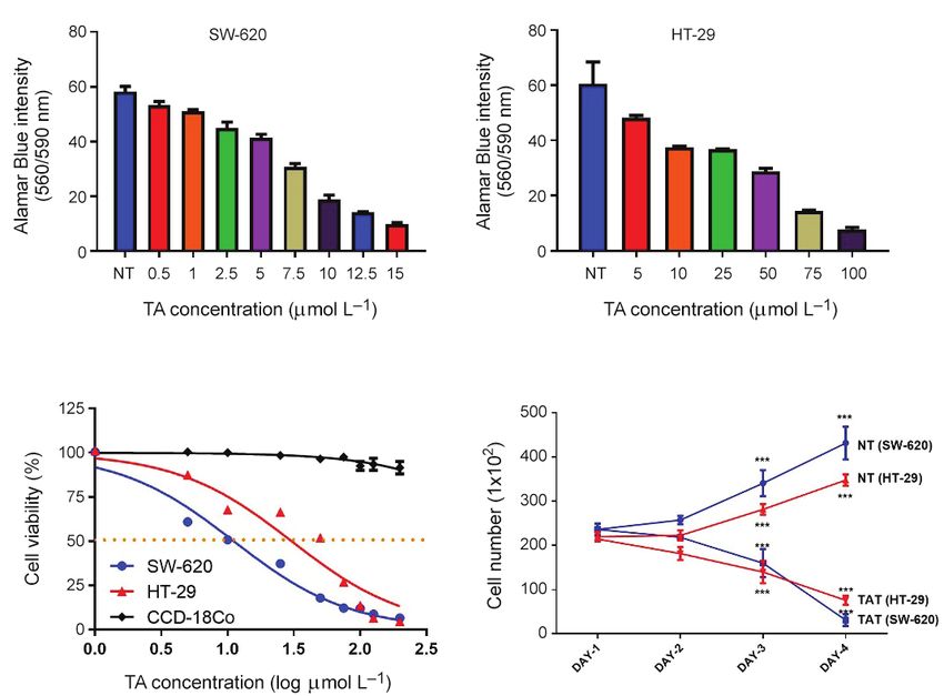
S. Karakurt et al.: Upregulation of p53 by tannic acid treatment suppresses the proliferation of human colorectal carcinoma, Acta Pharm.
71 (2021) ???–???.
overnight at 4 °C. Following TBST wash, the secondary antibody was added, and bands
were visualized by the enhanced chemiluminescence (ECL) system. The densitometric
analysis was performed with Image J software.
Determination of NQO1 activity. – NQO1 activity was measured using the 2,6 dichloro-
phenolindophenol (DCPIP) substrate, as described by Ernster et al. (22) using the spectro-
photometric method and the conditions optimized by Karakurt and Adali (13). This method
is based on the reduced absorption of the reduced form of DCPIP by NQO1 absorbing light
at 600 nm (Fig. 5a).
cDNA synthesis and quantitative mRNA expression by real-time PCR. – Total RNA from
SW-620 and HT-29 cells was extracted with QIAzol (Qiagen, USA), and the quality and
quantity of the RNA were analyzed with 2100 Bioanalyser instrument (Agilent Technolo-
gies, USA). The samples that have RIN (RNA Integrity Number) higher than 7 were used
for cDNA synthesis. The cDNA synthesis was performed by using iScript cDNA synthesis
kit (Bio-Rad Laboratories, Inc.), and the cDNA synthesis conditions included priming for
5 min at 25 °C and reverse transcription for 20 min at 46 °C, and the reaction was stopped
by incubation for 1 min at 95 °C. The samples were held at 4 °C until qRT-PCR studies. The
alterations in the mRNA expression of NQO1 were determined with qRT-PCR using Bio-
-Rad CFX Connect (Bio-Rad Laboratories, Inc.). Primers were designed with the primer and
controlled with Primer-Blast (NIH). The sequences of the forward (F) and reverse (R) primers
were: Specific for NQO1 is F-5’-AAG GCA GTG CTT TCC ATC AC-3’, and R-5’-AGG CTG
CTT GGA GCA AAA TA-3’. Specific for p53 is F-5’-CCT ATG GAA ACT ACT TCC TGA
AAA C-3’, and R-5’-GTA GAT TAC CAC TGG AGT CTT CC-3’. Specific for Bax is F-5’-ATG
GAC GGG TCC GGG GAG-3’, and R-5’-ATC CAG CCC AAC AGC CGC-3’. Specific for Bcl-2
is F-5’-AAG CCG GCG ACG ACT TCT-3’ and R-5’-GGT GCC GGT TCA GGT ACT CA-3’.
GAPDH was used as a housekeeping gene for normalization. Specific for GAPDH is F-5’-
GCC AAA AGG GTC ATC ATC TC-3’, and R-5’-TGA GTC CTT CCA CGA TAC CA-3’. 20 µL
of the reaction mixture was initially denaturated at 95 °C for 3 min, then 35 cycles of
following conditions; denaturation at 95 °C for 15 s, and annealing at 60 °C for 30 s. The
mRNA expression of specific genes was calculated by the 2−ΔΔCt method, which is exten-
sively used, and reliable data analysis (35).
Statistical analysis
GraphPad Prism v8.0 was used for the calculations and statistical analysis of the ob-
tained data. The results were expressed as mean ± standard deviation (SD) of three inde-
pendent experiments. The obtained results were analyzed using Two-Way repeated mea-
sure analysis of variance (ANOVA) and unpaired t-test. Anderson-Darling test was used
for assessing normality. The significance level was set to p < 0.05.
RESULTS AND DISCUSSION
Dynamic cellular localization of TA in human CRC cells was monitored with fluores-
cence microscopy technique, and it was found that TA may penetrate from the outer cell
membrane and localize both in cytoplasm and nucleus (Fig. 1b). TA was found to be a
5S. Karakurt et al.: Upregulation of p53 by tannic acid treatment suppresses the proliferation of human colorectal carcinoma, Acta Pharm.
71 (2021) ???–???.
stable compound in growth mediums of human CRC cells (Fig. 1d). Herein, UV spectra of
40 µg mL–1 of TA, and its monomeric subunit gallic acid (GA) were monitored up to 48 h at
37 °C with UV-spectrophotometer and no spectrum shift was observed during the
experiment. The results suggested that TA can remain relatively stable (10 % degradation)
in neutral pH (7.4) at 37 °C up to 48 h. The studies proved that especially alkaline conditions
lead to TA’s inactivation, and it can be stable at pH 6, 7, and 8 (36). Under alkaline condition
(pH > 10), TA is converted to GA that is esterified to TA’s glucose moiety. Analyses of TA
and GA UV spectrums proved that both compounds have different maximum absorbance.
TA’s maximum absorbance is at 280 nm, whereas GA’s maximum is at 260 nm. Therefore,
the conversion of TA to GA can be detected by measuring the absorbance changes at 280
nm and 260 nm (37).
The cytotoxic potency of TA was determined by Alamar Blue assay. Treatment of human
CRC cell lines, SW-620 (metastatic) and HT-29 (invasive), with TA leads to suppression
of cell growth in a dose-dependent manner (Fig. 2a and 2b). On the other hand, when
healthy colon epithelial cells (CCD-18Co) were treated with TA, the cell viability of CCD-
18Co was slightly reduced (IC50 > 200 µmol L–1) (Fig. 2c). Following the TA treatment of
SW-620 and HT-29 cells, the calculated IC50 values were 7.2 ± 0.8 and 37.6 ± 1.4 µmol L–1,
respectively (Fig. 2c). The viability of SW-620 and HT-29 cells was almost completely
reduced after 15 and 100 µmol L–1, respectively. The inhibitory potential of TA on human
a) b)
c) d)
Fig. 2. Cytotoxic effects of TA on SW-620 and HT-29 cells. a) and b) TA dose-dependently inhibits the
viability of SW-620 and HT-29 cells; c) determination of IC50 values of TA on human CRC cells, CCD-
18Co, was used as a healthy colon epithelial cell; d) growth curve SW-620 and HT-29 cells. Data are
displayed as mean ± SD. ***, p < 0.0001 for TA treated group (TAT), and non-treated group (NT).
6S. Karakurt et al.: Upregulation of p53 by tannic acid treatment suppresses the proliferation of human colorectal carcinoma, Acta Pharm.
71 (2021) ???–???.
CRC cells was observed at 24 h and increased time-dependently (Fig. 2d). Alteration of
gene and protein expressions varies from cell to cell and the compound used for treatment.
It may have taken 24 h for changes in gene and protein expressions to appear after treating
cells with tannic acid. As indicated in Fig. 2d, TA’s inhibitory effect on cell proliferation is
cytotoxic, not cytostatic.
Alteration in the SW-620 and HT-29 cell mobility was measured by wound healing
assay (Fig. 3a), a standard in vitro technique for determining two-dimension cell migration
a) b)
c) d)
e) f)
Fig. 3. Tannic acid modulates migration, invasion, and cellular anchorage-independent growth prop-
erties of CRC cells. Treated and non-treated (control) SW620 (a-1/a-2; c-1/c-2; e-1/e-2) and HT-29 (a-3/a4;
c-3/c-4; e-3/e4) cells were subject to wound healing, matrigel invasion, and soft agar colony formation
assays. a, c and e are the representative images, b, d, and f quantitate analysis of wound healing, inva-
sion, and colony formation assays. The representative images of control and TA treated groups for
wound healing, invasion and colony formation assay were shown in a-1(0 h)/a-2 (24 h), c-1(0 h)/c-2 (24
h), and e-2(0 h)/e-2(15 days) and a-3(0 h)/a-4 (24 h), c-3(0 h)/c-4 (24 h), and e-3(0 h)/e-4(15 days), respec-
tively. Data are expressed as mean ± SD. Asterisks signify the level of significance: ***p < 0.0001 (n = 6
per group).
7S. Karakurt et al.: Upregulation of p53 by tannic acid treatment suppresses the proliferation of human colorectal carcinoma, Acta Pharm.
71 (2021) ???–???.
(38, 39). The cells were pretreated with TA for 48 h and transferred and subjected to cell
scratching assay for 24 h. The number of migrated cells was significantly (p < 0.001) r educed
in SW-620 (82 %) and HT-29 (73 %) cell lines when compared to non-treated cells (Fig. 3b).
Mechanical forces, molecular interactions, and biochemical cascades are activated after
gap formation (40, 41). The other important issue during wound healing assay is TA’s
concentration since overdose may cause apoptosis and necrosis. Besides, the TA’s concen-
tration should not exceed the IC50 value to detect only migrating cells, not to proliferated
cells. To figure out the TA’s effect on CRC cells’ invasive potential, Matrigel invasion assay
was performed (Fig. 3c). Cells were forced to invade through a two-chamber system sepa-
rated by a cell-permeable membrane (42, 43). TA significantly inhibited the invasive poten-
tial of SW-620 and HT-29 cells by 73 % and 55 % (p < 0.0001) (Fig. 3d). To evaluate the cel-
lular transformation and cells’ ability to form colonies in vitro, Soft Agar Colony Formation
Assay (Fig. 3e) was performed (44, 45). TA significantly inhibited colony formation poten-
tial of SW-620 and HT-29 cell as 72 % (p < 0.0001) and 65 % (p = 0.0044), respectively (Fig.
3f). A significant correlation was observed between the migration, invasion, and colony
formation ratios of CRC cells (p < 0.05). These results indicate that TA reduces cell migra-
tion, invasion, and colony formation in CRC cells in vitro (Fig. 3). To elucidate TA’s contribu-
tion to the apoptotic process of SW-620 and HT-29 cells, phosphatidylserine residues exter-
nalization on the outer plasma membrane of apoptotic cells was monitored with flow
cytometry (Fig. 4a), and alteration in protein expression was measured by Western blot
(Fig. 4c). TA significantly trigger the cancerous cells to apoptosis in a different manner. As
shown in Fig. 4b, TA increased early rate apoptosis (13 %) and late apoptosis (6 %) in SW-
620 cells. On the other hand, in HT-29 cells, TA treatment increased not only early (6 %) and
late (12 %) apoptosis but also necrosis (3 %), which is a toxic process and energy-indepen-
dent mode of death. The apoptotic pathway includes many genes and proteins, and cas-
cades (46, 47). Cancerous cells have been developed many strategies such as up-regulation
of Bcl-2 and down-regulation of Bax to prevent apoptosis. Following the TA treatment, a
non-significant alteration was observed in Bcl-2 protein expressions neither SW-620 nor
HT-29 cells. On the other hand, the Bax protein expression was significantly increased in
SW-620 (p < 0.0001) and HT-29 (p < 0.005) cell following the TA treatment (Fig. 4d). SW-620
cells were obtained from lymph node where colon adenocarcinoma was metastasized. On
the other hand, HT-29 cells are less metastatic. The mRNA and protein expression studies
were found to be correlated with each other and flow cytometer analyses. TA treatment
increased the proapoptotic Bax protein level by 3.05-fold and gene expression as 4.02 fold
in SW-620 cells. Besides, they increased 1.86-fold and 2.0-fold, respectively, in HT-29 cells.
The obtained results demonstrated that protein and mRNA expressions were correlated
with each other.
It has been demonstrated that Bax and Bcl-2 expressions are regulated by tumor
suppressor gene; p53 (48). TP53 gene (expresses p53 protein) activates DNA repair pro-
teins and induces apoptosis is mutant in more than 50 % of human cancer (49–51).
Western blot and qRT-PCR studies showed that after TA treatment, protein and mRNA
expressions of P53 in SW-620 cells were significantly (p < 0.001) elevated as 1.54-fold and
1.78-fold, respectively (Fig. 5d and 5e). This alteration might explain the increased levels
of Bax gene expression since Bax is involved in p53-mediated apoptosis (52). To elucidate
the reason for p53, the NQO1 enzyme, protein, and mRNA expression were investigated.
NQO1 is responsible for detoxification of several natural and synthetic compounds by
two-electron reduction and the stability of tumor suppressor proteins p53 (53–55).
8S. Karakurt et al.: Upregulation of p53 by tannic acid treatment suppresses the proliferation of human colorectal carcinoma, Acta Pharm.
71 (2021) ???–???.
a)
b)
c) d)
e) f)
Fig. 4. Effects of TA on apoptosis of SW-620 and HT-29 cells. The cells were treated with an equivalent
concentration of IC50 values of TA for 24 h. a) representative charts of apoptosis after Annexin V-FITC
/7AAD stains with flow cytometry analyses; b) analyses and ratios of apoptosis of SW-620 and HT-29
cells; c) representative Western blot bands of Bax and Bcl-2 proteins in SW-620 and HT-29 cell; d)
analyses of Bax protein expression in SW-620 and HT-29 cells; e) heat map analyses of mRNA expres-
sion of Bax and Bcl-2 genes in SW-620 and HT-29 cells; f) analyses of Bax and Bcl-2 gene expression.
The results are expressed as mean ± SD of three different experiments (n = 6). **p < 0.001 and ***p <
0.0001).
9S. Karakurt et al.: Upregulation of p53 by tannic acid treatment suppresses the proliferation of human colorectal carcinoma, Acta Pharm.
71 (2021) ???–???.
a)
H
N Cl N Cl
NQO1
HO O HO OH
+ +
NADPH, H NADP
Cl Cl
2,6-dichlorophenolindophenol reduced form
b) c)
d) e)
Fig. 5. Effects of TA on the expression of NQO1 and p53. a) Reduction of DCPIP by NQO1; b) in vitro
effects of TA on NQO1 enzyme activity of SW-620 and HT-29 cells; c) and d) Effects of TA on NQO1
and p53 protein expressions of SW-620 and HT-29. Representative immunoblot and band density
analysis of protein expression of the non-treated and TA treated groups; e) mRNA relative expression
profiles of NQO1 and TP53 genes. The results are expressed as mean ± SD of three different experi-
ments (n = 6). *p < 0.05, **p < 0.001 and ***p < 0.0001.
10S. Karakurt et al.: Upregulation of p53 by tannic acid treatment suppresses the proliferation of human colorectal carcinoma, Acta Pharm.
71 (2021) ???–???.
olecular studies proved that NQO1 enzyme activity significantly elevated in many solid
M
tumors, which suggests its crucial importance in cancer therapy (56). NQO1 indirectly
inhibits the deacetylation of histone H3 lysine 9 which inhibits Bax transcription and
decreased apoptosis (57). Besides, overexpression of NQO1 decreased Bax mRNA expres
sion and stimulated Bax and Caspase-3 expressions in hepatocellular carcinoma (58). TA
treatment of SW-620 and HT-29 cells significantly (p < 0.0001) increased NQO1 enzyme
activity as 1.56-fold and 3.97-fold, respectively (Fig. 5b). Treatment with TA also increased
NQO1 protein expression in SW-620 and HT-29 cells 1.5-fold (p < 0.005) and 1.4-fold (p < 0.05),
respectively (Fig. 5c). When NQO1 mRNA expression was investigated in those cells, the
correlation between protein and mRNA expressions of NQO1 was observed in SW-620
(1.58-fold, p < 0.05). On the other hand, no significant correlation was observed between
protein and gene expressions as NQO1 mRNA change in HT-29 cells did not alter signi
ficantly in HT-29 cells (1.2-fold, p = 0.072) (Fig. 5e). Tannic acid treatment increased
very similarly in NQO1 mRNA, protein, and enzyme activity as 1.56-fold, 1.5-fold, and
1.58-fold, respectively. It was quite different in HT-29 cells since no alteration was observed
at the mRNA level, while protein and enzyme activities were increased as 1.4-fold and
3.97 fold, respectively. Studies have demonstrated that sometimes there is a poor correla-
tion between mRNA and enzyme activity results (59, 60). The alteration of enzyme
activity might be due to the alteration in protein expression and post-translational modi
fication, such as the addition of functional groups or proteolytic cleavage of regulatory
subunits (61).
Increased NQO1 protein expression and enzyme activity may be due to the tran-
scriptional modulation of the NQO1 gene; however, it may be a posttranscriptional modi
fication in HT-29 cells. NQO1 protein expression was also found significantly (p = 0.0002)
higher in SW-620 cells than HT-29 cells (2.05-fold). SW-620 cells are aggressive colorectal
carcinoma cells, whereas HT-29 cells are much more adherent. Therefore, SW-620 cells
have developed aggressive defense systems against chemotherapeutic agents. A single
nucleotide polymorphism (SNP) in NQO1, in which cysteine is converted to threonine at
the position 609 (C609T), is associated with low NQO1 activity in human CRC (62). There
is a direct correlation between NQO1 activity and tumorigenicity (24, 63). TA treatment
increased the mRNA and protein expressions of NQO1, and an increased level of NQO1
has been demonstrated to enhance p53 protein stability. p53 has a crucial role in the Arf/
p53/p21 and DDR pathway so that it can regulate growth arrest and apoptosis. The activ-
ity of p53 is highly connected to its structure. NQO1 directly binds to p53 whose regula-
tion is controlled by the E3 ubiquitin ligase MDM2 and protects it from 20S proteasomes
degradation. Decreased p53 activity, due to the presence of arginine instead of proline at
codon 72, has been linked to low chemotherapy-induced apoptosis (64). In vitro studies
are the first choice methods for explaining molecular mechanisms due to many samples,
rapid results, and animal ethics. However, in vitro results are not entirely reliable based
on the complex communication between cells and tissues and the inability to create the
microenvironment in vivo conditions such as the appropriate dose. The expression of
hundreds of genes is up or down-regulated when a primary hepatocyte cell is isolated
from its original environment. Besides, the mimic of xenobiotic metabolism is too com-
plex for in vitro conditions. There is no interaction between different cells, and it is dif-
ficult to understand the long term consequences of agents. Therefore, in further studies,
TA‘s in vitro effects on human CRC cells must be validated with in vivo experiments.
11S. Karakurt et al.: Upregulation of p53 by tannic acid treatment suppresses the proliferation of human colorectal carcinoma, Acta Pharm.
71 (2021) ???–???.
CONCLUSIONS
The present study concluded that TA has the potential for cancer chemoprevention. It
significantly inhibited the viability of human CRC and is stable up to 48 h at 37 °C. Active
phenolic groups of TA can bind to proteins and nucleic acids, which elevated activity and
stability of them. The treatment of TA promotes the activation of the NQO1 enzyme that
enhances the stability of p53 proteins. p53 protein is a crucial protein in cell arrest and
apoptosis; therefore, increased stability (activity) of p53 promotes apoptosis and causes
inhibition of human CRC cells. A decreased level of p53, either mutation or deletion, is an
effective strategy of cancerous cells against apoptosis, making p53 an ideal target during
anti-cancer drug design. Herein, efficient, reliable, and efficiently handled alternative com-
pounds gain vital importance. The modulatory effect of TA on gene and protein expres-
sions makes it a valuable chemo protectant against human CRC. TA either directly or in-
directly induces apoptosis and inhibits the viability of CRC cells. This in vitro study may
supply an essential background for in vivo studies in which the molecular mechanisms of
antioxidant and chemopreventive activities of TA will be clarified entirely.
Acknowledgments. – The Research Foundation of Selcuk University supported this study (Grant
Numbers 14401031 and 16401083).
REFERENCES
1. N. A. Alqallaf, H. A. G. Saleh, A. M. Abdu, S. H. Almuntaser, S. A. Bin Rakhis, A. A. Almughamis,
A. A. Ghanim, A. S. Alkhathami, N. A. Aldossari and G. M. Ahmad, Colon cancer screening and
prevention, Indo. Am. J. Pharm. Sci. 5 (2018) 13071–13078; https://doi.org/10.5281/zenodo.1495157
2. R. L. Siegel, K. D. Miller, S. A. Fedewa, D. J. Ahnen, R. G. S. Meester, A. Barzi and A. Jemal, Colorec-
tal cancer statistics, CA: Cancer J. Clin. 67 (2017) 177–193; https://doi.org/10.3322/caac.21395
3. H. S. Wong and W. C. Chang, Correlation of clinical features and genetic profiles of stromal interac-
tion molecule 1 (STIM1) in colorectal cancers, Oncotarget 6 (2015) 42169–42182; https://doi.org/10.18632/
oncotarget.5888
4. B. K. Edwards, E. Ward, B. A. Kohler, C. Eheman, A. G. Zauber, R. N. Anderson, A. Jemal, M. J.
Schymura, I. Lansdorp-Vogelaar, L. C. Seeff, M. van Ballegooijen, S. L. Goede and L. A. G. Ries,
Annual report to the nation on the status of cancer, 1975-2006, Featuring colorectal cancer trends
and impact of interventions (Risk factors, screening, and treatment) to reduce future rates, Cancer
116 (2010) 544–573; https://doi.org/10.1002/cncr.24760
5. M. Wang, Y. R. Li and X. D. Hu, Chebulinic acid derived from triphala is a promising antitumour
agent in human colorectal carcinoma cell lines, BMC Complement. Altern. Med. 18 (2018) 342; https://
doi.org/10.1186/s12906-018-2412-5
6. H. M. Li, S. Krstin and M. Wink, Modulation of multidrug resistant in cancer cells by EGCG, tannic
acid and curcumin, Phytomedicine 50 (2018) 213–222; https://doi.org/10.1016/j.phymed.2018.09.169
7. Y. M. Zheng, J. Z. Shen, Y. Wang, A. X. Lu and W. S. Ho, Anti-oxidant and anti-cancer activities of
Angelica dahurica extract via induction of apoptosis in colon cancer cells, Phytomedicine 23 (2016)
1267–1274; https://doi.org/10.1016/j.phymed.2015.11.008
8. S. Dasari and P. B. Tchounwou, Cisplatin in cancer therapy: molecular mechanisms of action, Eur. J.
Pharmacol. 740 (2014) 364–378; https://doi.org/10.1016/j.ejphar.2014.07.025
9. P. Apostolou, M. Toloudi, M. Chatziioannou, E. Ioannou, D. R. Knocke, J. Nester, D. Komiotis and I.
Papasotiriou, Anvirzel in combination with cisplatin in breast, colon, lung, prostate, melanoma and
pancreatic cancer cell lines, BMC Pharmacol. Toxicol. 14 (2013) 18; https://doi.org/10.1186/2050-6511-14-18
12S. Karakurt et al.: Upregulation of p53 by tannic acid treatment suppresses the proliferation of human colorectal carcinoma, Acta Pharm.
71 (2021) ???–???.
10. G. Maisetta, G. Batoni, P. Caboni, S. Esin, A. C. Rinaldi and P. Zucca, Tannin profile, antioxidant
properties, and antimicrobial activity of extracts from two Mediterranean species of parasitic plant
Cytinus, BMC Complement. Altern. Med. 19 (2019) 82; https://doi.org/10.1186/s12906-019-2487-7
11. J. Dai and R. J. Mumper, Plant phenolics: extraction, analysis and their antioxidant and anticancer
properties, Molecules 15 (2010) 7313–7352; https://doi.org/10.3390/molecules15107313
12. M. P. Borisova, A. A. Kataev and V. S. Sivozhelezov, Action of tannin on cellular membranes: Novel
insights from concerted studies on lipid bilayers and native cells, BBA – Biomembrane 1861 (2019)
1103–1111; https://doi.org/10.1016/j.bbamem.2019.03.017
13. S. Karakurt and O. Adali, Effect of tannic acid on glutathione S-transferase and NAD(P)H: Quinone
oxidoreductase 1 enzymes in rabbit liver and kidney, Fresen. Environ. Bull. 20 (2011) 1804–1811.
14. S. Quideau, D. Deffieux, C. Douat-Casassus and L. Pouysegu, Plant polyphenols: Chemical proper-
ties, biological activities, and synthesis, Angew. Chem. Int. Edit. 50 (2011) 586–621; https://doi.
org/10.1002/anie.201000044
15. J. Das, R. Ramani and M. O. Suraju, Polyphenol compounds and PKC signaling, Biochim. Biophys.
Acta 1860 (2016) 2107–2121; https://doi.org/10.1016/j.bbagen.2016.06.022
16. N. Sahiner, S. Sagbas, N. Aktas and C. Silan, Inherently antioxidant and antimicrobial tannic acid
release from poly(tannic acid) nanoparticles with controllable degradability, Colloid Surface B 142
(2016) 334–343; https://doi.org/10.1016/j.colsurfb.2016.03.006
17. J. Zhang, D. Chen, D. M. Han, Y. H. Cheng, C. Dai, X. J. Wu, F. Y. Che and X. Y. Heng, Tannic acid
mediated induction of apoptosis in human glioma Hs 683 cells, Oncol. Lett. 15 (2018) 6845–6850;
https://doi.org/10.3892/ol.2018.8197
18. Y. Ren, X. Li, B. Han, N. Zhao, M. Mu, C. Wang, Y. Du, Y. Wang, A. Tong, Y. Liu, L. Zhou, C. You and
G. Guo, Improved anti-colorectal carcinomatosis effect of tannic acid co-loaded with oxaliplatin in
nanoparticles encapsulated in thermosensitive hydrogel, Eur. J. Pharm. Sci. 128 (2019) 279–289;
https://doi.org/10.1016/j.ejps.2018.12.007
19. X. Zhang, H. Zhang, N. Zhou, J. Xu, M. Si, Z. Jia, X. Du and H. Zhang, Tannic acid modulates excit-
ability of sensory neurons and nociceptive behavior and the Ionic mechanism, Eur. J. Pharmacol. 764
(2015) 633–642; https://doi.org/10.1016/j.ejphar.2015.06.048
20 G. Goel, A. K. Puniya and K. Singh, Tannic acid resistance in ruminal streptococcal isolates, J. Basic
Microbiol. 45 (2005) 243–245; https://doi.org/10.1002/jobm.200410517
21. G. K. Lopes, H. M. Schulman and M. Hermes-Lima, Polyphenol tannic acid inhibits hydroxyl radi-
cal formation from Fenton reaction by complexing ferrous ions, Biochim. Biophys. Acta 1472 (1999)
142–152; https://doi.org/10.1016/s0304-4165(99)00117-8
22. L. Ernster, L. Danielson and M. Ljunggren, Dt diaphorase I. Purification from the soluble fraction
of rat-liver cytoplasm, and properties, Biochim. Biophys. Acta 58 (1962) 171–188; https://doi.
org/10.1016/0006-3002(62)90997-6
23. Z. Anusevicius, J. Sarlauskas and N. Cenas, Two-electron reduction of quinones by rat liver NAD(P)
H:quinone oxidoreductase: quantitative structure-activity relationships, Arch. Biochem. Biophys. 404
(2002) 254–262; https://doi.org/10.1016/S0003-9861(02)00273-4
24. N. Hamajima, K. Matsuo, H. Iwata, M. Shinoda, Y. Yamamura, T. Kato, S. Hatooka, T. Mitsudomi,
M. Suyama, Y. Kagami, M. Ogura, M. Ando, Y. Sugimura and K. Tajima, NAD(P)H: quinone oxido-
reductase 1 (NQO1) C609T polymorphism and the risk of eight cancers for Japanese, Int. J. Clin.
Oncol. 7 (2002) 103–108; https://doi.org/10.1007/s101470200013
25. H. J. Menzel, J. Sarmanova, P. Soucek, R. Berberich, K. Grunewald, M. Haun and H. G. Kraft, As-
sociation of NQO1 polymorphism with spontaneous breast cancer in two independent populations,
Br. J. Cancer 90 (2004) 1989–1994; https://doi.org/10.1038/sj.bjc.6601779
26. A. T. Dinkova-Kostova and P. Talalay, NAD(P)H:quinone acceptor oxidoreductase 1 (NQO1), a mul-
tifunctional antioxidant enzyme and exceptionally versatile cytoprotector, Arch. Biochem. Biophys.
501 (2010) 116–123; https://doi.org/10.1016/j.abb.2010.03.019
13S. Karakurt et al.: Upregulation of p53 by tannic acid treatment suppresses the proliferation of human colorectal carcinoma, Acta Pharm.
71 (2021) ???–???.
27. G. Asher, P. Tsvetkov, C. Kahana and Y. Shaul, A mechanism of ubiquitin-independent protea-
somal degradation of the tumor suppressors p53 and p73, Genes Dev. 19 (2005) 316–321; https://doi.
org/10.1101/gad.319905
28. G. Asher, Z. Bercovich, P. Tsvetkov, Y. Shaul and C. Kahana, 20S proteasomal degradation of orni-
thine decarboxylase is regulated by NQO1, Mol. Cell. 17 (2005) 645–655; https://doi.org/10.1016/j.
molcel.2005.01.020
29. K. Mikami, M. Naito, T. Ishiguro, H. Yano, A. Tomida, T. Yamada, N. Tanaka, T. Shirakusa and T.
Tsuruo, Immunological quantitation of DT-diaphorase in carcinoma cell lines and clinical colon
cancers: advanced tumors express greater levels of DT-diaphorase, Jpn. J. Cancer Res. 89 (1998) 910–
915; https://doi.org/10.1111/j.1349-7006.1998.tb00648.x
30. O. J. Achadu and N. Revaprasadu, Tannic acid-derivatized graphitic carbon nitride quantum dots
as an “on-off-on” fluorescent nanoprobe for ascorbic acid via copper(II) mediation, Mikrochim. Acta
186 (2019) 87; https://doi.org/10.1007/s00604-018-3203-x
31. S. Karakurt and O. Adali, Tannic acid inhibits proliferation, migration, invasion of prostate cancer
and modulates drug metabolizing and antioxidant enzymes, Anticancer Agents Med. Chem. 16 (2016)
781–789; https://doi.org/10.2174/1871520616666151111115809
32. S. Karakurt, G. Abuşoğlu and Z. C. Arituluk, Comparison of anticarcinogenic properties of Vibur-
num opulus and its active compound p-coumaric acid on human colorectal carcinoma, Turk. J. Biol.
44 (2020) 252–263; https://doi.org/10.3906/biy-2002-30
33. R. E. Brown, K. L. Jarvis and K. J. Hyland, Protein measurement using bicinchoninic acid – elimina-
tion of interfering substances, Anal. Biochem. 180 (1989) 136–139; https://doi.org/10.1016/0003-
2697(89)90101-2
34. P. K. Smith, R. I. Krohn, G. T. Hermanson, A. K. Mallia, F. H. Gartner, M. D. Provenzano, E. K. Fuji-
moto, N. M. Goeke, B. J. Olson and D. C. Klenk, Measurement of protein using bicinchoninic acid,
Anal. Biochem. 150 (1985) 76–85; https://doi.org/10.1016/0003-2697(85)90442-7
35. K. J. Livak and T. D. Schmittgen, Analysis of relative gene expression data using real-time quantita-
tive PCR and the 2(-Delta Delta C(T)) Method, Methods 25 (2001) 402–408; https://doi.org/10.1006/
meth.2001.1262
36. H. P. S. Makkar and K. Becker, Effect of pH, temperature, and time on inactivation of tannins and
possible implications in detannification studies, J. Agr. Food Chem. 44 (1996) 1291–1295; https://doi.
org/10.1021/jf9506287
37. L. C. Katwa, M. Ramakrishna and M. R. R. Rao, Spectrophotometric assay of immobilized tannase,
J. Biosci. 3 (1981) 135–142; https://doi.org/10.1007/BF02702656
38. S. R. Vedula, A. Ravasio, C. T. Lim and B. Ladoux, Collective cell migration: a mechanistic perspec-
tive, Physiology (Bethesda) 28 (2013) 370–379; https://doi.org/10.1152/physiol.00033.2013
39. O. Ilina and P. Friedl, Mechanisms of collective cell migration at a glance, J. Cell Sci. 122 (2009)
3203–3208; https://doi.org/10.1242/jcs.036525
40. P. Vitorino and T. Meyer, Modular control of endothelial sheet migration, Genes Dev. 22 (2008) 3268–
3281; https://doi.org/10.1101/gad.1725808
41. D. A. Chapnick and X. Liu, Leader cell positioning drives wound-directed collective migration in
TGFbeta-stimulated epithelial sheets, Mol. Biol. Cell 25 (2014) 1586–1593; https://doi.org/10.1091/mbc.
E14-01-0697
42. X. Liu and X. Wu, Utilizing matrigel transwell invasion assay to detect and enumerate circulating
tumor cells, Methods Mol. Biol. 1634 (2017) 277–282; https://doi.org/10.1007/978-1-4939-7144-2_23
43. K. Soejima, N. Mimura, M. Hirashima, H. Maeda, T. Hamamoto, T. Nakagaki and C. Nozaki, A
novel human metalloprotease synthesized in the liver and secreted into the blood: possibly, the von
Willebrand factor-cleaving protease?, J. Biochem. 130 (2001) 475–480; https://doi.org/10.1093/oxford-
journals.jbchem.a003009
14S. Karakurt et al.: Upregulation of p53 by tannic acid treatment suppresses the proliferation of human colorectal carcinoma, Acta Pharm.
71 (2021) ???–???.
44. S. Horibata, T. V. Vo, V. Subramanian, P. R. Thompson and S. A. Coonrod, Utilization of the soft agar
colony formation assay to identify inhibitors of tumorigenicity in breast cancer cells, J. Vis. Exp. 99
(2015) e52727; https://doi.org/10.3791/52727
45. S. Borowicz, M. Van Scoyk, S. Avasarala, M. K. Karuppusamy Rathinam, J. Tauler, R. K. Bikkavilli
and R. A. Winn, The soft agar colony formation assay, J. Vis. Exp. 92 (2014) e51998; https://doi.
org/10.3791/51998
46. S. Elmore, Apoptosis: a review of programmed cell death, Toxicol Pathol. 35 (2007) 495–516; https://
doi.org/10.1080/01926230701320337
47. M. M. Metzstein, G. M. Stanfield and H. R. Horvitz, Genetics of programmed cell death in C. ele-
gans: past, present and future, Trends Genet. 14 (1998) 410–416; https://doi.org/10.1016/s0168-
9525(98)01573-x
48. T. Miyashita, S. Krajewski, M. Krajewska, H. G. Wang, H. K. Lin, D. A. Liebermann, B. Hoffman and
J. C. Reed, Tumor suppressor p53 is a regulator of bcl-2 and bax gene expression in vitro and in vivo,
Oncogene 9 (1994) 1799–1805; https://doi.org/10.1016/0092-8674(95)90412-3
49. A. A. Roman-Rosales, E. Garcia-Villa, L. A. Herrera, P. Gariglio and J. Diaz-Chavez, Mutant p53 gain
of function induces HER2 over-expression in cancer cells, BMC Cancer 18 (2018) 709; https://doi.
org/10.1186/s12885-018-4613-1
50. H. Solomon, N. Dinowitz, I. S. Pateras, T. Cooks, Y. Shetzer, A. Molchadsky, M. Charni, S. Rabani, G.
Koifman, O. Tarcic, Z. Porat, I. Kogan-Sakin, N. Goldfinger, M. Oren, C. C. Harris, V. G. Gorgoulis
and V. Rotter, Mutant p53 gain of function underlies high expression levels of colorectal cancer stem
cells markers, Oncogene 37 (2018) 1669–1684; https://doi.org/10.1038/s41388-017-0060-8
51. N. C. Synnott, M. R. Bauer, S. Madden, A. Murray, R. Klinger, N. O’Donovan, D. O’Connor, W. M.
Gallagher, J. Crown, A. R. Fersht and M. J. Duffy, Mutant p53 as a therapeutic target for the treat-
ment of triple-negative breast cancer: Preclinical investigation with the anti-p53 drug, PK11007,
Cancer Lett. 414 (2018) 99–106; https://doi.org/10.1016/j.canlet.2017.09.053
52. H. Xiang, Y. Kinoshita, C. M. Knudson, S. J. Korsmeyer, P. A. Schwartzkroin and R. S. Morrison, Bax
involvement in p53-mediated neuronal cell death, J. Neurosci. 18 (1998) 1363–1373; https://doi.
org/10.1523/JNEUROSCI.18-04-01363.1998
53. J. H. Sun, Y. J. Wen, Y. Y. Zhou, Y. M. Jiang, Y. X. Chen, H. Z. Zhang, L. H. Guan, X. P. Yao, M. Huang
and H. C. Bi, p53 attenuates acetaminophen-induced hepatotoxicity by regulating drug-metaboliz-
ing enzymes and transporter expression, Cell Death Dis. 9 (2018); https://doi.org/10.1038/s41419-018-
0507-z
54. T. Maeda, C. Tanabe-Fujimura, Y. Fujita, C. Abe, Y. Nanakida, K. Zou, J. J. Liu, S. Y. Liu, T. Nakajima
and H. Komano, NAD(P)H quinone oxidoreductase 1 inhibits the proteasomal degradation of ho-
mocysteine-induced endoplasmic reticulum protein, Biochem. Bioph. Res. Co. 473 (2016) 1276–1280;
https://doi.org/10.1016/j.bbrc.2016.04.057
55. O. H. Rokah, O. Shpilberg and G. Granot, NAD(P)H quinone oxidoreductase protects TAp63 gam-
ma from proteasomal degradation and regulates TAp63 gamma-dependent growth arrest, Plos One
5 (2010); https://doi.org/10.1371/journal.pone.0011401
56. M. J. Lamberti, N. B. Vittar, C. da Silva Fde, V. F. Ferreira and V. A. Rivarola, Synergistic enhance-
ment of antitumor effect of beta-Lapachone by photodynamic induction of quinone oxidoreductase
(NQO1), Phytomedicine 20 (2013) 1007–1012; https://doi.org/10.1016/j.phymed.2013.04.018
57. H. Z. Zhou, H. Q. Zeng, D. Yuan, J. H. Ren, S. T. Cheng, H. B. Yu, F. Ren, Q. Wang, Y. P. Qin, A. L.
Huang and J. Chen, NQO1 potentiates apoptosis evasion and upregulates XIAP via inhibiting
proteasome-mediated degradation SIRT6 in hepatocellular carcinoma, Cell Commun. Signal 17 (2019)
168; https://doi.org/10.1186/s12964-019-0491-7
15S. Karakurt et al.: Upregulation of p53 by tannic acid treatment suppresses the proliferation of human colorectal carcinoma, Acta Pharm.
71 (2021) ???–???.
58. X. Zhang, K. Han, D.H. Yuan and C. Y. Meng, Overexpression of NAD(P)H: Quinone oxidoreduc-
tase 1 inhibits hepatocellular carcinoma cell proliferation and induced apoptosis by activating
AMPK/PGC-1alpha pathway, DNA Cell Biol. 36 (2017) 256–263; https://doi.org/10.1089/dna.2016.3588
59. M. Hayashi, N. Matsumoto, S. Takenoshita-Nakaya, Y. Takeba, M. Watanabe, T. Kumai, M. Takagi,
M. Tanaka, T. Otsubo and S. Kobayashi, Individual metabolic capacity evaluation of cytochrome
P450 2C19 by protein and activity in the small intestinal mucosa of Japanese pancreatoduodenec-
tomy patients, Biol. Pharm. Bull. 34 (2011) 71–76; https://doi.org/10.1248/bpb.34.71
60. S. Ohtsuki, O. Schaefer, H. Kawakami, T. Inoue, S. Liehner, A. Saito, N. Ishiguro, W. Kishimoto, E.
Ludwig-Schwellinger, T. Ebner and T. Terasaki, Simultaneous absolute protein quantification of
transporters, cytochromes P450, and UDP-glucuronosyltransferases as a novel approach for the
characterization of individual human liver: comparison with mRNA levels and activities, Drug
Metab. Dispos. 40 (2012) 83–92; https://doi.org/10.1124/dmd.111.042259
61. H. Lin and K. S. Caroll, Introduction: Posttranslational protein modification, Chem. Rev. 118 (2018)
887–888; https://doi.org/10.1021/acs.chemrev.7b00756
62. R. D. Traver, T. Horikoshi, K. D. Danenberg, T. H. W. Stadlbauer, P. V. Danenberg, D. Ross and N. W.
Gibson, NAD(P)H-quinone oxidoreductase gene-expression in human colon-carcinoma cells: char-
acterization of a mutation which modulates DT-diaphorase activity and mitomycin sensitivity,
Cancer Res. 52 (1992) 797–802.
63. X. Zhang, K. Han, D. H. Yuan and C. Y. Meng, Overexpression of NAD(P)H: quinone oxidoreduc-
tase 1 inhibits hepatocellular carcinoma cell proliferation and induced apoptosis by activating
AMPK/PGC-1alpha pathway, DNA Cell Biol. 36 (2017) 256–263; https://doi.org/10.1089/dna.2016.3588
64. D. Bergamaschi, M. Gasco, L. Hiller, A. Sullivan, N. Syed, G. Trigiante, I. Yulug, M. Merlano, G.
Numico, A. Comino, M. Attard, O. Reelfs, B. Gusterson, A. K. Bell, V. Heath, M. Tavassoli, P. J. Far-
rell, P. Smith, X. Lu and T. Crook, p53 polymorphism influences response in cancer chemotherapy
via modulation of p73-dependent apoptosis, Cancer Cell 3 (2003) 387–402; https://doi.org/10.1016/
s1535-6108(03)00079-5
16You can also read