Ivermectin Induces Cytostatic Autophagy by Blocking the PAK1/Akt Axis in Breast Cancer
←
→
Page content transcription
If your browser does not render page correctly, please read the page content below
Published OnlineFirst June 14, 2016; DOI: 10.1158/0008-5472.CAN-15-2887

                                                                                                                                   Cancer
  Therapeutics, Targets, and Chemical Biology                                                                                      Research

Ivermectin Induces Cytostatic Autophagy by
Blocking the PAK1/Akt Axis in Breast Cancer
Qianhui Dou1, Hai-Ning Chen2, Kui Wang1,3, Kefei Yuan1, Yunlong Lei4, Kai Li1, Jiang Lan1,3,
Yan Chen1, Zhao Huang1, Na Xie1, Lu Zhang1, Rong Xiang5, Edouard C. Nice6, Yuquan Wei1,
and Canhua Huang1,3

Abstract
   Breast cancer is the most common cancer among women                          activated kinase 1 (PAK1) expression via the ubiquitination-
worldwide, yet successful treatment remains a clinical challenge.               mediated degradation pathway. The inhibition of PAK1 decreases
Ivermectin, a broad-spectrum antiparasitic drug, has recently been              the phosphorylation level of Akt, resulting in the blockade of the
characterized as a potential anticancer agent due to observed                   Akt/mTOR signaling pathway. In breast cancer xenografts, the
antitumor effects. However, the molecular mechanisms involved                   ivermectin-induced cytostatic autophagy leads to suppression of
remain poorly understood. Here, we report a role for ivermectin in              tumor growth. Together, our results provide a molecular basis for
breast cancer suppression by activating cytostatic autophagy both               the use of ivermectin to inhibit the proliferation of breast cancer
in vitro and in vivo. Mechanistically, ivermectin-induced autop-                cells and indicate that ivermectin is a potential option for the
hagy in breast cancer cells is associated with decreased P21-                   treatment of breast cancer. Cancer Res; 76(15); 1–13. 2016 AACR.

Introduction                                                                    poor (3), highlighting the need for the development of poten-
                                                                                tial therapeutic agents.
   Breast cancer is the most frequent cancer among women and
                                                                                   Autophagy is a self-degrading process characterized by forma-
ranks as the fifth leading cause of cancer-related death world-
                                                                                tion of double-membrane autophagosomes, which sequester
wide with more than 1.67 million people diagnosed annually,
                                                                                excess or defective organelles and fuse with lysosomes for deg-
with over 522,000 deaths per year (1). Although surgical
                                                                                radation of enclosed materials (4). Although constitutively active
resection, in combination with radiotherapy when necessary,
                                                                                in cells, autophagy can be stimulated in response to multiple
affords curative treatment for early or local disease, approxi-
                                                                                cellular stresses, such as nutrient shortage, hypoxia, and oxidative
mately 70% patients with advanced breast cancer require cyto-
                                                                                stress. In the scenario of tumor development, autophagy elim-
toxic chemotherapy, endocrine therapy, biologic therapy, or
                                                                                inates the source of cellular damage and protects the cells from
combinations of these (2). Despite the diverse strategies that
                                                                                stress induced by chemotherapy or radiation, which represents a
have been proposed to improve the current situation, the
                                                                                fine mechanism of negative feedback regulation. However, in the
prognosis for patients with advanced breast cancer still remains
                                                                                recent decade, it has become apparent that the consequence of
                                                                                autophagy varies significantly under different circumstances
1
                                                                                (5, 6). To date, cytoprotective, cytostatic, cytotoxic, and nonpro-
  State Key Laboratory of Biotherapy and Cancer Center, West China
Hospital, Sichuan University, and Collaborative Innovation Center for           tective autophagy have been proposed as the four main functional
Biotherapy, Chengdu, P.R. China. 2Department of Gastrointestinal                forms of autophagy in the context of anticancer therapy (7).
Surgery, State Key Laboratory of Biotherapy and Cancer Center, West             Cytoprotective autophagy serves as a survival mechanism to
China Hospital, Sichuan University, and Collaborative Innovation Cen-
ter for Biotherapy, Chengdu, P.R. China. 3Key Laboratory of Tropical
                                                                                promote the nutrient cycle (8), which theoretically can be inhib-
Diseases and Translational Medicine of Ministry of Education & Depart-          ited to achieve therapeutic advantage by sensitizing cells to
ment of Neurology, the Affiliated Hospital of Hainan Medical College,            anticancer agents (9). Subsequent studies on autophagy in cancer
Haikou, China. 4Department of Biochemistry and Molecular Biology,
Molecular Medicine and Cancer Research Center, Chongqing Medical
                                                                                cells revealed the anticancer properties of autophagy, leading to
University, Chongqing, P.R. China. 5School of Medicine/Collaborative            the discovery of cytotoxic autophagy, cytostatic autophagy, and
Innovation Center of Biotherapy, Nankai University, Tianjin, China.
6
                                                                                nonprotective autophagy (6, 7, 10). In view of current clinical
  Department of Biochemistry and Molecular Biology, Monash Univer-              efforts to exploit autophagy as a therapeutic target for cancer
sity, Clayton, Victoria, Australia.
                                                                                treatment, the multiple roles of autophagy underscore the neces-
Note: Supplementary data for this article are available at Cancer Research
                                                                                sity to understand the mechanism of autophagy and the regula-
Online (http://cancerres.aacrjournals.org/).
                                                                                tory signaling pathways involved in cancer cells (11).
Q. Dou, H. Chen, K. Wang, and K. Yuan contributed equally to this article.
                                                                                   Avermectin was initially purified by Drs. Campbell and Omura
Corresponding Author: Canhua Huang, The State Key Laboratory of Biotherapy      (12), and implicated as an efficient agent against parasites, which
and Cancer Center, West China Hospital, Sichuan University, and Collaborative   earned a Nobel Prize for physiology or medicine in 2015. Sub-
Innovation Center for Biotherapy, Chengdu 610041, P.R. China. Phone: 86-
                                                                                sequently, ivermectin, an avermectin derivative, was chemically
13258370346; Fax: 8628-8516-4060; E-mail: hcanhua@scu.edu.cn
                                                                                modified and found to be a more effective compound against a
doi: 10.1158/0008-5472.CAN-15-2887                                              variety of parasites (13). Recently, ivermectin has been identified
2016 American Association for Cancer Research.                                 as a promising anticancer agent for colon cancer, ovarian cancer,

www.aacrjournals.org                                                                                                                                   OF1

       Downloaded from cancerres.aacrjournals.org on January 9, 2021. © 2016 American Association for Cancer
                                                    Research.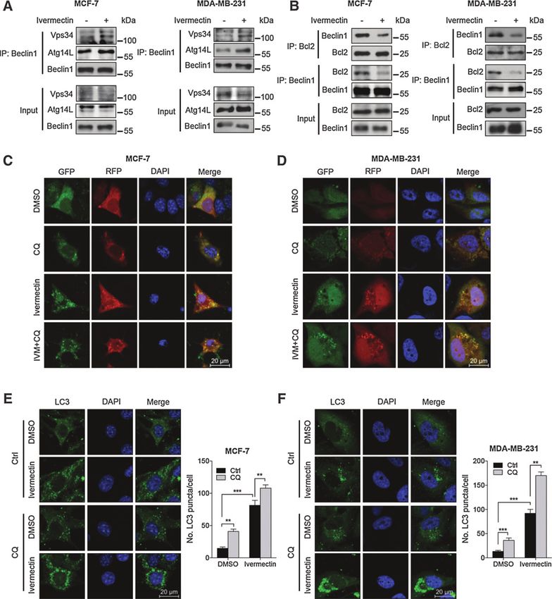
Published OnlineFirst June 14, 2016; DOI: 10.1158/0008-5472.CAN-15-2887

      Dou et al.

      melanoma, and leukemia (14–17). However, the detailed molec-          GE Healthcare) for 2 hours. The samples were analyzed by
      ular mechanisms underlying ivermectin-mediated suppression of         immunobloting with the indicated antibodies. The identity and
      tumor growth remain to be further elucidated.                         the suppliers of the antibodies are provided in Supplementary
        In this study, we show that ivermectin inhibits the growth of       Methods.
      breast cancer by stimulating autophagy. Ivermectin promotes
      ubiquitination-mediated degradation of PAK1, which results in         Measurement of cell viability
      the blockade of the Akt/mTOR signaling, and thereby activates            The short-term effects of ivermectin on tumor cell growth were
      autophagy in breast cancer cells. These findings demonstrate a         assessed using the 3-(4,5-dimethylthiazol-2-yl)-2,5-diphenylte-
      novel link between ivermectin and the autophagy machinery,            trazolium bromide (MTT; Sigma) assay, as described previously
      indicating that the use of ivermectin as an autophagy inducer may     (6). The long-term effects of ivermectin on tumor cell prolifera-
      constitute a new therapeutic approach for breast cancer.              tion were analyzed with a colony formation assay as described in
                                                                            Supplementary Methods.
      Materials and Methods
      Cell culture                                                          BrdUrd labeling assay
         Human breast cancer cell lines MDA-MB-435, HS578T, 4T1,               The bromodeoxyuridine (BrdUrd) labeling assay was per-
      and HEK 293T cell lines were purchased from the ATCC. MCF-7,          formed in 96-well plate using the BrdU Cell Proliferation Assay
      MDA-MB-231, MDA-MB-468, and MDA-MB-361 cell lines were                Kit (Roche). After ivermectin treatment, 10 mmol/L BrdUrd was
      kindly provided by Prof. Qiang Yu (Genome Institute of Singa-         added to each well, and the cells were incubated for 12 hours at
      pore, Singapore). All cell lines were cultured according to the       37 C. The BrdUrd signaling was determined using a Multiscan
      ATCC guidelines and used within 6 months. The last time of            MK3 ELISA reader (Thermo Scientific) at 450 nm.
      authentication was between December 2015 and February 2016
      using the short tandem repeat (STR) analysis. Cells were main-        TUNEL assays
      tained in DMEM or RPMI1640 supplemented with 100 U/mL                    Cells were plated on glass coverslips in 24-well plates, fixed in
      penicillin (Sigma), 100 mg/mL streptomycin (Sigma), and 10%           4% paraformaldehyde (Sigma), following incubation with iver-
      serum (Biowest) in a humidified incubator at 37 C under 5% CO2        mectin for 24 hours. Terminal deoxynucleotidyl transferase-medi-
      atmosphere.                                                           ated nick-end labeling (TUNEL) staining was performed using the
                                                                            DeadEnd Fluorometric TUNEL system (Promega). Two 40 fields
      Animal models                                                         of cells were imaged to evaluate the TUNEL-positive cells per
         Female NOD/SCID and Balb/c mice at 8 weeks of age were             coverslip in every independent experiment.
      purchased from HFK Bioscience Co., Ltd (Beijing). All studies
      were approved by the Institutional Animal Care and Treatment          Flow cytometry
      Committee of Sichuan University. For the orthotopic breast               Cells were harvested and washed once with PBS, and then
      cancer model, 1  106 MDA-MB-231-GFP cells were suspended             resuspended in PI/Annexin-V solution (KeyGEN Biotech) for
      in PBS and engrafted in the mammary fat pad of NOD/SCID mice.         apoptosis analysis. At least 10,000 live cells were analyzed on a
      For the syngeneic model, 1  106 4T1 cells were suspended in PBS      FACSCalibur flow cytometer (Becton Dickinson). Data were
      and injected subcutaneously into Balb/c mice. When the tumor          analyzed by using FlowJo software.
      volumes reached 100 mm3, mice were randomized into two
      groups receiving 0.1 mL of vehicle (10% ricinus oil) or 0.12 mg       Transmission electron microscopy
      ivermectin/mouse/day, respectively. Vehicle or ivermectin was           Transmission electron microscopy was performed as described
      injected intraperitoneally (i.p.) on the first 10 days. Mice were      previously (6). Briefly, MCF-7 cells were fixed in 4% glutaralde-
      euthanized for analysis after three weeks. Tumor tissues were         hyde (Sigma). A sorvall MT5000 microtome (DuPont Instru-
      isolated and frozen in liquid nitrogen or fixed in 10% formalin        ments, MT5000) was used to prepare ultrathin sections after
      immediately.                                                          dehydration. Lead citrate and /or 1% uranyl acetate were used
                                                                            to stain the sections, and the autophagic vacuoles in the cyto-
                                                                            plasmic area were calculated using Image Pro Plus version 3
      Breast cancer patients                                                software.
         All clinical breast cancer tissues were obtained from West China
      Hospital (Chengdu, P.R. China) with the approval of the Bio-          RT-PCR analysis
      medical Ethics Committee. A total of 20 patients with breast            RNA was prepared using TRIzol (Invitrogen). cDNA was pre-
      cancer who underwent radical mastectomy were involved in this         pared from 1 mg of total RNA, using reverse transcriptase and
      study. Tumor samples from these patients were collected for           random hexamers from RevertAid First Strand cDNA Synthesis Kit
      immunohistochemical analysis. Detailed clinicopathologic fea-         (Fermentas). The PAK1 primers are available in Supplementary
      tures including age, gender, and clinical stage were listed in        Methods.
      Supplementary Table S1.
                                                                            Immunohistochemistry
      Western blotting and immunoprecipitation                                 Immunohistochemical analysis was performed as described
        Cells were lysed with RIPA buffer supplemented with protease        previously (18). The immunostaining intensity (A) was indicated
      and phosphatase inhibitor cocktail (Sigma, p8340). For immu-          by four grades (0, negative; 1, weakly positive; 2, positive; 3,
      noprecipitations, whole cell lysates were subjected to immuno-        strongly positive) and the proportion of staining-positive cells (B)
      precipitation overnight at 4 C with 1 mg of the indicated anti-      was divided into five grades (0, < 5%; 1, 6%–25%; 2, 26%–50%; 3,
      bodies, followed by addition of protein A-Sepharose beads (40 mL,     51%–75%; 4, > 75%). The final score was calculated as A  B.

OF2 Cancer Res; 76(15) August 1, 2016                                                                                         Cancer Research

             Downloaded from cancerres.aacrjournals.org on January 9, 2021. © 2016 American Association for Cancer
                                                          Research.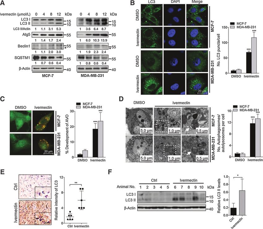
Published OnlineFirst June 14, 2016; DOI: 10.1158/0008-5472.CAN-15-2887

                                                                                                    Ivermectin Induces Cytostatic Autophagy

Images were captured using a DM2500 fluorescence microscope               (Supplementary Fig. S1G). Taken together, our data indicate that
(Leica).                                                                 short-term treatment (i.e., 24 hours) of ivermectin displays a
                                                                         profound antiproliferative effect on breast cancer cells, while such
Immunofluorescence                                                        a growth inhibition is independent of apoptosis.
   Cells were fixed with 4% paraformaldehyde (Sigma) for 30                  To evaluate the effect of ivermectin on breast cancer cell growth
minutes, washed three times with PBS and exposed to PBS                  in vivo, we employed an orthotopic breast cancer model by injecting
containing 0.2% Triton X-100 and 5% BSA for 30 minutes. The              human MDA-MB-231-GFP cells subcutaneously into the mam-
slides were then stained with antibody against LC3 at 4 C over-         mary fat pad of NOD-SCID mice. As shown in Fig. 1E, xenografts
night, and subsequently incubated with Alexa Fluor 488–conju-            treated with ivermectin grew at a slower rate than those treated with
gated goat anti-rabbit IgG (Molecular Probes) at 37 C for 1 hour.       placebo. Macroscopically, the size of control tumors was much
Nuclei were stained with Hoechst 33342 (Cell Signaling Tech-             larger than that of ivermectin-treated tumors (Fig. 1F). Consistent-
nology). Images were captured using a confocal laser scanning            ly, tumor weight was reduced in ivermectin-treated mice compared
microscopy (Zeiss).                                                      with that of the control group (Fig. 1G). To confirm the change in
                                                                         proliferation status of tumors, xenografts were stained for Ki67,
Acridine orange staining                                                 which is used clinically to assess the proliferative fraction in breast
   Evaluation of autophagy by acridine orange staining was per-          cancer (22). All control xenografts displayed stronger Ki67 staining
formed as described previously (5). Briefly, cells were treated with      than that of ivermectin-treated mice (Fig. 1H). Futhermore, we
or without ivermectin at indicated concentrations for 24 hours,          observed similar results in a syngeneic model using subcutaneous
and then stained with 1 mmol/L acridine orange (Sigma-Aldrich)           injection of 4T1 cells (Supplementary Fig. S2A–S2D). Taken
in PBS containing 5% FBS at 37 C for 15 minutes. Cells were             together, these data suggest that ivermectin inhibits the growth of
washed and then observed under fluorescence microscopy.                   breast cancer both in vitro and in vivo.

Statistical analysis                                                     Ivermectin stimulates autophagy in breast cancer cells
   Statistical analysis was performed using Prism 6. Statistical            As increasing evidence has highlighted the important roles of
differences were determined using a two-sample equal variance            drug-induced autophagy in anticancer therapies (6, 23), we inves-
Student t test. Data were deemed to be statistically significant if P <   tigated whether ivermectin regulated autophagy in breast cancer
0.05. Error bars indicate SEM unless otherwise indicated.                cells. We first evaluated the effect of ivermectin on the formation of
                                                                         the autophagosome membrane by detecting the conversion of
                                                                         LC3-I to lipidated LC3-II, and the distribution of endogenous LC3
Results                                                                  puncta, two classical markers of autophagy (24). Ivermectin treat-
Ivermectin inhibits breast cancer growth both in vitro and in vivo       ment resulted in marked autophagy induction as evidenced by
   To ascertain the anticancer effect of ivermectin in breast cancer     increased LC3-II conversion (Fig. 2A and Supplementary Fig. S3A)
cells, the MTT assay was conducted to assess the growth of six           and LC3 puncta (Fig. 2B). However, no apparent difference in LC3-
breast cancer cell lines (MCF-7, MDA-MB-231, MDA-MB-468,                 II conversion was detected between ivermectin-treated cells and
MDA-MB-361, MDA-MB-435, and HS578T) and a nontumori-                     controls in MCF-10A cells (Supplementary Fig. S3B). Cells were
genic human breast cell line (MCF-10A) following ivermectin              also stained with acridine orange to detect the formation of acidic
treatment. As shown in Fig. 1A and Supplementary Fig. S1A,               vesicular organelles (AVO), a characteristic of autophagy (24). As
ivermectin treatment for 24 hours markedly decreased the cell            shown in Fig. 2C, abundant cytoplasmic AVO formation was
viability of breast cancer cell lines in a dose-dependent manner,        readily observed in ivermectin-treated cells. To further corroborate
while the IC50 value in MCF-10A cells was much higher than those         ivermectin-induced autophagy, the appearance of double-mem-
in breast cancer cells. Consistently, ivermectin significantly sup-       braned autophagosomes was investigated by transmission elec-
pressed cell proliferation in MCF-7 and MDA-MB-231 cells, as             tronic microscopy. As shown in Fig. 2D, there was a significant
evidenced by reduced clonogenic survival (Fig. 1B). In addition, a       accumulation of autophagosomes/autolysosomes in ivermectin-
significantly lower percentage of BrdUrd-positive cells was               treated cells but not in control cells. In addition, mouse xenografts
observed in ivermectin-treated cells compared with controls (Fig.        were stained with LC3 to clarify whether ivermectin could induce
1C). Collectively, these results demonstrate that ivermectin inhi-       autophagy in vivo. As shown in Fig. 2E and Supplementary Fig. S4A,
bits the proliferation of breast cancer cells in vitro. Apoptosis is a   ivermectin-treated xenografts displayed stronger LC3 staining com-
major form of cell death induced by chemotherapeutic agents              pared with the control group. Consistently, a similar tendency was
(19). To determine whether ivermectin induces apoptosis in               observed in LC3-II conversion in ivermectin-treated tumors (Fig. 2F
breast cancer cells, we evaluated the apoptotic rate using both          and Supplementary Fig. S4B). Taken together, these data indicate
TUNEL and flow cytometry assays (doxorubicin or cisplatin was             that ivermectin stimulates autophagy in breast cancer cells both in
used as positive control; refs. 20, 21). Ivermectin treatment for 24     vitro and in vivo.
hours showed no obvious effect on apoptosis in either breast                The expression levels of Beclin 1 and Atg5, two autophagy-
cancer cells (MCF-7, MDA-MB-231, MDA-MB-468, MDA-MB-                     related proteins (24), were then examined to clarify whether
361, MDA-MB-435, and HS578T) or breast epithelial cells                  ivermectin promoted autophagosome formation. As shown
(MCF-10A; Fig. 1D and Supplementary Fig. S1B–S1D). This was              in Fig. 2A, ivermectin promoted the expression of both Beclin
further supported by equivalent levels of cleaved caspase-3 in           1 and Atg5 in a dose-dependent manner. To explore the mech-
ivermectin-treated cells and control cells (Supplementary Fig. S1E       anism by which ivermectin induces autophagy, we next investi-
and S1F). Of note, apoptotic induction, albeit at a relatively low       gated whether ivermectin could induce the formation of autop-
level, could be observed with the prolonged treatment of iver-           hagosome by enhancing the interaction of Beclin 1 with positive
mectin till 48 hours, in all the breast cancer cells examined            regulators such as Atg14L and Vps34, and diminishing the

www.aacrjournals.org                                                                                        Cancer Res; 76(15) August 1, 2016      OF3

       Downloaded from cancerres.aacrjournals.org on January 9, 2021. © 2016 American Association for Cancer
                                                    Research.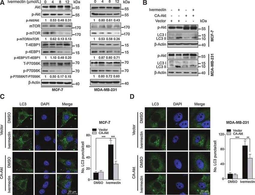
Published OnlineFirst June 14, 2016; DOI: 10.1158/0008-5472.CAN-15-2887

      Dou et al.

      Figure 1.
      Ivermectin inhibits the growth of breast cancer cells. A, ivermectin inhibited breast cancer cell viability. Cell viability was measured by the MTT assay in MCF-7,
      MDA-MB-231, and MCF-10A cells treated with the indicated concentrations of ivermectin for 24 hours. B, ivermectin suppressed colony formation in breast
      cancer cells. Cells were cultured in the indicated concentrations of ivermectin for 10 days. C, ivermectin inhibited breast cancer cell proliferation measured by BrdUrd
      labeling. Cells were treated as in A. D, the apoptosis rate was assessed by TUNEL assay. Cells were treated as in A. The TUNEL-positive cells were counted
      from at least 100 random fields. DNase, positive control.  , P < 0.01;    , P < 0.001. E–H, NOD-SCID mice were inoculated with MDA-MB-231-GFP cells and treated with
      ivermectin or vehicle. Tumor volumes were measured at indicated time points (E). Photograph of isolated tumors derived from control or ivermectin-treated
      mice (F). Tumor weights at time of sacrifice (G). Ki67 expression in tumor xenografts was examined by IHC (H). Representative images were provided as indicated.

          , P < 0.001; Scale bars, 20 mm.

      interaction of Beclin 1 with negative regulators such as Bcl-2 (24).                  mectin failed to stimulate autophagy (Supplementary Fig. S5D–
      As shown in Fig. 3A, ivermectin treatment increased coimmuno-                         S5F). Moreover, ivermectin treatment resulted in decreased levels
      precipitation of Beclin 1 with Vps34 or Atg14L, respectively.                         of SQSTM1, a well-known autophagic substrate, in a dose-depen-
      Conversely, cells treated with ivermectin showed decreased coim-                      dent manner (Fig. 2A). Using a tandem monomeric RFP-GFP–
      munoprecipitation of Beclin 1 with Bcl-2 (Fig. 3B). Silencing the                     tagged LC3, we found increased formation of yellow fluorescent
      expression of either Beclin 1 or Atg5 using siRNA partially blocked                   autophagosomes and red fluorescent autolysosomes (Fig. 3C and
      LC3 lipidation and endogenous LC3 puncta accumulation in                              D). Combinatorial treatment of chloroquine (a lysosomal inhib-
      ivermectin-treated cells (Supplementary Fig. S5A–S5C). Coad-                          itor) and ivermectin resulted in further accumulation of yellow
      ministration of wortmannin, a PI3K inhibitor (25), with iver-                         fluorescent autophagosomes, endogenous LC3 puncta, and

OF4 Cancer Res; 76(15) August 1, 2016                                                                                                                    Cancer Research

              Downloaded from cancerres.aacrjournals.org on January 9, 2021. © 2016 American Association for Cancer
                                                           Research.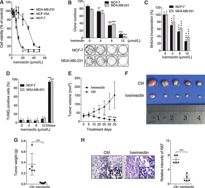
Published OnlineFirst June 14, 2016; DOI: 10.1158/0008-5472.CAN-15-2887

                                                                                                                  Ivermectin Induces Cytostatic Autophagy

Figure 2.
Ivermectin induces autophagy in breast cancer cells. A, immunoblot analysis of LC3, Atg5, Beclin 1, and SQSTM1 in cells treated with the indicated concentrations
of ivermectin for 24 hours.  , nonspecific band. B, left, the formation of endogenous LC3 puncta in cells treated with DMSO or 8 mmol/L ivermectin for
24 hours. Right, total number of endogenous LC3 puncta per cell. C, left, autophagy measured by acridine orange staining of cells treated as in B. Right, total
number of acidic vesicular organelles (AVO) per cell. D, left, autophagy measured by transmission electron microscopy in cells treated as in B. N, nucleus.
Arrows, autophagosomes/autolysosomes. Right, total number of autophagosomes per cell.   , P < 0.01;   , P < 0.001. E, LC3 expression in orthotopic xenografts
was examined by IHC. Representative images were provided as indicated.    , P < 0.001. Scale bars, 20 mm. F, left, orthotopic xenograft tissues were
extracted to assess the levels of LC3-II by Western blot analysis. Right, densitometry quantification of the band intensities in F was carried out using ImageJ
software and is presented as a percentage of relative densitometry normalized to actin.

increased LC3-II conversion (Fig. 3C–F and Supplementary                           of either Beclin 1 or Atg5 significantly restored cell growth in
Fig. S5G). These results indicate that ivermectin induces autop-                   ivermectin-treated cells. Consistently, similar results were
hagic flux in breast cancer cells.                                                  obtained by inhibition of autophagy using wortmannin or
                                                                                   chloroquine (Fig. 4D), indicating that ivermectin-inhibited
Autophagy is involved in ivermectin-inhibited cell proliferation                   breast cancer cell growth was autophagy dependent. In addi-
in breast cancer cells                                                             tion, combinatorial treatment of wortmannin or chloroquine
   To determine whether autophagy was involved in the anti-                        with ivermectin showed no obvious effect on apoptosis induc-
cancer effect of ivermectin, cells were transfected with Beclin 1                  tion in breast cancer cells (Supplementary Fig. S6). Thus, these
siRNA or Atg5 siRNA followed by treatment with ivermectin.                         findings suggest that ivermectin-induced autophagy is cytostat-
Cell growth was assessed by MTT assay, BrdUrd labeling, and                        ic in breast cancer cells, and suppression of autophagy may
colony formation analysis. As shown in Fig. 4A–C, knockdown                        attenuate the anticancer effect of ivermectin.

www.aacrjournals.org                                                                                                       Cancer Res; 76(15) August 1, 2016         OF5

        Downloaded from cancerres.aacrjournals.org on January 9, 2021. © 2016 American Association for Cancer
                                                     Research.
Published OnlineFirst June 14, 2016; DOI: 10.1158/0008-5472.CAN-15-2887
Dou et al.
Figure 3.
Ivermectin promotes autophagy flux in breast cells. A, interaction among Beclin 1, Atg14L, and Vps34 was determined by coimmunoprecipitation assay. B,
interaction between Beclin 1 and Bcl-2 was determined by coimmunoprecipitation assay. C and D, cells were transiently transfected with an RFP-GFP tandem
fluorescent-tagged LC3 (RFP-GFP-LC3). In addition, cells were treated with 8 mmol/L ivermectin (IVM) alone or in combination with 10 mmol/L chloroquine (CQ)
for 24 hours. E and F, left, immunofluorescence analysis of endogenous LC3 puncta in cells treated as in C. Right, total number of endogenous LC3 puncta per cell.
, P < 0.01; , P < 0.001. Scale bars, 20 mm.
Akt/mTOR signaling plays a major role in ivermectin-induced mTOR acts as a key negative modulator of autophagy (26).
autophagy Therefore, we examined whether the Akt/mTOR pathway was
It has previously been reported that constitutively activated inhibited in ivermectin-treated breast cancer cells. As shown
PI3K/Akt signaling is involved in breast carcinogenesis, and Akt/ in Fig. 5A and Supplementary Fig. S7A, ivermectin treatment
OF6 Cancer Res; 76(15) August 1, 2016 Cancer Research
Downloaded from cancerres.aacrjournals.org on January 9, 2021. © 2016 American Association for Cancer
Research.Published OnlineFirst June 14, 2016; DOI: 10.1158/0008-5472.CAN-15-2887
Ivermectin Induces Cytostatic Autophagy
Figure 4.
Inhibition of autophagy represses the antiproliferative effect of ivermectin in breast cancer cells. A–C, cells were transfected with siRNA against Atg5 or Beclin 1 or
control (50 nmol/L) for 48 hours, and then treated with ivermectin at 8 mmol/L for another 24 hours. Proliferation rate was detected by MTT assay (A),
BrdUrd labeling (B), and colony formation (C). D, cells were treated with DMSO, chloroquine (CQ), or wortamannin (Wort) in the presence or absence of ivermectin
(8 mmol/L) for 24 hours, and then the proliferation rate was measured by MTT assay. , P < 0.05; , P < 0.01; , P < 0.001.
www.aacrjournals.org Cancer Res; 76(15) August 1, 2016 OF7
Downloaded from cancerres.aacrjournals.org on January 9, 2021. © 2016 American Association for Cancer
Research.Published OnlineFirst June 14, 2016; DOI: 10.1158/0008-5472.CAN-15-2887
Dou et al.
resulted in inhibition of the Akt/mTOR pathway, as evidenced by these proteins, PAK1 has been reported to be a potential target of
decreased phosphorylation levels of Akt, mTOR, p70S6K, and 4E- ivermectin (15). In addition, PAK1 is associated with phosphor-
BP1. To determine whether the Akt/mTOR pathway is involved in ylation of Akt (29). These observations suggest that PAK1 might
ivermectin-induced autophagy, we transfected a constitutively be involved in ivermectin-induced autophagy through regulation
active form of Akt (CA-Akt) to restore ivermectin-induced Akt/ of Akt/mTOR pathway. It has been reported that PAK1 is abnor-
mTOR inhibition (27). Akt activation significantly reduced LC3-II mally expressed in a variety of tumor cells and accociated with
conversion and LC3 puncta accumulation in ivermectin-treated tumor cell proliferation and invasiveness (30, 31). Our data
cells (Fig. 5B–D), suggesting that the Akt/mTOR pathway is an showed that the basal levels of PAK1 in breast epithelial cells
important mediator in ivermectin-induced autophagy in breast (MCF-10A) were significantly lower than that in breast cancer
cancer cells. cells (MCF-7, MDA-MB-231, MDA-MB-361, MDA-MB-435,
HS578T, and MDA-MB-468; Supplementary Fig. S7B). In addi-
Ivermectin induces autophagy through the blocking tion, ivermectin treatment showed no notable effect on the
PAK1/Akt/mTOR axis expression of both PAK1 and p-Akt in MCF-10A cells (Supple-
To further investigate the mechanism underlying ivermectin- mentary Fig. S7C), while in breast cancer cells, ivermectin
induced autophagy, we identified the Akt-interacting proteins treatment decreased PAK1 expression in a dose-dependent
using a previously constructed global protein–protein interaction manner (Fig. 6A and Supplementary Fig. S7A). Following on
network (Supplementary Fig. S8A; ref. 28). Intriguingly, among from this, we examined the phosphorylation levels of Akt in
Figure 5.
Ivermectin induces autophagy by repressing the Akt/mTOR pathway in breast cancer cells. A, immunoblot analysis of phosphorylation of Akt (S473), mTOR (S2448),
p70S6K (S424/T421), and 4EBP1 (S65/T70) in cells treated with the indicated concentrations of ivermectin for 24 hours. Total Akt, mTOR, p70S6K,
and 4EBP1 expression was used as the internal control, respectively. B, cells were transfected with an empty vector (pECE) or with a constitutively active
CA-Akt for 48 hours, and then cells were treated with 8 mmol/L ivermectin for another 24 hours. Akt and mTOR phosphorylation, and LC3 lipidation were
determined by immunoblotting. C and D, left, the formation of endogenous LC3 puncta was assessed in cells treated as in B. Right, total number of endogenous
LC3 puncta per cell. , P < 0.01; , P < 0.001. Scale bars, 20 mm.
OF8 Cancer Res; 76(15) August 1, 2016 Cancer Research
Downloaded from cancerres.aacrjournals.org on January 9, 2021. © 2016 American Association for Cancer
Research.Published OnlineFirst June 14, 2016; DOI: 10.1158/0008-5472.CAN-15-2887
Ivermectin Induces Cytostatic Autophagy
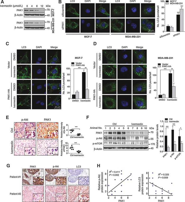
Figure 6.
Ivermectin induces autophagy through downregulation of PAK1 in breast cancer cells. A, immunoblot analysis of PAK1 protein expression in cells treated with the
indicated concentrations of ivermectin for 24 hours. B, left, the formation of endogenous LC3 puncta was analyzed in cells transfected with PAK1 siRNA or
control (50 nmol/L) for 48 hours. Right, total number of endogenous LC3 puncta per cell. C and D, left, cells were transfected with an empty vector (pCDNA-3.1-HA) or
with PAK1-HA for 48 hours, and then cells were treated with 8 mmol/L ivermectin for another 24 hours. The formation of endogenous LC3 puncta was
analyzed by immunofluorescence. Right, total number of endogenous LC3 puncta per cell. , P < 0.01; , P < 0.001. E, p-Akt and PAK1 expression in orthotopic
xenografts was examined by IHC. Scale bars, 20 mm. F, orthotopic xenograft tissues were extracted to assess the levels of p-Akt, p-mTOR, and PAK1 by
Western blot analysis. Densitometry quantification of the band intensities in Fig. 6F was carried out using ImageJ software and is shown as a percentage of relative
densitometry normalized to actin. G, immunohistochemical analyses of PAK1, p-Akt, and LC3 expression in breast cancer tissues. Scale bars, 20 mm.
H, correlation of immunostaining intensity between PAK1 and p-Akt or LC3, respectively.
www.aacrjournals.org Cancer Res; 76(15) August 1, 2016 OF9
Downloaded from cancerres.aacrjournals.org on January 9, 2021. © 2016 American Association for Cancer
Research.Published OnlineFirst June 14, 2016; DOI: 10.1158/0008-5472.CAN-15-2887
Dou et al.
Figure 7.
Ivermectin promotes ubiquitin degradation of PAK1. A, PAK1 mRNA expression in cells treated with the indicated concentrations of ivermectin for 24 hours was
evaluated by RT-PCR. B, immunoblot analysis of PAK1 expression in cells treated with 8 mmol/L ivermectin alone or pretreatment with MG132 (10 mmol/L,
2 hours) for 24 hours. C, immunoprecipitation (IP)–Western blots showing the ubiquitination of PAK1 after ivermectin treatment. HEK 293T cells were cotransfected
with PAK1-HA and Flag-tagged ubiquitin. D, HEK 293T cells were cotransfected with Flag-tagged ubiquitin and PAK1-HA (wild-type, WT; knockout, KO;
K11R; K29R; K39R; K148R; K162R; K256R). E, HEK 293T cells were cotransfected with Flag-tagged ubiquitin and PAK1-HA (WT; KO; Lys11, Lys29, Lys39, Lys148, Lys162,
or Lys256 only) as indicated.
PAK1 knockdown or PAK1-overexpressing cells, respectively, to and GFP-LC3 puncta (Fig. 6C and D and Supplementary Fig. S9B).
validate whether PAK1 regulates the Akt/mTOR signaling path- Consistently, control xenografts showed stronger phosphorylated
way. As shown in Supplementary Fig. S8B and S8C, knockdown of Akt and PAK1 staining compared to that in ivermectin-treated
PAK1 decreased Akt phosphorylation, while enforced expression xenografts (Fig. 6E and Supplementary Fig. S4C and S4D).
of PAK1 promoted Akt phosphorylation. To explore the mecha- Accordingly, immunoblot analysis displayed an apparent atten-
nism by which PAK1 promotes Akt phosphorylation, we per- uation of phosphorylated Akt, phosphorylated mTOR, and PAK1
formed a molecular docking calculation of Akt binding confor- in tumors from ivermectin-treated mice compared with controls
mation (Supplementary Fig. S8D). The results indicated that (Fig. 6F). Furthermore, we assessed the clinical relevance of the
PAK1 could interact with Akt directly, which was further corrob- PAK1/Akt axis and LC3 in breast cancer patient tissues (n ¼ 20). As
orated by coimmunoprecipitation analysis (Supplementary Fig. expected, PAK1 expression was positively correlated with p-Akt
S8E). Moreover, we found that this interaction was reduced in and negatively correlated with LC3, respectively (Fig. 6G and H).
ivermectin-treated cells (Supplementary Fig. S8E), suggesting that Together, our results indicate the PAK1/Akt/mTOR axis as a crucial
ivermectin regulates Akt/mTOR signaling by interfering with the pathway of ivermectin-induced autophagy in breast cancer.
interaction between PAK1 and Akt in breast cancer cells.
To further evaluate the role of PAK1 in ivermectin-induced Ubiquitination-mediated degradation of PAK1 results in
autophagy, LC3 lipidation and LC3 puncta were assessed in PAK1 autophagy activation in breast cancer cells
siRNA–transfected breast cancer cells followed by treatment with To gain insights into the mechanism underlying the regulation
or without ivermectin. PAK1 knockdown resulted in LC3 lipida- of PAK1 by ivermectin, we used reverse transcription PCR (RT-
tion (Supplementary Fig. S9A) and LC3 puncta accumulation PCR) analysis to quantify the mRNA level of PAK1. As shown
(Fig. 6B), while ivermectin treatment failed to induce further in Fig. 7A, ivermectin treatment showed no obvious effect on
lipidation of LC3 in PAK1 siRNA–treated cells (Supplementary PAK1 mRNA level, suggesting that transcription regulation might
Fig. S9A). In contrast, PAK1 overexpression suppressed ivermec- not account for the decreased PAK1 expression observed follow-
tin-induced LC3 lipidation and accumulation of endogenous LC3 ing ivermectin treatment. We next examined whether PAK1 was
OF10 Cancer Res; 76(15) August 1, 2016 Cancer Research
Downloaded from cancerres.aacrjournals.org on January 9, 2021. © 2016 American Association for Cancer
Research.Published OnlineFirst June 14, 2016; DOI: 10.1158/0008-5472.CAN-15-2887
Ivermectin Induces Cytostatic Autophagy
degraded through the proteasome/ubiquitination pathway. We ment, suggesting that short-term treatment of ivermectin induces
showed that treatment of MG132, a proteasome inhibitor, could cytostatic autophagy in breast cancer cells. Similar reports have
stabilize the protein levels of PAK1 in MCF-7 and MDA-MB-231 been documented that drug-induced autophagy may precede
cells (Fig. 7B), suggesting that PAK1 may be degraded by the apoptosis in certain cancer cells (35, 36), it would be of particular
proteasome/ubiquitination pathway. interest for us to consider optimal manipulation of autophagy for
To further determine whether ivermectin-induced PAK1 reduc- cancer treatment in combination with conventional apoptosis-
tion is due to proteasome-mediated degradation, we measured inducing agents. Because of the complex nature of apoptosis and
the effect of ivermectin on PAK1 ubiquitination by cotransfecting autophagy in cell fate determination (36), further studies are
PAK1-HA and Flag-ubiquitin expression vectors in human needed to investigate the crosstalk between these tightly regulated
HEK293T cells with or without MG132 treatment. As shown biological processes.
in Fig. 7C, ivermectin markedly induced PAK1–Ub conjugation Conventional anticancer therapies primarily trigger apoptosis
and this was further enhanced by MG132 treatment. Bioinfor- to promote cancer cell death. However, accumulating evidence
matics analysis was then used to identify the potential ubiquiti- suggests that cancer cells may deregulate apoptosis, leading to
nation site(s) of PAK1. As shown in Supplementary Table S2, 47 drug resistance and tumor recurrence (37, 38). Thus, with current
lysine residues in PAK1 were identified using the UbPred program chemotherapy regimens, apoptosis resistance has become a tre-
(www.ubpred.org), and six candidates (Lys11, Lys29, Lys39, mendous challenge in the development of novel anticancer
Lys148, Lys162, Lys256) were predicted with high confidence to therapies. As a backup strategy to inhibit tumor growth, cytostatic
be the ubiquitination sites (32). To test the contribution of these autophagy may overcome these barriers by inhibiting the growth
lysine residues to PAK1 ubiquitination, we constructed six single- of cancer cells regardless of their sensitivity to apoptosis (39, 40).
site mutants with each of the six lysine residues mutated to In support of this, very recently, it has been reported that the
arginine (K11R, K29R, K39R, K148R, K162R, and K256R). How- combination of radiotherapy with vitamin D inhibits cell prolif-
ever, the ubiquitination of these mutants was almost the same as eration through inducing cytostatic autophagy in non-small cell
the wild type (Fig. 7D). We further constructed another six lung cancer cells (41). Our results revealed that autophagy
mutants (K11, K29, K39, K148, K162, and K256) that each induced by ivermectin has the capacity to inhibit breast cancer
contained a single candidate lysine (e.g., K11 contained one lysine cell growth in a cytostatic way, suggesting that the use of iver-
at position 11 with other five lysines mutated to arginine). As mectin as an anticancer agent may reduce the self-renewal capacity
shown in Fig. 7E, ubiquitin was conjugated efficiently with PAK1 and proliferation recovery in breast cancer cells.
mutants containing K11, K29, K39, or K148, indicating that these The Akt/mTOR signaling pathway is a major pathway account-
four lysines might be potential ubiquitination sites. These results ing for autophagy activation and is also involved in regulating the
show that ivermectin downregulates the expression of PAK1 by proliferation and apoptosis of cancer cells (42). In this study, we
targeting the lysine residues at K11, K29, K39, or K148 and found that the Akt/mTOR signaling pathway was significantly
promoting the ubiquitin/proteasome–mediated degradation in inhibited by ivermectin. The inhibited Akt/mTOR signaling in
breast cancer cells. ivermectin-treated cells was attributed to down regulated PAK1
(15), whose expression correlates with the phosphorylation of Akt
(29). Our data showed that ivermectin could markedly decrease
Discussion the expression of PAK1 and inhibit the Akt/mTOR signaling
The use of ivermectin, a broad-spectrum antiparasitic drug, has pathway, implicating the PAK1/Akt/mTOR axis as a novel path-
now been extended to multiple disease models (14, 33). Recently, way in ivermectin-induced autophagy. Although previous studies
this antiparasitic drug has been proposed as a promising anti- have mentioned that ivermectin could decrease the expression of
cancer agent in several types of cancer due to its remarkable PAK1, the detailed mechanism underlying ivermectin-regulated
ability to inhibit tumor growth (16, 17). However, the mechan- PAK1 still remains unclear. We showed that ivermectin down-
isms underlying the growth-inhibitory effects of ivermectin are regulated PAK1 protein levels by targeting the lysine residues at
still elusive. In this study, our data revealed that ivermectin K11, K29, K39, or K148 and promoting ubiquitination-mediated
suppressed Akt/mTOR signaling by promoting ubiquitination degradation. These results support ivermectin as a potent agent in
degradation of PAK1, and thereby activated cytostatic autophagy, the induction of ubiquitination-mediated degradation of PAK1,
leading to inhibition of tumor growth in breast cancer cells. suggesting that ivermectin-mediated inhibition of the PAK1/Akt/
Studies on cancer treatment have identified autophagy activa- mTOR signaling pathway may merit exploration as a therapeutic
tion as a consequence of chemotherapy or radiotherapy; however, strategy for breast cancer treatment.
the role that autophagy plays in cancer progression is varied (34). Notably, PAK1 has been demonstrated to be increased in breast
Generally, autophagy is considered as a prosurvival mechanism in cancer and plays a key role in promoting tumor growth and drug
cancer cells by removing damaged organelles and recycling nutri- resistance (30, 43–45). The oncogenic function of PAK1 is also
ents upon anticancer treatment (10). However, a recent remark- observed in other cancers, such as colon cancer, neurofibroma-
able finding is that autophagy induced by certain chemothera- tosis, and ovarian cancer (31, 46, 47). Thus, further study targeting
peutic agents may have a suppressive role in cancer cells, revealing PAK1 may provide an optional therapeutic strategy for tumors
two additional functional forms of autophagy, of which one is the with PAK1 overexpression. On the basis of our findings, the
cytotoxic function that results in autophagic cell death or pro- combination of traditional anticancer treatment with ivermectin
motes apoptosis, the other is the cytostatic function that may seems to be a rational way to treat cancer cells with PAK1-involved
inhibit cell proliferation in an apoptosis-independent way (6, 7). drug resistance (48, 49). In line with our hypothesis, a recent study
In this study, we demonstrated that ivermectin-induced autop- showed that ivermectin could alleviate multidrug resistance in
hagy inhibited the growth of breast cancer cells while no signif- breast cancer and enhance the cytotoxicity of doxorubicin and
icant apoptosis was observed till 48 hours after ivermectin treat- paclitaxel (50). Further studies may focus on validating the effect
www.aacrjournals.org Cancer Res; 76(15) August 1, 2016 OF11
Downloaded from cancerres.aacrjournals.org on January 9, 2021. © 2016 American Association for Cancer
Research.Published OnlineFirst June 14, 2016; DOI: 10.1158/0008-5472.CAN-15-2887
Dou et al.
of ivermectin in conjunction with conventional anticancer ther- Administrative, technical, or material support (i.e., reporting or organizing
apies in drug-resistant tumors. data, constructing databases): K. Li, J. Lan
Study supervision: E.C. Nice, C. Huang
In summary, our study revealed that ivermectin inhibited the
tumor growth of breast cancer by inducing PAK1/Akt/mTOR axis-
Acknowledgments
mediated cytostatic autophagy. These findings provide insights The authors thank Dr. Changan Jiang for providing Flag-Ub plasmid and
into the anticancer efficacy of ivermectin, which support a pre- Dr. Qiang Yu and Dr. Ning Zhang for providing breast cancer cell lines used in
clinical rational to explore broadening the clinical evaluation of this study.
ivermectin for the treatment of breast tumor.
Grant Support
Disclosure of Potential Conflicts of Interest This work was supported by grants from the National 973 Basic Research
No potential conflicts of interest were disclosed. Program of China (no. 2013CB911300 to C. Huang), the National Science and
Technology Major Project (no. 2012ZX09501001-003 to C. Huang), the
Authors' Contributions Chinese NSFC (nos. 81225015 and 81430071 to C. Huang; no. 81502441
Conception and design: K. Li, Y.-Q. Wei, C. Huang to K. Yuan; no. 81401951 to Y. Lei; no. 81501743 to N. Xie), and Sichuan
Development of methodology: C. Huang Science-Technology Innovative Research Team for Young Scientist (no.
Acquisition of data (provided animals, acquired and managed patients, 2013TD0001 to C. Huang).
provided facilities, etc.): Q. Dou, H.-N. Chen, K. Li, Y. Chen, Z. Huang, The costs of publication of this article were defrayed in part by the payment of
C. Huang page charges. This article must therefore be hereby marked advertisement in
Analysis and interpretation of data (e.g., statistical analysis, biostatistics, accordance with 18 U.S.C. Section 1734 solely to indicate this fact.
computational analysis): Q. Dou, H.-N. Chen, K. Li, N. Xie, R. Xiang, C. Huang
Writing, review, and/or revision of the manuscript: Q. Dou, H.-N. Chen, Received October 20, 2015; revised April 22, 2016; accepted May 6, 2016;
K. Wang, K. Yuan, Y. Lei, L. Zhang, E.C. Nice, C. Huang published OnlineFirst June 14, 2016.
References
1. Ferlay J, Soerjomataram I, Ervik M, Dikshit R, Eser S, Mathers C, et al. 17. Melotti A, Mas C, Kuciak M, Lorente-Trigos A, Borges I, Ruiz i Altaba A. The
GLOBOCAN 2012 v1.0, Cancer Incidence and Mortality Worldwide: IARC river blindness drug Ivermectin and related macrocyclic lactones inhibit
CancerBase No. 11 [Internet]. Lyon, France: International Agency for WNT-TCF pathway responses in human cancer. EMBO Mol Med
Research on Cancer; 2013. Available from: http://globocan.iarc.fr. 2014;6:1263–78.
2. Siegel RL, Miller KD, Jemal A. Cancer statistics, 2015. CA Cancer J Clin 18. Noman MZ, Buart S, Romero P, Ketari S, Janji B, Mari B, et al. Hypoxia-
2015;65:5–29. inducible miR-210 regulates the susceptibility of tumor cells to lysis by
3. Sharon Y, Raz Y, Cohen N, Ben-Shmuel A, Schwartz H, Geiger T, et al. cytotoxic T cells. Cancer Res 2012;72:4629–41.
Tumor-derived osteopontin reprograms normal mammary fibroblasts to 19. Bai L, Wang S. Targeting apoptosis pathways for new cancer therapeutics.
promote inflammation and tumor growth in breast cancer. Cancer Res Annu Rev Med 2014;65:139–55.
2015;75:963–73. 20. Yang XH, Sladek TL, Liu X, Butler BR, Froelich CJ, Thor AD. Reconstitution
4. Boya P, Reggiori F, Codogno P. Emerging regulation and functions of of caspase 3 sensitizes MCF-7 breast cancer cells to doxorubicin- and
autophagy. Nat Cell Biol 2013;15:713–20. etoposide-induced apoptosis. Cancer Res 2001;61:348–54.
5. Wang K, Liu R, Li J, Mao J, Lei Y, Wu J, et al. Quercetin induces protective 21. Blanc C, Deveraux QL, Krajewski S, Janicke RU, Porter AG, Reed JC, et al.
autophagy in gastric cancer cells: involvement of Akt-mTOR- and Caspase-3 is essential for procaspase-9 processing and cisplatin-induced
hypoxia-induced factor 1alpha-mediated signaling. Autophagy 2011;7: apoptosis of MCF-7 breast cancer cells. Cancer Res 2000;60:4386–90.
966–78. 22. Klauschen F, Wienert S, Schmitt WD, Loibl S, Gerber B, Blohmer JU, et al.
6. Liu R, Li J, Zhang T, Zou L, Chen Y, Wang K, et al. Itraconazole suppresses Standardized Ki67 diagnostics using automated scoring-clinical validation
the growth of glioblastoma through induction of autophagy: involvement in the GeparTrio Breast Cancer Study. Clin Cancer Res 2015;21:3651–7.
of abnormal cholesterol trafficking. Autophagy 2014;10:1241–55. 23. Kim Y, Kim Y-S, Kim DE, Lee JS, Song JH, Kim H-G, et al. BIX-01294 induces
7. Gewirtz DA. The four faces of autophagy: implications for cancer therapy. autophagy-associated cell death via EHMT2/G9a dysfunction and intra-
Cancer Res 2014;74:647–51. cellular reactive oxygen species production. Autophagy 2014;9:2126–39.
8. Efeyan A, Comb WC, Sabatini DM. Nutrient-sensing mechanisms and 24. Klionsky DJ, Abdelmohsen K, Abe A, Abedin MJ, Abeliovich H, Acevedo
pathways. Nature 2015;517:302–10. Arozena A, et al. Guidelines for the use and interpretation of assays for
9. He C, Klionsky DJ. Regulation mechanisms and signaling pathways of monitoring autophagy (3rd edition). Autophagy 2016;12:1–222.
autophagy. Annu Rev Genet 2009;43:67–93. 25. Wu YT, Tan HL, Shui G, Bauvy C, Huang Q, Wenk MR, et al. Dual role of 3-
10. Fulda S, Kogel D. Cell death by autophagy: emerging molecular mechan- methyladenine in modulation of autophagy via different temporal pat-
isms and implications for cancer therapy. Oncogene 2015;34:5105–13. terns of inhibition on class I and III phosphoinositide 3-kinase. J Biol Chem
11. Levine B, Kroemer G. Autophagy in the pathogenesis of disease. Cell 2010;285:10850–61.
2008;132:27–42. 26. Schmelzle T, Hall MN. TOR, a central controller of cell growth. Cell
12. Ikeda H, Omura S. Avermectin biosynthesis. Chem Rev 1997;97:2591– 2000;103:253–62.
610. 27. Bodine SC, Stitt TN, Gonzalez M, Kline WO, Stover GL, Bauerlein R, et al.
13. Gonzalez Canga A, Sahagun Prieto AM, Jose Diez Liebana M, Martinez NF, Akt/mTOR pathway is a crucial regulator of skeletal muscle hypertrophy
Vega MS, Vieitez JJ. The pharmacokinetics and metabolism of ivermectin in and can prevent muscle atrophy in vivo. Nat Cell Biol 2001;3:1014–9.
domestic animal species. Vet J 2009;179:25–37. 28. Fu L, Zhang S, Zhang L, Tong X, Zhang J, Zhang Y, et al. Systems biology
14. Drinyaev VA, Mosin VA, Kruglyak EB, Novik TS, Sterlina TS, Ermakova NV, network-based discovery of a small molecule activator BL-AD008 targeting
et al. Antitumor effect of avermectins. Eur J Pharmacol 2004;501:19–23. AMPK/ZIPK and inducing apoptosis in cervical cancer. Oncotarget
15. Hashimoto H, Messerli SM, Sudo T, Maruta H. Ivermectin inactivates the 2015;6:8071–88.
kinase PAK1 and blocks the PAK1-dependent growth of human ovarian 29. Higuchi M, Onishi K, Kikuchi C, Gotoh Y. Scaffolding function of PAK in
cancer and NF2 tumor cell lines. Drug Discov Ther 2009;3:243–6. the PDK1-Akt pathway. Nat Cell Biol 2008;10:1356–64.
16. Sharmeen S, Skrtic M, Sukhai MA, Hurren R, Gronda M, Wang X, et al. The 30. Kumar R, Gururaj AE, Barnes CJ. p21-activated kinases in cancer. Nat Rev
antiparasitic agent ivermectin induces chloride-dependent membrane Cancer 2006;6:459–71.
hyperpolarization and cell death in leukemia cells. Blood 2010;116: 31. Dummler B, Ohshiro K, Kumar R, Field J. Pak protein kinases and their role
3593–603. in cancer. Cancer Metastasis Rev 2009;28:51–63.
OF12 Cancer Res; 76(15) August 1, 2016 Cancer Research
Downloaded from cancerres.aacrjournals.org on January 9, 2021. © 2016 American Association for Cancer
Research.Published OnlineFirst June 14, 2016; DOI: 10.1158/0008-5472.CAN-15-2887
Ivermectin Induces Cytostatic Autophagy
32. Zhang Z, Bao M, Lu N, Weng L, Yuan B, Liu YJ. The E3 ubiquitin ligase 42. Shinojima N, Yokoyama T, Kondo Y, Kondo S. Roles of the Akt/mTOR/
TRIM21 negatively regulates the innate immune response to intracellular p70S6K and ERK1/2 signaling pathways in curcumin-induced autophagy.
double-stranded DNA. Nat Immunol 2013;14:172–8. Autophagy 2007;3:635–7.
33. Jin L, Feng X, Rong H, Pan Z, Inaba Y, Qiu L, et al. The antiparasitic drug 43. Vadlamudi RK, Adam L, Wang RA, Mandal M, Nguyen D, Sahin A, et al.
ivermectin is a novel FXR ligand that regulates metabolism. Nat Commun Regulatable expression of p21-activated kinase-1 promotes anchorage-
2013;4:1937. independent growth and abnormal organization of mitotic spindles in
34. Kenific CM, Debnath J. Cellular and metabolic functions for autophagy in human epithelial breast cancer cells. J Biol Chem 2000;275:36238–44.
cancer cells. Trends Cell Biol 2015;25:37–45. 44. Bokoch GM. Biology of the p21-activated kinases. Annu Rev Biochem
35. Sy LK, Yan SC, Lok CN, Man RY, Che CM. Timosaponin A-III induces 2003;72:743–81.
autophagy preceding mitochondria-mediated apoptosis in HeLa cancer 45. Holm C, Rayala S, Jirstrom K, Stal O, Kumar R, Landberg G. Association
cells. Cancer Res 2008;68:10229–37. between Pak1 expression and subcellular localization and tamoxifen
36. Marino G, Niso-Santano M, Baehrecke EH, Kroemer G. Self-consumption: resistance in breast cancer patients. J Natl Cancer Inst 2006;98:671–80.
the interplay of autophagy and apoptosis. Nat Rev Mol Cell Biol 2014; 46. Siu MK, Wong ES, Chan HY, Kong DS, Woo NW, Tam KF, et al. Differential
15:81–94. expression and phosphorylation of Pak1 and Pak2 in ovarian cancer:
37. Ghobrial IM, Witzig TE, Adjei AA. Targeting apoptosis pathways in cancer effects on prognosis and cell invasion. Int J Cancer 2010;127:21–31.
therapy. CA Cancer J Clin 2005;55:178–94. 47. Zhu G, Wang Y, Huang B, Liang J, Ding Y, Xu A, et al. A Rac1/PAK1 cascade
38. de Bruin EC, Medema JP. Apoptosis and non-apoptotic deaths in cancer controls beta-catenin activation in colon cancer cells. Oncogene 2012;31:
development and treatment response. Cancer Treat Rev 2008;34:737–49. 1001–12.
39. Hippert MM, O'Toole PS, Thorburn A. Autophagy in cancer: good, bad, or 48. Rayala SK, Molli PR, Kumar R. Nuclear p21-activated kinase 1 in breast
both? Cancer Res 2006;66:9349–51. cancer packs off tamoxifen sensitivity. Cancer Res 2006;66:5985–8.
40. Lefranc F, Facchini V, Kiss R. Proautophagic drugs: a novel means to combat 49. Zwart W, Griekspoor A, Berno V, Lakeman K, Jalink K, Mancini M, et al.
apoptosis-resistant cancers, with a special emphasis on glioblastomas. PKA-induced resistance to tamoxifen is associated with an altered orien-
Oncologist 2007;12:1395–403. tation of ERalpha towards co-activator SRC-1. EMBO J 2007;26:3534–44.
41. Sharma K, Goehe RW, Di X, Hicks MA II, Torti SV, Torti FM, et al. A novel 50. Kwon Y-J, Leibovitch BA, Zeng L, Mezei M, Christova R, Yang S, et al.
cytostatic form of autophagy in sensitization of non-small cell lung cancer Selamectin and ivermectin are small molecule inhibitors that interfere with
cells to radiation by vitamin D and the vitamin D analog, EB 1089. Sin3A-PAH2 function and exert anti-tumor activity in triple-negative breast
Autophagy 2014;10:2346–61. cancer. Cancer Res 2014;74:807–07.
www.aacrjournals.org Cancer Res; 76(15) August 1, 2016 OF13
Downloaded from cancerres.aacrjournals.org on January 9, 2021. © 2016 American Association for Cancer
Research.Published OnlineFirst June 14, 2016; DOI: 10.1158/0008-5472.CAN-15-2887
Ivermectin Induces Cytostatic Autophagy by Blocking the
PAK1/Akt Axis in Breast Cancer
Qianhui Dou, Hai-Ning Chen, Kui Wang, et al.
Cancer Res Published OnlineFirst June 14, 2016.
Updated version Access the most recent version of this article at:
doi:10.1158/0008-5472.CAN-15-2887
Supplementary Access the most recent supplemental material at:
Material http://cancerres.aacrjournals.org/content/suppl/2016/09/21/0008-5472.CAN-15-2887.DC1
E-mail alerts Sign up to receive free email-alerts related to this article or journal.
Reprints and To order reprints of this article or to subscribe to the journal, contact the AACR Publications
Subscriptions Department at pubs@aacr.org.
Permissions To request permission to re-use all or part of this article, use this link
http://cancerres.aacrjournals.org/content/early/2016/07/17/0008-5472.CAN-15-2887.
Click on "Request Permissions" which will take you to the Copyright Clearance Center's
(CCC)
Rightslink site.
Downloaded from cancerres.aacrjournals.org on January 9, 2021. © 2016 American Association for Cancer
Research.You can also read