Chronic stress promotes glioma cell proliferation via the PI3K/Akt signaling pathway
←
→
Page content transcription
If your browser does not render page correctly, please read the page content below
ONCOLOGY REPORTS 46: 202, 2021

                       Chronic stress promotes glioma cell proliferation
                            via the PI3K/Akt signaling pathway
                       ZI‑QIAN ZHANG1,2*, XUE WANG2*, BING‑HUA XUE2, YUN ZHAO2, FANG XIE2,
                     SHI‑DA WANG2, CONG XUE2, YING WANG2, YAN‑SHU ZHANG1 and LING‑JIA QIAN2

              1
                  Laboratory Animal Center, North China University of Science and Technology, Tangshan, Hebei 063210;
                     2
                       Department of Stress Medicine, Institute of Basic Medical Sciences, Beijing 100850, P.R. China

                                          Received February 3, 2021; Accepted June 10, 2021

                                                       DOI: 10.3892/or.2021.8153

Abstract. High malignancy and high mortality of glioma                 receptors (ADRBs) were both required for the biological
render it urgent to elucidate the underlying mechanisms of             functions of GC and NE in glioma cells. In conclusion, these
glioma carcinogenesis and explore novel targets for therapy.           results indicated that chronic stress and the stress hormones
Epidemiologic and clinical studies have revealed that chronic          GC and NE activated PI3K/Akt signaling through binding to
stress promotes the progression of various solid tumors and            GR and ADRBs, thereby promoting glioma cell growth. Our
is correlated with poor prognosis; however, findings reporting         findings may provide potential therapeutic targets and pave the
the involvement of chronic stress in glioma are rare. In the           way for the development of new strategies to protect patients
present study, a chronic restraint animal model and a chronic          with glioma from the detrimental effects of stress on tumor
stress cell model were established to explore the effects of           progression.
chronic stress on glioma and its molecular mechanisms. The
results revealed that chronic stress promoted glioma growth            Introduction
in vivo, and the serum levels of the stress hormones gluco‑
corticoid (GC) and noradrenaline (NE) were significantly               Malignant glioma is the most common primary intracranial
increased. In addition, GC and NE were verified to accelerate          tumor, accounting for approximately 80% of central nervous
the proliferation of glioma cells in vitro. Mechanistically, the       system malignancies (1). At present, the treatment of glioma
phosphatidylinositol 3‑kinase (PI3K)/Akt signaling pathway             is still mainly based on surgical tumor removal supplemented
was revealed to be activated under stress conditions, and              by radiation therapy, chemotherapy, and other compre‑
inhibition of the expression of p‑Akt could restrain the stress        hensive treatment methods (2). Glioma is characterized by
hormone‑induced glioma cell proliferation. In addition, our            rapid proliferation, aggressive growth, and strong cellular
data indicated that the GC receptor (GR) and β ‑adrenergic             heterogeneity (3,4), which is the main reason why glioma is
                                                                       difficult to remove completely by surgery and why glioma
                                                                       is associated with high recurrence rates and poor prognosis.
                                                                       There are numerous measures of glioma prognosis, such as
Correspondence to: Professor Yan‑Shu Zhang, Laboratory                 overall survival (OS) at one year, progression‑free survival,
Animal Center, North China University of Science and Technology,       and median survival. Of these, median survival is the most
21 Bohai Road, Tangshan, Hebei 063210, P.R. China                      common and widely accepted metric for establishing the
E‑mail: yanshu_zhang@163.com                                           prognosis of glioma multiforme. The median survival of
Professor Ling‑Jia Qian, Department of Stress Medicine, Institute of   patients with glioblastoma multiforme (5‑8), the most malig‑
Basic Medical Sciences, 27 Taiping Road, Beijing 100850, P.R. China    nant glioma, is only 1 to 2 years (9). Therefore, it is urgent to
E‑mail: newjia@vip.sina.com                                            investigate the regulatory mechanisms of glioma growth and
                                                                       explore new therapeutic targets.
*
    Contributed equally                                                    Patients with cancer suffer from mental and physical
                                                                       stress, which causes an adverse stress response in the body
Abbreviations: GC, glucocorticoid; NE, noradrenaline; PI3K,            and seriously affects clinical treatment and prognosis.
phosphatidylinositol 3‑kinase; GR, glucocorticoid receptor; MR,
                                                                       Epidemiologic and clinical experimental studies have revealed
mineralocorticoid receptor; ADRA, α‑adrenergic receptor; ADRB,
β‑adrenergic receptor; HPA, hypothalamic‑pituitary‑adrenal; SNS,
                                                                       that chronic stress can promote the progression of tumor
sympathetic nervous system; FBS, fetal bovine serum; DMSO, dimethyl    and is closely related to poor prognosis (10‑12). The hypo‑
sulfoxide; SD, standard deviation; MR, mineralocorticoid receptor      thalamic‑pituitary‑adrenal (HPA) axis and the sympathetic
                                                                       nervous system (SNS) are activated by chronic stress, which
Key words: glioma, chronic stress, GC, NE, Akt, cell proliferation     is characterized by increased secretion of glucocorticoid (GC)
                                                                       and catecholamines (13). Numerous studies have revealed that
                                                                       GC signaling activation may contribute to progression of solid
                                                                       tumors, primarily through i) inducing anti‑apoptosis activity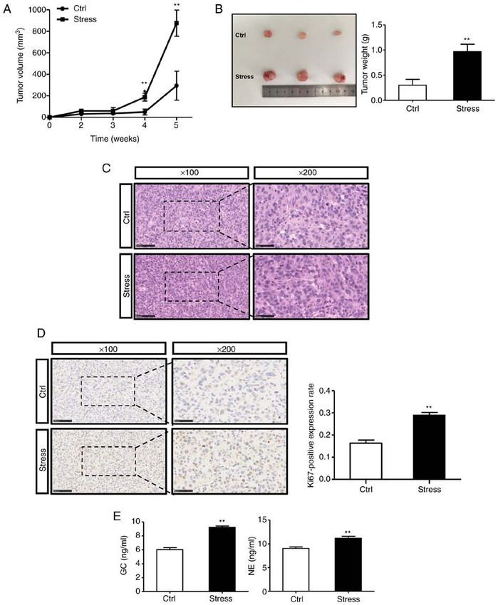
2                             ZHANG et al: CHRONIC STRESS PROMOTES GLIOMA CELL PROLIFERATION

and chemotherapy resistance and ii) disrupting antitumor               to restraint stress for 21 days, 6 h per day (9 am to 3 pm). Since
immunity (14‑16). It has been reported that the catecholamines,        the restrained group could not eat and drink normally during
especially noradrenaline (NE), can alter downstream signaling          restraint, the control mice were also deprived of water and
pathways by binding to membrane receptors and are involved             food during the restraining period. The animals in the control
in the regulation of tumor growth. It has been revealed that           group were also treated with water and food deprivation
NE binds to β ‑adrenergic receptors (ADRBs) and activates              during the restraint period. The health and behavior of animals
the cAMP/PKA signaling pathway in various cancers, such as             were monitored and observed every Wednesday and Saturday
ovarian, prostate, and pancreatic cancer (17‑20). Propranolol,         at 9 a.m. The checks included the growth of the xenograft
an ADRB inhibitor, has been demonstrated to inhibit tumor              (measurement of tumor size), the general condition of the mice
growth and metastasis (21,22), which provides a new strategy           (weight and mental status), abdominal breathing (breathing
for the treatment of tumors. In addition, catecholamines               rate), and paw and toe characteristics (whether the paws and
can also directly promote cancer cell proliferation through            toes had fight damage). Adequate food and water was ensured
α‑adrenergic receptors (ADRAs) (23). However, findings                 and mice were sacrificed after 5 weeks. The specific criteria
reporting the involvement of chronic stress in glioma are rare.        for euthanasia applied in this experiment were according to the
    Numerous studies have revealed the changes of some                 Guidelines for the Review of Humane Endpoints in Animal
genes and signaling pathways involved in the occurrence and            Experiments (GB/T1.1‑2009; www.chinesestandard.net/PDF.
development of glioma. The predominant signaling pathways              aspx/GBT1.1‑2009); rapid cervical dislocation for euthanasia
involved in glioma cell regulation include the RAS/RAF/ERK             when the tumor metastasizes or grows rapidly to the point of
pathway, the p53/MDM2/p21 pathway, and the phosphati‑                  ulceration, causing infection or necrosis, and observation of
dylinositol 3‑kinase (PI3K)/Akt/mTOR pathway (24‑26).                  the experimental mice for two to three min without voluntary
Studies have revealed that inhibition of PI3K/Akt signaling            respiration and no blink reflex, was considered as having
can increase autophagy levels of tumor cells, and thereby              succumbed. The tumor samples were collected and embedded
restrain cell proliferation and induce apoptosis (27‑29). Akt,         in paraffin for subsequent analysis.
also known as protein kinase B, is one of the most important
downstream target kinases in the PI3K signal transduction              Cell culture and intervention. The glioma cell lines U87MG
pathway. It is in the central link of the PI3K/Akt pathway, and        and LN229 were purchased from the Chinese National
thus it plays an important role in a series of biological activities   Infrastructure of Cell Line Resource. U87MG cells were
such as apoptosis, survival, and proliferation (30). However,          cultured in MEM (Sigma‑Aldrich; Merck KGaA) medium
there are few studies on the regulatory effects of the PI3K/Akt        containing 10% fetal bovine serum (FBS; Gibco; Thermo
signaling pathway on the chronic stress‑induced proliferation          Fisher Scientific, Inc.), 100 U/ml penicillin, 100 U/ml strep‑
of gliomas.                                                            tomycin, and 100 µg/ml nonessential amino acids. LN229
    In the present study, the effect of chronic stress on the          cells were cultured in DMEM (Sigma‑Aldrich; Merck KGaA)
malignant behavior of glioma was explored and its accelerative         medium containing 10% FBS, 100 U/ml penicillin, and
role in glioma cell proliferation was elucidated. To investigate       100 U/ml streptomycin. All cells were cultured in a humidified
whether chronic stress and stress hormone‑induced glioma               incubator containing 5% CO2 at 37˚C. U87MG cells were used
cell proliferation are regulated by the PI3K/Akt signaling             as ‘glioblastoma of unknown origin’ and identified by short
pathway in vivo and in vitro, chronic stress animal experi‑            tandem repeat analysis.
ments and cytological experiments were used. The present                   Stress hormones (GC and NE), an Akt‑specific inhibitor
study was performed to investigate the targets and molecular           (perifosine), or receptor antagonists were added to the
mechanisms of chronic stress in regulating glioma progression          culture medium for intervention. Based on successful
and to provide new targets for the treatment of glioma.                activation/inhibition in previous publications (32,33),
                                                                       the concentrations of all drugs were selected as follows:
Materials and methods                                                  GC (10 µmol/l), NE (10 µmol/l), perifosine (10 µmol/l;
                                                                       cat. no. S1037), eplerenone [mineralocorticoid receptor (MR)
Animal model. A total of 10 male BALB/c nude mice                      antagonist, 5.2 µmol/l; cat. no. S1707], mifepristone (GR
(6‑8 weeks old; weight, ~18 g) were used in the present                receptor antagonist, 5.2 µmol/l; cat. no. S2606), phentolamine
study. The feeding conditions were as follows: Temperature,            (ADRA antagonist, 0.2 µmol/l; cat. no. S2038), propranolol
18‑22˚C; relative humidity, 50‑60%; 10‑14 h light/dark cycle.          (ADRB antagonist, 24 µmol/l; cat. no. S4076), atenolol (ADRB1
Feed was added 3‑4 times a week and water was changed                  antagonist, 0.5 µmol/l; cat. no. S4817), and higenamine hydro‑
2‑3 times a week. All of the animal experiments were approved          chloride (ADRB2 antagonist, 1.4 µmol/l; cat. no. S3960). An
(approval no. 2016‑0002) by the Institutional Animal Care and          equal volume of dimethyl sulfoxide (DMSO) was used as the
Use Committee of the Academy of Military Medicine Sciences             control. The inhibitor and antagonists were purchased from
(Beijing, China). The experimental group was divided into the          Selleck Chemicals.
chronic restraint stress group and the control group, with 5 rats
in each group. A restraint stress procedure was adopted based          Immunohistochemistry. Hematoxylin‑eosin (H&E) staining
on previous studies (17,31). The animals in the stress group           and immunohistochemistry were performed as previously
were subjected to restraint adaptation for 1 week, which was           described (28). Briefly, paraffin‑embedded sections were
gradually extended to 6 h per day. Next, U87MG glioma cells            deparaffinized in xylene and rehydrated, followed by antigen
were injected subcutaneously into the mice of both groups              retrieval in sodium citrate. After blocking with 1% BSA
at 2x10⁶ cells per mouse. Then, the stress group was subjected         (BioFroxx), the sections were incubated with anti‑Ki67 (1:200;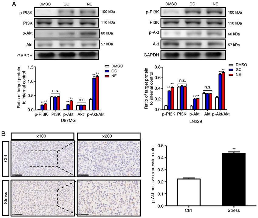
ONCOLOGY REPORTS 46: 202, 2021 3
cat. no. A11005) and anti‑p‑Akt (1:100; cat. no. AP0004; both Cell Signaling Technology, Inc.), rabbit anti‑p‑PI3K (1:1,000;
from ABclonal Biotech Co., Ltd.) overnight at 4˚C. A DAB product no. 13857; Cell Signaling Technology, Inc.), rabbit
kit (ZLI‑9018; ZSGB‑BIO; OriGene Technologies, Inc.) was anti‑Akt (1:1,000; cat. no. 51077‑1‑AP; ProteinTech Group,
utilized to stain until the desired stain intensity was devel‑ Inc.), mouse anti‑p‑Akt (1:1,000; cat. no. 66444‑1‑lg;
oped. Sections were then counterstained with hematoxylin, ProteinTech Group, Inc.), or mouse anti‑GAPDH (1:5,000;
dehydrated, and mounted. cat. no. 60004‑1‑lg; ProteinTech Group, Inc.). After washing
three times in 1X TBST, membranes were incubated with
ELISA. Serums were collected from the stressed mice and the HRP‑labeled goat anti‑rabbit IgG (1:5,000; cat. no. ZB‑2301;
control mice. The serum concentrations of GC and NE were ZSJQ‑Bio) or goat anti‑mouse IgG (1:5,000; cat. no. ZB‑2305;
determined using a mouse GC ELISA kit (cat. no. D721183) ZSJQ‑Bio) for 2 h at room temperature. Protein bands were
and a mouse NE ELISA kit (cat. no. D751020; both from visualized with an ECL western blotting substrate kit (Thermo
Sangon Biotech, Co., Ltd.), respectively, following the manu‑ Fisher Scientific, Inc.). The protein molecular weights of
facturer's instructions. The ELISA kits were purchased from PI3K (110 kDa) and Akt (57 kDa) were so close to their
Shanghai Guduo Biological Technology Co., Ltd. corresponding phosphorylated proteins, p‑PI3K (100 kDa)
and p‑Akt (60 kDa), that the same protein strip needed to
Colony formation assay. Glioma cells were inoculated into be washed with stripping buffer and then re‑incubated with
a 6‑well plate at 2,000 cells/well. GC, NE, Akt signaling the antibody as well as exposed, followed by image acquisi‑
inhibitor, or DMSO was added and the culture solution was tion and analysis by ImageQuant LAS 4000 (GE Healthcare
changed every 3 days. After culturing for 7‑10 days, the cells Bio‑Sciences; Cytiva).
were fixed with 2 ml of 4% paraformaldehyde for 15 min
at 4˚C and stained at room temperature with 10% crystal violet siRNA transfection and quantitative (q)PCR. The siRNAs
solution for 30 min. The colonies with >10 cells were counted targeting ADRB1 and ADRB2 were synthesized by TsingKe
under a low‑power light microscope and representative images Biological Technology. Sequences of siRNAs targeting
were captured. ADRB1 were as follows: Sense, 5'‑CCGAUAGCAGGUGAA
CUCGAA‑3' and antisense, 5'‑GGCUAUCGUCCACUUGAG
Cell Counting Kit‑8 (CCK‑8) assay. Cells in logarithmic CUU‑3'. Sequences of siRNAs targeting ADRB2 were as
growth phase were inoculated into a 96‑well plate at 500 follows: Sense, 5'‑CAGAGUGGAUAUCACAUGGAA‑3' and
cells/well in quintuplicate and incubated. After the cells antisense, 5'‑GUCUCACCUAUAGUGUACCUU‑3'. siRNA or
adhered to the wall, GC, NE, inhibitors, and antagonists siRNA negative control (2.2 pmol) was diluted in 200 µl of
were added. After continuous culture for 0, 24, and 48 h, serum‑free medium and mixed with a pipette. INTERFERin®
the drug‑containing medium was discarded, and freshly (Polyplus transfection) reagent (12 µl) was added, followed
prepared solution containing 10 µl CCK‑8 (Dojindo Molecular by vortex for 10 sec and spinning twice. After 10 min of
Technologies, Inc.) was added into each well. After incubation incubation at room temperature, the transfection mixture was
at 37˚C for 1 h, the OD value at 450 nm was measured by an added to the serum‑containing medium and the cells were
enzyme mapping instrument (Varioskan Flash; Thermo Fisher incubated for 48 h before performing the experiments, with
Scientific, Inc.). The experiment was repeated three times. the following groups: Ctrl group, siRNA negative control and
siRNA group. For siRNA against Akt, the product Akt1/2
Cell cycle experiment. The cells were digested to single siRNA (h) (product no. sc‑43609) was used, purchased from
cells and centrifuged at 1,000 rpm for 5 min to collect cell Santa Cruz Biotechnology, Inc. siRNA or siRNA negative
precipitates. Then 70% ethanol was added and cells were control was diluted to 20 µmol/l with sterilized ddH2O. The
fixed overnight at 4˚C. After washing with PBS, the cells were transfection mixture was added to serum‑containing medium,
resuspended in 500 µl PBS containing 50 µg/ml PI, 100 µg/ml and the cells were incubated for 12 h before performing the
RNase A, and 0.2% Triton X‑100 and incubated for 30 min experiments with the following groups: Ctrl group, siRNA
at room temperature. Flow cytometry (CytomicsFC500; negative control and Akt1/2 siRNA (h) group. The siRNA
Beckman Coulter) was used to detect 20,000‑30,000 cells. The negative control was purchased from TsingKe Biological
results were analyzed by cell cycle fitting software ModFit Technology. Sequences of siRNA negative control were as
LT 4.1 (Verity Software House). follows: SiRNA negative control forward, 5'‑UUCUCCGAA
CGUGUCACGU TT‑3' and reverse, 5'‑ACGUGACACGUU
Western blot analysis. The cells were lysed in RIPA buffer CGGAGAATT‑3'. Successful siRNA knockdown was verified
(Biosharp) containing protease inhibitors and centrifuged by qPCR. Total RNA from cells or tissues was extracted using
at 12,000 x g for 15 min at 4˚C. The pellet was discarded TRIzol reagent (Takara Biotechnology Co., Ltd.) according
and loading buffer (Tiangen Biotech Co., Ltd.) was added to the manufacturer's instructions. Reverse transcription
to the supernatant. Protein determination was performed by (ABScriptII One Step RT‑qPCR; Abclonal Biotech Co., Ltd.)
bicinchoninic acid assay. Samples were boiled for 5 min and reactions were performed with SYBR‑Green. Real‑time
proteins (30 µg) were separated by 10 or 12% SDS‑PAGE, PCR was performed using a LightCycler 96 Real‑time PCR
followed by transfer to a PVDF membrane using a dry System (Roche Diagnostics) with TB Green Premix Ex Taq kit
transfer system (Bio‑Rad Laboratories, Inc.). The membrane (Takara Biotechnology Co., Ltd.). The thermocycling condi‑
was blocked in 5% milk at room temperature for 1 h, washed tions were as follows: Initial denaturation at 95˚C for 10 min,
3 times in 1X TBST (0.5% Tween‑20), and incubated at 4˚C denaturation at 95˚C for 5 sec, annealing at 60˚C for 30 sec and
for 8 h with rabbit anti‑PI3K (1:1,000; product no. 4255; extension at 72˚C for 35 sec, for 40 cycles. β‑Actin was used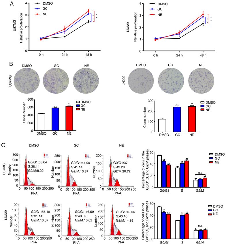
4 ZHANG et al: CHRONIC STRESS PROMOTES GLIOMA CELL PROLIFERATION
as the internal control. All samples were normalized to the flow cytometric analysis. The data revealed a decrease in
internal controls, and fold changes were calculated via the rela‑ G 0/G1‑phase cells and an enhanced S‑phase transition after
tive quantification method (2‑ΔΔCq) (34). The primer sequences GC or NE intervention (Fig. 2C). Collectively, these results
used were as follows: ADRB1 forward, 5'‑GACGCTCACCAA supported the theory that stress could accelerate glioma cell
CCTCTTCA‑3' and reverse, ACT TGG G GTCGT TGTAGC proliferation by increasing the concentrations of GC and
AG; ADRB2 forward, 5'‑TGATCGCAGTGGATCGCTAC‑3' NE.
and reverse, 5'‑CCACCTG GCTAAG GT TCTG G‑3'; AKT1
forward, 5'‑ACTGTCATCGAACGCACCT T‑3' and reverse, PI3K/Akt signaling is activated by chronic stress. Considering
5'‑TCGGAGCCCCCGAGTTG‑3'; AKT2 forward, 5'‑CCT PI3K/Akt signaling is hyperactivated in various cancers and
CTGCAA AGAG GGCATCA‑3' and reverse, 5'‑GAG GAT plays a critical role in cell proliferation and survival (27,28),
GAGCTCGAAGAGGC‑3'. β‑actin forward, 5'‑AATCGTGCG the influence of chronic stress on PI3K/Akt signaling was
TGACATTAAGGAG‑3', and reverse, 5'‑ACTGTGT TGGCG evaluated. Western blot results revealed that compared with
TACAGGTCTT‑3'. the DMSO group, the expression levels of p‑PI3K and p‑Akt
in U87MG and LN229 cells were increased after GC or NE
Statistical analysis. All of the data are listed as the intervention, indicating the PI3K/Akt pathway was activated
mean ± standard deviation (SD). The continuous variables (Fig. 3A). Moreover, the immunohistochemical results further
were evaluated for normality before comparison for statistical confirmed that the positive expression rate of p‑Akt in xeno‑
differences. Paired Student's t‑tests were used for comparisons grafts in stressed mice was higher than that in xenografts in
between two groups. One‑way analysis of variance (ANOVA) control mice (Fig. 3B). The aforementioned results indicated
was performed to compare the values among multiple groups. that the PI3K/Akt signaling pathway may have a regulatory
Significant results of ANOVA were subjected to Tukey's post role in chronic stress‑induced glioma proliferation.
hoc test. All of the differences were two‑sided. Statistical
analysis was conducted with GraphPad Prism 5.0 software Chronic stress promotes glioma cell proliferation via Akt
(GraphPad Software, Inc.) and P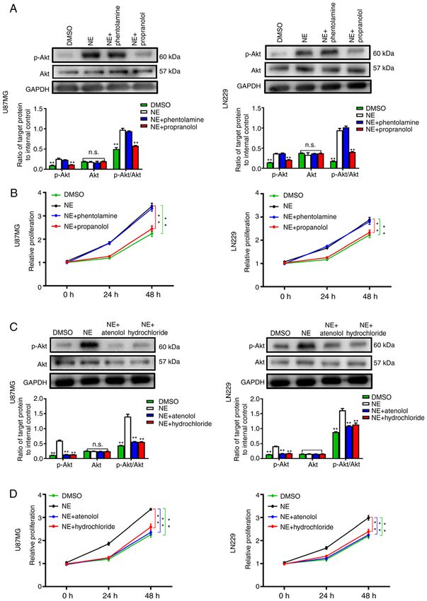
ONCOLOGY REPORTS 46: 202, 2021                                                                    5

Figure 1. Chronic stress promotes glioma growth in vivo. (A) Xenograft tumor growth was monitored. (B) The weight of xenograft tumors from control and
stress groups was compared. (C) Representative images of hematoxylin and eosin staining of xenograft tumors. Magnification, x100; scale bar, 100 µm.
Magnification x200; scale bar, 50 µm. (D) Representative images of immunohistochemical staining of Ki67 in xenograft tumors. Magnification, x100; scale
bar, 100 µm. Magnification, x200; scale bar, 50 µm. (E) Concentrations (ng/ml) of glucocorticoid and norepinephrine in serum of control and stress mice after
sacrifice. Data are presented as the mean ± SD (n=3). **P
6                                ZHANG et al: CHRONIC STRESS PROMOTES GLIOMA CELL PROLIFERATION

Figure 2. Stress hormones facilitate glioma cell proliferation. (A) Cell Counting Kit‑8 results of U87MG and LN229 cells treated with GC (10 µmol/l) or NE
(10 µmol/l). (B) Representative images and statistical analysis of colony formation assays in U87MG and LN229 cells treated with GC or NE. (C) Cell cycle
assay and statistical analysis of U87MG and LN229 cells treated with GC or NE. Data are presented as the mean ± SD (n=3). **P
ONCOLOGY REPORTS 46: 202, 2021                                                                    7

Figure 3. Chronic stress activates the PI3K/Akt signaling pathway. (A) Western blot assay and statistical analysis of PI3K/Akt signaling‑related proteins in
U87MG and LN229 cells treated with GC or NE. (B) Representative images and statistical analysis of immunohistochemical staining of p‑Akt in xenograft
tumors from control and stress mice. Magnification, x100; scale bar, 100 µm. Magnification, x200; scale bar, 50 µm. Data are presented as the mean ± SD (n=3).
**
  P
8                                  ZHANG et al: CHRONIC STRESS PROMOTES GLIOMA CELL PROLIFERATION

Figure 4. Inhibition of Akt signaling suppresses stress‑induced glioma cell proliferation. (A) Western blot assay and statistical analysis of p‑Akt levels in
U87MG and LN229 cells treated with GC or NE with or without Akt signaling inhibitor (perifosine, 10 µmol/l). (B) CCK8 results of U87MG and LN229
cells after the inhibition of Akt signaling. (C) Representative images and statistical analysis of colony formation assays in U87MG and LN229 cells after the
inhibition of Akt signaling. (D) Cell cycle assay and statistical analysis of U87MG and LN229 cells after the inhibition of Akt signaling. Data are presented as
the mean ± SD (n=3). **P
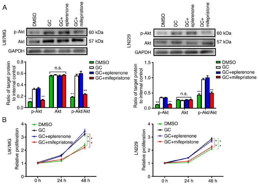
ONCOLOGY REPORTS 46: 202, 2021                                                                   9

Figure 5. GR contributes to GC‑enhanced glioma cell proliferation. (A) Western blot assay and statistical analysis of p‑Akt levels in U87MG and LN229
cells treated with GC with or without MR antagonist (eplerenone, 5.2 µmol/l) or GR antagonist (mifepristone, 5.2 µmol/l). (B) Cell Counting Kit‑8 results of
U87MG and LN229 cells with indicated treatment. Data are presented as the mean ± SD (n=3). **P
10                                ZHANG et al: CHRONIC STRESS PROMOTES GLIOMA CELL PROLIFERATION

Figure 6. NE promotes glioma cell progression via ADRBs. (A) Western blot assay and statistical analysis of p‑Akt levels in U87MG and LN229 cells treated
with NE with or without ADRA antagonist (phentolamine, 0.2 µmol/l) or ADRB antagonist (propranolol, 24 µmol/l). (B) CCK‑8 results of U87MG and
LN229 cells after NE treatment with or without adrenergic receptor antagonists. (C) Western blot assay and statistical analysis of p‑Akt levels in U87MG and
LN229 cells treated with NE with or without ADRB1 antagonist (atenolol, 0.5 µmol/l) or ADRB2 antagonist (hydrochloride, 1.4 µmol/l). (D) CCK‑8 results
of U87MG and LN229 cells after NE treatment with or without ADRB antagonists. Data are presented as the mean ± SD (n=3). **P
ONCOLOGY REPORTS 46: 202, 2021                                                          11

protect patients with glioma from the detrimental effects of           3. Cuddapah VA, Robel S, Watkins S and Sontheimer H: A neuro‑
                                                                          centric perspective on glioma invasion. Nat Rev Neurosci 15:
stress on tumor progression. However, there are still shortcom‑           455‑465, 2014.
ings in our experiments, such as not considering the sample            4. Chen J, McKay RM and Parada LF: Malignant glioma: Lessons
size adequately in order to adhere to the 3R principle of animal          from genomics, mouse models, and stem cells. Cell 149: 36‑47,
welfare (Reduction, Replacement, Refinement) (53), and thus               2012.
                                                                       5. Gately L, McLachlan SA, Dowling A and Philip J: Life beyond a
the sample size will be increased accordingly to render the               diagnosis of glioblastoma: A systematic review of the literature.
data more scientific and reliable when the same problems will             J Cancer Surviv 11: 447‑452, 2017.
be encountered in future experiments.                                  6. Lacroix M, Abi‑Said D, Fourney DR, Gokaslan ZL, Shi W,
                                                                          DeMonte F, Lang FF, McCutcheon IE, Hassenbusch SJ,
                                                                          Holland E, et al: A multivariate analysis of 416 patients with
Acknowledgements                                                          glioblastoma multiforme: Prognosis, extent of resection, and
                                                                          survival. J Neurosurg 95: 190‑198, 2001.
                                                                       7. Carlsson SK, Brothers SP and Wahlestedt C: Wahlestedt,
Not applicable.                                                           Emerging treatment strategies for glioblastoma multiforme.
                                                                          EMBO Mol Med 6: 1359‑1370, 2014.
Funding                                                                8. Witthayanuwat S, Pesee M, Supaadirek C, Supakalin N,
                                                                          Thamronganantasakul K and Krusun S: Survival analysis of glio‑
                                                                          blastoma multiforme. Asian Pac J Cancer Prev 19: 2613‑2617, 2018.
The present study was supported (grant nos. 81702454,                  9. Omuro A and DeAngelis LM: Glioblastoma and other malignant
31771290 and 31571173) by the National Natural Science                    gliomas: A clinical review. JAMA 310: 1842‑1850, 2013.
                                                                      10. Graham J, Ramirez A, Love S, Richards M and Burgess C:
Foundation of China.                                                      Stressful life experiences and risk of relapse of breast cancer:
                                                                          Observational cohort study. BMJ 324: 1420, 2002.
Availability of data and materials                                    11. Chida Y, Hamer M, Wardle J and Steptoe A: Do stress‑related
                                                                          psychosocial factors contribute to cancer incidence and survival?
                                                                          Nat Clin Pract Oncol 5: 466‑475, 2008.
The datasets used and/or analyzed during the current study are        12. Magnon C, Hall SJ, Lin J, Xue X, Gerber L, Freedland SJ and
available from the corresponding author on reasonable request.            Frenette PS: Autonomic nerve development contributes to pros‑
                                                                          tate cancer progression. Asian J Androl 15: 713‑714, 2013.
                                                                      13. Gray JD, Kogan JF, Marrocco J and McEwen BS: Genomic and
Authors' contributions                                                    epigenomic mechanisms of glucocorticoids in the brain. Nat Rev
                                                                          Endocrinol 13: 661‑673, 2017.
ZQZ performed the experiments. ZQZ and XW analyzed                    14. Yang H, Xia L, Chen J, Zhang S, Martin V, Li Q, Lin S, Chen J,
                                                                          Calmette J, Lu M, et al: Stress‑glucocorticoid‑TSC22D3 axis
and interpreted the data as well as critically revised the                compromises therapy‑induced antitumor immunity. Nat Med 25:
manuscript for important intellectual content. ZQZ performed              1428‑1441, 2019.
the statistical analysis and drafted the manuscript. YSZ and          15. Volden PA and Conzen SD: The influence of glucocorticoid
LJQ provided administrative and technical support and also                signaling on tumor progression. Brain Behav Immun 30 (Suppl):
                                                                          S26‑S31, 2013.
supervised the study. ZQZ, XW, BHX, YZ, FX, SDW, CX and               16. Skor MN, Wonder EL, Kocherginsky M, Goyal A, Hall BA,
YW contributed to the conception and design of the study. All             Cai Y and Conzen SD: Glucocorticoid receptor antagonism as
authors read and approved the final manuscript and agree to be            a novel therapy for triple‑negative breast cancer. Clin Cancer
                                                                          Res 19: 6163‑6172, 2013.
accountable for all aspects of the research.                          17. Thaker PH, Han LY, Kamat AA, Arevalo JM, Takahashi R, Lu C,
                                                                          Jennings NB, Armaiz‑Pena G, Bankson JA, Ravoori M, et al:
Ethics approval and consent to participate                                Chronic stress promotes tumor growth and angiogenesis in a
                                                                          mouse model of ovarian carcinoma. Nat Med 12: 939‑944, 2006.
                                                                      18. Park SY, Kang JH, Jeong KJ, Lee J, Han JW, Choi WS, Kim YK,
All of the animal experiments were approved (approval                     Kang J, Park CG and Lee HY: Norepinephrine induces VEGF
no. 2016‑0002) by the Institutional Animal Care and Use                   expression and angiogenesis by a hypoxia‑inducible factor‑1α
                                                                          protein‑dependent mechanism. Int J Cancer 128: 2306‑2316, 2011.
Committee of the Academy of Military Medicine Sciences                19. Radu M, Semenova G, Kosoff R and Chernoff J: PAK signal‑
(Beijing, China).                                                         ling during the development and progression of cancer. Nat Rev
                                                                          Cancer 14: 13‑25, 2014.
                                                                      20. Park MH, Lee HS, Lee CS, You ST, Kim DJ, Park BH, Kang MJ,
Patient consent for publication                                           Heo WD, Shin EY, Schwartz MA and Kim EG: p21‑Activated
                                                                          kinase 4 promotes prostate cancer progression through CREB.
Not applicable.                                                           Oncogene 32: 2475‑2482, 2013.
                                                                      21. Le CP, Nowell CJ, Kim‑Fuchs C, Botteri E, Hiller JG, Ismail H,
                                                                          Pimentel MA, Chai MG, Karnezis T, Rotmensz N, et al: Chronic
Competing interests                                                       stress in mice remodels lymph vasculature to promote tumour
                                                                          cell dissemination. Nat Commun 7: 10634, 2016.
                                                                      22. Na Z, Qiao X, Hao X, Fan L, Xiao Y, Shao Y, Sun M, Feng Z,
The authors declare that they have no competing interests.                Guo W, Li J, et al: The effects of beta‑blocker use on cancer prog‑
                                                                          nosis: A meta‑analysis based on 319,006 patients. Onco Targets
Reference                                                                 Ther 11: 4913‑4944, 2018.
                                                                      23. Lamkin DM, Sung HY, Yang GS, David JM, Ma JC, Cole SW
                                                                          and Sloan EK: α2‑Adrenergic blockade mimics the enhancing
 1. Ostrom QT, Gittleman H, Fulop J, Liu M, Blanda R, Kromer C,           effect of chronic stress on breast cancer progression.
    Wolinsky Y, Kruchko C and Barnholtz‑Sloan JS: CBTRUS                  Psychoneuroendocrinology 51: 262‑270, 2015.
    statistical report: Primary brain and central nervous system      24. Gao J, Liu X, Yang F, Liu T, Yan Q and Yang X: By inhibiting
    tumors diagnosed in the united states in 2008‑2012. Neuro             Ras/Raf/ERK and MMP‑9, knockdown of EpCAM inhibits
    Oncol 17 (Suppl 4): iv1‑iv62, 2015.                                   breast cancer cell growth and metastasis. Oncotarget 6:
 2. Chinot OL, Wick W, Mason W, Henriksson R, Saran F,                    27187‑27198, 2015.
    Nishikawa R, Carpentier AF, Hoang‑Xuan K, Kavan P,                25. Das S: MDM2 Inhibition in a subset of sarcoma cell lines
    Cernea D, et al: Bevacizumab plus radiotherapy‑temozolomide for       increases susceptibility to radiation therapy by inducing senes‑
    newly diagnosed glioblastoma. N Engl J Med 370: 709‑722, 2014.        cence in the polyploid cells. Adv Radiat Oncol 5: 250‑259, 2020.
12 ZHANG et al: CHRONIC STRESS PROMOTES GLIOMA CELL PROLIFERATION
26. O'Donnell JS, Massi D, Teng MWL and Mandala M: 40. Starr LR, Dienes K, Li YI and Shaw ZA: Chronic stress expo‑
PI3K‑AKT‑mTOR inhibition in cancer immunotherapy, redux. sure, diurnal cortisol slope, and implications for mood and
Semin Cancer Biol 48: 91‑103, 2018. fatigue: Moderation by multilocus HPA‑Axis genetic variation.
27. Jia X, Wen Z, Sun Q, Zhao X, Yang H, Shi X and Xin T: Apatinib Psychoneuroendocrinology 100: 156‑163, 2019.
suppresses the proliferation and apoptosis of gastric cancer 41. Verbeek E, Colditz I, Blache D and Lee C: Chronic stress influ‑
cells via the PI3K/Akt signaling pathway. J buon 24: 1985‑1991, ences attentional and judgement bias and the activity of the HPA
2019. axis in sheep. PLoS One 14: e0211363, 2019.
28. Chen H, Zhou L, Wu X, Li R, Wen J, Sha J and Wen X: The 42. Kvetnansky R, Sabban EL and Palkovits M: Catecholaminergic
PI3K/AKT pathway in the pathogenesis of prostate cancer. Front systems in stress: Structural and molecular genetic approaches.
Biosci (Landmark Ed) 21: 1084‑1091, 2016. Physiol Rev 89: 535‑606, 2009.
29. Costa RLB, Han HS and Gradishar WJ: Targeting the 43. Chetty S, Friedman AR, Taravosh‑Lahn K, Kirby ED,
PI3K/AKT/mTOR pathway in triple‑negative breast cancer: A Mirescu C, Guo F, Krupik D, Nicholas A, Geraghty A,
review. Breast Cancer Res Treat 169: 397‑406, 2018. Krishnamurthy A, et al: Stress and glucocorticoids promote
30. Ediriweera MK, Tennekoon KH and Samarakoon SR: Role of oligodendrogenesis in the adult hippocampus. Mol Psychiatry 19:
the PI3K/AKT/mTOR signaling pathway in ovarian cancer: 1275‑1283, 2014.
Biological and therapeutic significance. Semin Cancer Biol 59: 44. Pistritto G, Trisciuoglio D, Ceci C, Garufi A and D'Orazi G:
147‑160, 2019. Apoptosis as anticancer mechanism: Function and dysfunction
31. Cui B, Luo Y, Tian P, Peng F, Lu J, Yang Y, Su Q, Liu B, Yu J, of its modulators and targeted therapeutic strategies. Aging
Luo X, et al: Stress‑induced epinephrine enhances lactate dehy‑ (Albany NY) 8: 603‑619, 2016.
drogenase A and promotes breast cancer stem‑like cells. J Clin 45. D'Alterio C, Scala S, Sozzi G, Roz L and Bertolini G: Paradoxical
Invest 129: 1030‑1046, 2019. effects of chemotherapy on tumor relapse and metastasis promo‑
32. Chen D, Tan Y, Li Z, Li W, Yu L, Chen W, Liu Y, Liu L, Guo L, tion. Semin Cancer Biol 60: 351‑361, 2020.
Huang W and Zhao Y: Organoid cultures derived from patients 46. Obradović MMS, Hamelin B, Manevski N, Couto JP, Sethi A,
with papillary thyroid cancer. J Clin Endocrinol Metab 106: Coissieux MM, Münst S, Okamoto R, Kohler H, Schmidt A and
1410‑1426, 2021. Bentires‑Alj M: Glucocorticoids promote breast cancer metas‑
33. Hu R, Li X, Peng C, Gao R, Ma L, Hu J, Luo T, Qing H, Wang Y, tasis. Nature 567: 540‑544, 2019.
Ge Q, et al: miR‑196b‑5p‑enriched extracellular vesicles from 47. Shin KJ, Lee YJ, Yang YR, Park S, Suh PG, Follo MY, Cocco L
tubular epithelial cells mediated aldosterone‑induced renal fibrosis and Ryu SH: Molecular mechanisms underlying psychological
in mice with diabetes. BMJ Open Diabetes Res Care 8: e001101, stress and cancer. Curr Pharm Des 22: 2389‑2402, 2016.
2020. 48. Butler DE, Marlein C, Walker HF, Frame FM, Mann VM,
34. Livak KJ and Schmittgen TD: Analysis of relative gene expres‑ Simms MS, Davies BR, Collins AT and Maitland NJ: Inhibition
sion data using real‑time quantitative PCR and the 2(‑Delta Delta of the PI3K/AKT/mTOR pathway activates autophagy and
C(T)) method. Methods 25: 402‑408, 2001. compensatory Ras/Raf/MEK/ERK signalling in prostate cancer.
35. Zhe D, Fang H and Yuxiu S: Expressions of hippocampal miner‑ Oncotarget 8: 56698‑56713, 2017.
alocorticoid receptor (MR) and glucocorticoid receptor (GR) in 49. Zhang H, Zhu Y, Sun X, He X, Wang M, Wang Z, Wang Q,
the single‑prolonged stress‑rats. Acta Histochem Cytochem 28: Zhu R and Wang S: Curcumin‑loaded layered double hydroxide
41: 89‑95, 2008. nanoparticles‑induced autophagy for reducing glioma cell migra‑
36. Zhang D, Ma Q, Shen S and Hu H: Inhibition of pancreatic cancer tion and invasion. J Biomed Nanotechnol 12: 2051‑2062, 2016.
cell proliferation by propranolol occurs through apoptosis induc‑ 50. Cole SW and Sood AK: Molecular pathways: Beta‑adrenergic
tion: The study of beta‑adrenoceptor antagonist's anticancer signaling in cancer. Clin Cancer Res 18: 1201‑1206, 2012.
effect in pancreatic cancer cell. Pancreas 38: 94‑100, 2009. 51. Homburger V, Lucas M, Rosenbaum E, Vassent G and Bockaert J:
37. Surman M and Janik ME: Stress and its molecular consequences Presence of both beta1‑and beta2‑adrenergic receptors in a single
in cancer progression. Postepy Hig Med Dosw (Online) 71: cell type. Mol Pharmacol 20: 463‑469, 1981.
485‑499, 2017. 52. Schwalbe T, Huebner H and Gmeiner P: Development of cova‑
38. Umamaheswaran S, Dasari SK, Yang P, Lutgendorf SK and lent antagonists for β1‑and β2‑adrenergic receptors. Bioorg Med
Sood AK: Stress, inflammation, and eicosanoids: An emerging Chem 27: 2959‑2971, 2019.
perspective. Cancer Metastasis Rev 37: 203‑211, 2018. 53. O'Connor MD: The 3R principle: Advancing clinical application
39. Zhang X, Zhang Y, He Z, Yin K, Li B, Zhang L and Xu Z: Chronic of human pluripotent stem cells. Stem Cell Res Ther 4: 21, 2013.
stress promotes gastric cancer progression and metastasis: An
essential role for ADRB2. Cell Death Dis 10: 788, 2019. This work is licensed under a Creative Commons
Attribution-NonCommercial-NoDerivatives 4.0
International (CC BY-NC-ND 4.0) License.You can also read