Histone H3 lysine 9 trimethylation and HP1γ favor inclusion of alternative exons

Page created by Rose Conner
 
CONTINUE READING
Histone H3 lysine 9 trimethylation and HP1γ favor inclusion of alternative exons
articles

                                                   Histone H3 lysine 9 trimethylation and HP1γ favor
                                                   inclusion of alternative exons
                                                   Violaine Saint-André1–3, Eric Batsché1–3, Christophe Rachez1–3 & Christian Muchardt1–3

                                                   Pre-messenger RNAs (pre-mRNAs) maturation is initiated cotranscriptionally. It is therefore conceivable that chromatin-borne
                                                   information participates in alternative splicing. Here we find that elevated levels of trimethylation of histone H3 on Lys9
                                                   (H3K9me3) are a characteristic of the alternative exons of several genes including CD44. On this gene the chromodomain
© 2011 Nature America, Inc. All rights reserved.

                                                   protein HP1g, frequently defined as a transcriptional repressor, facilitates inclusion of the alternative exons via a mechanism
                                                   involving decreased RNA polymerase II elongation rate. In addition, accumulation of HP1g on the variant region of the CD44
                                                   gene stabilizes association of the pre-mRNA with the chromatin. Altogether, our data provide evidence for localized histone
                                                   modifications impacting alternative splicing. They further implicate HP1g as a possible bridging molecule between the chromatin
                                                   and the maturating mRNA, with a general impact on splicing decisions.

                                                   Alternative splicing is a regulated process by which pre-mRNAs are               by regulating the charges of the tails and thereby modifying their
                                                   differentially spliced, leading to expression of several mRNAs from              contacts with neighboring nucleosomes. Histone modifications also
                                                   a single gene. It provides a mechanism for producing a wide variety              function as flags, recruiting chromatin factors. For instance, histone
                                                   of proteins from a small number of transcription units. It is now esti-          acetylation is detected by bromodomain-containing proteins, whereas
                                                   mated that about 90% of human pre-mRNAs are alternatively spliced,               methylation of lysines can create a binding site for proteins contain-
                                                   such that the product of a gene will frequently vary from one tissue to          ing chromodomains13,14. These proteins in turn affect the level of
                                                   the other, dictated by the balance of splicing enhancer and repressor            chromatin condensation and DNA accessibility15.
                                                   activities that are present1,2.                                                      Several reports suggest that such histone modifications may
                                                      Transcription factors recruited to promoter sequences can also                ­influence the regulation of alternative splicing. In particular, treatment
                                                   affect the outcome of alternative splicing3. This is understandable               of cells with HDAC inhibitors, which favor acetylation of ­histones,
                                                   because splicing of pre-mRNA is initiated cotranscriptionally (see,               results in increased exon skipping on several genes that have been
                                                   for example, refs. 4,5). Consistent with this, RNA polymerase II                  monitored (see, for example, ref. 16). Furthermore, trimethylation
                                                   (RNAP II) participates in the recruitment of splicing factors to                  of histone H3 on Lys4 (H3K4me3; see related abbreviations below
                                                   nascent transcripts6, and on some genes Ser2 phosphorylation of the               reflecting different positions of lysine and di- or trimethylation)
                                                   poly­merase C-terminal domain (CTD) heptamer repeat is required                   leads to the recruitment of the CHD1 ATPase with consequences
                                                   for the processing of the pre-mRNA by the splicing machinery7.                    on pre-mRNA splicing efficiencies17. An enrichment of H3K36
                                                      The involvement of the human SWI/SNF chromatin remodeling                      trimethylation has also been detected inside exons12,18,19. This modi-
                                                   complex in the inclusion of alternative exons has revealed that splicing          fication facilitates recruitment of the polypyrimidine tract binding
                                                   decisions are also governed by chromatin and its components8. Exons               protein involved in repression of specific alternative exons 20. Finally,
                                                   are on average 140–150 nucleotides long, a length strikingly similar              on the fibronectin gene, local dimethylation of histone H3 on Lys9
                                                   to the 147 nucleotides necessary to accommodate a nucleosome,                     induced by short interfering RNAs (siRNAs) directed against intronic
                                                   which is the fundamental building block of chromatin9. In agreement               sequences can favor the inclusion of alternative exons21.
                                                   with this, genome-wide analyses show that nucleosomes are preferen-                  It is possible that chromatin factors and histone modifications
                                                   tially located on exons and may participate in the definition of splice           affect inclusion of alternative exons by modulating the recruitment
                                                   sites10–12. These studies also establish a positive correlation between           of the spliceosome. For example, depletion of CHD1 leads to reduced
                                                   nucleosome occupancy and the degree of exon inclusion, consistent                 recruitment of the U2 small nuclear ribonuclear protein component17.
                                                   with the impact of chromatin on alternative splicing.                             Likewise, the SWI/SNF complex interacts with, and possibly recruits,
                                                      The histones that compose the nucleosomes are substrates for                   several components of the spliceosome 8,22. In addition, chromatin
                                                   post-translational modifications on their lysine- and arginine-rich               may affect the RNAP II elongation rate, a parameter that regulates
                                                   N-terminal tails. These modifications, including lysine acetylation               inclusion of alternative exons23. The rationale here is that the slowing
                                                   and serine/threonine phosphorylation, affect chromatin structure                  of the RNAP II increases the chances for the spliceosome to recognize

                                                   1Institut
                                                           Pasteur, Département de Biologie du Développement, Unité de Régulation Epigénétique, Paris, France. 2Centre National de la Recherche Scientifique
                                                   (CNRS), URA2578, Paris, France. 3INSERM Avenir, Paris, France. Correspondence should be addressed to C.M. (muchardt@pasteur.fr).

                                                   Received 25 February 2010; accepted 2 December 2010; published online 27 February 2011; doi:10.1038/nsmb.1995

                                                   nature structural & molecular biology                   VOLUME 18       NUMBER 3      MARCH 2011                                                            337
Histone H3 lysine 9 trimethylation and HP1γ favor inclusion of alternative exons
articles

                                                   Figure 1 Histone H3K9 trimethylation (H3K9me3) is enriched on
                                                   the CD44 alternative exons. (a,b) ChIP assays with antibodies to H3,
                                                                                                                                 a                    6                                                                  H3K4me3

                                                                                                                                Relative enrichment
                                                                                                                                                      5                                                                  H3K4me2
                                                   H3K4me2, H3K4me3 and H3K36me3 (a) and antibodies to H3K9me2
                                                                                                                                                                                                                         H3K36me3

                                                                                                                                    (versus H3)
                                                   and H3K9me3 (b), and chromatin from HeLa cells. The relative                                       4
                                                                                                                                                                                                                         IgG
                                                   enrichment of each modification along the CD44 gene was measured by                                3
                                                   qPCR using primer sets targeting the indicated exons and introns and is                            2
                                                   expressed as a fraction of the signal obtained with antibody to H3. The                            1
                                                   signal obtained upon immunoprecipitation with nonimmune IgG indicates                              0
                                                   the level of background (dotted line). Values are means ± s.e.m. of six                                PP C1     C2 C3      C4 C5   v2 v3v4v5v6 v7 v8 v9 v10 C16    C17 C18   C19
                                                   qPCR values from one representative experiment of five independent
                                                                                                                                               CD44               Constant exons (C)      Variant exons (v)
                                                   experiments. Constant and variant exons are represented as black
                                                   and gray boxes, respectively, on the CD44 gene map below the graphs.          b                    2                                                                  H3K9me3
                                                   The proximal promoter (PP) corresponds to the transcriptional start site.                                                                                             H3K9me2

                                                                                                                                Relative enrichment
                                                                                                                                                                                                                         IgG

                                                                                                                                    (versus H3)
                                                                                                                                                      1
                                                   splice sites as they appear and thus minimizes the probability of skip-
                                                   ping to stronger splice sites farther down the gene. Our earlier data
                                                   show that the SWI/SNF-dependent inclusion of CD44 variant exons                                    0
                                                   observed upon activation of protein kinase C (PKC) is accompanied                                      PP C1     C2 C3      C4 C5   v2 v3v4v5v6 v7 v8 v9 v10 C16    C17 C18   C19
                                                   by a local accumulation of RNAP II on the regions encoding the vari-
                                                                                                                                               CD44               Constant exons (C)      Variant exons (v)           3.5 kb:
                                                   ant domain of the CD44 protein8. This suggests that the SWI/SNF
                                                   complex affects variant exon inclusion in part by modulating the
© 2011 Nature America, Inc. All rights reserved.

                                                   RNAP II elongation rate.                                                    amplitude (Fig. 1b). Conversely, H3K27me3, another chromatin modi-
                                                      The human CD44 gene consists of two series of constant exons             fication associated with transcriptional repression in mammals, was
                                                   (C1–C5 and C16–C19) that frame a large cluster of nine variant              sparsely distributed within the gene, with no significant differences
                                                   exons (v2–v10). By this spatial organization, the gene is amena-            between constant and variant exons (Supplementary Fig. 1b).
                                                   ble for detecting changes in chromatin structure between constant
                                                   and variant exons. Therefore, we have used CD44 to carefully map            H3K9 methylation favors inclusion of CD44 variant exons
                                                   a series of histone modifications potentially involved in regulation        We next investigated whether the H3K9me3 mark detected on CD44
                                                   of alternative splicing, including H3K4me2, H3K4me3, H3K36me3,              was involved in regulation of the alternative splicing of this gene.
                                                   H3K9me2 and H3K9me3. Notably, H3K9me3 is enriched inside the                To this end, we used siRNAs to transiently deplete HeLa cells from
                                                   variant region, and the presence of this mark can be correlated with        several histone lysine methyltransferases (HKMTs) known to ­modify
                                                   an increased inclusion of the alternative exons in the mature CD44          H3K9, including SUV39H1, SUV39H2, EHMT1 (also known as
                                                   mRNA. Furthermore, we find that upon activation of PKC, HP1γ, a             GLP) and EHMT2 (also known as G9A) (Supplementary Fig. 2a).
                                                   chromodomain protein with specificity for histone H3 methylated             Depletion of SUV39H1 or SUV39H2 had only minor effects on
                                                   on K9, accumulates in the CD44 variant region where it is found             amounts of H3K9me3 inside the CD44 gene. In contrast, upon deple-
                                                   associated with both the chromatin and the nascent pre-mRNA, and            tion of EHMT1 or EHMT2, accumulation of H3K9me3 was substan-
                                                   colocalized with splicing factors. Genome-wide analysis further shows       tially affected, with a reduction by approximately a factor of 2 of the
                                                   that HP1γ has a role in the regulation of alternative splicing on a large   methylation peak on v9 and v10 (Supplementary Fig. 2b). In these
                                                   number of genes. Thus, our data suggest that the chromatin of vari-         experiments, amounts of H3K27me3 on the CD44 gene remained
                                                   ant exons carries information conveyed from the nucleosomes to the          unaffected (data not shown).
                                                   splicing factors, by proteins binding both histones and RNA.                   ChIP assays further confirmed the presence of EHMT1 or EHMT2
                                                                                                                               on the CD44 gene, with a notable accumulation on the variant exons
                                                   RESULTS                                                                     v9 and v10. On these exons, depletion of the HKMTs resulted in their
                                                   Increased histone H3K9 trimethylation on CD44 variant exons                 decreased accumulation, evidence for the specificity of the ChIP data
                                                   We have probed the coding region of the CD44 gene for differences in        (Supplementary Fig. 2c,d). ChIP assays also showed that depletion of
                                                   histone modifications between constant and variant exons. Chromatin         EHMT1 or EHMT2 reduced accumulation of RNAP II on the CD44
                                                   immunoprecipitation (ChIP) walking in HeLa cells detecting histone          gene. The effect was stronger on v9 and v10 than on C2 or C5, suggest-
                                                   H3 showed a preferential positioning of nucleosomes on exons ­versus        ing that the depletion of the HKMTs increased the elongation rate of
                                                   introns but no substantial difference in this positioning between con-      the polymerase inside the variant region (Supplementary Fig. 2e).
                                                   stant and variant exons (Supplementary Fig. 1a). We detected the               We next probed the impact of the HKMTs on exon inclusion.
                                                   expected elevated levels of H3K4me2 and H3K4me3 at the promoter,            Depletion of EHMT1 and EHMT2 reduced inclusion of variant exon
                                                   and of H3K36me3 inside the coding region of the gene (Fig. 1a).             couples v8v9 and v9v10 in the mature CD44 transcript by a factor of
                                                   H3K36me3 showed a minor enrichment on variant versus constant               2 or more, whereas depletion of SUV39H1 and SUV39H2 had only
                                                   exons, and on exons versus their surrounding introns, in the vari-          marginal effects on splicing (Supplementary Fig. 2f). We conclude
                                                   ant region of the gene. These observations are consistent with earlier      from these experiments that methylation of histone H3 in the variant
                                                   reports18,20. H3K9me3 was enriched on the promoter of CD44. Notably,        region of CD44 involves the histone methyltransferases EHMT1 and
                                                   this modification also accumulated on the cluster of variant exons          EHMT2, and that high levels of H3K9me3 favor RNAP II accumula-
                                                   inside the gene (Fig. 1b). Peaks of H3K9me3 on the variant region of        tion and inclusion of CD44 variant exons.
                                                   CD44 were also visible in the data from a genome-wide study carried
                                                   out on human T cells, illustrating that it is not a phenomenon specific     HP1g is involved in efficient inclusion of CD44 variant exons
                                                   to HeLa cells24. Expression of H3K9me2 was also higher on the vari-         H3K9 methylation creates a binding site for the chromodomain of
                                                   ant compared to the constant exons of CD44, although with reduced           HP1 proteins. In mammalian cells, this family of proteins includes

                                                   338                                                    VOLUME 18      NUMBER 3                     MARCH 2011         nature structural & molecular biology
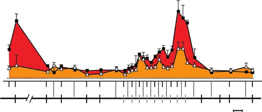
Histone H3 lysine 9 trimethylation and HP1γ favor inclusion of alternative exons
articles

                                                    a                                                                          b                     1.5
                                                                                                                                                           siGAPDH      siHP1β                     c1.5                               siGAPDH          siHP1β                                   d                    e                            HeLa S3   HeLa S3-HP1γ
                                                                                                                                                           siHP1α       siHP1γ                                                        siHP1α           siHP1γ                                                                               2
                                                                     si DH

                                                                     si DH

                                                                     si DH

                                                                                                                                                                                                  Splicing fold effect
                                                                     si α

                                                                     si β

                                                                           γ
                                                                        AP

                                                                         AP

                                                                         AP

                                                                                                                               Relative expression
                                                                         P1

                                                                         P1

                                                                         P1
                                                                                                                                                                                                                                                                                                HeLa S3

                                                                                                                                                                                                                                                                                                                     Splicing fold effect
                                                                       H

                                                                       H

                                                                       H
                                                                 G

                                                                       G

                                                                       G
                                                              si

                                                                                                                                                     1.0                                                                 1.0                                                                    Ctl HP1γ
                                                    H3
                                                                                                                                                                                                                                                                                                           H3                               1
                                                                                                                                                     0.5                                                                 0.5                                                                               HP1γ-HA
                                                                  HP1α                                 HP1β        HP1γ                                                                                                                                                                                    HP1γ

                                                                                                                                                      0                                                                      0                                                                                                              0
                                                                                                                                                           RLP0      CD44                                                         C4C5          v2v3        v4v5   v8v9     v9v10 C16C17                                                        C4C5 v2v3 v4v5 v8v9 v9v10 C16C17
                                                                                                                                                                     C2C3

                                                   Figure 2 HP1γ regulates alternative splicing of the CD44 transcript. (a) HeLa cells were transfected with siHP1α, siHP1β, siHP1γ or siGAPDH as a
                                                   control. Western blots on nuclear extracts were carried out with antibodies to HP1α (anti-HP1α), anti-HP1β or anti-HP1γ (lower panels) or anti-H3
                                                   (H3 used as a loading control, upper panels). Blots are representative of the experimental replicates. (b,c) RNAs from transfected cells were quantified
                                                   by RT-qPCR. (b) Expression level of the CD44 C2C3 constant exons relative to that of RLP0 (set to 1) for each transfection condition. (c) Exon inclusion
                                                   is measured as the ratio between the indicated CD44 exon couples and the constant exon couple C2C3 and expressed relatively to the values obtained
                                                   upon transfection of siGAPDH (set to 1). Values are means ± s.e.m. of six qPCR values from three experimental replicates. (d) Western blots on nuclear
                                                   extracts from HeLa S3 and HeLa S3-overexpressing HP1γ-HA (HeLa S3-HP1γ) cells were carried out with anti-HP1γ or anti-H3 (H3 used as a loading
                                                   control). Blots are representatives of the replicates. (e) RNAs from the corresponding HeLa S3 or HeLa S3-HP1γ cells were quantified by RT-qPCR. Exon
                                                   inclusion is measured as the ratio between the indicated CD44 exon couples and the constant exon couple C2C3, and expressed relatively to the values
                                                   obtained with the HeLa S3 cell line (set to 1). Values are means ± s.e.m. from four experimental replicates.

                                                   three members, namely HP1α, HP1β and HP1γ. Of these, only HP1γ                                                                                                                                             (Fig. 3c, ). This led us to explore the distribution of H3K9me3
© 2011 Nature America, Inc. All rights reserved.

                                                   has been reported to be on the coding region of active genes25–27.                                                                                                                                         and HP1γ upon activation of the PKC pathway by the phorbol ester
                                                   To investigate the possible involvement of HP1 proteins in regula-                                                                                                                                         PMA. This treatment enhances accumulation of RNAP II inside the
                                                   tion of CD44 variant exon inclusion, we carried out siRNA-mediated                                                                                                                                         CD44 variant region and stimulates inclusion of the variant exons
                                                   depletion of each HP1 isoform in HeLa cells (Fig. 2a; abbreviated as                                                                                                                                       (Fig. 3a,f)8,28. Under these conditions, amounts of H3K9me3 were
                                                   siHP1α, etc.). These siRNAs did not affect expression of the CD44                                                                                                                                          increased throughout the gene, but the profile was unchanged as
                                                   gene (Fig. 2b). Inactivation of HP1α and HP1β had little or no effect                                                                                                                                      a peak inside the variant region of the CD44 gene was maintained
                                                   on splicing, whereas inactivation of HP1γ resulted in decreased inclu-                                                                                                                                     (Fig. 3b). In contrast, HP1γ distribution was markedly changed by
                                                   sion of all the variant exons examined (Fig. 2c). Conversely, stable                                                                                                                                       the PMA, which induces a clear accumulation of the protein inside the
                                                   moderate overexpression of HP1γ in HeLa cells (Fig. 2d) resulted in                                                                                                                                        variant region of CD44 (Fig. 3c), largely overlapping with the peak of
                                                   increased inclusion of the same variant exons (Fig. 2e).                                                                                                                                                   RNAP II (Fig. 3f). The PMA also induces phosphorylation of HP1γ
                                                                                                                                                                                                                                                              on its Ser83 (HP1γS83p, Fig. 3g). The profile of HP1γ ­distribution
                                                   HP1g slows RNAP II                                                                                                                                                                                         was essentially reproduced in ChIP experiments with an anti–
                                                   The impact of HP1γ on inclusion of CD44 variant exons prompted us                                                                                                                                          phospho-S83 HP1γ (Fig. 3d). Although this antibody was of sub-
                                                   to explore the distribution of this protein within the chromatin of the                                                                                                                                    optimal quality, these data suggest that the HP1γ recruited to the
                                                   CD44 gene. As shown in Figure 3, ChIP walking revealed that HP1γ                                                                                                                                           CD44 gene upon treatment with PMA is phosphorylated. This is
                                                   is present inside the gene but with no obvious sites of ­accumulation                                                                                                                                      consistent with an earlier study showing that HP1γ is modified by

                                                    a                                            3.5
                                                                                                          – PMA
                                                                                                                                                                             d                                                                                     g                                Figure 3 HP1γ, RNAP II and U2AF65
                                                                          Splicing fold effect

                                                                                                 3.0      + PMA                                                                                                                                                                                     accumulate on the variant region of CD44
                                                                                                                                                                              Relative enrichment

                                                                                                 2.5                                                                                                                             HP1γS38p
                                                                                                                                                                                                                                                                           PMA – +                  chromatin upon PKC stimulation. (a) HeLa
                                                                                                                                                                                                                         3                       – PMA
                                                                                                 2.0
                                                                                                                                                                                 (versus IgG)

                                                                                                 1.5                                                                                                                     2
                                                                                                                                                                                                                                                 + PMA             HP1γS38p                         cells were incubated (+PMA) or not (−PMA)
                                                                                                 1.0                                                                                                                                                                                                overnight with PMA. After extraction of
                                                                                                                                                                                                                         1                                                HP1γ
                                                                                                 0.5                                                                                                                                                                                                cytoplasmic RNA and DNase treatment, each
                                                                                                   0                                                                                                                     0
                                                                                                       C2 C5 v2 v3 v4 v5 v6 v7 v8 v9 v10C19                                                                                  C2 v4 v9 C19                                         1   2             CD44 exon was quantified by RT-qPCR.
                                                                                                                                                                                                                                                                                                    Exon inclusion is represented as fold change
                                                                                                                                                                             e                                           7            16                                                            ± s.e.m. between stimulated and nonstimulated
                                                                      CD44
                                                                                                                                                                              Relative enrichment

                                                                                                                                                                                                                         6            14                                         – PMA              cells, with the signal from nonstimulated
                                                                          Constant exons (C) Variant exons (v)                                                                                                                        12        U2AF65                           + PMA
                                                                                                                                                                                 (versus IgG)

                                                                                                                                                                                                                         5
                                                    b                                                              – PMA                                                                                                 4
                                                                                                                                                                                                                                      10
                                                                                                                                                                                                                                       8
                                                                                                                                                                                                                                                                                                    samples being set to 1. (b–f) ChIP walking
                                                                                                                                                                                                                                                                                                    experiments were carried out with antibodies
                                                                                                                   + PMA                                                                                                 3
                                                                                30                                                                                                                                                     6                                                            to H3K9me3 (b), HP1γ (c), HP1γS83p (d),
                                                                                                        16                                                                                                               2
                                                    Relative enrichment

                                                                                                        14                                                                                                                             4
                                                                                25                                 H3K9me3                                                                                               1             2                                                            U2AF65 (e) and RNAP II (8WG16) (f) and
                                                                                                        12                                                                                                                                                                                IgG
                                                                                20                                                                                                                                       0             0                                                            chromatin from HeLa cells treated (black bars
                                                           (% H3)

                                                                                                        10
                                                                                                                                                                                                                                 PP        C2          C5                        i18 C19
                                                                                                                                                                                                                                                                 5
                                                                                                                                                                                                                                                                 0
                                                                                                                                                                                                                                                               v3
                                                                                                                                                                                                                                                               v4
                                                                                                                                                                                                                                                               v5
                                                                                                                                                                                                                                                               v7
                                                                                                                                                                                                                                                               v8
                                                                                                                                                                                                                                                             v19
                                                                                                                                                                                                                                                              i1

                                                                                                                                                                                                                                                                                                    and squares) or not (white bars and squares)
                                                                                                                                                                                                                                                               v

                                                                                15                       8
                                                                                10                       6
                                                                                                         4
                                                                                                                                                                             f                               50                        8
                                                                                                                                                                                                                                                                                                    for 2 h with PMA. Amounts of H3K9me3 are
                                                                                          5                                                                                                                                                                                                         expressed in percent of H3, and amounts of
                                                                                                                                                                            Relative enrichment

                                                                                                         2                                                           IgG                                                               7         RNAP II
                                                                                          0              0                                                                                                   40                                                                                     HP1γ, U2AF65, RNAP II and HP1γS83p are
                                                    c                                                                                                                                                                                  6        (8WG16)
                                                                                                                                                                               (versus IgG)

                                                                                                 PP           C2          C5                                  i18 C19
                                                                                                                                   5
                                                                                                                                   0
                                                                                                                                 v2
                                                                                                                                 v3
                                                                                                                                 v4
                                                                                                                                 v5
                                                                                                                                 v6
                                                                                                                                 v7
                                                                                                                                 v8
                                                                                                                               v19
                                                                                                                                i1

                                                                                                                                                                                                                                       5
                                                                                                                                 v

                                                                                       7                 30
                                                                                                                                                                                                             30                                                                                     expressed relatively to the signal obtained for
                                                                                                                                                                                                                                       4
                                                    Relative enrichment

                                                                                       6                 25        HP1γ                                                                                      20                        3                                                            ChIP using nonimmune IgG. Values are means
                                                                                                                                                                                                                                                                                                    ± s.e.m. of three independent experiments.
                                                       (versus IgG)

                                                                                       5                 20                                                                                                  10                        2
                                                                                                                                                                                                                                       1                                               IgG
                                                                                       4
                                                                                                         15                                                                                                              0
                                                                                                                                                                                                                                                                                                    (g) HeLa cells were stimulated with PMA for
                                                                                       3                                                                                                                                               0
                                                                                                         10                                                                                                                      PP        C2          C5                        i18 C19            2 h. Western blots on total protein extracts
                                                                                                                                                                                                                                                                0
                                                                                                                                                                                                                                                                5
                                                                                                                                                                                                                                                              v3
                                                                                                                                                                                                                                                              v4
                                                                                                                                                                                                                                                              v5
                                                                                                                                                                                                                                                              v7
                                                                                                                                                                                                                                                              v8
                                                                                                                                                                                                                                                             v19
                                                                                                                                                                                                                                                             i1
                                                                                                                                                                                                                                                              v

                                                                                       2
                                                                                       1                  5                                                                                                                                                                                         were carried out with the indicated antibodies.
                                                                                                                                                                     IgG
                                                                                       0                  0                                                                                 CD44 Constant exons (C) Variant exons (v)                                                               PP, proximal promoter.
                                                                                                 PP           C2          C5                                  i18 C19
                                                                                                                                   5
                                                                                                                                   0
                                                                                                                                 v3
                                                                                                                                 v4
                                                                                                                                 v5
                                                                                                                                 v7
                                                                                                                                 v8
                                                                                                                               v19
                                                                                                                                i1
                                                                                                                                 v

                                                   nature structural & molecular biology                                                                                                 VOLUME 18                                          NUMBER 3               MARCH 2011                                                                                               339
Histone H3 lysine 9 trimethylation and HP1γ favor inclusion of alternative exons
articles

                                                   a                                            Input + PMA
                                                                                                                        b                        90   HP1γ
                                                                                                                                                                                        c                        16
                                                                                                                                                                                                                                                        d                              siGAPDH
                                                                            100

                                                                                                                        Relative enrichment of
                                                                                                                                                                                                                      H3                                                               siHP1γ
                                                   Relative enrichment of

                                                                                                                                                 80

                                                                                                                                                                                        Relative enrichment of
                                                                                                Input – PMA

                                                                                                                         RNA (versus DNA)
                                                                                                                                                                                                                 14
                                                    RNA (versus DNA)

                                                                                                                                                                                         RNA (versus DNA)
                                                                                                                                                 70        – PMA                                                           – PMA                                                    RNA-ChlP with anti-H3
                                                                                                                                                                                                                 12        + PMA

                                                                                                                                                                                                                                                        Relative enrichment
                                                                            10                                                                   60        + PMA
                                                                                                                                                                                                                 10                                                           0.8
                                                                                                                                                 50
                                                                                                                                                 40                                                               8

                                                                                                                                                                                                                                                             (% input)
                                                                             1                                                                   30                                                               6
                                                                                                                                                                                                                                                                              0.4
                                                                                                                                                 20                                                               4
                                                                                                                                                 10                                                               2                               lgG
                                                                            0.1                                                                   0                                                               0                                                            0
                                                                                  C2   C4 C5                  i18 C19                                 C2     C5               i18 C19                                 C2     C5              i18 C19                                  C2    v5     v8
                                                                                                  v4
                                                                                                  v5
                                                                                                  v6

                                                                                                v19
                                                                                                    0
                                                                                                   5
                                                                                                  6b

                                                                                                                                                                     v4
                                                                                                                                                                     v5
                                                                                                                                                                     v6
                                                                                                                                                                   v19
                                                                                                                                                                    i10
                                                                                                                                                                       5

                                                                                                                                                                                                                                     v4
                                                                                                                                                                                                                                     v5
                                                                                                                                                                                                                                     v6

                                                                                                                                                                                                                                   v19
                                                                                                                                                                                                                                      0
                                                                                                                                                                                                                                       5
                                                                                                  v

                                                                                                 i1

                                                                                                                                                                     v

                                                                                                                                                                                                                                     v

                                                                                                                                                                                                                                    i1
                                                                                               i1
                                                                  CD44                                                               CD44                                                             CD44
                                                                  Constant exons (C) Variant exons (v)                               Constant exons (C) Variant exons (v)                             Constant exons (C) Variant exons (v)

                                                   Figure 4 HP1γ bridges chromatin to pre-mRNA. (a) Quantification by qPCR of CD44 pre-mRNA exons retained on chromatin. Non-cross-linked DNase-treated
                                                   chromatin from HeLa cells stimulated (+PMA) or not (−PMA) with PMA for 2 h was extracted as described in Online Methods. Graph shows means ± s.e.m.
                                                   of chromatin-bound RNA (quantified by RT-qPCR) relative to the DNA in the input (quantified by qPCR). (b,c) Quantification of CD44 pre-mRNA exons
                                                   associated with HP1γ (b) and H3 (c) in the non-cross-linked chromatin fraction. RNA-ChIP assays were carried out with the indicated antibodies and chromatin
                                                   prepared as in a. Levels of immunoprecipitated RNA are expressed relatively to the levels of corresponding DNA sequences present in the extracted chromatin
                                                   fraction. (d) Association of histone H3 with the CD44 pre-mRNA requires HP1γ. RNA-ChIPs were carried out with anti-H3 and cross-linked chromatin from
                                                   HeLa cells transfected with the indicated siRNAs and stimulated with PMA for 2 h. The qPCR values are from a representative experiment and are expressed as
                                                   a percent of input for each indicated amplicon. For all panels, values are means ± s.e.m. from two independent experiments.

                                                   ­ hosphorylation when it is present inside active genes29. Finally, we
                                                   p                                                                                                                            (Supplementary Fig. 5b). We also carefully verified that the signals
                                                   noted that the PMA treatment also causes accumulation of the splic-                                                          were due to detection of pre-mRNA and not ­contaminating mature
© 2011 Nature America, Inc. All rights reserved.

                                                   ing factor U2AF65 on the variant region of CD44 (Fig. 3e) in a pattern                                                       mRNA (Supplementary Fig. 5c,d). We observed that, although the
                                                   similar to that of HP1γ and the RNAP II.                                                                                     variant exons are enriched in RNAP II (Fig. 3d), RNA encoded by
                                                      We next depleted the HeLa cells of HP1γ using siRNAs. This                                                                these exons is less abundant on the chromatin than that encoded by
                                                   resulted in decreased accumulation of RNAP II on the coding region                                                           constant exons (Fig. 4a, m). In addition, amounts of RNA encoded
                                                   of CD44, suggestive of an increased elongation rate of the poly­merase                                                       by variant exons and neighboring introns increase on the chromatin
                                                   (Supplementary Fig. 3d). In these knockdown experiments, the                                                                 by a factor about 10 upon treatment of the cells with PMA, whereas
                                                   transcriptional activity of the CD44 gene was unaffected (as illus-                                                          amounts of RNA encoded by constant exons are increased only by
                                                   trated by levels of constant exons in Fig. 2c), consistent with the                                                          a factor of 2 (Fig. 4a; compare m and ; Supplementary Fig. 5e).
                                                   essentially unchanged levels of RNAP II accumulation on its pro-                                                             These observations suggest a correlation between chromatin ­retention
                                                   moter region (Supplementary Fig. 3d, left). Depletion of HP1γ also                                                           of a pre-mRNA sequence and its presence in the mature message.
                                                   reduced recruitment of U2AF65 (Supplementary Fig. 3c). Likewise,                                                             They also suggest that proteins other than the RNAP II bridge the
                                                   recruitment of the splicing factor PRP8, involved in later stages of                                                         ­chromatin to the pre-mRNA.
                                                   the spliceosome assembly30, was also affected by reduced levels                                                                  To investigate this issue, we used the chromatin fraction described
                                                   of HP1γ (Supplementary Fig. 4). Finally, we noted that depletion of                                                           above to carry out RNA-ChIP experiments. With antibodies to RNAP II
                                                   HP1γ moderately decreased levels of H3K9me3 within the gene                                                                   (anti-RNAP II), surprisingly little CD44 pre-mRNA was immuno-
                                                   (Supplementary Fig. 3a). A similar link between levels of HP1γ and                                                            precipitated, suggesting that the interaction between the polymer-
                                                   H3K9me3 had already been observed31–33 and may illustrate the                                                                 ase and the RNA was largely disrupted by the extraction protocol.
                                                   implication of HP1 proteins in the recruitment of HKMTs34.                                                                    Nevertheless, after treatment of the cells with PMA we observed a
                                                      Altogether our experiments show that activation of the PKC path-                                                           peak of interaction between the RNAP II and the RNA encoded by the
                                                   way causes HP1γ and U2AF65 to largely distribute together with the                                                            CD44 variant exons (Supplementary Fig. 5g). This is consistent with
                                                   RNAP II, with a peak of accumulation appearing inside the CD44                                                                the accumulation of polymerase observed on these exons.
                                                   variant region. In addition, depletion of HP1γ reduces accumulation                                                              In contrast to the anti–RNAP II, antibody to HP1γ (anti-HP1γ)
                                                   of RNAP II and spliceosome components throughout the gene. These                                                              immunoprecipitated a large fraction of the input CD44 pre-mRNA.
                                                   observations suggest that HP1γ participate in the PMA-induced inclu-                                                          The HP1γ-RNA interaction required induction of the cells with PMA
                                                   sion of CD44 variant exons by slowing the RNAP II and by favoring                                                             and peaked inside the region encoded by the variant exons (Fig. 4b).
                                                   recruitment of splicing factors either directly or via the RNAP II.                                                           Histone H3 and H3K9me3 were also found associated with the
                                                                                                                                                                                 CD44 pre-mRNA upon PMA treatment (Fig. 4c and Supplementary
                                                   HP1g binds CD44 pre-mRNA inside the variant region of CD44                                                                    Fig. 5f). But as with the RNAP II, we observed weaker PCR signals
                                                   HP1γ is a chromatin protein that binds RNA35. This prompted                                                                   for H3 than for HP1γ, although the antibody to H3 (anti-H3) is more
                                                   us to examine the contacts between the chromatin and the CD44                                                                 efficient at recruiting DNA material in ChIP than the antibody to
                                                   pre-mRNA. To this end, we extracted native non-cross-linked chro-                                                             HP1γ. In addition, siRNA-mediated depletion of HP1γ reduced the
                                                   matin from HeLa cells. From this chromatin we collected a fraction                                                            interaction of H3 with the CD44 pre-mRNA, indicating that HP1γ is
                                                   solubilized by deoxyribonuclease (DNase) that was then mildly soni-                                                           in between the transcript and histone H3 (Fig. 4d).
                                                   cated so as to fragment larger RNA molecules. This fraction is strongly                                                          These observations suggest that HP1γ, through its dual function as
                                                   enriched in RNAP II and U2AF65, suggesting that it contains tran-                                                             both a chromatin- and an RNA-binding protein, is involved in linking the
                                                   scriptionally active chromatin (Supplementary Fig. 5a). Using PCR                                                             nascent CD44 transcript to the chromatin template during elongation.
                                                   after reverse transcription (RT-PCR) we quantified CD44 message
                                                   present in this fraction at different positions throughout the CD44                                                          RNAP II accumulation and variant exon inclusion
                                                   gene. Because the RNA was fragmented, this analysis reflected the                                                            To identify additional situations connecting amounts of H3K9me3
                                                   retention on chromatin of nascent transcripts. In these experiments we                                                       and HP1γ with exon inclusion, we characterized the CD44 locus in
                                                   verified that DNA was uniformly extracted throughout the CD44 gene                                                           SW626 and SKOV3 cells, two cell lines derived from ovarian tumors.

                                                   340                                                                                                       VOLUME 18     NUMBER 3                 MARCH 2011                    nature structural & molecular biology
articles

                                                   Figure 5 Cell type–specific distribution of               2.5       a     SW626                                                                 d                         e                     f

                                                                                                                                                                                                                                         γ
                                                                                                                                                                                                                  v9v10 / C16C17
                                                   H3K9me3 and HP1γ correlates with variant

                                                                                                                                                                                                                                       P1
                                                                                                                             SKOV3                                                                            25

                                                                                                                                                                                                                                       -H
                                                                                                                                    Relative expression
                                                                                                             2.0

                                                                                                                                                                                                                                             Splicing fold effect
                                                                                                                                                                                                                                     V3
                                                                                                                                                                                                        V3
                                                                                                                                                                                                    SK 626

                                                                                                                                                                                                                                 SK 3
                                                   exon inclusion and RNAP II accumulation.

                                                                                                                                                                                                                                     V
                                                                                                                                                                                                                      – PMA

                                                                                                                                                                                                                                   O
                                                                                                                                                                                                              20

                                                                                                                                                                                                      O

                                                                                                                                                                                                                                   O
                                                                                                                                                                                                    SW

                                                                                                                                                                                                                             SK
                                                                                                                                                                                                                      + PMA
                                                   (a) Each CD44 exon was quantified by                      1.5                                                                 HP1α
                                                                                                                                                                                                   HP1γ-HA
                                                                                                                                                                                                              15
                                                                                                                                                                                 HP1β
                                                   RT-qPCR after DNase treatment of SKOV3                    1.0
                                                                                                                                                                                 HP1γ
                                                                                                                                                                                                   HP1γ       10
                                                                                                                                                                                                               5
                                                   (white) or SW626 (black) RNAs. Relative                   0.5                                                                 H3                H3
                                                                                                                                                                                                               0
                                                   abundance of each exon on the mature                                                                                                                           SKOV3 SKOV3-

                                                                                                                                                                                                             g
                                                                                                               0
                                                                                                                 C2 C5 v2 v3 v4 v5 v6 v7 v8 v9 v10 C19                                                                      HP1γ
                                                   transcript was measured by using the same
                                                   external reference for each amplicon. Values                                                                                      HP1γ                         SKOV3-HP1γ

                                                                                                                                                                                                       Relative enrichment
                                                                                                          CD44                                                                    6         5                           SKOV3
                                                   are means ± s.e.m. from three independent

                                                                                                                                                                                                          (versus IgG)
                                                                                                                                                                                  5         4                                     –     +
                                                                                                                   Constant exons (C)   Variant exons (v)                         4                                              PMA PMA
                                                   experiments. (b,c) ChIP assays with antibodies                                                                                           3

                                                   to H3K9me3, H3 and HP1γ and chromatin                       4       b
                                                                                                                       0.6 H3K9me3                                     SW626
                                                                                                                                                                                  3
                                                                                                                                                                                  2
                                                                                                                                                                                  1
                                                                                                                                                                                            2
                                                                                                                                                                                            1                                        lgG

                                                                                                                                    Relative enrichment
                                                                                                                                                                       SKOV3
                                                   from SW626 or SKOV3 cells. The relative                     3                                                                  0
                                                                                                                                                                                      – +
                                                                                                                                                                                            0
                                                                                                                                                                                               C2       C5                   i18 C19

                                                                                                                                        (versus H3)

                                                                                                                                                                                                                                                   5
                                                                                                                                                                                                             h

                                                                                                                                                                                                                                                   0
                                                                                                                                                                                                                                                 v4
                                                                                                                                                                                                                                                 v5
                                                                                                                                                                                                                                                 v8
                                                                                                                                                                                                                                               v19
                                                   enrichment on IL8 and GAPDH promoters,

                                                                                                                                                                                                                                                i1
                                                                                                                       0.4                                                            PP

                                                                                                                                                                                                                                                 v
                                                                                                               2                                                                    RNAPII (N20)
                                                   on satellite sequences (hSat) or along the

                                                                                                                                                                                                       Relative enrichment
                                                                                                                                                                                 20         6
                                                                                                                       0.2
                                                   CD44 gene was quantified by qPCR using                      1

                                                                                                                                                                                                          (versus IgG)
                                                                                                                                                                                 15
                                                                                                                                                                                            4
                                                   primer sets targeting the exons and introns                           0
                                                                                                                                                                             lgG 10
                                                                                                               0
                                                   indicated on the map of the gene and expressed                hSat              C2       C5   v2 v4 v5 v7 v8 v9 v10    C19     5         2

                                                   as a fraction of histone H3 in b or relatively          4           c       HP1γ
                                                                                                                                                                                  0 – +     0
                                                                                                                                                                                              C2        C5                   i18 C19
                                                                                                                                                                                                                                    lgG

                                                                                                                                                                                                                                                                        5
                                                                                                                                                                                                                                                                        0
                                                                                                                                                                                                                                             v4
                                                                                                                                                                                                                                             v5
                                                                                                                                                                                                                                                                      v8
                                                                                                                                                                                                                                                                    v19
                                                                                                                        3.0
                                                                                                                  Relative enrichment

                                                                                                                                                                                      PP

                                                                                                                                                                                                                                                                     i1
                                                                                                                                                                                                                                                                      v
                                                                                                                                                                       SW626
                                                   to IgG in c (set to 1 for each cell line). Values       3                                                           SKOV3                                   i
                                                                                                                     (versus IgG)

                                                                                                                        2.5
                                                   are means ± s.e.m. of three independent                                                                                          H3K9me3

                                                                                                                                                                                                       Relative enrichment
                                                                                                           2            2.0                                                       6         3
                                                   qPCRs from one representative experiment.                            1.5
                                                                                                                                                                                  4         2

                                                                                                                                                                                                              (% H3)
                                                   (d,e) Western blots with the indicated antibodies
© 2011 Nature America, Inc. All rights reserved.

                                                                                                           1
                                                                                                                        1.0
                                                                                                                                                                            lgG
                                                   and nuclear protein extracts from SW626 or              0            0.5
                                                                                                                                                                                  2         1
                                                                                                                                                                                                                                    lgG
                                                   SKOV3 (d) or from SKOV3 and SKOV3 stably                                                                                       0 – + 0
                                                                                                                                                                H
                                                                                                                                                               at
                                                                                                                                                          G IL8

                                                                                                                          0
                                                                                                                                                              D
                                                                                                                                                            hS

                                                                                                                            PP     C2       C5      v4       v9 v10       C19                 C2        C5                   i18 C19
                                                                                                                                                           AP

                                                                                                                                                                                                                                                                        5
                                                                                                                                                                                                                                                                        0
                                                                                                                                                                                                                                               v4
                                                                                                                                                                                                                                               v5
                                                                                                                                                                                                                                                                      v8
                                                                                                                                                                                                                                                                    v19
                                                                                                                                                                                       PP

                                                                                                                                                                                                                                                                     i1
                                                   overexpressing HA-tagged HP1γ (SKOV3-HP1γ)

                                                                                                                                                                                                                                                                      v
                                                                                                                     CD44                                                        CD44
                                                   (e). (f) RT-qPCR quantification of RNAs from                               Constant exons (C)     Variant exons (v)                   Constant exons (C) Variant exons (v)

                                                   SKOV3 and SKOV3-HP1γ cells stimulated
                                                   (+PMA) or not (−PMA) overnight with PMA. Exon inclusion is measured as the ratio between the variant v9v10 and the C16C17 exon couples. Values are
                                                   means ± s.e.m. of three RT-qPCR from one representative experiment. (g–i) ChIP assays with antibodies to RNAP II (N20) (g), HP1γ (h) and H3K9me3
                                                   and H3 (i), and chromatin from SKOV3 or SKOV3-HP1γ transfected for 48 h with either siHP1γ or siGAPDH, then stimulated (+PMA) or not (−PMA)
                                                   with PMA for 2 h. The relative enrichment along the CD44 gene was measured by qPCR and expressed in percent of H3 (for H3K9me3) or relatively to
                                                   values obtained with IgG (for RNAP II and HP1γ). PP, proximal promoter.

                                                   Earlier studies have shown differential CD44 variant exon inclusion                                                    observed only after stimulation of the cells with PMA. Under these
                                                   between these two cell lines36. In our hands, the inclusion ratio of                                                   conditions, HP1γ peaked on the variant region in a distribution simi-
                                                   CD44 variant to constant exons in SKOV3 is about 1:100, whereas it                                                     lar to that observed in HeLa cells (see Fig. 5g, ). In the presence of
                                                   is about 1:2 in SW626 cells (Fig. 5a).                                                                                 the exogenous HP1γ, PMA stimulation had a stronger effect both on
                                                      ChIP walking along the CD44 gene in the SW626 cells line revealed a                                                 inclusion of the variant exon couple v9v10 (Fig. 5f) and on accumu-
                                                   H3K9 trimethylation pattern similar to that observed in HeLa cells with                                                lation of the RNAP II (Fig. 5h; compare  and ). Overexpression
                                                   an increased accumulation of this modification on the variant region                                                   of HP1γ also regenerated a peak of H3K9me3 on the variant region
                                                   (Fig. 5b, ). This observation further documents that the presence of                                                  of CD44 (Fig. 5i,  and ), possibly explained by the interaction of
                                                   the H3K9me3 mark inside the CD44 gene is not specific to HeLa cells. In                                                HP1 proteins with HKMTs34. These experiments confirm that HP1γ
                                                   this cell line we also observed clear HP1γ recruitment inside the coding                                               affects RNAP II elongation rate, inclusion of CD44 variant exons, and
                                                   region of the gene, with a moderate enrichment inside the variant region                                               amounts of H3K9me3 inside the CD44 gene.
                                                   (Fig. 5c, ). In contrast, we observed neither H3K9me3 nor HP1γ accu-
                                                   mulation on the CD44 gene of the SKOV3 cell line (Fig. 5b,c, ). In                                                    HP1g affects alternative splicing of multiple genes
                                                   this cell line, HP1γ is also poorly recruited to another of its target genes,                                          To address the global impact of HP1γ on alternative splicing, we trans-
                                                   IL8, whereas its recruitment on satellite sequences (hSat) is equivalent                                               fected HeLa cells with siRNAs targeting either HP1γ or GAPDH (as a
                                                   to that observed in SW626 cells. Consistent with the low abundance of                                                  control) in triplicates, and used total RNAs from these cells for hybridi-
                                                   HP1γ, we also observed no or little accumulation of RNAP II on the                                                     zation of Affymetrix exon arrays. In this approach, variations in exon
                                                   coding region of CD44 (see Fig. 5h, ◊). These observations suggest                                                     composition of more than a factor of 1.2 in the absence of HP1γ were
                                                   that amounts of CD44 variant exon inclusion in different cell lines can                                                observed in 10.5% of the genes analyzed on the arrays and expressed in
                                                   be correlated with amounts of H3K9me3 and recruitment of HP1γ to                                                       the HeLa cells either in the presence or in the absence of HP1γ (detailed
                                                   the gene. We noted, however, that in the SKOV3 cells, PMA treatment                                                    in Supplementary Table 1), whereas only 6.3% of the genes were affected
                                                   increases inclusion of variant exon couple v9v10, whereas ­distribution                                                in their expression level. In addition, two-thirds of the genes regulated at
                                                   of HP1γ, RNAP II and H3K9me3 remain essentially unaffected                                                             the level of splicing were not affected in their expression. We also noted
                                                   (see Fig. 5f–i; compare  and ). This possibly illustrates earlier data                                               that depletion of HP1γ did not substantially affect expression of any main
                                                   showing that proteins other than HP1γ can respond to activation of PKC                                                 splicing factors, suggesting that its effect on splicing is direct.
                                                   and favor inclusion of CD44 variant exons8,28.                                                                            To investigate whether the presence of H3K9me3 could be corre-
                                                      In the SKOV3 cells the abundance of HP1γ, but not that of HP1α                                                      lated with HP1γ-dependent inclusion of variant exons on genes other
                                                   and HP1β, is relatively low compared to SW626 cells (Fig. 5d). We                                                      than CD44, we carried out ChIP assays on two of the HP1γ splicing
                                                   therefore tested the effect of augmented amounts of HP1γ. Upon sta-                                                    targets revealed by the microarrays, namely the serine/threonine pro-
                                                   ble overexpression, this protein was reliably detected at some posi-                                                   tein kinase N2 (PKN2) and transcription initiation factor TFIID sub­
                                                   tions inside the CD44 gene (see Fig. 5g, ). Clear accumulation was                                                    unit 4B (TAF4B). For PKN2, depletion of HP1γ favors the production

                                                   nature structural & molecular biology                            VOLUME 18                                       NUMBER 3   MARCH 2011                                                                                   341
articles

                                                          a                                                                                           c
                                                                                  H3K9me3
                                                          Rel. enrichment
                                                                            0.8
                                                                                  ChIP
                                                           (versus H3)

                                                                            0.4                                                                                   1.0   Transcripts

                                                                                                                                                      inclusion
                                                                                                                                                       Relative
                                                                             0                                                                                    0.5
                                                                                  Transcripts
                                                                            1.0
                                                                                                                                                                   0
                                                          inclusion
                                                           Relative

                                                                            0.5                                                                           GLS           1     2   3       4   5       6    7 8 9 1011 12   13 14 15 1617 18

                                                                             0
                                                             PKN2 1                2   3       4   5   6     7   8   9 10 11 12 13 1415   22          d           1.0 Transcripts
                                                          b

                                                                                                                                                      inclusion
                                                                                                                                                       Relative
                                                                                  H3K9me3                                                                         0.5
                                                          Rel. enrichment

                                                                            1.2
                                                                                  ChIP
                                                           (versus H3)

                                                                                                                                                                   0
                                                                            0.6
                                                                                                                                                       BRCA1            1     4   5       6       7       10   11   12   13 14      15     16 24
                                                                             0
                                                                            1.5 Transcripts
                                                                                                                                                      e                 Transcripts                                              siGAPDH
                                                                                                                                                                  1.0                                                            siHP1γ
                                                          inclusion

                                                                            1.0
                                                           Relative

                                                                                                                                                      inclusion
                                                                                                                                                       Relative
                                                                                                                                                                                                                                 Forward primer
                                                                            0.5                                                                                   0.5                                                            Reverse primer
                                                                                                                                                                                                                            1 Exon
© 2011 Nature America, Inc. All rights reserved.

                                                                             0                                                                                     0
                                                                                                                                                                                                                            1 Downregulated exon
                                                              TAF4B               1        2   3   4   5 6       7       8   9 10 11 121314 15          DSN1            1     2       3       4 5 6 7 8        9 10 11

                                                   Figure 6 HP1γ-dependent exon inclusion is guided by an H3K9me3 mark on several genes. HeLa cells were transfected with siHP1γ (black) or siGAPDH
                                                   (gray) used as control. RNAs from the transfected cells were quantified by RT-qPCR using primer sets designed to amplify exon couples of which one exon
                                                   (in black) is identified on GeneChip Human Exon 1.0 ST Arrays as less included upon depletion of HP1γ. (a–e) H3K9me3 ChIP (a,b, top). Panels show
                                                   amounts of H3K9me3 expressed as a fraction of H3 estimated by ChIP at the indicated position. Transcripts are shown in a,b, bottom, and c–e. Panels
                                                   show the relative RNA amounts at the indicated position. Schematic of the mRNA represents to scale the coding exons of the longest isoform of the
                                                   mRNA and the regulated splicing event for the indicated genes. Values were normalized to those for control genes HPRT and RLP0, whose expression
                                                   is not affected by HP1γ depletion, and are means ± s.e.m. of three experimental replicates.

                                                   of an isoform lacking the kinase domain encoded by exon 13 (Fig. 6a,                              affected transcriptionally. HP1α and HP1β did not affect CD44
                                                   Transcripts). For TAF4B it results in increased amounts of transcripts                            ­splicing, but we cannot exclude the possibility that they may regulate
                                                   (as shown by exon couples 1-2, 4-5 and 5-6), with reduced inclusion                                splicing of other genes; for example, HP1β interacts with ASF–SF2
                                                   of exons 3 and 8 (Fig. 6b, Transcripts). On both of these genes the                                (ref. 41), whereas depletion of HP1α compromises the effect of siR-
                                                   H3K9me3 mark is enriched on the regions encoding HP1γ-sensitive                                    NAs on exon inclusion on the fibronectin gene21. In this context, we
                                                   exons (Fig. 6a,b, H3K9me3 ChIP).                                                                   note that Drosophila dHP1a may also be involved in splicing, in that it
                                                      Other interesting genes validated by quantitative PCR (qPCR)                                    shows a preference for exon-dense regions on highly expressed puffs
                                                   included GLS, which is one of the two genes encoding glutaminases,                                 on polytene chromosomes, and interacts with heterogeneous nuclear
                                                   a family of proteins involved in the synthesis of the neurotransmitter                             ribonucleoproteins and many pre-mRNAs42–44.
                                                   glutamate. At least two different transcripts arise from this gene. The                               We find that methylation of histone H3 on lysine 9 (H3K9me3), the
                                                   longer isoform (KGA), skipping exon 15, is expressed predominantly in                              modification bound by HP1 proteins, is a characteristic of the region
                                                   brain and kidney, whereas the shorter isoform (GAC), including exon 15,                            coding for the variant exons of the CD44 gene. Likewise, this modi-
                                                   is mainly expressed in cardiac muscle and pancreas37. Knockdown of                                 fication is enriched on DNA encoding HP1-sensitive variant exons
                                                   HP1γ reduces inclusion of exon 15 and therefore impedes the synthesis                              of the PKN2 and TAF4B genes. Thus, H3K9me3 is the first histone
                                                   of the GAC isoform (Fig. 6c). Finally, the genes encoding the breast                               modification found to function as a marker of alternatively spliced
                                                   cancer type 1 susceptibility protein BRCA1 and the kinetochore com-                                exons. We observed the marking of the CD44 variant region in several
                                                   plex component DSN1 also contain HP1γ-sensitive exons (Fig. 6d,e).                                 cell types, including the cervical carcinoma–derived HeLa cells and
                                                                                                                                                      the SW626 of ovarian origin (this study) and T cells24. Analysis of
                                                   DISCUSSION                                                                                         genome-wide H3K9me3 localization data indicates that this modifica-
                                                   The results presented here define HP1γ as a regulator of alternative                               tion is present mainly at satellite, telomeric and active long ­terminal
                                                   splicing. Within the HP1 family of proteins, HP1γ appears as an                                    repeats45. However, other studies have revealed the presence of
                                                   exception. Like HP1α and HP1β, it has been described as a repressor                                H3K9me3 inside the coding region of genes25,46. Possibly, the function
                                                   (see, for example, ref. 38). However, it clearly also has a role after the ini-                    of the H3K9me3 mark depends on neighboring histone modifications.
                                                   tiation of transcription. In particular, it is recruited to some inducible                         Indeed, H3K9me3 is associated with H3K27me3 on transcriptionally
                                                   promoters at the time of transcriptional activation, in replacement for                            silenced regions, whereas it is associated with H3K36me3 on “less
                                                   HP1α and HP1β26,27,39. Likewise, it is detected on the coding region of                            expressed” exons19. This second combination matches well the situ-
                                                   several transcribed genes in an RNAP II–dependent manner25,40. Our                                 ation on the CD44 variant exons, where, together with H3K9me3, we
                                                   data show that the presence of HP1γ inside the coding region of CD44                               find a moderate enrichment of the H3K36me3 mark.
                                                   favors inclusion of alternative exons. More generally, we find that                                   Depletion of the EHMT1 or EHMT2 histone methyltransferases
                                                   depletion of HP1γ affects alternative splicing of 10.5% of the expressed                           reduced amounts of H3K9me3 and decreased inclusion of the CD44
                                                   genes analyzed on the arrays, which is more than the number of genes                              variant exons. This shows that these enzymes are involved in ­setting the

                                                   342                                                                         VOLUME 18         NUMBER 3          MARCH 2011             nature structural & molecular biology
articles

                                                                                                                                Figure 7 Model for the regulation of CD44 alternative splicing by
                                                    a                                                                           H3K9me3 and HP1γ. (a) In the absence of the H3K9me3 mark, HP1γ is
                                                    Without H3K9me3
                                                                                                                                present at low levels on the chromatin, possibly via its interactions with the
                                                                                                                                RNAP II or the globular domain of histone H3. Under these conditions the
                                                                                                                                RNAP II elongates at high rates through the variant region of the gene, and
                                                                                                                                the spliceosome recognizes only the splice sites of constant exons. These
                                                                                                                                constant exons remain in contact with the chromatin via the spliceosome
                                                                                                                                and the RNAP II, whereas the variant exons not bound by spliceosomes
                                                                                                                                keep contact with the chromatin only via the RNAP II. Altogether, this
                                                    b                                                                           results in inclusion of only the constant exons in the mature mRNA.
                                                                                                                                (b) When levels of H3K9me3 increase inside the CD44 gene and/or when
                                                    With H3K9me3
                                                                                                                                HP1γ becomes more available (possibly released from the heterochromatin
                                                                                                                                by phosphorylation), HP1γ forms an additional link with chromatin via its
                                                                                                                                chromodomain. It accumulates on the coding region where it associates
                                                                                                                                with the pre-mRNA and favors its transient retention on the chromatin.
                                                                                                                                The generated chromatin structures slow down the RNAP II, which in turn
                                                                                                                                facilitates recruitment of splicing factors such as U2AF65 and PRP8 on the
                                                                                                                                pre-mRNA. This leads to increased inclusion of CD44 variant exons.

                                                                   Constant exons     Variant exons         Constant exons

                                                         mRNA constant exon         Spliceosome
                                                                                                                                   Because HP1γ interacts with the CTD of the RNAP II25,26, it is
                                                                                                        Nucleosome
                                                         mRNA variant exon                                                      very tempting to suggest that it functions as the ‘code reader’, detect-
                                                                                                        Nucleosome with K9me3
                                                                                    U2AF65                                      ing the H3K9me3 modification and relaying the information to the
© 2011 Nature America, Inc. All rights reserved.

                                                         Intron
                                                                                    HP1γ                Slow RNAPII             elongating polymerase. However, the H3K9me3 mark may not be the
                                                         Constant exon
                                                         Variant exon                                   Fast RNAPII             only determinant of HP1γ recruitment. In particular, the SWI/SNF
                                                                                    HP1γS83p
                                                                                                                                complex that accumulates inside the variant region of CD44 (ref. 8)
                                                                                                                                can also facilitate HP1γ binding to the chromatin of the CD44 gene
                                                   H3K9me3 mark inside the CD44 gene. We note, however, that EHMT1              (Supplementary Fig. 6). Earlier we described a similar observation
                                                   and EHMT2 are known to dimethylate lysines and are therefore prob-           on interferon-responsive genes39. This would explain why the distribu-
                                                   ably assisted in H3K9 trimethylation by other HKMTs47. The EHMT1-            tions of HP1γ and H3K9me3 are not strictly identical inside the variant
                                                   EHMT2 depletion experiments also clearly illustrate that information         region of CD44.
                                                   carried by the chromatin can be used by the machinery regulating alter-         The HP1γ-dependent accumulation of RNAP II on the variant region
                                                   native splicing (see model, Fig. 7). The differential accumulation of the    of CD44 gives an important insight on the mechanism allowing this
                                                   H3K9me3 mark and HP1γ that we observe between the SKOV3 and                  protein to regulate alternative splicing. RNAP II accumulation reflects
                                                   SW626 tumor cell lines, and that correlates with their respective level of   a decreased elongation rate of the enzymes, a pivotal parameter for
                                                   variant exon inclusion, further suggests that chromatin-borne informa-       splicing that favors the use of the suboptimal splice sites present on
                                                   tion relevant for splicing is precisely regulated and possibly unstable in   alternative exons23. Possibly, HP1γ may also affect splicing by recruiting
                                                   diseases. The targeting of ­histone methyltransferases specifically to the   the spliceosome. Indeed, accumulation of HP1γ on the variant region of
                                                   variant exons remains to be explored. In yeast, histone methyltransferases   CD44 correlates with the presence of U2AF65 and PRP8. Likewise, an
                                                   can be targeted to parti­cular chromatin domains by micro-RNAs48.            earlier study showed an interaction between HP1γ and the SR protein
                                                   A similar machinery may exist in mammals, in that several of the             ASF/SF2 (ref. 41). However, HP1γ has never been associated with the
                                                   involved yeast proteins have known homologs in higher eukaryotes49.          spliceosome in any of the several proteomic studies of this complex
                                                   In addition, an earlier study has shown that alternative splicing is         (reviewed in ref. 54). We therefore favor a model where HP1γ functions
                                                   controlled through siRNA-mediated transcriptional gene silencing21.          as a hub, connecting chromatin and pre-mRNA to create structures
                                                   Alternatively, the H3K9me3 mark may be set during early rounds of tran-      slowing the elongating RNAP II, which in turn facilitates recruitment
                                                   scription at a specific phase of development, possibly guided by splicing    of the spliceosome (see model, Fig. 7).
                                                   factors expressed at that time. The mark may then be maintained in the
                                                   adult by complexes containing both HP1 and HKMT proteins34,50,51.            Methods
                                                      Activation of PKC and the downstream mitogen-activated pro-               Methods and any associated references are available in the online
                                                   tein kinase ERK results in increased inclusion of alternative CD44           version of the paper at http://www.nature.com/nsmb/.
                                                   exons. This effect is mediated in part by Sam68 (approved symbol
                                                   KHDRBS1), which upon phosphorylation binds the CD44 mRNA                     Accession codes. Exon array data can be accessed on GEO (code:
                                                   inside exon v5 (ref. 52) and favors its inclusion in part by interacting     GSE25282).
                                                   with the SWI/SNF complex8. Activated ERK also phosphorylates the
                                                   RNAP II on the Ser5 in the CTD repeats53 and may thereby affect              Note: Supplementary information is available on the Nature Structural & Molecular
                                                                                                                                Biology website.
                                                   the elongation rate of the polymerase. In the present study we find
                                                   that activation of PKC also has a marked effect on the accumula-
                                                                                                                                Acknowledgments
                                                   tion of HP1γ on the variant region of CD44. This accumulation can            We thank E. Allemand and J. Seeler for critical reading of the manuscript,
                                                   be correlated with phosphorylation of HP1γ on Ser83 (HP1γS83p).              and D. Auboeuf (INSERM U590, Centre Léon Bérard, France), P de la Grange
                                                   HP1γS83p was unaffected in its interaction with the RNAP II (data            (Genosplice Technology, France), V. Ogryzko (CNRS, IGR, Université Paris-XI,
                                                   not shown). However, an earlier study has shown that HP1γS83p                France), L. Fritsch and S. Ait-Si-Ali (Université Paris–Diderot, Paris, France)
                                                                                                                                for advice and gifts of reagents. V.S.-A. received fellowships from Région Ile-
                                                   is strictly euchromatic29. We therefore suggest that PKC activation          de-France and L’Association pour la Recherche sur le Cancer. The work was
                                                   allows HP1γ retrieval from storage in the pericentromeric heterochro-        supported by grants from the Agence National de la Recherche and Cancéropôle
                                                   matin, for it to be recruited on transcribed genes.                          Ile-de-France.

                                                   nature structural & molecular biology                VOLUME 18       NUMBER 3    MARCH 2011                                                              343
articles

                                                   AUTHOR CONTRIBUTIONS                                                                              27. Smallwood, A., Esteve, P.O., Pradhan, S. & Carey, M. Functional cooperation
                                                   V.S.-A., E.B. and C.R. designed, performed and analyzed the experiments and                           between HP1 and DNMT1 mediates gene silencing. Genes Dev. 21, 1169–1178
                                                   prepared the manuscript. C.M. conceived the project and wrote the manuscript.                         (2007).
                                                                                                                                                     28. König, H., Ponta, H. & Herrlich, P. Coupling of signal transduction to alternative
                                                   COMPETING FINANCIAL INTERESTS                                                                         pre-mRNA splicing by a composite splice regulator. EMBO J. 17, 2904–2913
                                                                                                                                                         (1998).
                                                   The authors declare no competing financial interests.
                                                                                                                                                     29. Lomberk, G., Bensi, D., Fernandez-Zapico, M.E. & Urrutia, R. Evidence for the
                                                                                                                                                         existence of an HP1-mediated subcode within the histone code. Nat. Cell Biol. 8,
                                                   Published online at http://www.nature.com/nsmb/.                                                      407–415 (2006).
                                                   Reprints and permissions information is available online at http://npg.nature.com/                30. Newman, A.J. & Nagai, K. Structural studies of the spliceosome: blind men and
                                                   reprintsandpermissions/.                                                                              an elephant. Curr. Opin. Struct. Biol. 20, 82–89 (2010).
                                                                                                                                                     31. du Chéné, I. et al. Suv39H1 and HP1gamma are responsible for chromatin-mediated
                                                                                                                                                         HIV-1 transcriptional silencing and post-integration latency. EMBO J. 26, 424–435
                                                   1. Wang, E.T. et al. Alternative isoform regulation in human tissue transcriptomes.                   (2007).
                                                       Nature 456, 470–476 (2008).                                                                   32. Rowe, H.M. et al. KAP1 controls endogenous retroviruses in embryonic stem cells.
                                                   2. Allemand, E., Batsche, E. & Muchardt, C. Splicing, transcription, and chromatin:                   Nature 463, 237–240 (2010).
                                                       a menage a trois. Curr. Opin. Genet. Dev. 18, 145–151 (2008).                                 33. Stewart, M.D., Li, J. & Wong, J. Relationship between histone H3 lysine 9
                                                   3. Auboeuf, D. et al. Differential recruitment of nuclear receptor coactivators may                   methylation, transcription repression, and heterochromatin protein 1 recruitment.
                                                       determine alternative RNA splice site choice in target genes. Proc. Natl. Acad.                   Mol. Cell. Biol. 25, 2525–2538 (2005).
                                                       Sci. USA 101, 2270–2274 (2004).                                                               34. Fritsch, L. et al. A subset of the histone H3 lysine 9 methyltransferases Suv39h1,
                                                   4. Singh, J. & Padgett, R.A. Rates of in situ transcription and splicing in large human               G9a, GLP, and SETDB1 participate in a multimeric complex. Mol. Cell 37, 46–56
                                                       genes. Nat. Struct. Mol. Biol. 16, 1128–1133 (2009).                                              (2010).
                                                   5. Listerman, I., Sapra, A.K. & Neugebauer, K.M. Cotranscriptional coupling of splicing           35. Muchardt, C. et al. Coordinated methyl and RNA binding is required for
                                                       factor recruitment and precursor messenger RNA splicing in mammalian cells.                       heterochromatin localization of mammalian HP1alpha. EMBO Rep. 3, 975–981
                                                       Nat. Struct. Mol. Biol. 13, 815–822 (2006).                                                       (2002).
                                                   6. Das, R. et al. SR proteins function in coupling RNAP II transcription to pre-mRNA              36. Cannistra, S.A., DeFranzo, B., Niloff, J. & Ottensmeir, C. Functional heterogeneity
                                                       splicing. Mol. Cell 26, 867–881 (2007).                                                           of CD44 molecules in ovarian cancer cell lines. Clin. Cancer Res. 1, 333–342
© 2011 Nature America, Inc. All rights reserved.

                                                   7. Hargreaves, D.C., Horng, T. & Medzhitov, R. Control of inducible gene expression                   (1995).
                                                       by signal-dependent transcriptional elongation. Cell 138, 129–145 (2009).                     37. Elgadi, K.M., Meguid, R.A., Qian, M., Souba, W.W. & Abcouwer, S.F. Cloning and
                                                   8. Batsché, E., Yaniv, M. & Muchardt, C. The human SWI/SNF subunit Brm is a                           analysis of unique human glutaminase isoforms generated by tissue-specific
                                                       regulator of alternative splicing. Nat. Struct. Mol. Biol. 13, 22–29 (2006).                      alternative splicing. Physiol. Genomics 1, 51–62 (1999).
                                                   9. Kornblihtt, A.R., Schor, I.E., Allo, M. & Blencowe, B.J. When chromatin meets                  38. Vicent, G.P. et al. Induction of progesterone target genes requires activation of erk and
                                                       splicing. Nat. Struct. Mol. Biol. 16, 902–903 (2009).                                             msk kinases and phosphorylation of histone H3. Mol. Cell 24, 367–381 (2006).
                                                   10. Schwartz, S., Meshorer, E. & Ast, G. Chromatin organization marks exon-intron                 39. Lavigne, M. et al. Interaction of HP1 and Brg1/Brm with the globular domain of histone
                                                       structure. Nat. Struct. Mol. Biol. 16, 990–995 (2009).                                            H3 is required for HP1-mediated repression. PLoS Genet. 5, e1000769 (2009).
                                                   11. Tilgner, H. et al. Nucleosome positioning as a determinant of exon recognition.               40. Flanagin, S., Nelson, J.D., Castner, D.G., Denisenko, O. & Bomsztyk, K. Microplate-
                                                       Nat. Struct. Mol. Biol. 16, 996–1001 (2009).                                                      based chromatin immunoprecipitation method, Matrix ChIP: a platform to study
                                                   12. Andersson, R., Enroth, S., Rada-Iglesias, A., Wadelius, C. & Komorowski, J.                       signaling of complex genomic events. Nucleic Acids Res. 36, e17 (2008).
                                                       Nucleosomes are well positioned in exons and carry characteristic histone                     41. Loomis, R.J. et al. Chromatin binding of SRp20 and ASF/SF2 and dissociation from
                                                       modifications. Genome Res. 19, 1732–1741 (2009).                                                  mitotic chromosomes is modulated by histone H3 serine 10 phosphorylation.
                                                   13. Brehm, A., Tufteland, K.R., Aasland, R. & Becker, P.B. The many colours of                        Mol. Cell 33, 450–461 (2009).
                                                       chromodomains. Bioessays 26, 133–140 (2004).                                                  42. de Wit, E., Greil, F. & van Steensel, B. High-resolution mapping reveals links of
                                                   14. Mujtaba, S., Zeng, L. & Zhou, M.M. Structure and acetyl-lysine recognition of the                 HP1 with active and inactive chromatin components. PLoS Genet. 3, e38.
                                                       bromodomain. Oncogene 26, 5521–5527 (2007).                                                       doi:10.1371/journal.pgen.0030038.
                                                   15. Ruthenburg, A.J., Li, H., Patel, D.J. & Allis, C.D. Multivalent engagement of                 43. Piacentini, L. et al. Heterochromatin protein 1 (HP1a) positively regulates
                                                       chromatin modifications by linked binding modules. Nat. Rev. Mol. Cell Biol. 8,                   euchromatic gene expression through RNA transcript association and interaction
                                                       983–994 (2007).                                                                                   with hnRNPs in Drosophila. PLoS Genet. 5, e1000670 (2009).
                                                   16. Schor, I.E., Rascovan, N., Pelisch, F., Allo, M. & Kornblihtt, A.R. Neuronal cell             44. Piacentini, L., Fanti, L., Berloco, M., Perrini, B. & Pimpinelli, S. Heterochromatin
                                                       depolarization induces intragenic chromatin modifications affecting NCAM                          protein 1 (HP1) is associated with induced gene expression in Drosophila
                                                       alternative splicing. Proc. Natl. Acad. Sci. USA 106, 4325–4330 (2009).                           euchromatin. J. Cell Biol. 161, 707–714 (2003).
                                                   17. Sims, R.J. III et al. Recognition of trimethylated histone H3 lysine 4 facilitates the        45. Mikkelsen, T.S. et al. Genome-wide maps of chromatin state in pluripotent and
                                                       recruitment of transcription postinitiation factors and pre-mRNA splicing. Mol. Cell              lineage-committed cells. Nature 448, 553–560 (2007).
                                                       28, 665–676 (2007).                                                                           46. Brinkman, A.B. et al. Histone modification patterns associated with the human
                                                   18. Kolasinska-Zwierz, P. et al. Differential chromatin marking of introns and expressed              X chromosome. EMBO Rep. 7, 628–634 (2006).
                                                       exons by H3K36me3. Nat. Genet. 41, 376–381 (2009).                                            47. Tachibana, M. et al. Histone methyltransferases G9a and GLP form heteromeric
                                                   19. Hon, G., Wang, W. & Ren, B. Discovery and annotation of functional chromatin                      complexes and are both crucial for methylation of euchromatin at H3–K9.
                                                       signatures in the human genome. PLOS Comput. Biol. 5, e1000566 (2009).                            Genes Dev. 19, 815–826 (2005).
                                                   20. Luco, R.F. et al. Regulation of alternative splicing by histone modifications. Science 327,   48. Verdel, A. et al. RNAi-mediated targeting of heterochromatin by the RITS complex.
                                                       996–1000 (2010).                                                                                  Science 303, 672–676 (2004).
                                                   21. Alló, M. et al. Control of alternative splicing through siRNA-mediated transcriptional        49. Moazed, D. Small RNAs in transcriptional gene silencing and genome defence.
                                                       gene silencing. Nat. Struct. Mol. Biol. 16, 717–724 (2009).                                       Nature 457, 413–420 (2009).
                                                   22. Tyagi, A., Ryme, J., Brodin, D., Ostlund Farrants, A.K. & Visa, N. SWI/SNF associates         50. Melcher, M. et al. Structure-function analysis of SUV39H1 reveals a dominant role
                                                       with nascent pre-mRNPs and regulates alternative pre-mRNA processing.                             in heterochromatin organization, chromosome segregation, and mitotic progression.
                                                       PLoS Genet. 5, e1000470 (2009).                                                                   Mol. Cell. Biol. 20, 3728–3741 (2000).
                                                   23. de la Mata, M. et al. A slow RNA polymerase II affects alternative splicing in vivo.          51. Sampath, S.C. et al. Methylation of a histone mimic within the histone
                                                       Mol. Cell 12, 525–532 (2003).                                                                     methyltransferase G9a regulates protein complex assembly. Mol. Cell 27, 596–608
                                                   24. Barski, A. et al. High-resolution profiling of histone methylations in the human                  (2007).
                                                       genome. Cell 129, 823–837 (2007).                                                             52. Matter, N., Herrlich, P. & Konig, H. Signal-dependent regulation of splicing via
                                                   25. Vakoc, C.R., Mandat, S.A., Olenchock, B.A. & Blobel, G.A. Histone H3 lysine 9                     phosphorylation of Sam68. Nature 420, 691–695 (2002).
                                                       methylation and HP1gamma are associated with transcription elongation through                 53. Bonnet, F., Vigneron, M., Bensaude, O. & Dubois, M.F. Transcription-independent
                                                       mammalian chromatin. Mol. Cell 19, 381–391 (2005).                                                phosphorylation of the RNA polymerase II C-terminal domain (CTD) involves ERK
                                                   26. Mateescu, B., Bourachot, B., Rachez, C., Ogryzko, V. & Muchardt, C. Regulation                    kinases (MEK1/2). Nucleic Acids Res. 27, 4399–4404 (1999).
                                                       of an inducible promoter by an HP1beta-HP1gamma switch. EMBO Rep. 9,                          54. Wahl, M.C., Will, C.L. & Luhrmann, R. The spliceosome: design principles of a
                                                       267–272 (2008).                                                                                   dynamic RNP machine. Cell 136, 701–718 (2009).

                                                   344                                                                    VOLUME 18          NUMBER 3        MARCH 2011          nature structural & molecular biology
ONLINE METHODS                                                                     a­ ntibodies. Saturated magnetic beads coupled to protein A or G or to anti-­rabbit
                                                   Cells culture. SKOV3 cells (HTB-77; American Type Culture Collection, ATCC)         or anti-mouse antibodies (Dynabeads; Invitrogen) were used to recover the com-
                                                   stably overexpressing HP1γ were constructed by infection55 with a Flag-HA-His6–     plexes. After 2 h of incubation the bound complexes were washed extensively and
                                                   tagged HP1γ-containing retroviral vector pOZ-FHHN-HP1γ. (HA, ­hemagglutinin).       nucleic acids were purified. DNA bound to the immunoprecipitated proteins was
                                                   HeLa (ATCC), HeLa S3– and HeLa S3 HP1γ–overexpressing cells26, and SKOV3            quantified by qPCR. Values were measured relatively to the input and expressed
                                                   (ATCC), SKOV3-HP1γ and SW626 (HTB-78; ATCC) cells were cultured in                  ­relatively to the signal obtained for the immunoprecipitation with nonimmune
                                                   DMEM (Invitrogen) supplemented with 7% (v/v) FCS and 100 U ml−1 ­penicillin-         IgG (set to1 or as a fraction of H3). For RNA-ChIP (Fig. 4d), quickly after ­elution,
                                                   streptomycin. When indicated, 40 ng ml−1 of phorbol 12-myristate 13-acetate          samples were treated with DNase I (10 U; Roche) for 1 h before reverse transcrip-
                                                   (PMA) in dimethyl sulfoxide (DMSO) were added on confluent cells.                    tion and qPCR.

                                                   siRNAs and transfection. siRNAs targeting HP1α, HP1β, HP1γ and BRM were            Isolation of native non-cross-linked chromatin and immunoprecipitation.
                                                   described earlier8,26. The siRNAs targeting SUV39H1, SUV39H2, EHMT1,               Non-cross-linked chromatin was isolated as described56, except that micrococcal
                                                   EHMT2 and GAPDH were designed by Dharmacon. Target sequences are avail-            nuclease digestion was replaced by a combination of TURBO DNase (Ambion)
                                                   able upon request. All siRNAs were synthesized as ON-TARGETplus grade from         digestion, according to the manufacturer’s conditions, and sonication (three
                                                   Dharmacon. siRNAs were transfected with Dharmafect no. 1 (Dharmacon) for           cycles of 10 s on a Diagenode Bioruptor). For RNA-ChIP with non-cross-linked
                                                   48 h or 72 h according to the manufacturer’s instructions.                         chromatin, the chromatin from 106 cells was incubated for 2 h with 1 µg of
                                                                                                                                      indicated antibodies or nonimmune IgG as negative control, and processed as
                                                   Antibodies. Anti-H3 (Abcam 1791), anti-H3K36me3 (Abcam 9050), anti-                described for ChIP (see above).
                                                   H3K4me2 (Upstate 07-030), anti-H3K4me3 (Abcam 8580), anti-H3K27me3
                                                   (Upstate 07-448), anti-H3K9me2 (Upstate 07-441), anti-H3K9me3 (Abcam               Exon microarray. HeLa cells were transfected twice consecutively with siHP1α,
                                                   8898), anti-HP1γ (42s2; Upstate 05-690), anti–RNAP II (N20; Santa Cruz 890         siHP1β, siHP1γ or siGAPDH. mRNAs from these cells were hybridized on
                                                   and 8WG16, Covance), anti-U2AF65 (MC3; Santa Cruz 53942), and nonim-               GeneChip Human Exon 1.0 ST Arrays (Affymetrix), in triplicates, following the
© 2011 Nature America, Inc. All rights reserved.

                                                   mune isotype-matched IgG antibodies (Sigma) were used for ChIP and RNA-            manufacturer’s instructions. Scanning was performed on an Affymetrix station.
                                                   ChIP assays. Anti-HP1α (Euromedex 2G9), anti-HP1β (Euromedex 1A9),                 Analysis was performed by using GenoSplice technology (http://www.genosplice.
                                                   anti-HP1γ (Euromedex 1G6) and anti-H3 (Abcam 1791) were used for western           com) EASANA visualization interface. Only genes expressed in the cells for at
                                                   blot assays.                                                                       least one of the two compared conditions and giving a signal of good ­quality
                                                                                                                                      as described57 were considered for this analysis. Only significant variations
                                                   RNA extracts. Total RNA was extracted using commercially available kits (Qiagen    (P < 0.05) in exon inclusion above 20% were taken into account.
                                                   or Macherey-Nagel) with DNase treatment. Reverse transcription was carried out
                                                   with SuperScript III (Invitrogen) and random hexanucleotides for 1 h at 50 °C on   Equipment and settings. Quantitative PCR data were processed with Stratagene
                                                   1 µg RNA, quantified with a nanodrop (Thermo Scientific).                          MxPro 4.01. Graphs were generated with Microsoft Excel. Gel images were
                                                                                                                                      acquired in Adobe Photoshop and saturated. Figures were mounted in Microsoft
                                                   Quantitative PCR. Real-time qPCR was carried out on a Stratagene Mx3005p           PowerPoint. The figures comply with Nature Publishing Group policy concern-
                                                   with Brilliant II SYBR Green kits (Stratagene) according to the manufacturer’s     ing image integrity.
                                                   instructions. Data were computed as described 8. Primer sequences are described
                                                   in Supplementary Table 2.                                                          55. Morgenstern, J.P. & Land, H. Advanced mammalian gene transfer: high titre
                                                                                                                                          retroviral vectors with multiple drug selection markers and a complementary helper-
                                                                                                                                          free packaging cell line. Nucleic Acids Res. 18, 3587–3596 (1990).
                                                   ChIP and RNA-ChIP. Briefly, cells were fixed in 1% (v/v) formaldehyde for          56. Méndez, J. & Stillman, B. Chromatin association of human origin recognition
                                                   10 min at room temperature (20–25 °C). The chromatin preparation was proc-             complex, cdc6, and minichromosome maintenance proteins during the cell cycle:
                                                   essed as described8. Chromatin was incubated overnight with 1 µg of the spe-           assembly of prereplication complexes in late mitosis. Mol. Cell. Biol. 20,
                                                                                                                                          8602–8612 (2000).
                                                   cific antibodies or nonimmune IgG as negative control. 106 cells were used to
                                                                                                                                      57. de la Grange, P., Gratadou, L., Delord, M., Dutertre, M. & Auboeuf, D. Splicing
                                                   prepare chromatin for immunoprecipitation with antibodies to histone, HP1γ             factor and exon profiling across human tissues. Nucleic Acids Res. 38, 2825–2838
                                                   and RNAP II and 3 × 10⁶ to 5 × 10⁶ cells for immunoprecipitation with other            (2010).

                                                   doi:10.1038/nsmb.1995                                                                                        nature structural & molecular biology
You can also read