Pharmacokinetics and Pharmacodynamics of Amphotericin B Deoxycholate, Liposomal Amphotericin B, and Amphotericin B Lipid Complex in an In Vitro ...
←
→
Page content transcription
If your browser does not render page correctly, please read the page content below
ANTIMICROBIAL AGENTS AND CHEMOTHERAPY, Aug. 2010, p. 3432–3441                                                                               Vol. 54, No. 8
0066-4804/10/$12.00 doi:10.1128/AAC.01586-09
Copyright © 2010, American Society for Microbiology. All Rights Reserved.

          Pharmacokinetics and Pharmacodynamics of Amphotericin B
           Deoxycholate, Liposomal Amphotericin B, and Amphotericin
              B Lipid Complex in an In Vitro Model of Invasive
                            Pulmonary Aspergillosis䌤
          Jodi M. Lestner,1 Susan J. Howard,1 Joanne Goodwin,1 Lea Gregson,1 Jayesh Majithiya,1
                      Thomas J. Walsh,2 Gerard M. Jensen,3 and William W. Hope1*
        The University of Manchester, Manchester Academic Health Science Centre, NIHR Translational Research Facility in
               Respiratory Medicine, University Hospital of South Manchester NHS Foundation Trust, Manchester,
                  United Kingdom1; Pediatric Oncology Branch, National Cancer Institute, National Institutes of
                            Health, Bethesda, Maryland2; and Gilead Sciences, San Dimas, California3
                         Received 9 November 2009/Returned for modification 5 March 2010/Accepted 22 April 2010

             The pharmacodynamic and pharmacokinetic (PK-PD) properties of amphotericin B (AmB) formula-
          tions against invasive pulmonary aspergillosis (IPA) are not well understood. We used an in vitro model
          of IPA to further elucidate the PK-PD of amphotericin B deoxycholate (DAmB), liposomal amphotericin
          B (LAmB) and amphotericin B lipid complex (ABLC). The pharmacokinetics of these formulations for
          endovascular fluid, endothelial cells, and alveolar cells were estimated. Pharmacodynamic relationships
          were defined by measuring concentrations of galactomannan in endovascular and alveolar compartments.
          Confocal microscopy was used to visualize fungal biomass. A mathematical model was used to calculate
          the area under the concentration-time curve (AUC) in each compartment and estimate the extent of drug
          penetration. The interaction of LAmB with host cells and hyphae was visualized using sulforhodamine
          B-labeled liposomes. The MICs for the pure compound and the three formulations were comparable (0.125
          to 0.25 mg/liter). For all formulations, concentrations of AmB progressively declined in the endovascular
          fluid as the drug distributed into the cellular bilayer. Depending on the formulation, the AUCs for AmB
          were 10 to 300 times higher within the cells than within endovascular fluid. The concentrations producing
          a 50% maximal effect (EC50) in the endovascular compartment were 0.12, 1.03, and 4.41 mg/liter for
          DAmB, LAmB, and ABLC, respectively, whereas, the EC50 in the alveolar compartment were 0.17, 7.76,
          and 39.34 mg/liter, respectively. Confocal microscopy suggested that liposomes interacted directly with
          hyphae and host cells. The PK-PD relationships of the three most widely used formulations of AmB differ
          markedly within an in vitro lung model of IPA.

   Aspergillus fumigatus is an environmentally ubiquitous mold                  adverse effects, such as infusional toxicity and nephrotoxicity
that is a leading cause of morbidity and mortality in immuno-                   (3, 27). Lipid formulations are better tolerated than DAmB
compromised patients (18). Despite the advent of newer diag-                    and are increasingly used for the treatment of invasive pulmo-
nostic and therapeutic modalities, the mortality rate remains                   nary aspergillosis (IPA). Three licensed lipid-based formula-
approximately 50% (22). An improved understanding of the                        tions have been developed for clinical use: liposomal ampho-
pharmacology of existing agents represents an important strat-                  tericin (LAmB), amphotericin B lipid complex (ABLC), and
egy to improve the outcomes of patients with this rapidly pro-                  amphotericin B colloidal dispersion (ABCD). These formula-
gressive and frequently lethal infectious syndrome.                             tions differ significantly in their structures and pharmacological
   Amphotericin B (AmB) is a polyene derived from Strepto-                      properties (1).
myces nodosus. This compound was discovered in the mid-                            Here, we describe the pharmacokinetics and pharmacody-
1950s and remains a first-line agent for the treatment of inva-                 namics (PK-PD) of the frequently used clinical formulations of
sive aspergillosis and other life-threatening invasive fungal                   amphotericin B by the use of an in vitro model of IPA. This
infections (23, 24). Amphotericin B is amphipathic; i.e., it has                model enabled assessment of the extent of drug penetration
both hydrophilic and hydrophobic moieties that render it in-                    into a number of tissue subcompartments that are relevant to
soluble in water. Aqueous solubility is achieved by formulation                 the pathogenesis of IPA.
with deoxycholate or a variety of lipid carriers. Amphotericin B
deoxycholate (DAmB) is a highly potent antifungal formula-                                             MATERIALS AND METHODS
tion, but its clinical utility is limited by a high frequency of                   Construction of the air-liquid model of the human alveolus. Cell culture
                                                                                models that have been described previously were modified to produce an air-
                                                                                liquid interface model of the human alveolus (6, 15). A cellular bilayer was
   * Corresponding author. Mailing address: The University of Manches-          constructed using human pulmonary artery endothelial cells (HPAEC; Lonza
ter, 1.800 Stopford Building, Oxford Road, Manchester M13 9PT, United           Biologics, Slough, United Kingdom) and human alveolar epithelial cells (A549;
Kingdom. Phone: 44 161 275 3918. Fax: 44 161 275 5656. E-mail: william          LGC Standards, Middlesex, United Kingdom). HPAEC and A549 cells were
.hope@manchester.ac.uk.                                                         used in passages 4 and 79 to 86, respectively. HPAECs were grown to near-
   䌤
     Published ahead of print on 3 May 2010.                                    confluence in endothelial basal medium (EBM-2) supplemented with 2% fetal

                                                                         3432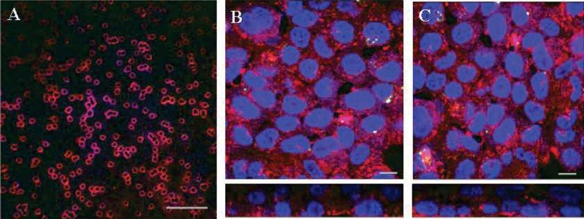
VOL. 54, 2010 PHARMACOKINETICS AND PHARMACODYNAMICS OF AmB 3433 bovine serum (FBS), ascorbic acid, heparin, hydrocortisone, human endothelial LAmB, 1, 10, 50, and 150 mg/liter; and ABLC, 1, 10, 50, and 150 mg/liter; these growth factor, vascular endothelial growth factor, human fibroblast growth factor encompassed the known concentration-effect relationships of each compound B, and R3-insulin-like growth factor-1 according to the manufacturer’s instruc- defined from preliminary experiments. Endovascular fluid, endothelial cells, and tions, to produce endothelial growth medium (EGM-2). Amphotericin B and alveolar cells were sampled at 3, 6, 12, 18, 24, and 30 h posttreatment for each gentamicin, which are ordinarily constituents of EGM-2, were omitted. A549 formulation. cells were grown to near-confluence in EBM-2 supplemented with 10% FBS Pharmacodynamics and pharmacodynamic modeling. The antifungal effect of (Lonza Biologics, Slough, United Kingdom) without antimicrobial agents. the three formulations was estimated by measuring the concentrations of galac- HPAEC and A549 cells were harvested using warmed 0.25% trypsin-EDTA tomannan in alveolar lavage and endovascular fluid using a commercially avail- (Lonza Biologics), centrifuged, and resuspended in warmed fresh media. Final able double-sandwich enzyme-linked immunosorbent assay (ELISA) (Platelia densities of HPAEC and A549 cells of 1 ⫻ 106 and 5 ⫻ 105 cells/ml, respectively, Aspergillus kit; Bio-Rad Laboratories). The use of the green fluorescent protein were obtained by serial dilution in their respective growth media. transformant also enabled visualization of hyphal invasion using confocal mi- One hundred microliters of the HPAEC suspension was seeded onto the croscopy and therefore provided a complementary measure of fungal burden and bottom of polyester Transwell inserts (6.5-mm-diameter membrane, 3-m pores; antifungal effect. Concentration-response relationships were initially defined fol- Corning Life Sciences, Lowell, MA). The inverted inserts were incubated for 2 h lowing 24 h of drug exposure (i.e., 30 h postinoculation). Based on preliminary before being righted and placed in 24-well tissue culture plates containing 600 l dose-finding experiments, the initial concentration ranges of DAmB, LAmB, and EGM. One hundred microliters of EBM-2 supplemented with 10% FBS was ABLC within the endovascular compartment were 0 to 2 mg/liter, 0 to 75 then added to the upper chamber and incubated at 37°C in humidified 5% CO2 mg/liter, and 0 to 75 mg/liter, respectively. for 24 h. To construct the bilayer, spent medium from the upper chamber was The concentrations of galactomannan in the alveolar and endovascular com- removed, and 100 l of the A549 suspension was added and incubated for 2 h to partments were modeled using an inhibitory sigmoid maximal effect (Emax) enable cellular adhesion to the polyester membrane. Medium from the alveolar model that took the following form: Effect ⫽ Econ ⫺ (Emax ⫻ ExpH)/(EC50H ⫹ compartment was then removed to create an air-liquid interface, and the inserts ExpH), where Econ is the fungal burden in the absence of drug, Emax is the were transferred to trays containing 600 l EGM-2. Medium in the endovascular asymptotic reduction in fungal burden induced by antifungal drug exposure, Exp compartment was changed daily, and any medium that accumulated in the is a measure of drug exposure (i.e., initial concentration of drug in endovascular alveolar compartment was also removed. fluid or AUC/MIC ratio), EC50 is the concentration resulting in half-maximal Cellular confluence was assessed at time points between zero and 120 h by effect, and H is the slope (or Hill) function. The model was implemented within placing 100 l of 1% (wt/vol) dextran blue (Sigma-Aldrich, Exeter, United the identification module of the pharmacokinetic program ADAPT II (9), and Kingdom) in the alveolar compartment and placing inserts in tissue culture plates the data were weighted by the inverse of the observed variance. containing 600 l of warmed phosphate-buffered saline (PBS; Invitrogen Ltd., The inhibitory sigmoid Emax model was used to identify concentrations that Renfrew, United Kingdom) for 2 h. Transgression of dye through the cellular produced zero, EC20, EC50, and near-maximal effect (0, 0.1, 0.15, and 2.0 mg/ bilayer and into the endothelial compartment containing PBS was measured liter, respectively, for DAmB; 0, 1, 2, and 75 mg/liter, respectively, for LAmB and spectrophotometrically using a wavelength of 620 nm. To ensure that drug ABLC). These values were subsequently used to examine the time course of penetration was not due to direct cellular toxicity and loss of cellular confluence, antifungal effect following a range of effective and ineffective drug concentra- the transgression of dextran blue was assessed at the end of experiments using tions. For these experiments, samples of alveolar lavage and endovascular fluid the highest concentration for each formulation of AmB. were collected at 0, 12, 18, 24, 26, and 30 h postinoculation, and the concentra- Organism, inoculation, and MICs. An A. fumigatus transformant expressing tion of galactomannan was determined. green fluorescent protein (GFP) was used for all experiments, as previously HPLC. Concentrations of amphotericin B were measured using high-perfor- described (15). Prior to each experiment, the organism was subcultured from mance liquid chromatography (HPLC) as previously described (15). Briefly, 100 beads to a potato dextrose agar slope (Oxoid, Basingstoke, United Kingdom) and l of each sample was analyzed using a C18 5-m column (Varian Ltd., Oxford, incubated at 37°C for 7 days. A conidial suspension was prepared by flooding the United Kingdom), Shimadzu SIL20A AutoSampler, LC 20AD pump, and SPD slope with 15 ml PBS (without Tween 80). The suspension was centrifuged at 20A UV/VIS detector (Shimadzu UK Ltd., Milton Keynes, United Kingdom). 1,000 ⫻ g, and the pellet resuspended in PBS; this process was repeated three Amphotericin B powder (Sigma Aldrich) was solubilized in 1:1 (vol/vol) meth- times. The final inoculum was prepared in cell culture medium EBM-2 without anol-dimethyl sulfoxide (DMSO), and a four-point standard curve prepared in FBS using a hemocytometer and checked with quantitative cultures. the respective matrix (i.e., media, PBS, cell suspension). Piroxicam (1 mg/liter) Experiments were performed 5 days after seeding A549 cells. Immediately was used as the internal standard. The dynamic range of the assay was 0.01 to 200 prior to inoculation, the medium in the endovascular compartment was changed mg/liter. to warmed EBM-2 supplemented with 2% FBS (i.e., without the additional Construction of sulforhodamine-labeled liposomes and confocal microscopy. growth factors that constitute EGM-2). One hundred microliters of a suspension Hydrogenated soy phosphatidylcholine (HSPC), cholesterol, distearoylphos- containing 1 ⫻ 104 conidia/ml was placed in the alveolar compartment. Six hours phatidylglycerol (DSPG), amphotericin B, and alpha-tocopherol were dissolved later, all fluid within the alveolar compartment was removed, and the inserts in a 2:1:0.8:0.4:0.01 molar ratio in a 1:1 mixture of methanol and chloroform (or, were transferred to fresh tissue culture plates containing 600 l of EBM-2 for placebo, the same formula without amphotericin B). Once all components supplemented with 2% FBS along with the desired concentration of DAmB, were dissolved, solvents were removed by evaporation under continuous nitrogen LAmB, or ABLC (i.e., drug was administered within the endovascular compart- flow. Residual solvent was removed by storing the container containing the ment). The 6-h delay in the administration of drug was based on a previous study material in a desiccator under vacuum for at least 48 h. The dried lipid was and mimics the treatment of early IPA (15). hydrated in a buffer containing 9% sucrose and 10 mM succinate at desired drug MICs for each of the clinical formulations were determined on three separate concentrations, and the hydrated material was processed through a high-shear occasions using National Committee for Clinical Laboratory Standards M38-A2 homogenizer to form liposomes. For sulforhodamine-labeled material, the buffer methodology (7). in this step was supplemented with sulforhodamine B at 600 mg/liter. Unen- Antifungal compounds. LAmB (Gilead Sciences, Cambridge, United King- trapped sulforhodamine was removed by ultrafiltration against a Millipore 100- dom) and DAmB (Bristol-Myers Squibb, Uxbridge, United Kingdom) were kDa polyethersulfone (PES) membrane. The resulting solution was filtered reconstituted with sterile water to produce stock solutions of 500 mg/liter. ABLC through a 0.2-m filter (also PES). Drug-containing preparations had a final pH (Cephalon, Hertfordshire, United Kingdom) was obtained as a lipid complex of 5.4 and were freeze-dried. Formulations without drug were stored as a liquid solution containing 500 mg/liter amphotericin B. and had a final pH of 6.4. Samples were confirmed to have a median particle size Collection of pharmacokinetic and pharmacodynamic data. The air-liquid of ⬍100 nm by dynamic light scattering. Amphotericin B concentration was interface model enabled sampling from a number of compartments relevant to confirmed by reversed-phase HPLC using a C18 column and isocratic elution the pathogenesis of IPA. The surface of the alveolar cells was washed with 300 against acetonitrile-methanol-2.5 mM EDTA (25:50:30 [vol-vol-vol]) and using l PBS to mimic a bronchoalveolar lavage, while the endovascular fluid was the USP standard. Sulforhodamine concentration was determined by UV-visible sampled directly to simulate a blood sample. Endothelial and alveolar epithelial (Vis) spectroscopy, and preparations contained between 0.1 mM and 1 mM cells were physically removed by abrading the polyester membrane with a mi- sulforhodamine. crobiological loop and then suspended in 300 l PBS. Liposomes were reconstituted in 6 ml sterile water per vial. Further dilutions Pharmacokinetic experiments. The pharmacokinetics of the three formula- were prepared in EBM-2 supplemented with 2% FBS. Six hundred microliters of tions within the endovascular fluid endothelial cells and alveolar cells were media containing LAmB-containing sulforhodamine-labeled liposomes (LAmB- defined in the presence of infection. The following amphotericin B concentra- Rho) or drug-free sulforhodamine-labeled liposomes (LPlac-Rho) was placed in tions for each formulation were studied: DAmB, 0.1, 0.5, 1.0, and 2.0 mg/liter; tissue culture plates, and membranes were transferred 6 h after conidial inocu-
3434 LESTNER ET AL. ANTIMICROB. AGENTS CHEMOTHER.
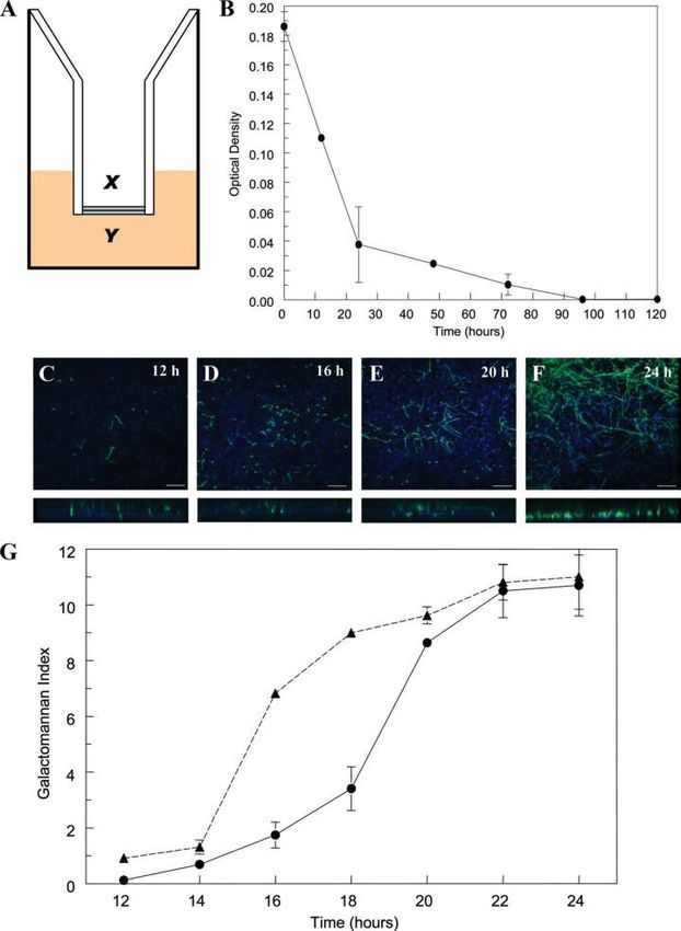
FIG. 1. (A) A schematic representation of the air-liquid interface (x, alveolar compartment; y, endovascular compartment); (B) the decline in
the transgression of dextran blue into the endovascular compartment as a function of time, reflecting the attainment of cellular confluence. (C to
F) Confocal images at 12, 16, 20, and 24 h postinoculation are shown; the lower panes depict the corresponding cross-sectional images. Scale bar,
10 m. (G) The kinetics of galactomannan in infected but untreated inserts. The solid line represents the time course of galactomannan in the
endovascular compartment, whereas the dashed line is the time course in the alveolar compartment. Data are means ⫾ standard deviations of three
inserts.
lation as described above for other AmB preparations. Samples taken from a Mathematical modeling. The total concentrations of amphotericin B associated
range of time points 0 to 24 h posttreatment were fixed using 4% paraformal- with each formulation were modeled using a population methodology which em-
dehyde (Sigma-Aldrich). Six hundred microliters of 5 g/ml 4⬘,6-diamidino-2- ployed the Big version of the program Nonparametric Adaptive Grid (BIG NPAG)
phenylindole solution (DAPI; Sigma-Aldrich) was instilled into the alveolar and (19). The movement of drug from the endovascular fluid into the endothelial and
endovascular compartment, incubated for 30 min at room temperature, and then alveolar cells was described using the following three inhomogeneous differential
washed twice with PBS. equations:
Confocal microscopy. A Nikon Eclipse C1-Plus inverted confocal microscope
(Nikon UK Limited, Surrey United Kingdom) with a 20⫻, 40⫻, or 100⫻ Apo-
XP(1) ⫽ R(1) ⫺ K12 ⫻ X(1) ⫹ K21 ⫻ X(2) (1)
chromat objective lens was used. Image z stacks with 0.3-m x-y pixel size and an
optical slice of 0.34- to 1.00-m thicknesses were collected and analyzed using
EZ-C1 FreeViewer (v3.9) software (Nikon UK Limited). XP(2) ⫽ K12 ⫻ X(1) ⫹ K32 ⫻ X(3) ⫺ K21 ⫻ X(2)⫺ K23 ⫻ X(2) (2)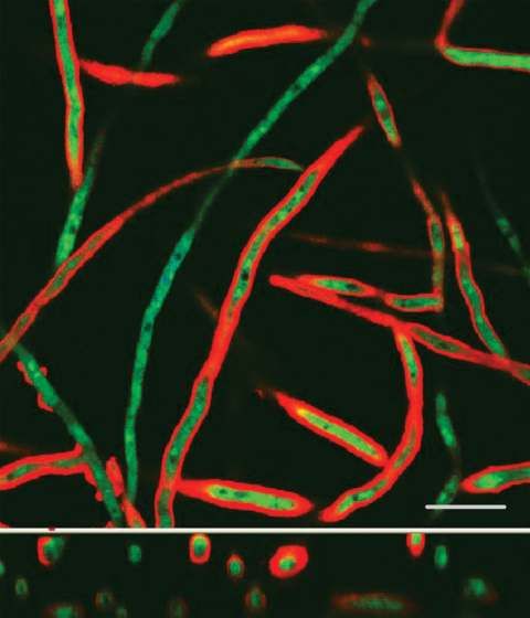
VOL. 54, 2010 PHARMACOKINETICS AND PHARMACODYNAMICS OF AmB 3435
FIG. 2. The pharmacokinetics of amphotericin B deoxycholate (DAmB), liposomal amphotericin (LAmB), and amphotericin B lipid complex
(ABLC) in the endovascular fluid (top row), endothelial cells (middle row), and alveolar epithelial cells (bottom row). Four concentrations are
displayed in each panel. Data are mean ⫾ standard deviation of three inserts.
XP(3) ⫽ K23 ⫻ X(2) ⫺ K32 ⫻ X(3) (3) RESULTS
Equations 1, 2, and 3 describe the movement of drug (XP) into and out of Formulation-specific MICs. The MICs from the three rep-
endovascular fluid, endothelial cells, and alveolar cells, respectively. R(1) repre- licate experiments for the three AmB formulations were as
sents the bolus injection of drug into the endovascular compartment; K12, K21,
follows: DAmB, 0.25, 0.25, and 0.5; ABLC, 0.125, 0.125, and
K23, and K32 are the first-order intercompartmental rate constants between
compartment 1 (endovascular fluid), compartment 2 (endothelial cells), and 0.125; LAmB, 0.25, 0.125, and 0.25; and LAmB-Rho, 0.25,
compartment 3 (alveolar cells). X(1), X(2), and X(3) represent the amount of 0.25, and 0.25 mg/liter. The MIC for pure amphotericin B was
drug (mg) in the respective compartments. The volume of each compartment 0.25 mg/liter on three separate occasions.
(liters) was estimated in the output equations that described the time course of
Air-liquid interface model. A schematic representation of
concentrations (not shown). The concentration of drug in the lavage fluid from
the surface of the (relatively dry) alveolar cells was not modeled because of the in vitro air-liquid model is shown in Fig. 1A. There was a
difficulties in accurately estimating the volume of this compartment. progressive decline in the extent that dextran blue traversed
The mean drug concentrations from each compartment from three inserts the cellular bilayer, becoming negligible 96 h post-seeding of
were modeled. The data were weighted by the inverse of the observed variance.
A549 cells (Fig. 1B). The addition of DAmB, LAmB, or ABLC
The fit of the model to the data was assessed using measures of precision and bias
along with the coefficient of determination (r2) and visual inspection of the did not affect the transgression of dextran blue across the
observed-versus-predicted relationships after the Bayesian step. To assess the bilayer (data not shown). Progressive hyphal growth and inva-
extent of drug penetration into each of the pharmacokinetic compartments, sion through the cellular bilayer were reflected by changes in
the mean parameter values were inserted into the simulation module of
galactomannan concentrations in both the alveolar and endo-
ADAPT II (9), and the AUC in each compartment was calculated by inte-
gration. The inhibitory sigmoid Emax model was refitted to the data using the vascular compartments (Fig. 1C to F). The kinetics of galac-
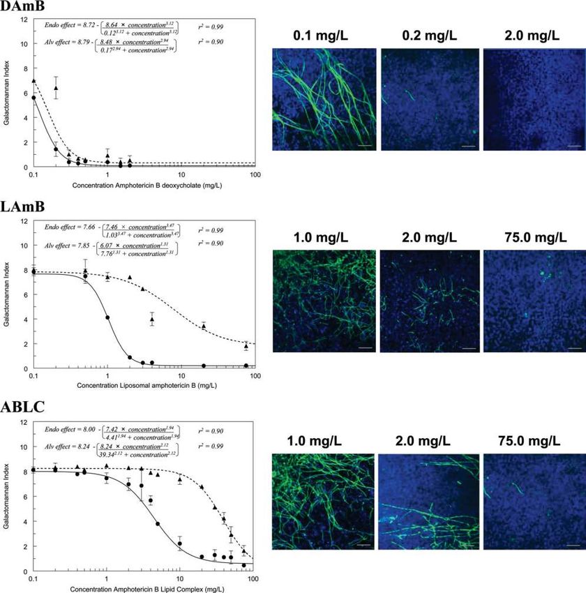
AUC/MIC ratio as the independent variable. tomannan concentrations in the alveolar and endovascular3436 LESTNER ET AL. ANTIMICROB. AGENTS CHEMOTHER. FIG. 3. Concentration-response relationships at 24 h posttreatment for amphotericin B deoxycholate (DAmB), liposomal amphotericin B (LAmB), and amphotericin B lipid complex (ABLC). Dashed and solid lines represent galactomannan concentrations in the alveolar lavage and endovascular fluid, respectively. The confocal images on the right enable the fungal biomass associated with various concentrations to be visualized. Data are means ⫾ standard deviations of three inserts. compartments were discordant, reflecting the time required for Pharmacokinetics. The concentration-time profiles of each hyphae to invade across the cellular bilayer (Fig. 1G). The of the AmB formulations are shown in Fig. 2. The concentra- kinetics of galactomannan in the endovascular compartment were tions of amphotericin B associated with each of the formula- mirrored by progressive hyphal invasion observed with confocal tions declined in the endovascular compartment throughout microscopy. Both galactomannan concentrations and confocal the experimental period. Concomitantly, concentrations of am- microscopy suggested that hyphae emerged within the endovas- photericin B in both the endothelial and alveolar cells rose cular compartment 14 to 16 h postinoculation (Fig. 1C to F). steeply and attained concentrations far in excess of those ob-
VOL. 54, 2010                                                            PHARMACOKINETICS AND PHARMACODYNAMICS OF AmB                                            3437

TABLE 1. The estimates for EC50 and EC90 quantified in terms of the initial concentration and the area under the concentration-time curve/
                                    MIC ratio (AUC/MIC) for each amphotericin B formulation
                                Amphotericin B deoxycholate                    Liposomal amphotericin B                         Amphotericin B lipid complex
       Parametera
                               Endovascular           Alveolar           Endovascular                Alveolar              Endovascular               Alveolar

Initial concn (mg/liter)
  EC50 (95% confidence        0.12 (0.11–0.13)    0.17 (0.13–0.19)      1.03 (0.94–1.12            7.76 (2.40–13.19)      4.41 (4.13–4.70)       39.34 (35.61–43.03)
      interval)
  EC90                        0.24                0.36                  1.94                     41.48                   13.63                  110.82

AUC/MIC
 EC50 (95% confidence 6.4 (6.14–6.66)             8.35 (7.99–8.71)     67.73 (65.72–69.75)      505.2 (384.7–625.7)     210.2 (201.6–218.8) 1,874 (1,751–1,996)
   interval)
 EC90                 12.93                      17.62               127.46                   2,705.41                  651.3                 5,280.56
  a
    Initial concentration is the concentration of the respective formulations (mg/liter) administered to the endothelial compartment 6 h postinoculation; EC50 and EC90
represent the drug exposure required to induce 50 and 90% of the maximal antifungal effect, respectively.

served with the endovascular compartment. The highest con-                            shown in Fig. 7. For a given concentration, the relative pene-
centrations were seen with LAmB and ABLC.                                             tration into the endothelial cells was 305.2, 206.0, and 438.1
   Concentration-response relationships for amphotericin B                            and into the alveolar cells was 150.1, 14.3, and 21.4 for DAmB,
formulations. The concentration-response relationships for                            LAmB, and ABLC, respectively.
the three formulations were initially examined in detail 24 h
posttreatment (30 h postinoculation) (Fig. 3). DAmB induced
                                                                                                                       DISCUSSION
a steep exposure-response relationship in both the alveolar and
endovascular compartments over a very narrow concentration                               Despite intensive efforts, IPA remains a rapidly progressive
range. In contrast, LAmB and ABLC induced more languid                                and frequently lethal infectious syndrome for which there are
concentration-response relationships, with incomplete sup-                            relatively few therapeutic options. The lipid preparations of
pression of galactomannan in the alveolar compartment,                                amphotericin B have an established role for the treatment of
even at high concentrations. Marked differences in the con-                           proven and suspected Aspergillus infection (8, 25), and they are
centration-response and AUC-response relationships were ob-                           consistently less toxic than DAmB (3, 27). The air-liquid in-
served for DAmB, LAmB, and ABLC. These differences are                                terface model of the human alveolus provides an ideal con-
reflected in the estimates for EC50 and EC90, despite compa-                          struct to examine the extent of penetration for each of the
rable MICs (Table 1). These findings were further supported                           amphotericin B formulations into various subcompartments of
by confocal microscopy, which also demonstrated progressive                           the lung that are relevant for events in the pathogenesis of
reduction in hyphal penetration with increasing concentrations                        early IPA.
of all formulations (Fig. 3).                                                            The formulation of amphotericin B has a profound effect on
   Similar findings were apparent with the temporal galac-                            the disposition, elimination, and activity of the pure compound
tomannan concentrations for each of the formulations. For                             in both laboratory animal models and in humans (1, 2). Our
each compound, increasing drug concentrations resulted in                             results also suggest that the specific formulation influences the
increased suppression of fungal growth (Fig. 4). Consistent                           pharmacokinetic and pharmacodynamic relationships at a cel-
with previous experiments at a single time point, LAmB and                            lular level. Consistent with a previous study, the MICs of am-
ABLC both resulted in incomplete suppression of galactoman-                           photericin B and lipid formulations of amphotericin B were
nan in the alveolar compartment.                                                      comparable (17). Despite this, the exposure-response relation-
   Interaction of LAmB with human cells and hyphae. Confo-                            ships for DAmB versus LAmB and ABLC were strikingly dif-
cal images of endovascular fluid showed the size and spherical                        ferent, with estimates for EC50 for both lipid formulations
structure of free sulforhodamine-labeled liposomes (LAmB-                             being significantly higher than those for DAmB. These results
Rho and LPlac-Rho). Over the subsequent 24 h, there was                               suggest that the MIC of lipid preparations transmits relatively
progressive accumulation of sulforhodamine within the cellu-                          little information that can be used to predict exposure-re-
lar bilayer (Fig. 5). The majority of sulforhodamine was asso-                        sponse relationships in experimental systems and in humans.
ciated with endothelial cells with a less intense signal emanat-                         The formulation-specific pharmacodynamic relationships
ing from the alveolar epithelial cells (Fig. 5). There was an                         likely result from thermodynamic constraints that govern the
intense signal from sulforhodamine-encapsulated hyphae as                             release and transfer of pure amphotericin B from its lipid
they penetrated into the endovascular compartment (Fig. 6).                           carrier. Despite the likely interaction between liposomes and
   Mathematical modeling. The estimates of the means and                              human cells, the majority of pure amphotericin B probably
dispersion for the model parameters are summarized in Table                           remains preferentially complexed within the liposome, rather
2. The fit of the model to the data for all three formulations                        than engaging with mammalian lipids, because the former is a
was acceptable, with r2 values of ⬎99% for the endovascular                           more energetically favorable state. Indeed, this phenomenon is
fluid and endothelial cells, and ⬎89% for the alveolar cells for                      used for quality control processes for the manufacture of
observed-versus-predicted values, and with acceptable mea-                            LAmB. The incubation of defective liposomes with human
sures of precision and bias. The ratio of AUCs in the endo-                           erythrocytes results in the release of amphotericin B at
thelial and alveolar cells to those of the endovascular fluid is                      relatively low drug concentrations, and this causes potas-
3438 LESTNER ET AL. ANTIMICROB. AGENTS CHEMOTHER. FIG. 4. Serially collected pharmacodynamic data for the endovascular and alveolar compartments following exposure to a range of concentrations of amphotericin B deoxycholate (DAmB), liposomal amphotericin B (LAmB), and amphotericin B lipid complex (ABLC). Data are means ⫾ standard deviations of three inserts. sium leakage from damaged erythrocyte membranes (16). In which amphotericin B within LAmB engages hyphae and the presence of a higher-affinity target (i.e., ergosterol in conidia of Aspergillus within the lung during invasive pulmo- fungal membranes), it becomes energetically more favorable nary aspergillosis: (i) the liposome directly engages with hy- for amphotericin B to disengage from the liposome and aggre- phae (without initially interacting with host cells) as they in- gate within fungal membranes. Presumably these conclusions vade into the lumen of blood vessels and (ii) LAmB initially are also (qualitatively) applicable to ABLC, although this is associates with mammalian cell membranes and is then able to difficult to confirm in the absence of a similarly labeled prep- engage with conidia or hyphae as they, in turn, interact with aration. host cells in the course of invasive infection. Both points of Our results suggest two potential cellular mechanisms by cellular interaction permit the selective transfer of amphoter-
VOL. 54, 2010 PHARMACOKINETICS AND PHARMACODYNAMICS OF AmB 3439
FIG. 5. Confocal images of host cell-liposome interactions. (A) Liposomes within the endovascular compartment immediately after adminis-
tration; scale bar, 1 m. (B) Endothelial aspect of the cellular bilayer demonstrating the interaction of sulforhodamine-labeled liposomes devoid
of amphotericin B with endothelial cells after incubation for 24 h; scale bar, 10 m. (C) Endothelial aspect of the cellular bilayer demonstrating
the interaction of sulforhodamine-labeled liposomes containing amphotericin B; scale bar, 10 m. The second rows of images in panels B and C
represent a cross-sectional view of the cellular bilayer showing a gradient of liposomal deposition from endothelial cells (lower layer) to alveolar
cells (upper layer).
icin B from liposomes to fungal cell membranes. Amphotericin The location of sulforhodamine is within the enclosed aqueous
B remains strongly associated with the liposome of LAmB, space of the LAmB liposome structure, and one expects sul-
such that only a small fraction of “free” drug (⬍1%) is detect- forhodamine to be carried with the liposome so long as the
able in biological media (4). These hypotheses require an as- liposome remains intact.
sumption that the sulforhodamine used to visualize liposomal All three formulations achieve significantly higher concen-
distribution largely remains preferentially complexed within trations in the cells than in the contiguous endovascular fluid.
the liposomal structure and does not itself freely distribute. The proportional concentrations in endothelial and alveolar
cells relative to endovascular fluid are similar to those ob-
served with epithelial lining fluid and pulmonary alveolar mac-
rophages relative to serum in rabbits (11). The potential sig-
nificance of drug concentrations in tissues is inextricably linked
with the pathogenesis of the infectious diseases (13). IPA be-
gins with the inhalation of conidia into the lung, a proportion
of which contact alveolar epithelial cells and undergo phago-
cytosis (12). The resulting phagolysosome is enveloped by the
cell membrane. If this membrane contains amphotericin B,
then this may contribute to the killing of conidia within the
phagolysosome. Similarly, following germination, hyphae in-
vade through cell membranes of alveolar and endothelial cells,
at which time they are exposed to high concentrations of drug,
which may lead to hyphal damage and death.
The immune status of the host and innate immunological
effectors are critical determinants of the outcome of invasive
fungal diseases (14). A limitation of this in vitro model is that
the potential antifungal effect of immune effector cells, such as
circulating monocytes or pulmonary alveolar macrophages,
cannot be estimated. Furthermore, the reticuloendothelial sys-
tem may influence the serum concentration-time profile of the
lipid formulations of amphotericin B (26). Relatively large
concentrations of amphotericin B from LAmB and ABLC ac-
cumulate within pulmonary alveolar macrophages. This addi-
tional compartment provides another point of potential cellu-
lar interaction between fungal elements and amphotericin
within the alveolus. Empty liposomes have an antifungal effect
in laboratory animal models of IPA that presumably reflects
FIG. 6. The interaction of hyphae with sulforhodamine B-labeled
liposomal amphotericin B. The lower image represents a cross-sec- immunomodulation that is favorable for the host (20). A fur-
tional image. Scale bar, 10 m. ther possibility is that immune effector cells laden with drug3440 LESTNER ET AL. ANTIMICROB. AGENTS CHEMOTHER.
TABLE 2. Parameter means and standard deviations from the mathematical model describing the
pharmacokinetics of each of the amphotericin B formulations
Mean (SD)
Parameter, unitsa
b
DAmB LAmBc ABLCd
K12, h⫺1 4.97 (4.31) 3.01 (1.51) 5.65 (7.21)
K21, h⫺1 19.83 (8.21) 20.4 (5.36) 16.79 (8.46)
K23, h⫺1 7.13 (10.28) 1.404 (1.84) 1.04 (1.15)
K32, h⫺1 14.99 (7.83) 20.07 (6.73) 20.12 (7.59)
Vendo fluid, liters 7.89 ⫻ 10⫺4 (9.92 ⫻ 10⫺5) 7.60 ⫻ 10⫺4 (1.15 ⫻ 10⫺4) 6.9 ⫻ 10⫺4 (1.18 ⫻ 10⫺4)
Vendo cells, liters 6.47 ⫻ 10⫺7 (2.06 ⫻ 10⫺7) 5.56 ⫻ 10⫺7 (1.13 ⫻ 10⫺7) 5.29 ⫻ 10⫺7 (2.79 ⫻ 10⫺7)
Valv cells, liters 5.81 ⫻ 10⫺7 (2.80 ⫻ 10⫺7) 7.47 ⫻ 10⫺7 (2.65 ⫻ 10⫺7) 6.48 ⫻ 10⫺7 (2.71 ⫻ 10⫺7)
a
K12, K21, K23, and K32 are the first-order intercompartmental rate constants describing the movement of drug between compartment 1 (endovascular fluid),
compartment 2 (endothelial cells), and compartment 3 (alveolar cells). Vendo fluid is the volume of the endovascular compartment in liters, Vendo cells is the volume of
the entire endothelial cell layer, and Valv cells is the volume of the entire alveolar cell layer.
b
DAmB, amphotericin B deoxycholate.
c
LAmB, liposomal amphotericin B.
d
ABLC, amphotericin B lipid complex.
may traffic into the alveolar space and deliver drug to the extend and improve their clinical utility and serve as a valuable
fungal target. This so-called “dump truck” phenomenon also mechanism to further improve the safety and efficacy for a
has been postulated to account for the action of the macrolides persistently lethal infectious syndrome.
because this class of compounds does not achieve high con-
centrations within the epithelial lining fluid of the lung (10). ACKNOWLEDGMENTS
Mehta and colleagues found that an alternative formulation of This work was funded, in part, by the Fungal Research Trust and
amphotericin B accumulated in inflammatory peritoneal cells Gilead Sciences. William Hope is supported by a National Institute of
after intravenous administration of fluorescence-labeled Health Research (NIHR) Clinician Scientist Fellowship. This study
was supported, in part, by the intramural research program of the
L-AmB, suggesting that macrophages play an important role in National Cancer Institute, National Institutes of Health.
the transport of the intravenously administered lipid formula- We thank David Perlin and Jill Adler-Moore for their comments
tion to inflammatory sites (21). This transport process is likely and insights. We also thank Robert Fernandez (Biomedical Imaging,
during invasive pulmonary aspergillosis but has not been well Faculty of Life Sciences, University of Manchester, United Kingdom)
and Huy Pham and Tark Bunch (Gilead Sciences, San Dimas) for their
studied to any extent. The extent to which these kinetics may outstanding technical assistance.
be altered in neutropenic versus nonneutropenic hosts, in
which the inflammatory responses differ markedly (5), merits REFERENCES
further study. 1. Adler-Moore, J. P., and R. T. Proffitt. 2008. Amphotericin B lipid prepara-
tions: what are the differences? Clin. Microbiol. Infect. 14(Suppl. 4):25–36.
An improved understanding of the intrapulmonary pharma- 2. Andes, D., N. Safdar, K. Marchillo, and R. Conklin. 2006. Pharmacokinetic-
cokinetics and pharmacodynamics of AmB is required for the pharmacodynamic comparison of amphotericin B (AMB) and two lipid-
associated AMB preparations, liposomal AMB and AMB lipid complex, in
design of optimal (and innovative) dosing regimens for the murine candidiasis models. Antimicrob. Agents Chemother. 50:674–684.
prevention and treatment of IPA. Furthermore, novel formu- 3. Bates, D. W., L. Su, D. T. Yu, G. M. Chertow, D. L. Seger, D. R. Gomes, E. J.
lations of existing antifungal compounds may provide a way to Dasbach, and R. Platt. 2001. Mortality and costs of acute renal failure
associated with amphotericin B therapy. Clin. Infect. Dis. 32:686–693.
4. Bekersky, I., R. M. Fielding, D. E. Dressler, J. W. Lee, D. N. Buell, and T. J.
Walsh. 2002. Plasma protein binding of amphotericin B and pharmacokinet-
ics of bound versus unbound amphotericin B after administration of intra-
venous liposomal amphotericin B (AmBisome) and amphotericin B deoxy-
cholate. Antimicrob. Agents Chemother. 46:834–840.
5. Berenguer, J., M. C. Allende, J. W. Lee, K. Garrett, C. Lyman, N. M. Ali, J.
Bacher, P. A. Pizzo, and T. J. Walsh. 1995. Pathogenesis of pulmonary
aspergillosis. Granulocytopenia versus cyclosporine and methylprednisolone-
induced immunosuppression. Am. J. Respir. Crit. Care Med. 152:1079–1086.
6. Bermudez, L. E., F. J. Sangari, P. Kolonoski, M. Petrofsky, and J. Goodman.
2002. The efficiency of the translocation of Mycobacterium tuberculosis
across a bilayer of epithelial and endothelial cells as a model of the alveolar
wall is a consequence of transport within mononuclear phagocytes and in-
vasion of alveolar epithelial cells. Infect. Immun. 70:140–146.
7. CLSI/NCCLS. 2002. Reference method for broth dilution antifungal suscep-
tibility testing of filamentous fungi. Approved standard M38-A. National
Committee for Clinical Laboratory Standards, Wayne, PA.
8. Cornely, O. A., J. Maertens, M. Bresnik, R. Ebrahimi, A. J. Ullmann, E.
Bouza, C. P. Heussel, O. Lortholary, C. Rieger, A. Boehme, M. Aoun, H. A.
Horst, A. Thiebaut, M. Ruhnke, D. Reichert, N. Vianelli, S. W. Krause, E.
Olavarria, and R. Herbrecht. 2007. Liposomal amphotericin B as initial
therapy for invasive mold infection: a randomized trial comparing a high-
loading dose regimen with standard dosing (AmBiLoad trial). Clin. Infect.
Dis. 44:1289–1297.
FIG. 7. The proportional penetration of AmB in the endothelial 9. D’Argenio, D. Z., and A. Schumitzky. 1997. ADAPT II. A program for
and alveolar cells compared with endovascular fluid for DAmB, simulation, identification, and optimal experimental design. User manual.
LAmB, and ABLC). The estimates for the area under the concentra- Biomedical Simulations Resource, University of Southern California, Los
tion-time curve are derived from the mathematical model. Angeles, CA. http://bmsr.esc.edu/.VOL. 54, 2010 PHARMACOKINETICS AND PHARMACODYNAMICS OF AmB 3441
10. Drusano, G. L. 2005. Infection site concentrations: their therapeutic impor- models, p. 389–394. In Proceedings of the 14th IEEE Symposium on Com-
tance and the macrolide and macrolide-like class of antibiotics. Pharmaco- puter-Based Medical Systems. IEEE Computer Society, Bethesda, MD.
therapy 25:150S–158S. 20. Lewis, R. E., G. Chamilos, R. A. Prince, and D. P. Kontoyiannis. 2007.
11. Groll, A. H., C. A. Lyman, V. Petraitis, R. Petraitiene, D. Armstrong, D. Pretreatment with empty liposomes attenuates the immunopathology of
Mickiene, R. M. Alfaro, R. L. Schaufele, T. Sein, J. Bacher, and T. J. Walsh. invasive pulmonary aspergillosis in corticosteroid-immunosuppressed mice.
2006. Compartmentalized intrapulmonary pharmacokinetics of amphoteri- Antimicrob. Agents Chemother. 51:1078–1081.
cin B and its lipid formulations. Antimicrob. Agents Chemother. 50:3418– 21. Mehta, R. T., T. J. McQueen, A. Keyhani, and G. Lopez-Berestein. 1994.
3423. Phagocyte transport as mechanism for enhanced therapeutic activity of li-
12. Hope, W. W. 2009. Invasion of the alveolar-capillary barrier by Aspergillus posomal amphotericin B. Chemotherapy 40:256–264.
spp.: therapeutic and diagnostic implications for immunocompromised pa- 22. Nivoix, Y., M. Velten, V. Letscher-Bru, A. Moghaddam, S. Natarajan-Ame,
tients with invasive pulmonary aspergillosis. Med. Mycol. 47(Suppl. 1):S291– C. Fohrer, B. Lioure, K. Bilger, P. Lutun, L. Marcellin, A. Launoy, G. Freys,
S298. J. P. Bergerat, and R. Herbrecht. 2008. Factors associated with overall and
13. Hope, W. W., and G. L. Drusano. 2009. Antifungal pharmacokinetics and attributable mortality in invasive aspergillosis. Clin. Infect. Dis. 47:1176–
pharmacodynamics: bridging from the bench to bedside. Clin. Microbiol. 1184.
Infect. 15:602–612. 23. Pappas, P. G., C. A. Kauffman, D. Andes, D. K. Benjamin, Jr., T. F. Ca-
14. Hope, W. W., G. L. Drusano, C. B. Moore, A. Sharp, A. Louie, T. J. Walsh, landra, J. E. Edwards, Jr., S. G. Filler, J. F. Fisher, B. J. Kullberg, L.
D. W. Denning, and P. A. Warn. 2007. Effect of neutropenia and treatment Ostrosky-Zeichner, A. C. Reboli, J. H. Rex, T. J. Walsh, and J. D. Sobel.
2009. Clinical practice guidelines for the management of candidiasis: 2009
delay on the response to antifungal agents in experimental disseminated
update by the Infectious Diseases Society of America. Clin. Infect. Dis.
candidiasis. Antimicrob. Agents Chemother. 51:285–295.
48:503–535.
15. Hope, W. W., M. J. Kruhlak, C. A. Lyman, R. Petraitiene, V. Petraitis, A.
24. Walsh, T. J., E. J. Anaissie, D. W. Denning, R. Herbrecht, D. P. Kontoyian-
Francesconi, M. Kasai, D. Mickiene, T. Sein, J. Peter, A. M. Kelaher, J. E.
nis, K. A. Marr, V. A. Morrison, B. H. Segal, W. J. Steinbach, D. A. Stevens,
Hughes, M. P. Cotton, C. J. Cotten, J. Bacher, S. Tripathi, L. Bermudez,
J. A. van Burik, J. R. Wingard, and T. F. Patterson. 2008. Treatment of
T. K. Maugel, P. M. Zerfas, J. R. Wingard, G. L. Drusano, and T. J. Walsh. aspergillosis: clinical practice guidelines of the Infectious Diseases Society of
2007. Pathogenesis of Aspergillus fumigatus and the kinetics of galactoman- America. Clin. Infect. Dis. 46:327–360.
nan in an in vitro model of early invasive pulmonary aspergillosis: implica- 25. Walsh, T. J., R. W. Finberg, C. Arndt, J. Hiemenz, C. Schwartz, D. Boden-
tions for antifungal therapy. J. Infect. Dis. 195:455–466. steiner, P. Pappas, N. Seibel, R. N. Greenberg, S. Dummer, M. Schuster, and
16. Jensen, J. M., C. R. Skenes, T. H. Bunch, N. Weissman, N. Amirghahari, A. J. S. Holcenberg. 1999. Liposomal amphotericin B for empirical therapy in
Satorius, K. L. Moynihan, and C. G. S. Eley. 1999. Determination of the patients with persistent fever and neutropenia. National Institute of Allergy
relative toxicity of amphotericin B formulations: a red blood cell potassium and Infectious Diseases Mycoses Study Group. N. Engl. J. Med. 340:764–
release assay. Drug Delivery 6:81–88. 771.
17. Lass-Flörl, C., A. Mayr, S. Perkhofer, G. Hinterberger, J. Hausdorfer, C. 26. Walsh, T. J., J. L. Goodman, P. Pappas, I. Bekersky, D. N. Buell, M. Roden,
Speth, and M. Fille. 2008. Activities of antifungal agents against yeasts and J. Barrett, and E. J. Anaissie. 2001. Safety, tolerance, and pharmacokinetics
filamentous fungi: assessment according to the methodology of the Euro- of high-dose liposomal amphotericin B (AmBisome) in patients infected
pean Committee on Antimicrobial Susceptibility Testing. Antimicrob. with Aspergillus species and other filamentous fungi: maximum tolerated
Agents Chemother. 52:3637–3641. dose study. Antimicrob. Agents Chemother. 45:3487–3496.
18. Latgé, J. P. 1999. Aspergillus fumigatus and aspergillosis. Clin. Microbiol. 27. Wingard, J. R., P. Kubilis, L. Lee, G. Yee, M. White, L. Walshe, R. Bowden,
Rev. 12:310–350. E. Anaissie, J. Hiemenz, and J. Lister. 1999. Clinical significance of neph-
19. Leary, R., R. Jelliffe, A. Schumitzky, and M. van Guilder. 2001. An adaptive rotoxicity in patients treated with amphotericin B for suspected or proven
grid, non-parametric approach to pharmacokinetic and dynamic (PK/PD) aspergillosis. Clin. Infect. Dis. 29:1402–1407.You can also read