Banting Lecture 2004 The Pathobiology of Diabetic Complications A Unifying Mechanism
←
→
Page content transcription
If your browser does not render page correctly, please read the page content below
Banting Lecture 2004
The Pathobiology of Diabetic Complications
A Unifying Mechanism
Michael Brownlee
PIECES OF THE PUZZLE
The general features of hyperglycemia-induced tissue
I
t’s a great honor to join the exceptional club of damage are shown schematically in Fig. 1. The DCCT
Banting Award winners, many of whom were my (Diabetes Control and Complications Trial) and the
role models and mentors. In addition, giving the UKPDS (U.K. Prospective Diabetes Study) established that
Banting Lecture also has a very personal meaning to hyperglycemia, shown on the far left of the figure, is the
me, because without Frederick Banting, I would have died initiating cause of the diabetic tissue damage that we see
from type 1 diabetes when I was 8 years old. However, it clinically, shown on the far right (1,2). Although this
was already apparent at the time I was diagnosed that for too process is modified by both genetic determinants of indi-
many people like me, Banting’s discovery of insulin only vidual susceptibility, shown in the top box, and by inde-
allowed them to live just long enough to develop blindness, pendent accelerating factors such as hypertension, shown
renal failure, and coronary disease. For example, when I in the bottom box, today I will concentrate on the inner
started college, the American Diabetes Association’s Diabe- boxes, the mechanisms that mediate the tissue-damaging
effects of hyperglycemia.
tes Textbook had this to say to my parents: “The person with
When I refer to the tissue-damaging effects of hypergly-
type 1 diabetes can be reassured that it is highly likely that he
cemia, of course, I mean damage to a particular subset of
will live at least into his 30s.” Not surprisingly, my parents did cell types: capillary endothelial cells in the retina, mesan-
not find this particularly reassuring. gial cells in the renal glomerulus, and neurons and
At the same time we were reading this in 1967, however, Schwann cells in peripheral nerves. What is distinct about
the first basic research discovery about the pathobiology these cells that makes them so vulnerable to hyperglyce-
of diabetic complications had just been published in mia? We know that in diabetes, hyperglycemia is bathing
Science the previous year. In my Banting Lecture today, I all the cells of every tissue. So why does damage occur
am thus going to tell you a scientific story that is also only in the few cell types involved in diabetic complica-
profoundly personal. tions? The answer is that most cells are able to reduce the
I’ve divided my talk into three parts. The first part is called transport of glucose inside the cell when they are exposed
“pieces of the puzzle,” and in it I describe what was learned to hyperglycemia, so that their internal glucose concentra-
about the pathobiology of diabetic complications starting tion stays constant. In contrast, the cells damaged by
with that 1966 Science paper and continuing through the end
of the 1990s. In the second part, I present a unified mecha-
nism that links together all of the seemingly unconnected
pieces of the puzzle. Finally, in the third part, I focus on three
examples of novel therapeutic approaches for the prevention
and treatment of diabetic complications, which are all based
on the new paradigm of a unifying mechanism for the
pathogenesis of diabetic complications.
From the Departments of Medicine and Pathology, Albert Einstein College of
Medicine, Bronx, New York.
Address correspondence and reprint requests to Michael Brownlee, Anita
and Jack Saltz Professor of Diabetes Research, Departments of Medicine and
Pathology, Albert Einstein College of Medicine, F-531 1300 Morris Park Ave.,
Bronx, NY 10461-1602. E-mail: brownlee@aecom.yu.edu.
AGE, advanced glycation end product; eNOS, endothelial nitric oxide
synthase; FFA, free fatty acid; GAPDH, glyceraldehyde-3 phosphate dehydro-
genase; MnSOD, manganese superoxide dismutase; NFB, nuclear factor B;
PARP, poly(ADP-ribose) polymerase; PKC, protein kinase C; ROS, reactive
oxygen species; SOD, superoxide dismutase; TCA, tricarboxylic acid; UCP,
uncoupling protein.
© 2005 by the American Diabetes Association. FIG. 1. General features of hyperglycemia-induced tissue damage.
DIABETES, VOL. 54, JUNE 2005 1615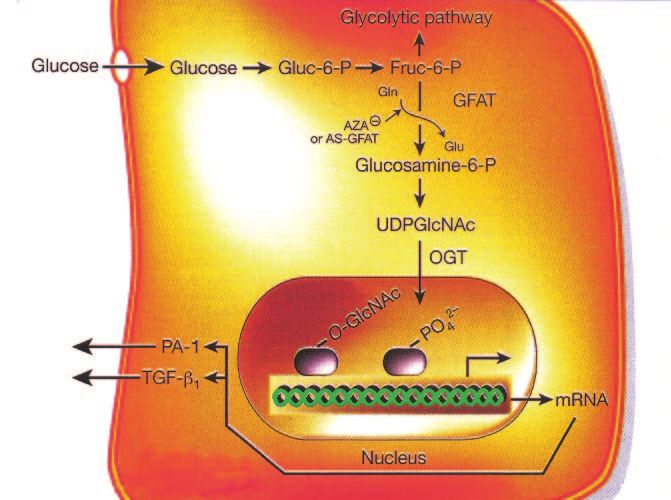
PATHOBIOLOGY OF DIABETIC COMPLICATIONS
FIG. 2. Hyperglycemia increases flux through the
polyol pathway. From Brownlee M: Biochemistry and
molecular cell biology of diabetic complications. Na-
ture 414:813– 820, 2001.
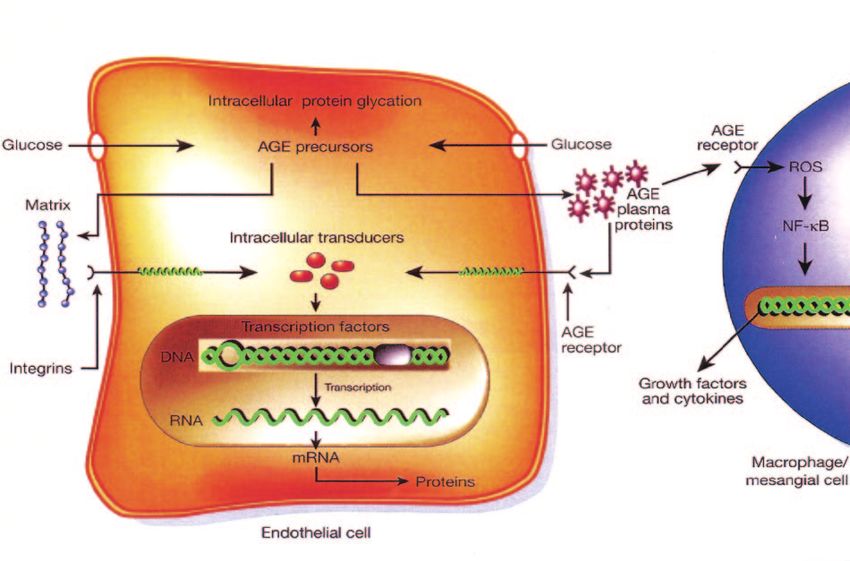
hyperglycemia are those that cannot do this efficiently Intracellular production of AGE precursors. The sec-
(3,4). Thus, diabetes selectively damages cells, like endo- ond discovery listed on my pieces of the puzzle list is the
thelial cells and mesangial cells, whose glucose transport intracellular production of AGE precursors. As shown
rate does not decline rapidly as a result of hyperglycemia, schematically in Fig. 3, these appear to damage cells by
leading to high glucose inside the cell. This is important, three mechanisms. The first mechanism, shown at the top
because it tells us that the explanation for what causes of the endothelial cell, is the modification of intracellular
complications must involve mechanisms going on inside proteins including, most importantly, proteins involved in
these cells, rather than outside. the regulation of gene transcription (8,9 and M.B., unpub-
The first such mechanism that was discovered was the lished observations). The second mechanism, shown on
polyol pathway and increased polyol pathway flux, de- the left, is that these AGE precursors can diffuse out of the
scribed in peripheral nerve in the 1966 Science paper I cell and modify extracellular matrix molecules nearby
refer to above (5). This was the first piece of the puzzle. (10), which changes signaling between the matrix and the
Then, ⬃10 years later, in the late 1970s, a second piece of cell and causes cellular dysfunction (11). The third mech-
the puzzle emerged: increased formation of advanced anism, shown on the right of Fig. 3, is that these AGE
glycation end products (AGEs). In the late 1980s and early precursors diffuse out of the cell and modify circulating
1990s, a third piece of the puzzle was discovered: hyper- proteins in the blood such as albumin. These modified
glycemia-induced activation of protein kinase C (PKC) iso- circulating proteins can then bind to AGE receptors and
forms. And in the late 1990s, a fourth piece of the puzzle was activate them, thereby causing the production of inflam-
discovered: increased hexosamine pathway flux and conse- matory cytokines and growth factors, which in turn cause
quent overmodification of proteins by N-acetylglucosamine. vascular pathology (12–21). Again, how do we know that
I’m going to very briefly review each of these, because I this piece of the puzzle is really important? From many
think each one is an important piece of the puzzle. animal studies such as one done by Hans-Peter Hammes
Increased flux through the polyol pathway. The polyol (22), showing that that pharmacologic inhibition of AGEs
pathway, shown schematically in Fig. 2, focuses on the prevents late structural changes of experimental diabetic
enzyme aldose reductase. Aldose reductase normally has retinopathy.
the function of reducing toxic aldehydes in the cell to PKC activation. The third mechanism on my “pieces of
inactive alcohols, but when the glucose concentration in the puzzle” list was the PKC pathway. In this pathway,
the cell becomes too high, aldose reductase also reduces shown schematically in Fig. 4, hyperglycemia inside the
that glucose to sorbitol, which is later oxidized to fructose. cell increases the synthesis of a molecule called diacyl-
In the process of reducing high intracellular glucose to glycerol, which is a critical activating cofactor for the
sorbitol, the aldose reductase consumes the cofactor classic isoforms of protein kinase-C, -, -␦, and -␣ (23–26).
NADPH (6). But as shown in Fig. 2, NADPH is also the When PKC is activated by intracellular hyperglycemia, it
essential cofactor for regenerating a critical intracellular has a variety of effects on gene expression, examples of
antioxidant, reduced glutathione. By reducing the amount which are shown in the row of open boxes in Fig. 4. In
of reduced glutathione, the polyol pathway increases each case, the things that are good for normal function are
susceptibility to intracellular oxidative stress. decreased and the things that are bad are increased. For
How do we know that this piece of the puzzle is really example, starting from the far left of Fig. 4, the vasodilator
important? From studies like one conducted by Ron Enger- producing endothelial nitric oxide (NO) synthase (eNOS)
man and Tim Kern (7), in which diabetic dogs were treated is decreased, while the vasoconstrictor endothelin-1 is
for 5 years with an aldose reductase inhibitor. Nerve conduc- increased. Transforming growth factor- and plasminogen
tion velocity in the diabetic dogs decreased over time as it activator inhibitor-1 are also increased. At the bottom of
does in patients. In contrast, in diabetic dogs treated with an the figure, the row of black boxes lists the pathological
aldose reductase inhibitor, the diabetes-induced defect in effects that may result from the abnormalities in the open
nerve conduction velocity was prevented. boxes (26 –30). Again, how do we know that this piece of
1616 DIABETES, VOL. 54, JUNE 2005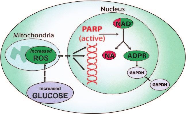
M. BROWNLEE

FIG. 3. Increased production of AGE precursors and its pathologic consequences. From Brownlee M: Biochemistry and molecular cell biology of
diabetic complications. Nature 414:813– 820, 2001.

the puzzle is really important? We know that this is                   ever, some of that fructose-6-phosphate gets diverted into
important from many animal studies such as several                     a signaling pathway in which an enzyme called GFAT
published by George King, showing that inhibition of PKC               (glutamine:fructose-6 phosphate amidotransferase) con-
prevented early changes in the diabetic retina and kidney              verts the fructose-6 phosphate to glucosamine-6 phos-
(27,31,32).                                                            phate and finally to UDP (uridine diphosphate) N-acetyl
Increased hexosamine pathway activity. The last                        glucosamine.
mechanism on the “pieces of the puzzle” list was increased                What happens after that is the N-acetyl glucosamine gets
flux through the hexosamine pathway. As shown schemat-                 put onto serine and threonine residues of transcription
ically in Fig. 5, when glucose is high inside a cell, most of          factors, just like the more familiar process of phosphory-
that glucose is metabolized through glycolysis, going first            lation, and overmodification by this glucosamine often
to glucose-6 phosphate, then fructose-6 phosphate, and                 results in pathologic changes in gene expression (33–35).
then on through the rest of the glycolytic pathway. How-               For example, in Fig. 5, increased modification of the

FIG. 4. Consequences of hyperglycemia-induced activation of PKC.

DIABETES, VOL. 54, JUNE 2005                                                                                                          1617
PATHOBIOLOGY OF DIABETIC COMPLICATIONS
FIG. 5. Hyperglycemia increases flux through the
hexosamine pathway. From Brownlee M: Biochem-
istry and molecular cell biology of diabetic com-
plications. Nature 414:813– 820, 2001.
transcription factor Sp1 results in increased expression of (ROS) (36,39). Although hyperglycemia had been associ-
transforming growth factor-1 and plasminogen activator ated with oxidative stress in the early 1960s (40), neither
inhibitor-1, both of which are bad for diabetic blood the underlying mechanism that produced it nor its conse-
vessels (36). Again, how do we know that this piece of the quences for pathways of hyperglycemic damage were
puzzle is really important? Although this piece of the known.
puzzle is the most recent to be recognized as a factor in the How does hyperglycemia increase superoxide pro-
pathogenesis of diabetic complications, it has been shown duction by the mitochondria? There are four protein
to play a role both in hyperglycemia-induced abnormalities complexes in the mitochondrial electron transport chain,
of glomerular cell gene expression (33) and in hypergly- called complex I, II, III, and IV (Fig. 6). When glucose is
cemia-induced cardiomyocyte dysfunction in cell culture metabolized through the tricarboxylic acid (TCA) cycle, it
(37). In carotid artery plaques from type 2 diabetic sub- generates electron donors. The main electron donor is
jects, modification of endothelial cell proteins by the NADH, which gives electrons to complex I. The other
hexosamine pathway is also significantly increased (38). electron donor generated by the TCA cycle is FADH2,
formed by succinate dehydrogenase, which donates elec-
A UNIFIED MECHANISM trons to complex II. Electrons from both these complexes
Over 13,000 articles published since 1966 seemed to show are passed to coenzyme Q, and then from coenzyme Q they
that all of these pieces of the puzzle were important in the are transferred to complex III, cytochrome-C, complex IV,
pathogenesis of diabetic complications, yet two things and finally to molecular oxygen, which they reduce to
suggested that something major was missing. First, there water.
was no apparent common element linking these mecha- The electron transport system is organized in this way
nisms to each other. Second, clinical trials of inhibitors of so that the level of ATP can be precisely regulated. As
these pathways in patients were all disappointing. Trying electrons are transported from left to right in Fig. 6, some
to make sense of all this, we hypothesized that all of these of the energy of those electrons is used to pump protons
mechanisms were in fact linked to a common upstream across the membrane at complexes I, III, and IV. This
event and that the failure to block all of the downstream generates what is in effect a voltage across the mitochon-
pathways could explain the disappointing clinical trials drial membrane. The energy from this voltage gradient
with single-pathway inhibitors. What we discovered is that drives the synthesis of ATP by ATP synthase (41,42).
all of these different pathogenic mechanisms do reflect a Alternatively, uncoupling proteins (UCPs; Fig. 6) can bleed
single hyperglycemia-induced process and that this single down the voltage gradient to generate heat as a way of
unifying process is the overproduction of superoxide by keeping the rate of ATP generation constant.
the mitochondrial electron transport chain. That’s what happens in normal cells. In contrast, in
We began by asking the following question: What pro- diabetic cells with high glucose inside, there is more
cesses are increased by intracellular hyperglycemia in glucose being oxidized in the TCA cycle, which in effect
cells whose glucose transport rate is not downregulated by pushes more electron donors (NADH and FADH2) into the
hyperglycemia but not increased in cells whose glucose electron transport chain. As a result of this, the voltage
transport rate is downregulated by hyperglycemia? gradient across the mitochondrial membrane increases
We discovered that a consistent differentiating feature until a critical threshold is reached. At this point, electron
common to all cell types that are damaged by hyperglyce- transfer inside complex III is blocked (43), causing the
mia is an increased production of reactive oxygen species electrons to back up to coenzyme Q, which donates the
1618 DIABETES, VOL. 54, JUNE 2005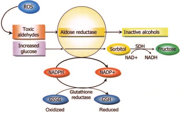
M. BROWNLEE
FIG. 6. Hyperglycemia-induced production of superoxide by the mitochondrial electron transport chain.
electrons one at a time to molecular oxygen, thereby
generating superoxide (Fig. 6). The mitochondrial isoform
of the enzyme superoxide dismutase degrades this oxygen
free radical to hydrogen peroxide, which is then converted
to H2O and O2 by other enzymes.
How do we know that this really happens in cells known
to be damaged by hyperglycemia? First, we looked at such
cells with a dye that changes color with increasing voltage
of the mitochondrial membrane and found that intracellu-
lar hyperglycemia did indeed increase the voltage across
the mitochondrial membrane above the critical threshold
necessary to increase superoxide formation (44). In order
to prove that the electron transport chain indeed produces
superoxide by the mechanism we proposed, we examined
the effect of overexpressing either UCP-1 or manganese
superoxide dismutase (MnSOD) on hyperlglycemia-in-
duced ROS generation (Fig. 7A). Hyperglycemia caused a
big increase in production of ROS. In contrast, an identical
level of hyperglycemia does not increase ROS at all when
we also collapse the mitochondrial voltage gradient by
overexpressing UCP (39). Similarly, hyperglycemia does
not increase ROS at all when we degrade superoxide by
overexpressing the enzyme MnSOD. These data demon-
strate two things. First, the UCP effect shows that the
mitochondrial electron transport chain is the source of the
hyperglycemia-induced superoxide. Second, the MnSOD
effect shows that the initial ROS formed is indeed super-
oxide.
To confirm these findings by an independent experimen-
tal approach, we depleted mitochondrial DNA from nor-
mal endothelial cells to form so-called 0 endothelial cells,
which lack a functional mitochondrial electron transport
chain (Fig. 7B). When the mitochondrial electron transport
chain is removed, the effect of hyperglycemia on ROS
production is completely lost (M.B., unpublished observa-
FIG. 7. Hyperglycemia-induced ROS production in wild type and 0
tions). Similarly, in 0 endothelial cells, hyperglycemia endothelial cells. A: From ref. 39. B: From M.B., M.H. Zou, X. Du, D.
completely fails to activate the polyol pathway, AGE Edelstein, unpublished data.
DIABETES, VOL. 54, JUNE 2005 1619PATHOBIOLOGY OF DIABETIC COMPLICATIONS
FIG. 8. Mitochondrial overpro-
duction of superoxide activates
four major pathways of hypergly-
cemic damage by inhibiting
GAPDH. From Brownlee M: Bio-
chemistry and molecular cell bi-
ology of diabetic complications.
Nature 414:813– 820, 2001.
formation, PKC, or the hexosamine pathway (M.B., unpub- when GAPDH activity is inhibited? As shown in Fig. 8, the
lished observations). level of all the glycolytic intermediates that are upstream
We also looked at the effect of either UCP-1 overexpres- of GAPDH increase. Increased levels of the upstream
sion or MnSOD overexpression on each of these four glycolytic metabolite glyceraldehyde-3-phosphate acti-
hyperglycemia-activated pathways. Hyperglycemia did not vates two of the four pathways. It activates the AGE
activate any of the pathways when either the voltage pathway because the major intracellular AGE precursor
gradient across the mitochondrial membrane was col- methylglyoxal is formed from glyceraldehyde-3 phosphate.
lapsed by UCP-1 or when the superoxide produced was It also activates the classic PKC pathway, since the
degraded by MnSOD (39). We have verified all of these activator of PKC, diacylglycerol, is also formed from
endothelial cell culture experiments in transgenic mice glyceraldehyde-3 phosphate.
that overexpress MnSOD (M.B., unpublished observa- Further upstream, levels of the glycolytic metabolite
tions). When wild-type animals are made diabetic, all four fructose-6 phosphate increase, which increases flux
of the pathways are activated in tissues where diabetic through the hexosamine pathway, where fructose-6 phos-
complications occur. In contrast, when MnSOD transgenic phate is converted by the enzyme GFAT to UDP–N-
mice are made diabetic, there is no activation of any of the acetylglucosamine (UDP-GlcNAc). Finally, inhibition of
four pathways. GAPDH increases intracellular levels of the first glycolytic
In endothelial cells, PKC also activates nuclear factor metabolite, glucose. This increases flux through the polyol
B (NFB), a transcription factor that itself activates pathway, where the enzyme aldose reductase reduces it,
many proinflammatory genes in the vasculature. As ex- consuming NADPH in the process. To rule out the possi-
pected, hyperglycemia-induced PKC activation is pre- bility that any other hyperglycemia-induced metabolic
vented by either UCP-1 or MnSOD, both in cells and in change accounted for these observations, we inhibited
animals. GAPDH activity using antisense DNA, so that the level of
Importantly, inhibition of hyperglycemia-induced super- GAPDH activity in cells cultured in 5 mmol/l (90 mg/dl)
oxide overproduction using a transgenic approach (su- glucose was reduced to that normally found in cells
peroxide dismutase [SOD]) also prevents long-term exposed to hyperglycemia. With reduced GAPDH activity
experimental diabetic nephropathy in the best animal the only perturbation in these cells, the activity of each of
model of this complication: the db/db diabetic mouse (45). the four pathways in 5 mmol/l glucose was elevated to the
Hyperglycemia-induced mitochondrial superoxide same extent as that induced by hyperglycemia (46).
production activates the four damaging pathways by Hyperglycemia-induced mitochondrial superoxide
inhibiting GAPDH. Figure 8 shows the scheme we pro- production inhibits GAPDH by activating poly(ADP-
posed for how all of these data link together. This model is ribose) polymerase. At this point, we knew that the way
based on a critical observation we made: diabetes in that hyperglycemia activates the four major pathways of
animals and patients, and hyperglycemia in cells, all hyperglycemic damage is by the overproduction of super-
decrease the activity of the key glycolytic enzyme glycer- oxide by the mitochondria, which then decreases GAPDH
aldehyde-3 phosphate dehydrogenase (GAPDH). Inhibi- activity. In a test tube, superoxide itself directly inacti-
tion of GAPDH activity by hyperglycemia does not occur vates GAPDH, but only at concentrations that far exceed
when mitochondrial overproduction of superoxide is pre- levels found in hyperglycemic cells. We therefore asked
vented by either UCP-1 or MnSOD (36). What happens the following question: In cells and tissues, how do ROS
1620 DIABETES, VOL. 54, JUNE 2005M. BROWNLEE
FIG. 9. ROS-induced DNA damage activates PARP and
modifies GAPDH.
actually inhibit GAPDH activity? To answer this question, ADP-ribose. PARP then proceeds to make polymers of
we looked for chemical modifications of GAPDH that ADP-ribose, which accumulate on GAPDH and other nu-
correlated with the hyperglycemia-induced decrease in clear proteins. What is GAPDH doing in the nucleus?
GAPDH activity. As shown in Fig. 9, we found that Although GAPDH is commonly thought to reside exclu-
hyperglycemia-induced superoxide inhibits GAPDH activ- sively in the cytosol, in fact it normally shuttles in and out
ity in vivo by modifying the enzyme with polymers of of the nucleus, where it plays a critical role in DNA repair
ADP-ribose (46). By inhibiting mitochondrial superoxide (47,48).
production with either UCP-1 or MnSOD, we prevented A schematic summary showing the elements of the
both modification of GAPDH by ADP-ribose and reduction unified mechanism of hyperglycemia-induced cellular
of its activity by hyperglycemia. Most importantly, both damage is shown in Fig. 10. When intracellular hypergly-
modification of GAPDH by ADP-ribose and reduction of its cemia develops in target cells of diabetic complications, it
activity by hyperglycemia were also prevented by a spe- causes increased mitochondrial production of ROS. The
cific inhibitor of poly(ADP-ribose) polymerase (PARP), ROS cause strand breaks in nuclear DNA, which activate
the enzyme that makes these polymers of ADP-ribose. This PARP. PARP then modifies GAPDH, thereby reducing its
established a cause-and-effect relationship between PARP activity. Finally, decreased GAPDH activity activates the
activation and the changes in GAPDH. polyol pathway, increases intracellular AGE formation,
How does hyperglycemia activate PARP, a DNA repair activates PKC and subsequently NFB, and activates hex-
enzyme that’s found exclusively in the nucleus, and how osamine pathway flux.
does nuclear PARP get together with GAPDH, a glycolytic How does the unifying mechanism explain diabetic
enzyme commonly thought to reside in the cytosol? macrovascular disease? We now had a unifying mecha-
Normally, PARP resides in the nucleus in an inactive nism that explains the pathogenesis of diabetic microvas-
form, waiting for DNA damage to activate it (Fig. 9). When cular disease. But then we thought: What about diabetic
increased intracellular glucose generates increased ROS in macrovascular disease? In contrast to diabetic microvas-
the mitochondria, we found that these free radicals induce cular disease, data from the UKPDS have shown that
DNA strand breaks, thereby activating PARP. Both hyper- hyperglycemia is not the major determinant of diabetic
glycemia-induced processes are prevented by either UCP-1 macrovascular disease. For microvascular disease end
or MnSOD (46). Once activated, PARP splits the NAD⫹ points, there is a nearly 10-fold increase in risk as HbA1c
molecule into its two component parts: nicotinic acid and increases from 5.5 to 9.5%. In contrast, over the same
FIG. 10. The unifying mechanism of hyperglycemia-
induced cellular damage.
DIABETES, VOL. 54, JUNE 2005 1621PATHOBIOLOGY OF DIABETIC COMPLICATIONS
FIG. 11. Insulin resistance causes mitochondrial
overproduction of ROS in macrovascular endothe-
lial cells by increasing FFA flux and oxidation.
From Hofmann S, Brownlee M: Biochemistry and
molecular cell biology of diabetic complications: a
unifying mechanism. In Diabetes Mellitus: A Fun-
damental and Clinical Text. 3rd ed. LeRoith D,
Taylor SI, Olefsky JM, Eds. Philadelphia, Lippin-
cott Williams & Wilkins, p. 1441–1457, 2004.
HbA1c range, macrovascular risk increases only about the new paradigm of a unifying mechanism for the patho-
twofold (2). genesis of diabetic complications.
If hyperglycemia is not the major determinant of dia- Transketolase activators. The first new class of poten-
betic macrovascular disease, what about the constellation tial therapeutic agents is transketolase activators. This
of risk factors associated with insulin resistance and the concept originated from an obvious feature of the unifying
metabolic syndrome? In order to separate increased mac- mechanism (Fig. 8). When increased superoxide inhibits
rovascular disease risk due to insulin resistance and its GAPDH activity, the glycolytic intermediates above the
associated abnormalities from increased risk due to hyper- enzyme accumulate and are then shunted into the four
glycemia, the San Antonio Heart Study studied men with- pathways of hyperglycemic damage. We noted that two of
out diabetes or impaired glucose tolerance (49). Not these glycolytic intermediates, fructose-6-phosphate and
surprisingly, high insulin resistance increases cardiovas- glyceraldehyde-3-phosphate, are also the final products of
cular risk by 2.5-fold. What is surprising, though, is that the transketolase reaction, which is the rate-limiting en-
after adjustment for 11 known cardiovascular risk factors, zyme in another metabolic pathway, the pentose phos-
including LDL, HDL, triglycerides, systolic blood pressure, phate pathway (50). Although this pathway is traditionally
and smoking, the insulin-resistant subjects still had a taught as flowing from pentose phosphates to glycolytic
twofold increased risk of cardiovascular disease. This intermediates, in fact it can also flow from glycolytic
suggests that a large part of cardiovascular disease risk intermediates to pentose phosphates, depending on the
due to insulin resistance reflects a previously unappreci- concentrations of substrate presented to the transketolase
ated consequence of insulin resistance. enzyme. Since we know that in diabetes, the concentration
Using both cell culture and animal models, we found of the glycolytic intermediates is high, we reasoned that if
that the unappreciated consequence of insulin resistance we could activate transketolase, then we could decrease
is increased free fatty acid (FFA) flux from adipocytes into the concentration of these two glycolytic metabolites and
arterial endothelial cells, shown schematically in Fig. 11. thus divert their flux away from three of the damaging
In macrovascular, but not in microvascular endothelial pathways normally activated by hyperglycemia.
cells, we found that this increased flux results in increased But how could we activate transketolase? Since this
FFA oxidation by the mitochondria. Since both -oxida- enzyme requires the vitamin thiamine as a cofactor, we
tion of fatty acids and oxidation of FFA-derived acetyl CoA tried different thiamine derivatives and measured their
by the TCA cycle generate the same electron donors effects. While thiamine itself only activated transketolase
(NADH and FADH2) generated by glucose oxidation, in- ⬃25% in arterial endothelial cells, the thiamine derivative
creased FFA oxidation causes mitochondrial overproduc- called benfotiamine activated transketolase 250% in arte-
tion of ROS by exactly the same mechanism described rial endothelial cells. Based on such cell culture experi-
above for hyperglycemia. And, as with hyperglycemia, this ments, we treated diabetic rats for 9 months with
FFA-induced increase in ROS activates the same damaging benfotiamine and then evaluated the effect of this treat-
pathways: AGEs, PKC, the hexosamine pathway (GlcNAc), ment in the retina. After 9 months of diabetes, there was a
and NFB. In insulin-resistant nondiabetic animals mod- threefold increase in hexosamine pathway activity. In
els, inhibition of either FFA release from adipocytes or contrast, in diabetic animals treated with benfotiamine,
FFA oxidation in arterial endothelium prevents the in- there was a complete prevention of hexosamine pathway
creased production of ROS and its damaging effects (M.B., activation. The results were identical for diabetes-induced
unpublished observations). increases in intracellular AGE formation, PKC activation,
While more work certainly needs to be done, these data and NFB activation. Most importantly, benfotiamine
support a major role for the unifying mechanism in the treatment completely prevented the major structural le-
pathogenesis of diabetic macrovascular, as well as micro- sion of both human nonproliferative retinopathy and ex-
vascular, complications. perimental diabetic retinopathy: acellular capillaries (50).
PARP inhibitors. The second new class of potential
NOVEL THERAPEUTIC APPROACHES therapeutic agents based on the unified mechanism is
In the last part of my talk, I’m going to describe three PARP inhibitors. Since we had shown that increased
examples of novel therapeutic approaches for the preven- superoxide produced by the mitochondria in response to
tion and treatment of diabetic complications, all based on both hyperglycemia and increased FFA activates PARP,
1622 DIABETES, VOL. 54, JUNE 2005M. BROWNLEE
FIG. 12. Excess superoxide indepen-
dently inhibits activity of two critical
antiatherogenic enzymes without involve-
ment of the four pathways of hyperglyce-
mic damage. From refs. 51 and 52.
and that this PARP activation then modifies and inhibits ide. However, conventional antioxidants are unlikely to do
GAPDH (Fig. 9), we predicted that that PARP inhibition this effectively. Why? Because conventional antioxidants
would block the four major pathways of hyperglycemic neutralize reactive oxygen molecules on a one-for-one
damage that are activated by GAPDH inhibition. In cul- basis, while hyperglycemia-induced overproduction of su-
tured endothelial cells, a specific PARP inhibitor prevents peroxide is a continuous process. What is needed, then, is
hyperglycemia-induced activation of PKC, NFB, intracel- a new type of antioxidant, a catalytic antioxidant, such as
lular AGE formation, and the hexosamine pathway (46). In an SOD/catalase mimetic (53), that works continuously
long-term experimental diabetes, treatment with a PARP like the enzymes for which these compounds are named.
inhibitor also completely prevented the major structural Hyperglycemia-induced reactive oxygen overproduction
lesion of both human nonproliferative retinopathy and directly reduces eNOS activity in diabetic aortas by 65%.
experimental diabetic retinopathy: acellular capillaries However, when these diabetic animals are treated with an
(H.-P. Hammes, M.B., unpublished observations). SOD/catalase mimetic, there is no reduction in activity of
Catalytic antioxidants. Although the four damaging this antiatherogenic enzyme. Similarly, but more dramati-
pathways, which I have described as “pieces of the puz- cally, hyperglycemia-induced reactive oxygen overproduc-
zle,” have been the major focus of complications research tion directly reduces prostacyclin synthase activity in
over the past 40 years, it is important to recognize that diabetic aortas by 95%. Treatment of these diabetic ani-
excess superoxide itself can also directly inhibit critical mals with an SOD/catalase mimetic completely prevents
endothelial enzymes without any involvement of these diabetes-induced oxidative inactivation of aortic prostacy-
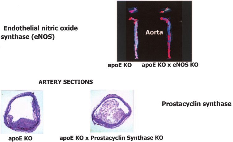
four mechanisms. Two of these enzymes that are particu- clin synthase.
larly important for vascular biology are eNOS and prosta- These data strongly suggest that therapeutic correction
cyclin synthase (Fig. 12). Both are dramatically inhibited of diabetes-induced superoxide production may be a pow-
in diabetic patients and diabetic animals. erful new approach for preventing diabetic complications.
eNOS is a very important antiatherogenic enzyme with
great relevance to diabetic macrovascular disease. This is
illustrated in Fig. 12. The upper panel of Fig. 12 shows that CONCLUSION: PERSONAL REFLECTIONS
in the aorta of a standard mouse model of atherosclerosis, In this Banting Lecture, I’ve told you a scientific story
the apoE knockout mouse, increased lipid-rich atheroscle- about diabetic complications. I’d like to close by telling
rotic lesions (red staining) are evident by week 16. How- you a human story about diabetic complications. Paul
ever, when the apoE knockout mouse is crossed with an Abercombie was an acquaintance of mine. Like all of us,
eNOS knockout mouse, the early atherosclerotic lesions he had hopes, fears, and dreams about the future. But
are nearly doubled simply by loss of eNOS activity (51). unfortunately, Paul was different from many of us here
Prostacyclin synthase is another critical endothelial because he also had type 1 diabetes. He did very well for
antiatherosclerotic enzyme. At the bottom of Fig. 12, a number of years, but then developed one complication
long-term mature atherosclerotic plaques are shown in a after another, and finally, he died.
vessel from an apoE knockout mouse and in a vessel from Like Paul, I and my family have struggled with diabetes
an apoE knockout mouse crossed with a prostacyclin for nearly all of my life. That struggle occupied a large part
synthase knockout mouse. The increase in lesion volume of my childhood, where everything I ate was weighed and
caused by lack of prostacyclin synthase is dramatic and calculated on a gram scale, and the doses of the few
very relevant to diabetic macrovascular disease (52). insulins that were then available were adjusted without
To prevent direct oxidative inactivation of these key the ability to know what my blood glucose values were.
enzymes, we must directly reduce the amount of superox- My parents did this despite the fact that most doctors
DIABETES, VOL. 54, JUNE 2005 1623PATHOBIOLOGY OF DIABETIC COMPLICATIONS
believed that hyperglycemia had nothing to do with the and p90 to 80K-H membrane proteins. Proc Natl Acad Sci U S A
93:11047–11052, 1996
pathogenesis of diabetic complications, because my par-
13. Neeper M, Schmidt AM, Brett J, Yan SD, Wang F, Pan YC, Elliston K, Stern
ents believed that abnormal glucose must be bad. I am D, Shaw A: Cloning and expression of a cell surface receptor for advanced
here today, and have accomplished what I have, in large glycosylation end products of proteins. J Biol Chem 267:14998 –15004,
part because of them. As a doctor, I’ve also had to struggle 1992
with the additional burden of knowing too much about 14. Smedsrod B, Melkko J, Araki N, Sano H, Horiuchi S: Advanced glycation
diabetic complications. Both my wife Karen and my sister end products are eliminated by scavenger-receptor-mediated endocytosis
in hepatic sinusoidal Kupffer and endothelial cells. Biochem J 322:567–573,
Martha taught me how to fully appreciate each day, 1997
despite my knowledge, and they both continue to fill my 15. Vlassara H, Li YM, Imani F, Wojciechowicz D, Yang Z, Liu FT, Cerami A:
life with joy and grace. Identification of galectin-3 as a high-affinity binding protein for advanced
Today, I have never been more excited, more optimistic, glycation end products (AGE): a new member of the AGE-receptor
or more certain that we can, and will, solve the remaining complex. Mol Med 1:634 – 646, 1995
16. Abordo EA, Thornalley PJ: Synthesis and secretion of tumour necrosis
important scientific questions about diabetic complica- factor-alpha by human monocytic THP-1 cells and chemotaxis induced by
tions, so that all people with diabetes, now and in the human serum albumin derivatives modified with methylglyoxal and glu-
future, can live full lives free of the fears and uncertainties cose-derived advanced glycation endproducts. Immunol Lett 58:139 –147,
that Paul Abercrombie knew all too well. 1997
17. Doi T, Vlassara H, Kirstein M, Yamada Y, Striker GE, Striker LJ: Receptor-
specific increase in extracellular matrix production in mouse mesangial
ACKNOWLEDGMENTS cells by advanced glycosylation end products is mediated via platelet-
derived growth factor. Proc Natl Acad Sci U S A 89:2873–2877, 1992
None of my own work or the work cited in this lecture
18. Kirstein M, Aston C, Hintz R, Vlassara H: Receptor-specific induction of
would be possible without the generous support of the insulin-like growth factor I in human monocytes by advanced glycosylation
National Institutes of Health, the American Diabetes As- end product-modified proteins. J Clin Invest 90:439 – 446, 1992
sociation, and the Juvenile Diabetes Research Foundation. 19. Schmidt AM, Hori O, Chen JX, Li JF, Crandall J, Zhang J, Cao R, Yan SD,
I want to thank all of my collaborators over the years Brett J, Stern D: Advanced glycation endproducts interacting with their
and my many colleagues, both local and all over the world, endothelial receptor induce expression of vascular cell adhesion mole-
cule-1 (VCAM-1) in cultured human endothelial cells and in mice: a
who have been and continue to be a constant source of potential mechanism for the accelerated vasculopathy of diabetes. J Clin
inspiration and insight. Invest 96:1395–1403, 1995
20. Skolnik EY, Yang Z, Makita Z, Radoff S, Kirstein M, Vlassara H: Human and
rat mesangial cell receptors for glucose-modified proteins: potential role in
REFERENCES kidney tissue remodelling and diabetic nephropathy. J Exp Med 174:931–
1. The Diabetes Control and Complications Trial Research Group: The effect 939, 1991
of intensive treatment of diabetes on the development and progression of 21. Vlassara H, Brownlee M, Manogue KR, Dinarello CA, Pasagian A: Cachec-
long-term complications in insulin-dependent diabetes mellitus. N Engl tin/TNF and IL-1 induced by glucose-modified proteins: role in normal
J Med 329:977–986, 1993 tissue remodeling. Science 240:1546 –1548, 1988
2. UK Prospective Diabetes Study (UKPDS) Group: Intensive blood-glucose 22. Hammes HP, Martin S, Federlin K, Geisen K, Brownlee M: Aminoguanidine
control with sulphonylureas or insulin compared with conventional treat- treatment inhibits the development of experimental diabetic retinopathy.
ment and risk of complications in patients with type 2 diabetes (UKPDS Proc Natl Acad Sci U S A 88:11555–11558, 1991
33). Lancet 352:837– 853, 1998 23. Koya D, King GL: Protein kinase C activation and the development of
3. Kaiser N, Sasson S, Feener EP, Boukobza-Vardi N, Higashi S, Moller DE, diabetic complications. Diabetes 47:859 – 866, 1998
Davidheiser S, Przybylski RJ, King GL: Differential regulation of glucose 24. DeRubertis FR, Craven PA: Activation of protein kinase C in glomerular
transport and transporters by glucose in vascular endothelial and smooth cells in diabetes: mechanisms and potential links to the pathogenesis of
muscle cells. Diabetes 42:80 – 89, 1993 diabetic glomerulopathy. Diabetes 43:1– 8, 1994
4. Heilig CW, Concepcion LA, Riser BL, Freytag SO, Zhu M, Cortes P: 25. Xia P, Inoguchi T, Kern TS, Engerman RL, Oates PJ, King GL: Character-
Overexpression of glucose transporters in rat mesangial cells cultured in a ization of the mechanism for the chronic activation of diacylglycerol-
normal glucose milieu mimics the diabetic phenotype. J Clin Invest protein kinase C pathway in diabetes and hypergalactosemia. Diabetes
96:1802–1814, 1995 43:1122–1129, 1994
5. Gabbay KH, Merola LO, Field RA: Sorbitol pathway: presence in nerve and 26. Koya D, Jirousek MR, Lin YW, Ishii H, Kuboki K, King GL: Characterization
cord with substrate accumulation in diabetes. Science 151:209 –210, 1966 of protein kinase C beta isoform activation on the gene expression of
6. Lee AY, Chung SS: Contributions of polyol pathway to oxidative stress in transforming growth factor-beta, extracellular matrix components, and
diabetic cataract. FASEB J 13:23–30, 1999 prostanoids in the glomeruli of diabetic rats. J Clin Invest 100:115–126,
7. Engerman RL, Kern TS, Larson ME: Nerve conduction and aldose reduc- 1997
tase inhibition during 5 years of diabetes or galactosaemia in dogs. 27. Ishii H, Jirousek MR, Koya D, Takagi C, Xia P, Clermont A, Bursell SE, Kern
Diabetologia 37:141–144, 1994 TS, Ballas LM, Heath WF, Stramm LE, Feener EP, King GL: Amelioration of
8. Giardino I, Edelstein D, Brownlee M: Nonenzymatic glycosylation in vitro vascular dysfunctions in diabetic rats by an oral PKC beta inhibitor.
and in bovine endothelial cells alters basic fibroblast growth factor Science 272:728 –731, 1996
activity: a model for intracellular glycosylation in diabetes. J Clin Invest 28. Kuboki K, Jiang ZY, Takahara N, Ha SW, Igarashi M, Yamauchi T, Feener
94:110 –117, 1994 EP, Herbert TP, Rhodes CJ, King GL: Regulation of endothelial constitutive
9. Shinohara M, Thornalley PJ, Giardino I, Beisswenger P, Thorpe SR, nitric oxide synthase gene expression in endothelial cells and in vivo : a
Onorato J, Brownlee M: Overexpression of glyoxalase-I in bovine endo- specific vascular action of insulin. Circulation 101:676 – 681, 2000
thelial cells inhibits intracellular advanced glycation endproduct formation 29. Studer RK, Craven PA, Derubertis FR: Role for protein kinase C in the
and prevents hyperglycemia-induced increases in macromolecular endo- mediation of increased fibronectin accumulation by mesangial cells grown
cytosis. J Clin Invest 101:1142–1147, 1998 in high-glucose medium. Diabetes 42:118 –126, 1993
10. McLellan AC, Thornalley PJ, Benn J, Sonksen PH: Glyoxalase system in 30. Feener EP, Xia P, Inoguchi T, Shiba T, Kunisaki M, King GL: Role of protein
clinical diabetes mellitus and correlation with diabetic complications. Clin kinase C in glucose- and angiotensin II-induced plasminogen activator
Sci (Lond) 87:21–29, 1994 inhibitor expression. Contrib Nephrol 118:180 –187, 1996
11. Charonis AS, Reger LA, Dege JE, Kouzi-Koliakos K, Furcht LT, Wohlhueter 31. Bishara NB, Dunlop ME, Murphy TV, Darby IA, Sharmini Rajanayagam MA,
RM, Tsilibary EC: Laminin alterations after in vitro nonenzymatic glyco- Hill MA: Matrix protein glycation impairs agonist-induced intracellular
sylation. Diabetes 39:807– 814, 1990 Ca2⫹ signaling in endothelial cells. J Cell Physiol 193:80 –92, 2002
12. Li YM, Mitsuhashi T, Wojciechowicz D, Shimizu N, Li J, Stitt A, He C, 32. Koya D, Haneda M, Nakagawa H, Isshiki K, Sato H, Maeda S, Sugimoto T,
Banerjee D, Vlassara H: Molecular identity and cellular distribution of Yasuda H, Kashiwagi A, Ways DK, King GL, Kikkawa R: Amelioration of
advanced glycation endproduct receptors: relationship of p60 to OST-48 accelerated diabetic mesangial expansion by treatment with a PKC beta
1624 DIABETES, VOL. 54, JUNE 2005M. BROWNLEE
inhibitor in diabetic db/db mice, a rodent model for type 2 diabetes. FASEB a mechanism of production of reactive oxygen species in mitochondria.
J 14:439 – 447, 2000 FEBS Lett 416:15–18, 1997
33. Kolm-Litty V, Sauer U, Nerlich A, Lehmann R, Schleicher ED: High 44. Du XL, Edelstein D, Dimmeler S, Ju Q, Sui C, Brownlee M: Hyperglycemia
glucose-induced transforming growth factor beta1 production is mediated inhibits endothelial nitric oxide synthase activity by posttranslational
by the hexosamine pathway in porcine glomerular mesangial cells. J Clin modification at the Akt site. J Clin Invest 108:1341–1348, 2001
Invest 101:160 –169, 1998 45. DeRubertis FR, Craven PA, Melhem MF, Salah EM: Attenuation of renal
34. Sayeski PP, Kudlow JE: Glucose metabolism to glucosamine is necessary injury in db/db mice overexpressing superoxide dismutase: evidence for
for glucose stimulation of transforming growth factor-alpha gene tran- reduced superoxide–nitric oxide interaction. Diabetes 53:762–768, 2004
scription. J Biol Chem 271:15237–15243, 1996 46. Du X, Matsumura T, Edelstein D, Rossetti L, Zsengeller Z, Szabo C,
35. Wells L, Hart G: O-GlcNAc turns twenty: functional implications for Brownlee M: Inhibition of GAPDH activity by poly(ADP-ribose) polymer-
posttranslational modification of nuclear and cytosolic protein with a ase activates three major pathways of hyperglycemic damage in endothe-
sugar. FEBS Lett 546:154 –158, 2003 lial cells. J Clin Invest 112:1049 –1057, 2003
36. Du XL, Edelstein D, Rossetti L, Fantus IG, Goldberg H, Ziyadeh F, Wu J, 47. Sawa A, Khan AA, Hester LD, Snyder SH: Glyceraldehyde-3-phosphate
Brownlee M: Hyperglycemia-induced mitochondrial superoxide overpro- dehydrogenase: nuclear translocation participates in neuronal and non-
duction activates the hexosamine pathway and induces plasminogen neuronal cell death. Proc Natl Acad Sci U S A 94:11669 –11674, 1997
48. Schmidtz HD: Reversible nuclear translocation of glyceraldehyde-3-phos-
activator inhibitor-1 expression by increasing Sp1 glycosylation. Proc Natl
phate dehydrogenase upon serum depletion. Eur J Cell Biol 80:419 – 427,
Acad Sci U S A 97:12222–12226, 2000
2001
37. Clark RJ, McDonough PM, Swanson E, Trost SU, Suzuki M, Fukuda M,
49. Hanley AJ, Williams K, Stern MP, Haffner SM: Homeostasis model assess-
Dillmann WH: Diabetes and the accompanying hyperglycemia impairs
ment of insulin resistance in relation to the incidence of cardiovascular
cardiomyocyte calcium cycling through increased nuclear O-GlcNAcyla-
disease: the San Antonio Heart Study. Diabetes Care 25:1177–1184, 2002
tion. J Biol Chem 278:44230 – 44237, 2003
50. Hammes HP, Du X, Edelstein D, Taguchi T, Matsumura T, Ju Q, Lin J,
38. Federici M, Menghini R, Mauriello A, Hribal ML, Ferrelli F, Lauro D, Bierhaus A, Nawroth P, Hannak D, Neumaier M, Bergfeld R, Giardino I,
Sbraccia P, Spagnoli LG, Sesti G, Lauro R: Insulin-dependent activation of Brownlee M: Benfotiamine blocks three major pathways of hyperglycemic
endothelial nitric oxide synthase is impaired by O-linked glycosylation damage and prevents experimental diabetic retinopathy. Nat Med 9:294 –
modification of signaling proteins in human coronary endothelial cells. 299, 2003
Circulation 106:466 – 472, 2002 51. Kuhlencordt PJ, Gyurko R, Han F, Scherrer-Crosbie M, Aretz TH, Hajjar R,
39. Nishikawa T, Edelstein D, Du XL, Yamagishi S, Matsumura T, Kaneda Y, Picard MH, Huang PL: Accelerated atherosclerosis, aortic aneurysm for-
Yorek MA, Beebe D, Oates PJ, Hammes HP, Giardino I, Brownlee M: mation, and ischemic heart disease in apolipoprotein E/endothelial nitric
Normalizing mitochondrial superoxide production blocks three pathways oxide synthase double-knockout mice. Circulation 104:448 – 454, 2001
of hyperglycaemic damage. Nature 404:787–790, 2000 52. Kobayashi T, Tahara Y, Matsumoto M, Iguchi M, Sano H, Murayama T, Arai
40. Giugliano D, Ceriello A, Paolisso G: Oxidative stress and diabetic vascular H, Oida H, Yurugi-Kobayashi T, Yamashita JK, Katagiri H, Majima M,
complications. Diabetes Care 19:257–267, 1996 Yokode M, Kita T, Narumiya S: Roles of thromboxane A(2) and prostacy-
41. Wallace DC: Diseases of the mitochondrial DNA (Review). Annu Rev clin in the development of atherosclerosis in apoE-deficient mice. J Clin
Biochem 61:1175–1212, 1992 Invest 114:784 –794, 2004
42. Trumpower BL: The protonmotive Q cycle: energy transduction by cou- 53. Salvemini D, Wang ZQ, Zweier JL, Samouilov A, Macarthur H, Misko TP,
pling of proton translocation to electron transfer by the cytochrome bc1 Currie MG, Cuzzocrea S, Sikorski JA, Riley DP: A nonpeptidyl mimic of
complex. J Biol Chem 265:11409 –11412, 1990 superoxide dismutase with therapeutic activity in rats. Science 286:304 –
43. Korshunov SS, Skulachev VP, Starkov AA: High protonic potential actuates 306, 1999
DIABETES, VOL. 54, JUNE 2005 1625You can also read


