Efficacy of a Cathepsin K Inhibitor in a Preclinical Model for Prevention and Treatment of Breast Cancer Bone Metastasis
←
→
Page content transcription
If your browser does not render page correctly, please read the page content below
Published OnlineFirst September 23, 2014; DOI: 10.1158/1535-7163.MCT-14-0253

                                                                                                                     Molecular
                                                                                                                        Cancer
Small Molecule Therapeutics                                                                                        Therapeutics

Efficacy of a Cathepsin K Inhibitor in a Preclinical Model for
Prevention and Treatment of Breast Cancer Bone
Metastasis
Le T. Duong1, Gregg A. Wesolowski1, Patrick Leung1, Renata Oballa2, and Maureen Pickarski1

Abstract
              Cathepsin K (CatK) is essential for osteoclast-mediated bone resorption. CatK expression is also detected in
           breast cancer cells that metastasize to bone. Here, the CatK inhibitor L-235 dosed in prevention (10, 30, and
           100 mg/kg, p.o., b.i.d.) or treatment regimen (30 mg/kg) was compared with the bisphosphonate zoledronic
           acid (ZOL, 7.5 mg/kg/wk, s.c.) in the intratibial injection model of MDA-MB-231 breast carcinoma in nude rats.
           Progression of osteolysis, skeletal tumor burden, and local metastasis was evaluated by radiography through
           42 days and ex vivo mCT and histology. IHC and RT-PCR confirmed the increases in CatK protein and mRNA
           levels in human breast cancer primary and metastatic tumors. In the experimental model of breast cancer bone
           metastasis, L-235 dosed in preventive mode resulted in a dose-related reduction of osteolysis of 72%, 75%, and
           87% respectively, compared with ZOL by 86% versus intact. Similarly, L-235 significantly reduced intratibial
           tumor volume by 29%, 40%, and 63%, respectively, compared with 56% by ZOL versus vehicle. Efficacy of
           L-235 and ZOL on reduction of osteolytic lesions and tumor burden was comparable in treatment versus
           preventive regimens. All L-235 doses inhibited cortical disruption and extraskeletal tumor growth to a level
           comparable with ZOL. Assessment of local metastasis demonstrated that treatment with the CatK inhibitor
           was more effective than ZOL in reducing breast cancer invasion. These data support the role of CatK in breast
           cancer skeletal growth and metastasis and CatK inhibitors may represent a novel oral therapy for treatment of
           metastatic breast cancer. Mol Cancer Ther; 13(12); 1–12. 2014 AACR.

Introduction                                                              tumor cells. This produces a self-sustaining cycle that
   The most common metastatic site for breast carcinoma                   eventually leads to bone destruction (3).
is bone (1, 2). The development of metastatic bone disease                   Bisphosphonates (BP) are the current standard of care
(MBD) in breast cancer is associated with morbidity and                   for the treatment of patients with breast cancer with
skeletal-related events (SRE), including pathologic frac-                 skeletal metastases (4). BPs disrupt the destructive cycle
tures, spinal cord compression, hypercalcemia, and bone                   in the bone microenvironment primarily by blocking bone
pain (2). Established MBD is considered an incurable                      resorption, leading to reduction of the release of local
disease and the current therapeutic options include pal-                  growth factors resulting in retardation of tumor growth
liation and prevention of SREs. MBD is predominantly                      and protection against osteolysis. Clinical trials with the
osteolytic in breast cancer, but it is also infrequently                  nitrogen-containing BPs (pamidronate, zoledronate, or
associated with osteoblastic lesions (3). Bone-residing                   ibandronate), predominantly as intravenous therapies,
tumor cells produce parathyroid hormone–related pro-                      demonstrated their efficacy in reduction of SREs, morbid-
tein (PTHrP), multiple interleukins (IL6, IL8, IL11), cyto-               ity rate, and prolonging the time to first SREs. Although
kines (TNF and M-CSF), and prostaglandins, all of which                   potential direct antitumor effects of the BPs have been
stimulate osteoclastogenesis. Osteoclast (OC) bone                        proposed, recent large clinical trials have demonstrated
resorption, in turn, releases local growth factors (TGFb,                 that this class of antiresorptives does not provide life-
insulin—like growth factors, FGFs, platelet—derived                       prolonging benefit to the patients with advanced cancer
growth factors, and BMPs) to promote proliferation of                     (5–7). Furthermore, the intravenous BPs are often associ-
                                                                          ated with adverse experiences such as renal toxicity and
                                                                          acute phase reactions (8). Hence, there is a need for
1
 Merck & Co. Inc., Whitehouse Station, New Jersey. 2Inception Sciences
                                                                          alternative bone agents with a better tolerability profile
Canada Inc., Vancouver, BC, Canada.                                       and perhaps greater efficacy in reduction of SREs and
Corresponding Author: Le T. Duong, Bone Biology, Merck & Co., Inc., 770   disease reoccurrence.
Sumneytown Pike, West Point, PA 19486. Phone: 215-652-7574; Fax: 215-        Cathepsin K (CatK), a lysosomal cysteine protease,
652-4328; E-mail: le_duong@merck.com                                      is expressed predominantly in OCs (9). CatK degrades
doi: 10.1158/1535-7163.MCT-14-0253                                        type I collagen by cleaving the triple helical domains at
2014 American Association for Cancer Research.                           multiple sites and directly releasing the telopeptides (10).

www.aacrjournals.org                                                                                                                     OF1

           Downloaded from mct.aacrjournals.org on December 24, 2020. © 2014 American Association for Cancer
                                                      Research.
Published OnlineFirst September 23, 2014; DOI: 10.1158/1535-7163.MCT-14-0253

      Duong et al.

      Modulation of CatK activity by small-molecule inhibitors         Materials and Methods
      reduces bone resorption activity of OCs in vitro and in vivo     Breast cancer tissue specimens
      (11, 12). In preclinical models of estrogen deficiency-             Human breast tumor RNA matched pairs and tissue
      induced bone loss, multiple CatK inhibitors have been            microarray (TMA) slides containing malignant and non-
      demonstrated to effectively prevent loss of bone mineral         malignant breast tumor tissue (n ¼ 88) were obtained from
      density, increase cortical thickness, and improve bone           AccuMax Array (ISU Abxis Co., Ltd.). The breast tumor
      strength in ovariectomized (OVX) rodents, rabbits, and           RNA pairs included 12 primary tumors (n ¼ 12) and four
      monkeys (11). Expression of CatK mRNA or protein has             pairs of primary/metastatic tumors. Each pair consisted
      been reported in breast and prostate cancers (13, 14). Le        of primary breast tumor and corresponding adjacent
      Gall and colleagues previously reported the expression of        normal tissue; the metastasis tumor pairs consisted of
      CatK in breast tumors and its expression is further              primary tumor and corresponding metastatic tissue. Total
      increased in breast cancer cells that metastasize to bone        RNA in each pair was obtained from the same donor.
      (15). In that study, a preclinical model of skeletal metas-      Donors were diagnosed with invasive ductal carcinoma
      tasis utilized the intratibially implanted CatK-expressing       with poor to moderate differentiation. The TMA slides
      human BT474 breast cancer cells in nude mice, and intra-         were constructed from tissue donors and included at least
      peritoneal dosing of the CatK inhibitor (CatKi) AFG-495          two cores (1 mm in diameter) per patient. Samples were
      was shown to reduce osteolytic lesions and skeletal tumor        reviewed by the vendor’s pathologist for classification
      burden (15).                                                     about the histopathology, the class, lymph node involve-
         The selective CatKi odanacatib (ODN) is currently in          ment, and the grade of the tumor. Samples were catego-
      large phase III fracture prevention trials as an orally active   rized on the basis of tumor–node–metastasis (TNM) grad-
      50 mg once-weekly drug for the treatment of postmeno-            ing of: the size of primary tumor (T), degree of regional
      pausal women and men with osteoporosis (12). Recently,           lymph node involvement (N), and presence of distal
      ODN was also evaluated in a small clinical study involv-         metastasis (M).
      ing patients with breast cancer with established MBD,               Ethics approval was not obtained for the use of the array
      who received either ODN (5 mg, once-daily) or zoledronic         from a commercial source. ISU Abxis provided documen-
      acid (ZOL, a single intravenous infusion of 4 mg) for 4          tation that the vendor obtained informed consent from all
      weeks of treatment. ODN suppressed the bone resorption           donors before tissue collection and confirmed that the
      marker, urinary N-telopeptide similarly to ZOL (16). This        patient’s personal information is kept strictly confidential
      result demonstrated efficacy of ODN as an effective inhib-       according to their privacy policies. We only received
      itor of bone resorption in this patient population, suggest-     blinded pathologic information including age, sex, histo-
      ing that the CatK inhibitors offer a potentially important,      logic diagnosis, and TNM stage to increase effectiveness
      novel oral therapeutic approach for the treatment of MBD.        of experiments.
         Because of the species differences between the rat/
      mouse CatK (87%–88% homology) and human CatK
      enzyme, ODN displays a low potency against the rodent            Reverse transcription and PCR
      enzyme. To further our understanding on the potential                Reverse transcription (RT) reactions were carried out in
      benefits and mechanisms of the CatK inhibitors compared          MicroAmp reaction tubes using TaqMan reagents and
      with the bisphosphonates for the treatment of MBD, we            containing 250 ng of total RNA in 50 mL of 1 TaqMan RT
      selected L-006235 (L-235) as a proof-of-concept CatKi for        buffer, 5.5 mmol/L MgCl2, 500 mmol/L of each dNTP, 2.5
      evaluation in various rodent models of metastatic disease.       mmol/L of oligo-d(T)16 primers, 2.5 mmol/L of random
      The reversible CatKi, L-235, is structurally related to ODN      hexamers, 0.4 U/mL RNase inhibitor and 1.25 U/mL Multi-
      (17, 18). This compound inhibits human CatK with a Ki of         Scribe Reverse Transcriptase, at 25 C for 10 minutes, 48 C
      0.25 nmol/L, and is >4,000-fold selective against other          for 30 minutes and 95 C for 5 minutes. Primers and
      human cathepsins L, B, and S (17). However, unlike ODN,          fluorogenic probes for CatK were designed using Primer
      L-235 shows relatively good potency against mouse                Express v.1.0 (Applied Biosystems) using the forward
      (IC50 ¼ 20 nmol/L) and rat (IC50 ¼ 12 nmol/L) CatK               primer (50 -CTGGCTATGAACCACCTGGG-30 ), reverse
      activity (19, 20). Efficacy of L-235 as a bone resorption        primer (50 -TGCGGGAATGAGA CAGGG-30 ), and fluoro-
      inhibitor has been well established in rabbits, it inhibits      genic probe (50 -AAGAGGTGGTTCAGAAGATGACTG-
      rabbit CatK (IC50 ¼ 0.5 nmol/L) and bone resorption in           GACTCAAAGT A-30 ). GAPDH primers and fluorogenic
      vitro by rabbit OCs (IC50 ¼ 5 nmol/L; ref. 17). When dosed       probes were from Applied Biosystems with the fluores-
      orally once-daily for 27 weeks in OVX- rabbits, L-235 at 10      cent reporter dye FAM (6-carboxy-fluorescein) at the
      mg/kg prevented estrogen deficiency-induced osteope-             50 -end and the quencher dye TAMRA (6-carboxy-tetra-
      nia (21). Hence, the aim of this study was to evaluate the       methyl-rhodamine) at the 30 -end. Real-time PCR (50 mL)
      efficacy of L-235 as a preclinical proof-of-concept CatKi,       included 10 mL of RT product (50 ng total RNA), 100
      compared with ZOL in the prevention and treatment of             nmol/L forward-primer, 100 nmol/L reverse-primer,
      osteolysis and skeletal tumor progression and local metas-       200 nmol/L probe, and 1 Universal Master Mix
      tasis in the intratibial injection of the breast cancer MDA-     (Applied Biosystems) for 2 minutes at 50 C, 10 min at
      MB-231 cells in nu/nu rats.                                      95 C followed by 40 cycles at 95 C for 15 seconds, 60 C for

OF2   Mol Cancer Ther; 13(12) December 2014                                                      Molecular Cancer Therapeutics

            Downloaded from mct.aacrjournals.org on December 24, 2020. © 2014 American Association for Cancer
                                                       Research.
Published OnlineFirst September 23, 2014; DOI: 10.1158/1535-7163.MCT-14-0253
Cathepsin K Inhibitor for Metastatic Bone Disease
1 minute. All reactions were performed in ABI Prism 7700 implantation, MDA-MB-231 cells (1.2 106 cells/mL)
Sequence Detection System. Expression levels of mRNA were harvested during log phase growth and suspended
were expressed relative to GAPDH. in PBS. Animals were sedated with acepromazine maleate
(2.5 mg/kg, i.p.) and anesthetized with isoflurane/O2
IHC during the engraftment procedure. The tumor cell sus-
The TMA slides were deparaffinated with xylene, rehy- pension was injected with a 25-gauge needle and at a 45
drated with ethanol, and incubated in 0.3% H2O2 in PBS. angle, from distal to proximal, through the medial aspect
Tissue sections were probed with anti-human CatK mAb of the tibia. The animals received 6 105 cells bilaterally
(Oncogene) overnight at 4 C. After washing with PBS on day 0. After tumor cell implantation, animals were
containing 0.03% Tween-20 (PBS-T), sections were incu- randomized into five groups, and dosed with: vehicle
bated with secondary antibody followed by washing with of 0.5% carboxymethylcellulose/1% fructose (Sigma-
PBS-T. IHC reaction color was developed with 3,30 -dia- Aldrich); L-006235 (L-235) in the same carboxy-methycel-
minobenzidine (DAB) substrate (DAB substrate kit, Vec- lulose vehicle at 10, 30, or 100 mg/kg, p.o., twice-daily; or
tor Laboratories). Slides were washed and counterstained with zoledronic acid (ZOL) in PBS at 7.5 mg/kg/week, s.c.,
with hematoxylin. The percentage of tumors positive for 3 per week, starting on day 1 (prevention regimen) or on
CatK was then assessed within each group according to day 7 (treatment regimen) up to day 42 (n ¼ 6/group). To
the TNM grade. To detect the injected MDA-MB-231 cells, determine drug exposure levels, plasma was obtained
sections of decalcified tibia were deparaffinated and trea- from L-235–treated rats at 1, 2, 4, 8, and 24 hours after
ted with 0.5% pepsin in 5 mmol/L HCl for 30 minutes at the final dose and analyzed from drug concentration.
37 C and washed in PBS-T, followed by incubation with After intratibial injection of the cells, body weights were
0.3% H2O2 in methanol. Slides were blocked with goat monitored on a daily basis for the first week, and then
serum followed by biotin-avidin blocking reagent. Tissue switched to weekly basis until the study ended. Animals
was then incubated with biotinylated mouse-anti-human were sacrificed on day 42 and the hind limbs were har-
cytokeratin AE1/AE3 (Dako, Cat# M3515), and then avi- vested and fixed in 10% buffered formalin for histologic
din-HRP conjugate. Positive reaction was visualized with analyses and mCT imaging.
DAB as described above. OCs in the same sections were To monitor osteolysis on days 0, 7, 14, 21, 28, and 35,
localized by staining the tissues for tartrate-resistant acid the rats were sedated, anesthetized, and subjected to
phosphatase (TRAP) activity. digital X-ray imaging of the mid region of the hindlimbs
using a Faxitron MX-20 (Field Emission Co.) with a
Animal model of intratibial tumorigenesis 3-second exposure time on calibrated settings (25 kV).
The in life portion of the studies were conducted at A fixed square 100 100 pixel region of interest (ROI)
Piedmont Research Center (PRC). PRC complies with the within the proximal tibial image was processed for
recommendations of the Guide for Care and Use of Lab- pixel intensity on a scale of 0–255 using Image Tool
oratory Animals with respect to restraint, husbandry, Processing Kit 3.0 plug-in filters. The average pixel
surgical procedure, feed, and fluid regulation. All proto- intensity was computed over the entire ROI for each
cols and procedures were approved by the PRC Institu- tibia image.
tional Animal Care and Use Committee. These studies
were monitored on a routine basis by the attending vet- Analyses of tumor progression and osteolysis
erinarian to ensure continued compliance with the pro- Microcomputed tomography (mCT). Bones were
posed protocols. scanned in a SCANCO mCT-40 (Scanco Medical AG) with
Drugs. Structure, potency, and selectivity profile of a 20-mm isotropic voxel size (cone beam X-ray 55 kV, 72 ma,
the cathepsin K inhibitor L-006,235 has previously been 1024 samples, 250 projections/180 , 60 ms integration
reported (17). L-235 [N-(1-((cyanomethyl)carbamoyl)cyc- time) for determining bone volume fraction. To evaluate
lohexyl)-4-(2-(4-methylpiperazin-1-yl)thiazol-4-yl)benza- osteolytic lesions, the bone volume was determined for all
mide] and zoledronic acid [1-hydroxy-2-(1H-imidazole-1- samples from the tibial plateau distally to the junction
yl)ethylidene-bisphophonic acid] was synthesized by with the fibula. Volume of interest (VOI) was selected by
Merck Res. Labs. according to previous reports (17, 22). drawing contours to include the tibia, but exclude the
In vitro tumor cell proliferation and invasion assays. fibula. Bone volume (BV) was determined from the VOI;
Human MDA-MB-231 breast carcinoma cells (ATCC) however, actual tissue volume (TV) could not be deter-
were maintained in RPMI-1640 medium containing 10% mined in many samples due to severe loss of cortical bone.
heat-inactivated FBS, 2 mmol/L glutamine, 100 U/mL Hence, TV of the same region was determined in age
penicillin-G sodium, 100 mg/mL streptomycin sulfate, matched intact animals (n ¼ 8) and the average TV was
0.25 mg/mL amphotericin B, and 25 mg/mL gentamicin used to calculate BV/TV for all samples. To estimate
at 37 C and 5% CO2. The cell proliferation and invasion tumor volume from mCT images from sagittal plane,
assays were done essentially as previously described (15). contours were drawn every 5 to 15 planes and "morphed"
Intratibial injection of breast cancer cells. Female through the entire sample to obtain a 3-D volume repre-
nude rats (rnu/rnu, Harlan) were 6 weeks of age with a senting space occupied by tumor within the confines of
body weight range 127 to 160 g on day 0 of the study. For the resorbed cortical bone. The bone component was
www.aacrjournals.org Mol Cancer Ther; 13(12) December 2014 OF3
Downloaded from mct.aacrjournals.org on December 24, 2020. © 2014 American Association for Cancer
Research.Published OnlineFirst September 23, 2014; DOI: 10.1158/1535-7163.MCT-14-0253
Duong et al.
subtracted from this volume to yield an estimate of actual metastasis and no distal metastasis (N ¼ 0, M ¼ 0), 27
space occupied by the tumor cells. patients with metastasis in 1 or more regional lymph
Histology and histomorphometry. Bones were decal- nodes without distal metastasis (N 1, M ¼ 0), and 12
cified with 0.5 mol/L EDTA and embedded in paraffin. patients with lymph node involvement as well as presence
Sagittal planes of the entire length of the rat tibia were of distal metastasis (M > 0). As shown in Fig. 1, high CatK
collected as serial sections at 5 mm thickness and 19 to expression was specifically detected in the epithelial cells
20 mm in the mean length and subjected to either hemo- of breast ductal carcinoma, while the surrounding basal
toxylin and eosin (H&E) staining or IHC evaluation. cells showed little to no expression of this protease
Tumor areas in the tibial sections included the areas of (Fig. 1A). At high magnification, CatK was localized in
"solid" tumor devoid of trabecular bone, and "infiltrated" the cytoplasm and with punctate appearance (Fig. 1A,
tumor areas where tumor cells invaded to the surround- d). Approximately 40% of breast tumor tissue biopsies
ing bone marrow space (23). The sagittal H&E-stained surveyed were positive for CatK expression (42%–48%)
sections were obtained as close to the central tibial plateau regardless of TMN grade in breast cancer severity
as possible. Images of the slides were captured using a (Table 1).
1.25X objective on an Olympus IX-51 microscope We also correlated CatK mRNA levels in human met-
equipped with a Spot Slider digital camera (Diagnostic astatic ductal carcinoma (Fig. 1B). Note that each
Instruments, Sterling Heights) and saved as JPEG files. matched pair was from the same donor. The primary
From these images, areas of tumor and bone were eval- pairs (n ¼ 12) consisted of primary breast tumor and
uated using ImagePro imaging software (Media Cyber- adjacent normal tissue, whereas the metastatic pairs (n ¼
netics). Areas for "solid" and "infiltrated" tumor were 4) consisted of primary breast tumor and corresponding
drawn manually. Extraskeletal tumor area outside of the metastatic tissue. CatK expression was frequently upre-
bone cortical envelope was also contoured. gulated (7 of 12 samples) in primary tumors versus the
To evaluate the effect of ZOL and the CatKi on cortical respective adjacent normal tissue (Fig. 1B, a). Primary
bone, the degree of cortical disruption was evaluated in tumor tissue demonstrated a 55% increase (P < 0.05) in
H&E-stained sagittal sections by manually tracing the CatK expression as compared with normal adjacent tis-
gaps or disrupted lengths of cortical bone of the proximal sue (Fig. 1B, a). CatK mRNA level was more dramatically
tibia (10 mm length from the growth plate) using Image- increased when comparing primary tumor with that in
Pro software. Cortical disruption (%) was determined metastatic tissue (Fig. 1B, b). CatK mRNA in metastatic
from the ratio of lengths of cortical gaps to total length tissue was elevated approximately 3-fold (P < 0.05) as
of two cortical surfaces. compared with its matched primary tumor in all 4 donors
To determine distal metastases within tibial marrow (Fig. 1B, b).
and away from the original injected site, the slides were
scanned on a Hewlett Packard Scanjet 8200 flatbed scan- Cathepsin K inhibitor reduced osteolysis in an
ner (Hewlett Packard) and imported into ImagePro. Distal experimental model of human breast cancer
tumor tissue was identified along the marrow of the tibial metastasis
shaft. Matching the microscopic observation with the We conducted two different studies that included dose
scanned image, the distance from the growth plate to the ranging of L-235, comparing with ZOL, in prevention
most distal tumor tissue was measured using the Image- (dosing began day-1) or treatment mode (dosing started
Pro software. day-7 after injection). The structure of L-235 is shown
in Fig. 2A. L-235 orally dosed at 10, 30, and 100 mg/kg
Statistical analysis twice daily provided mean plasma exposure levels of 10.4,
All data were analyzed with StatView software (version 35.6, and 166.2 (mmol/L)h, respectively. ZOL was sub-
5.0; SAS Institute, Inc.). Statistical analyses were carried cutaneously dosed at 7.5 mg/kg weekly. For both studies,
out by an unpaired Student t test or ANOVA, followed by there were no treatment-related adverse effects observed.
a Fisher protected least significant difference test. P < 0.05 There were no differences in body weights of animals
was considered statistically significant. The differences treated with either L-235 or ZOL as compared with vehi-
between the average pixel intensity from Faxitron images cle-treated controls over the study duration of 42 days
were determined by ANOVA with a Tukey multiple (data not shown).
comparison test for analysis of means. Progression of osteolytic lesions was followed by Fax-
itron analysis and quantified every week up to day-35 as
bone density (mean pixel intensity). In the prevention
Results regimen, bone density measurements showed significant
Expression of cathepsin K in breast cancer osteolysis in vehicle controls at days 7, 21, and 35 after
The breast cancer TMA slides contained malignant and injection (Fig. 2B, a), while treatment with ZOL and L-235
nonmalignant breast tumor samples from 88 patients protected against focal osteolysis (Fig. 2B, b and c). From
diagnosed with breast ductal carcinoma. The tissue array density measurements within a defined ROI in the prox-
grading included breast biopsies from: 49 patients with imal tibia, the vehicle controls showed 3.3% change
primary breast tumor who had no regional lymph node from baseline intensity measured at day 0. L-235, 10,
OF4 Mol Cancer Ther; 13(12) December 2014 Molecular Cancer Therapeutics
Downloaded from mct.aacrjournals.org on December 24, 2020. © 2014 American Association for Cancer
Research.Published OnlineFirst September 23, 2014; DOI: 10.1158/1535-7163.MCT-14-0253
Cathepsin K Inhibitor for Metastatic Bone Disease
A a b
Figure 1. Expression of cathepsin K
in breast tumors. A, localization of
CatK in breast carcinoma by IHC.
Representative images of primary
breast tumors. a, a negative control
with breast cancer tumor section
without anti-CatK antibody; b,
CatK expression was highly
induced in breast ductal epithelial
cells as compared with adjacent c d
stromal cells from a patient
diagnosed with high-grade
invasive ductal carcinoma, and (c)
another patient with infiltrated
breast carcinoma. d, the same
image of the inset in c (dashed box)
is displayed at higher magnification
demonstrating intense and
punctated intracellular CatK
expression. Bars, (a–c)
Expression relative to GAPDH
Expression relative to GAPDH
100 mm and (d) 10 mm. B, CatK
B 0.25 a 0.10
b
* *
mRNA determined by RT-PCR
elevated in breast cancer tissue 0.20 0.08
RNA extracted from: (a) primary
tumors compared with adjacent 0.15 0.06
normal tissues (n ¼ 12 matched
pairs), and (b) metastatic tissues 0.10 0.04
versus primary tumors from the
same donor (n ¼ 4 matched pairs). 0.05 0.02
, P < 0.05 versus matched pair
controls. 0.00 0.00
Normal Primary Primary Metastatic
(n = 12) (n = 12) (n = 4) (n = 4)
30, and 100 mg/kg, b.i.d., resulted in 2.5%, 6.5%, and controls. L-235 dose dependently reduced osteolysis,
9.5% change in bone density at day 35 versus baseline. with BV/TV of 72%, 75%, and 87% of intact controls,
ZOL at 7.5 mg/kg (s.c., weekly) showed comparable effi- respectively. Similar results were obtained in the treat-
cacy to L-235 100 mg/kg (p.o., b.i.d.) with a 9.5% increase ment experiment (Fig. 2C, b), where osteolysis was
in bone pixel density (P < 0.05). In the treatment regimen, significantly inhibited by either ZOL or L-235 30 mg/kg,
vehicle-treated animals showed 5.9% change of bone resulting in BV/TV of 101% and 80%, respectively, sig-
density versus baseline, whereas L-235 30 mg/kg and nificantly above vehicle.
ZOL increased bone density 1.9% and 7.3%, respectively.
To obtain a more sensitive measurement of osteolysis, Characterization of tumor growth after intratibial
bone volume/total tissue volume (BV/TV) was evaluated injection
from proximal tibia postnecropsy (day 42) by ex-vivo mCT. To evaluate the effects of the CatKi and ZOL on tumor
Note, BV/TV (%) is expressed relative to that of age progression, sagittal sections of the tibia from vehicle were
matched intact nude rats. In the prevention regimen subjected to IHC staining for cytokeratin to visualize the
(Fig. 2C, a), BV/TV of vehicle-treated proximal tibia was breast cancer tumor cells (Fig. 3A, a). The tibial medullary
61%, whereas the ZOL-treated tibia was 86% of intact space contained areas of normal bone marrow and
Table 1. Correlation of cathepsin K expression to histologic grading of a breast tumor TMA
Local Lymph node involvement Distal metastasis
TMN score N¼0M¼0 N 1 and M ¼ 0 M>0
Total tumors 49 27 12
CatK (þ) tumor 22 13 5
CatK (þ) tumor (%) 45 48 42
www.aacrjournals.org Mol Cancer Ther; 13(12) December 2014 OF5
Downloaded from mct.aacrjournals.org on December 24, 2020. © 2014 American Association for Cancer
Research.Published OnlineFirst September 23, 2014; DOI: 10.1158/1535-7163.MCT-14-0253
Duong et al.
D7 D21 D35 D42
A B
a
L-006,235
C b
120 a 120 b
100 *** *** 100 ***
**
BV/TV (%) **
BV/TV (%)
80 80
60 60
40 40 c
20 20
0 0
Veh ZOL L-235 Veh ZOL L-235
10 30 100 mg/kg (b.i.d.) 30 mg/kg (b.i.d.)
**, P < 0.01; ***, P < 0.001 vs. vehicle
Figure 2. Treatment with the CatKi or zoledronic acid prevented osteolysis and reduced cortical disruption induced by breast cancer cells in nude rats.
A, structure of L-235. B, human MDA-MB-231 breast cancer cells were injected in the tibial bone marrow cavity of nude rats. Animals were treated 1 day
after intratibial injection of breast cancer cells with: (a) vehicle (veh); (b) ZOL 7.5 mg/kg/wk, s.c.; or (c) L-235 30 mg/kg, p.o., b.i.d. In vivo Faxitron
monitoring weekly up to day-35 showed progression of osteolytic lesions in vehicle. Ex vivo mCT imaging of the same tibia harvested at day 42. C, bone volume
fraction (BV/TV, %) of the proximal tibia expressed relative to total tissue volume (TV) of the same region from the age-matched intact nude rats. L-235
(10, 30, and 100 mg/kg, p.o., b.i.d.) and ZOL (7.5 mg/kg/wk, s.c.) were dosed in (a) prevention and (b) treatment mode after cell injection versus vehicle.
, P < 0.01; , P < 0.001 versus vehicle.
intratibial solid tumor mass originating from the injection the invasive activity of MDA-MB-231 breast cancer tumor
site of MDA-MB-231 cells, and led to aggressive trabecular cells.
osteolysis. Extraskeletal tumor mass was also detected To further characterize the observations in cytokeratin-
outside of the cortical envelope and in contact with the stained sections, measurements on tumor areas were
surrounding soft tissue. Abundant TRAP-positive OCs determined from the H&E-stained tibial sections from
were recruited to endosteal (Fig. 3A, b) and periosteal (Fig. vehicle-, ZOL-, and L-235–treated rats as described
3A, c) cortical surfaces, as well as trabecular surfaces (data in Fig. 3B, a–c. We examined treatment-related effects on
not shown) in proximity to tumor cells. In addition, breast cancer tumor growth within the tibia at higher
cytokeratin positive cells were also detected in the bone magnification (Fig. 3B, d–f). Tumor growth was exten-
marrow space distant from the solid tumor mass. We sively detected in the vehicle-treated tibia, even at the site
characterized the positive cellular mass as "solid" and distal from the injected site (Fig. 3B, d, same region as the
"infiltrated" tumor and micrometastasis. Solid tumor was white box in Fig. 3B, a). ZOL treatment resulted in pro-
characterized by a solid mass of tumor cells with no bone tection of trabecular and cortical structures in the breast
within the mass, whereas the "infiltrated" tumor area cancer cell injected tibia (Fig. 3B, b); however, tumor (T)
represented breast cancer cells growing amongst the tra- cell growth was found to infiltrate among the trabeculae
becular bone and into the surrounding bone marrow (Fig. 3B, e, same region as the white box in Fig. 3B, b). L-235
space (Fig. 3A, d). It should be noted that the infiltrated treatment protected both trabecular and cortical struc-
tumor area included some degree of host fibroblastic tures, and significantly reduced breast cancer tumor cell
tissue as demonstrated by cytokeratin staining. Interest- infiltration even at a site proximal to the injected site
ingly, small discrete micrometastases were frequently (Fig. 3B, f, same region as the white box in Fig. 3B, c).
detected toward the distal tibia, a site distant from the The parameters described in the following sections were
original injection site (Fig. 3A, e). Using the growth plate determined using image analyses, including the intrati-
as a consistent landmark, we suggest that the distance to bial solid tumor area, extraskeletal tumor area, infiltrated
the micrometastasis from the landmark site represented tumor area, and distance to micrometastasis.
OF6 Mol Cancer Ther; 13(12) December 2014 Molecular Cancer Therapeutics
Downloaded from mct.aacrjournals.org on December 24, 2020. © 2014 American Association for Cancer
Research.Published OnlineFirst September 23, 2014; DOI: 10.1158/1535-7163.MCT-14-0253
Cathepsin K Inhibitor for Metastatic Bone Disease
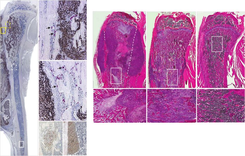
A a b B
Ct a b c
T T
OC
Tb
T *
* Ct *
T Ct
BM
T c
T
Ct
BM Ct d e f
T Tb Tb
T
T BM
d e
Ct BM
T
T
Figure 3. Histologic characterization of breast cancer tumor growth in the human MDA-MB-231 intratibial injection model of bone metastasis in nude rats. A,
a representative image of a sagittal view of a tibia engrafted with MDA-MB-231 breast cancer cells costained for TRAP(þ) OCs (red, arrows) and for tumor cells
using anti-cytokeratin 7 Ab (brown). The regions of interest included: (a and b) intratibial solid tumor area; b, enlarged view of the yellow dashed lined inset.
a and c, extraskeletal solid tumor area outside of the cortical envelope; c, same as yellow solid lined inset. a and d, area of tumor cells infiltrated in
between trabecular bone spicules; d, same as white dashed lined inset. a and e, distance of tumor cells metastasized from the implantation site toward distal
tibial end; e, same as white solid lined inset. Magnification, a, 0.6; b and c, 10; d and e, 20. B, representative images of the H&E-stained sections of tibia
engrafted with breast cancer cells at day 42 (a and d). In the vehicle-treated tibia, injected breast cancer cells developed into solid tumor mass in the
marrow cavity and extraskeletal outgrowth. Regions of disrupted cortical bone (dashed lines) are shown. d, same as inset in a; b and e, ZOL (7.5 mg/kg/wk, s.c.)
reduced loss of cortical and trabecular bone structures, and decreased areas of infiltrated tumor cells; e, same as inset in b; c and f, L-235 (100 mg/kg, p.o.,
b.i.d.)-treated tibia showed normal morphologies of bone marrow, trabecular, and cortical bone; f, same as inset in c. Magnification, a–c, 1; d–f, 4;
bar, 500 mm. , the injected site; T, tumor; Ct, cortical; Tb, trabecular bone; BM, bone marrow.
Cathepsin K inhibitor protected against cortical Cathepsin K inhibitor reduced skeletal breast cancer
disruption tumor burden
The data on cortical integrity are expressed here as the Reduction of intratibial breast cancer tumor. Exten-
percentage of the combined length of the eroded cortex for sive tumor growth was evident in the vehicle group
the anterior and posterior cortical surfaces to the total in both the prevention (Fig. 3B, a and Fig. 5A, a) and
cortical length. When the tibial cortical envelope was treatment protocol (Fig. 5A, b). Treatment with L-235
completely disrupted (Fig. 3B, a), the cortical length was (up to 10 mmol/L) did not change the rate of MDA-MB-
estimated by image analysis (Fig. 3B, a; dash lines). In the 231 cell proliferation in vitro (data not shown). On the
prevention study, approximately 70% of the cortical bone other hand, histologic examination of the in vivo study
was eroded in untreated animals. L-235 dose dependently clearly showed that the intratibial tumor burden was
preserved cortical bone and reduced cortical disruption to significantly reduced by treatment with ZOL (Fig. 3B,
36%, 17%, and 11% respectively, versus vehicle, whereas b) or L-235 100 mg/kg dose (Fig. 3B, c). In the preven-
ZOL resulted in 25% cortical disruption (P < 0.05; Fig. 4A). tion protocol, the vehicle group showed solid tumor
Similar results were seen in the treatment regimen where area of 24 1.2 mm2 (Fig. 5A, a). Although ZOL
L-235 30 mg/kg and ZOL showed 31% and 24% cortical potently reduced solid tumor area by more than 85%
disruption, respectively, compared with vehicle that versus vehicle, the CatKi at 30 and 100 mg/kg marked-
showed 58% cortical disruption (P < 0.05; Fig. 4B). The ly reduced solid tumor area by 70% to 75% versus
effects of L-235 versus ZOL on the preservation of cortical vehicle. Similarly, in treatment mode, efficacy of
bone correlated well with the reduction of tumor burden L-235 30 mg/kg was comparable with ZOL in reducing
as described in the section below. solid tumor burden (Fig. 5A, b). Intratibial solid tumor
www.aacrjournals.org Mol Cancer Ther; 13(12) December 2014 OF7
Downloaded from mct.aacrjournals.org on December 24, 2020. © 2014 American Association for Cancer
Research.Published OnlineFirst September 23, 2014; DOI: 10.1158/1535-7163.MCT-14-0253
Duong et al.
Figure 4. Treatment with the CatKi
A B L-235 compared with Zoledronic
Disrupted surface/Cortical surface (%)
Disrupted surface/Cortical surface (%)
90 80 acid inhibited cortical disruption in
80 the experimental model of breast
70 cancer bone metastasis. Cortical
70 disruption was measured from
60
60 H&E-stained sections as the
50 percentage of cortical surface
50 ** *
40 completely eroded through from
40 the estimated total length of both
** **
*** 30 cortical envelopes. A, L-235 at
30
*** 20 30 mg/kg, p.o., b.i.d. preserved
20
cortical bone in the prevention
10 10
regimen compared to ZOL (7.5
0 0 mg/kg/wk, s.c.); B, L-235 at 30
Veh ZOL L-235 Veh ZOL L-235 mg/kg, p.o., b.i.d. showed
30 mg/kg (b.i.d.) comparable efficacy as ZOL (7.5
10 30 100 mg/kg (b.i.d.) mg/kg/wk, s.c.) in the treatment
*, P < 0.05; **, P < 0.01; ***, P < 0.001 vs. vehicle regimen.
volume was also determined from the mCT-based 3-D as compared with ZOL that reduced tumor volume by
reconstructed images of the osteolytic void volume 56% (Fig. 5B, a). Similar responses were observed in the
(Fig. 5B). In the prevention study, tumor volume treatment experiment where L-235 30 mg/kg and ZOL
was reduced by treatment with L-235 at 10, 30, and resulted in 37% and 72% reduction of tumor volume,
100 mg/kg doses by 29%, 40%, and 63%, respectively, respectively (Fig. 5B, b).
A
30 a 35 b
25 30
Tumor area (mm2)
Tumor area (mm2)
25
20
20
15 ** Figure 5. Treatment with the CatKi
*** 15 or zoledronic acid inhibited
10 *** intratibial human breast cancer
10 *** tumor growth in nude rats. A, solid
5 *** tumor area was manually
5
contoured from histologic images
0 0 of sagittal tibial sections stained
Veh ZOL L-235 Veh ZOL L-235 with H&E. L-235 (10, 30, and
100 mg/kg, p.o., b.i.d.) and ZOL
10 30 100 mg/kg (b.i.d.) 30 mg/kg (b.i.d.)
(7.5 mg/kg/wk, s.c.) versus vehicle
were dosed in (a) prevention and (b)
B 200 a 120 b treatment mode. B, tumor volume
was estimated from mCT-based
volume as the "non-bone"
100
osteolytic regions within the
Tumor volume (mm3)
150
Tumor volume (mm3)
proximal tibia from the same
* 80 **
* studies. With the same dosages as
described in A, L-235 and ZOL
100 60 versus vehicle were dosed in (a)
***
*** *** prevention and (b) treatment mode.
40
, P < 0.05; , P < 0.01; P < 0.001
50 versus vehicle.
20
0 0
Veh ZOL L-235 Veh ZOL L-235
10 30 100 mg/kg (b.i.d.) 30 mg/kg (b.i.d.)
*, P < 0.05; **, P < 0.01; ***, P < 0.001 vs. vehicle
OF8 Mol Cancer Ther; 13(12) December 2014 Molecular Cancer Therapeutics
Downloaded from mct.aacrjournals.org on December 24, 2020. © 2014 American Association for Cancer
Research.Published OnlineFirst September 23, 2014; DOI: 10.1158/1535-7163.MCT-14-0253
Cathepsin K Inhibitor for Metastatic Bone Disease
Reduction of extraskeletal breast cancer tumor. Tumor Cathepsin K inhibitor reduced breast cancer local
area outside of periosteal surface of the proximal tibia was metastasis
also measured from the H&E sagittal sections. In the case Infiltrated tumor. Efficacy of L-235 in inhibiting the
that the tibial cortical envelope was completely disrupted breast cancer MDA-MB-231 cell invasion in vitro was
(Fig. 3B, a), tumor area was estimated on the basis of the evaluated essentially as previously described (15). This
predicted cortical outlines (Fig. 3B, a; dash lines). All CatKi potently inhibited the breast cancer Matrigel inva-
treatments significantly reduced tumor area outside of sion with an IC50 of 3.2 nmol/L (data not shown) as
the cortical bone envelope (Fig. 6A). In the prevention compared with its activity in blocking osteoclastic bone
study, the CatKi at all doses significantly reduced tumor resorption (IC50 ¼ 5 nmol/L; ref. 17). Therefore, the ability
burden in soft tissue by 60% to 80% versus vehicle, of the intratibially injected MDA-MB-231 cells to invade to
whereas ZOL resulted in a 68% reduction (Fig. 6A, a). sites distant from the original injected site was character-
Similar results were seen in the treatment study where ized in vivo by immunohistochemically staining the sec-
L-235 at 30 mg/kg and ZOL reduced tumor area outside tions from injected tibia for cytokeratin, a marker of the
of bone by 73% (P < 0.05) and 62% (NS), respectively, breast cancer cells (Fig. 3A, d and e). ZOL markedly
versus vehicle (Fig. 6A, b). reduced solid tumor area, and protected trabecular and
A 30
a 25 b
25
Tumor area (mm 2)
20
Tumor area (mm 2)
20
15
15
Figure 6. Treatment with the CatKi *
10
reduced local breast cancer 10 ** * *
invasiveness in the experimental
** 5
model of human breast cancer 5
bone metastasis in nude rats. A,
area of solid tumor developed 0 0
outside of the tibial cortical Veh ZOL L-235 Veh ZOL L-235
envelope, surrounding the injected 10 30 100 mg/kg (b.i.d.) 30 mg/kg (b.i.d.)
site was manually contoured from
H&E-stained histologic tibial B
images of nude rats dosed with 30
a 18
b
vehicle, L-235 (10, 30, and 100
Inf. tumor area (mm2)
Inf. tumor area (mm2)
16
25
mg/kg, p.o., b.i.d.), and ZOL (7.5 14
mg/kg/wk, s.c.) in (a) prevention and
20 12
(b) treatment mode. B, area of
10 §
infiltrated tumor cells detected in 15
bone marrow space and amongst 8
the trabecular networks was 10 6
measured from H&E-stained *** 4
sections obtained from tibia of 5 *** *** 2
animals dosed with vehicle, L-235
0 0
and ZOL at the doses as indicated, Veh ZOL L-235 Veh ZOL L-235
in (a) prevention and (b) treatment
10 30 100 mg/kg (b.i.d.) 30 mg/kg (b.i.d.)
mode. C, the longest distance from
the growth plate to detected breast
cancer micrometastasis was
C
determined as described in 18 a 18 b
Materials and Methods. The mean 16 16
distances of micrometastasis were 14 14
Distance (mm)
Distance (mm)
determined from tibial of nude rats 12 * 12
** *
dosed with vehicle, L-235, and ZOL 10 * 10
at the doses indicated, in (a)
8 8
prevention and (b) treatment mode.
6 6
, P < 0.05; , P < 0.01; P < 0.001
versus vehicle. 4 4
2 2
0 0
Veh ZOL L-235 Veh ZOL L-235
10 30 100 mg/kg (b.i.d.) 30 mg/kg (b.i.d.)
*, P < 0.05; **, P < 0.01; ***, P < 0.001 vs. vehicle; §, P = 0.533 vs. ZOL
www.aacrjournals.org Mol Cancer Ther; 13(12) December 2014 OF9
Downloaded from mct.aacrjournals.org on December 24, 2020. © 2014 American Association for Cancer
Research.Published OnlineFirst September 23, 2014; DOI: 10.1158/1535-7163.MCT-14-0253
Duong et al.
cortical bone. However, closer examination of the ZOL- 4 mg, i.v. after 4 weeks of treatment (16). Bone destruction
treated tibia revealed that breast cancer cells infiltrated the associated with skeletal metastasis is known to be medi-
bone marrow among the preserved trabeculae. Small ated by OC bone resorption (2) and the clinical finding
tumors could be detected further down the tibial shaft with ODN in patients with breast cancer supports the
(Fig. 3B, b). In contrast, the L-235–treated tibia showed notion that CatK inhibition may be a novel therapeutic
high density of trabecular spicules associated with normal approach for treating bone metastasis. However, there is
bone marrow integrity (Fig. 3B, c). The "infiltrated" tumor still very limited understanding on whether the orally
area was determined by measuring the total tumor-con- active CatK inhibitor(s) could be safe, efficacious, and
taining trabecular area and subtracting out the trabecular readily differentiated from the current injectable agents
bone area. In the prevention protocol (Fig. 6B, a), the such as the bisphosphonates for the chronic treatment of
majority of tumor burden in vehicle-treated animals was MBD. Hence, studies in animals have been thus con-
solid tumor (24.1 1.2 mm2) as shown above, with very ducted to further validate CatK as a target for the devel-
little infiltrated tumor (7.2 1.6 mm2), resulting in overall opment of an oral inhibitor of osteoclastic bone resorption
intratibial tumor burden of 31.3 1.4 mm2. ZOL drasti- and for exploring its potential direct effects on tumor cell
cally reduced solid tumor area to 2.6 1.1 mm2; but the metastasis.
infiltrated area was 18.3 2.6 mm2 resulting in a tumor Previously, treatment with the CatKi AFG-495 was
burden of 20.9 2.0 mm2 within the medullary space. reported to significantly reduce cancer-induced osteolysis
Meanwhile, L-235 at all doses markedly reduced infiltrat- and skeletal tumor burden in an intratibial injection with
ed tumor area (Fig. 6B, a). L-235 100 mg/kg reduced solid human BT474 breast cancer cells expressing CatK into
and infiltrated tumor areas to 6.5 2.8 and 1.9 0.7 mm2, nude mice (24). AFG-495 was reported to inhibit human
respectively, resulting in an overall tumor burden of CatK with an IC50 of 3 to 6 nmol/L and an excellent
8.4 3.1 mm2 in tibial bone marrow space. In the treat- selectivity profile versus human cathepsins L and S;
ment experiment, neither ZOL nor L-235 treatment was as however, its potency and selectivity profiles against
effective at reducing tumor cells infiltrated into the tra- rodent cathepsins were not available (25). Because of
becular bone as compared with that in the preventive species differences between the binding sites of human
regimen (Fig. 6B, b). versus rodent CatK, there are usually significant reduc-
Local metastasis. On the basis of our examination of tions in potency against the rodent CatK with the inhibi-
the tibial sections stained with cytokeratin, small discrete tors developed against the human enzyme. Even if the
tumor areas were detected toward the distal tibial end. human osteoporosis dose of AFG-495 has not been deter-
Because bone marrow is known for high vascularization mined, it was dosed via intraperitoneal injection twice
activity, we believe these small islands of breast cancer daily to achieve sufficient high drug exposure in mice (15).
cells represent local metastatic activity, and thus the Interestingly in that study, AFG-495 at 50 mg/kg, i.p.,
distance from growth plate to the most distal site of twice daily for 39 days reduced tumor burden by 62%,
observable micromass of tumor was determined to assess whereas a single high bolus subcutaneous injection of
treatment-related effects on local metastasis. In the pre- ZOL 100 mg/kg did not inhibit skeletal tumor burden (15).
vention protocol (Fig. 6C, a), L-235 at 30 and 100 mg/kg Of note, Le Gall and colleagues also confirmed that
doses significantly reduced this metastatic distance by AFG-495 did not inhibit subcutaneous growth of breast
38% and 51%, respectively versus vehicle (P < 0.05). ZOL cancer B02 tumor xenografts in nude mice at a dosing
and the lowest dose of the CatKi did not appear to show regimen that was demonstrated to inhibit skeletal tumor
significant effect on this parameter. On the other hand, in burden, demonstrating that CatK inhibition does not
the treatment protocol, both L-235 30 mg/kg and ZOL affect breast cancer cell proliferation (15, 24).
reduced breast cancer tumor metastasis by 42% and 47%, Here, we evaluated the CatKi L-235 orally at 10, 30, and
respectively, versus vehicle (Fig. 6C, b, P < 0.05). 100 mg/kg twice daily for 42 days in an intratibial injec-
tion of the breast cancer MDA-MB-231 cells in nude rats.
Efficacy of L-235 was evaluated in this preclinical model of
Discussion
bone metastasis in both prevention and treatment modes
The pivotal role of CatK in OC bone resorption has been in comparison with ZOL. L-235 is selective against other
well established by both genetic and pharmacologic evi- human cathepsins (17) and retains good selectivity (>30-
dence in animals and humans (11, 12). CatK-deficient mice fold) toward rat CatB, CatL, and CatS (data not shown).
develop osteopetrosis associated with impaired OC func- Similar to AFG-495, L-235 exhibits lysosomotropic prop-
tion in degrading bone matrix. Loss-of-function muta- erties; that is, these compounds accumulate within acidic
tions in the CatK gene lead to pycnodysostosis, a rare subcellular organelles such as lysosomes or the resorption
autosomal recessive disorder associated with bone scle- lacunae. This feature may compromise selectivity of the
rosis in humans (11, 12). In a small clinical study with 43 CatKi in vivo by inhibiting other lysosomal cathepsins at
breast cancer women with bone metastasis, the CatKi the doses used in this study (19, 20). Several cysteine
ODN dosed as 5 mg daily was recently demonstrated to cathepsins, particularly CatB, have been shown to be
be well tolerated and to suppress the bone resorption causally involved in the migration and invasion of tumor
marker uNTX similarly to the clinical dose of ZOL at cells (26, 27); the lysosomotropic property of L-235 may in
OF10 Mol Cancer Ther; 13(12) December 2014 Molecular Cancer Therapeutics
Downloaded from mct.aacrjournals.org on December 24, 2020. © 2014 American Association for Cancer
Research.Published OnlineFirst September 23, 2014; DOI: 10.1158/1535-7163.MCT-14-0253
Cathepsin K Inhibitor for Metastatic Bone Disease
part contribute to its anti-invasive activity in this model of cancer bone metastasis in nude rats, we speculate that
bone metastasis as discussed below. Despite the above the ability of tumor cells to infiltrate into the surrounding
drawback of L-235, this inhibitor showed potency equiv- bone marrow and among the trabecular spicules repre-
alent to our clinical candidate ODN against the human sented the local invasive activity of the breast cancer cells.
CatK (IC50 of 0.2 nmol/L; ref. 18), but has an approxi- Although ZOL showed no effect, even the low dose of
mately 50-fold shift in potency against the rat enzyme L-235 at 10 mg/kg significantly reduced the area of infil-
while ODN is not active in rodent. Because the effective trated tumor when dosed in prevention mode. This find-
dose of ODN at 5 mg p.o., daily provided mean plasma ing was supported by the direct action of this CatKi in
concentration of 512 203 nmol/L in patients with breast inhibiting the human breast cancer MDA-MB-231 cell
cancer (16) and due to the relatively short half-life of L-235 Matrigel invasion assay in vitro. Curiously, this anti-inva-
in rodent, we thus selected L-235 at 10, 30, and 100 mg/ sive activity of the CatKi seemed to be reduced when
kg twice-daily, which provided the mean plasma con- dosed in treatment mode, although the effect of CatKi was
centrations of 10.4, 35.6, and 166.2 (mmol/L)h, respec- still differentiated from ZOL. As an exploratory approach,
tively, in the rats. At this range of drug exposures, L-235 we confirmed the potential of injected human tumor cells
should display full CatK enzyme inhibition activity for in the proximal tibia to develop micrometastasis at the
studying the differential biologic effects of inhibiting distal end of the rat tibia. Consistent with the effects of
human CatK on MDA-MB-231–mediated cell invasion CatKi in reduction of local tumor infiltration, L-235 treat-
as well as blocking rat CatK activity of host osteoclastic ment significantly reduced the incidence of distant metas-
bone resorption. tasis of human breast cancer cells.
Interestingly, L-235 at the selected doses seemed to be a In summary, we confirmed the high expression of CatK
potent inhibitor of cortical disruption. Although the low- in primary and metastatic breast cancer tumors and dem-
est dose of L-235 at 10 mg/kg showed partial protection of onstrated that an orally active CatKi in both prevention
bone volume fraction and reduction on intratibial tumor and treatment modes protected against tumor-induced
growth, this dose showed substantial inhibition of cortical osteolytic lesions and cortical disruption, and reduced
disruption in the same group. This observation is aligned skeletal tumor burden in an experimental model of breast
with previous findings on the favorable efficacy of CatK cancer bone metastasis. The potential of the CatKi to
inhibitors on enhancing cortical bone formation while reduce local tumor invasion implicated an upside poten-
reducing endocortical and intracortical bone remodeling tial of the mechanism of CatK inhibition over the standard
(28). This unique mechanism of CatK inhibition on cortical antiresorptives such as the bisphosphonates, for the long-
protection is also highly correlated with the ability of term care of patients with breast cancer with established
CatKi to reduce extraskeletal tumor growth, suggesting bone metastasis. Additional studies on the molecular and
that the cortical envelope may serve as a barrier to prevent cellular changes within the skeletal environment in the
the breast cancer tumor overgrowth into the extramedul- L-235–treated intratibial injected MDA-MB-231 model of
lary soft tissue in this model of bone metastasis. MBD would be subjects of future investigation. Taken
The function of CatK in breast cancer is presently together, our results from this study support the role of
unknown. Besides its expression in OCs, there is limited CatK in breast cancer skeletal metastasis and CatK inhi-
evidence that CatK is also expressed in human breast bitors may represent a novel oral therapy for treatment of
carcinomas (14, 15). In addition, breast cancer cells resid- metastatic breast cancer.
ing in bone metastases overproduce CatK relative to the
expression levels in primary tumors and soft tissue metas- Disclosure of Potential Conflicts of Interest
No potential conflicts of interest were disclosed.
tases (15). These findings suggest that cancer cells metas-
tasizing to bone express bone-related genes to adapt and
Authors' Contributions
thrive in the bone microenvironment (29). We confirmed Conception and design: L.T. Duong, R. Oballa
high expression of CatK in primary breast cancer tumors Development of methodology: G.A. Wesolowski
and its upregulation in metastatic tumors as previously Acquisition of data (provided animals, acquired and managed patients,
provided facilities, etc.: G.A. Wesolowski, P. Leung, R. Oballa, M. Pickarski
reported (15). To further examine the role of CatK in breast Analysis and interpretation of data (e.g., statistical analysis, biostatis-
cancer metastasis, Le Gall and colleagues demonstrated tics, computational analysis): L.T. Duong, G.A. Wesolowski, M. Pickarski
Writing, review, and/or revision of the manuscript: L.T. Duong, G.A.
that the CatKi, AFG-495, did not directly inhibit breast Wesolowski, R. Oballa, M. Pickarski
cancer cell proliferation while reduced Matrigel cell inva- Administrative, technical, or material support (i.e., reporting or orga-
sion in vitro. However, these authors did not further nizing data, constructing databases): L.T. Duong, M. Pickarski
Study supervision: L.T. Duong, G.A. Wesolowski
examine the effects of CatK inhibition on the local tumor
metastasis in vivo. Acknowledgments
Secretion of the active CatK enzyme from macrophages The authors thank the staff at Piedmont Research Center, Morrisville,
and prostate cancer cells has been demonstrated (13, 26). NC for carrying out the in-life portion of the studies, and G. Neusch and A.
Cheema (Merck & Co. Inc. employees) for their excellent technical support
This pool of secreted enzyme could potentially participate to the early phase of study execution.
in local invasion of tumor cells by mediating the extra-
cellular or intracellular degradation or both of matrix Grant Support
proteins. In the experimental model of human breast This work was funded by Merck & Co.
www.aacrjournals.org Mol Cancer Ther; 13(12) December 2014 OF11
Downloaded from mct.aacrjournals.org on December 24, 2020. © 2014 American Association for Cancer
Research.Published OnlineFirst September 23, 2014; DOI: 10.1158/1535-7163.MCT-14-0253
Duong et al.
The costs of publication of this article were defrayed in part by the pay- Received March 24, 2014; revised August 13, 2014; accepted September 3,
ment of page charges. This article must therefore be hereby marked adver- 2014; published OnlineFirst September 23, 2014.
tisement in accordance with 18 U.S.C. Section 1734 solely to indicate this fact.
References
1. Coleman RE. Skeletal complications of malignancy. Cancer 1997;80: 16. Jensen AB, Wynne C, Ramirez G, He W, Song Y, Berd Y, et al. The
1588–94. cathepsin K inhibitor odanacatib suppresses bone resorption in wom-
2. Mundy GR. Metastasis to bone: causes, consequences and thera- en with breast cancer and established bone metastases: results of a 4-
peutic opportunities. Nat Rev Cancer 2002;2:584–93. week, double-blind, randomized, controlled trial. Clin Breast Cancer
3. Onishi T, Hayashi N, Theriault RL, Hortobagyi GN, Ueno NT. Future 2010;10:452–8.
directions of bone-targeted therapy for metastatic breast cancer. Nat 17. Palmer JT, Bryant C, Wang DX, Davis DE, Setti EL, Rydzewski RM,
Rev Clin Oncol 2010;7:641–51. et al. Design and synthesis of tri-ring P3 benzamide-containing ami-
4. Pavlakis N, Stockler M. Bisphosphonates for breast cancer. Cochrane nonitriles as potent, selective, orally effective inhibitors of cathepsin K.
Database Syst Rev 2012;2:CD003474. J Med Chem 2005;48:7520–34.
5. Coleman RE, Costa AL, Cook RJ, Lee K-A, Saad F, Brown JE, et al. 18. Gauthier JY, Chauret N, Cromlish W, Desmarais S, Duong LT, Fal-
Possible survival benefits from zoledronic acid treatment in patients gueyret JP, et al. The discovery of odanacatib (MK-0822), a selective
with bone metastases from solid tumours and poor prognostic fea- inhibitor of cathepsin K. Bioorg Med Chem Lett 2008;18:923–8.
tures - An exploratory analysis of placebo-controlled trials. J Bone 19. Falgueyret JP, Desmarais S, Oballa R, Black WC, Cromlish W, Khougaz
Oncol 2013;2:70–6. K, et al. Lysosomotropism of basic cathepsin K inhibitors contributes
6. Rosen LS, Gordon D, Tchekmedyian S, Yanagihara R, Hirsh V, Krza- to increased cellular potencies against off-target cathepsins and
kowski M, et al. Zoledronic acid versus placebo in the treatment of reduced functional selectivity. J Med Chem 2005;48:7535–43.
skeletal metastases in patients with lung cancer and other solid 20. Desmarais S, Black WC, Oballa R, Lamontagne S, Riendeau D, Tawa P,
tumors: a phase III, double-blind, randomized trial - the Zoledronic et al. Effect of cathepsin k inhibitor basicity on in vivo off-target
Acid Lung Cancer and Other Solid Tumors Study Group. J Clin Oncol activities. Mol Pharmacol 2008;73:147–56.
2003;21:3150–7. 21. Pennypacker BL, Duong LT, Cusick TE, Masarachia PJ, Gentile MA,
7. Saad F, Gleason DM, Murray R, Tchekmedyian S, Venner P, Lacombe Gauthier JY, et al. Cathepsin K inhibitors prevent bone loss in estro-
L, et al. A randomized, placebo-controlled trial of zoledronic acid in gen-deficient rabbits. J Bone Miner Res 2011;26:252–62.
patients with hormone-refractory metastatic prostate carcinoma. 22. Simoni D, Gebbia N, Invidiata FP, Eleopra M, Marchetti P, Rondanin R,
J Natl Cancer Inst 2002;94:1458–68. et al. Design, synthesis, and biological evaluation of novel aminobi-
8. Perazella MA, Markowitz GS. Bisphosphonate nephrotoxicity. Kidney sphosphonates possessing an in vivo antitumor activity through a
Int 2008;74:1385–93. gammadelta-T lymphocytes-mediated activation mechanism. J Med
9. Bromme D, Okamoto K, Wang BB, Biroc S. Human cathepsin O2, a Chem 2008;51:6800–7.
matrix protein-degrading cysteine protease expressed in osteoclasts. 23. Ferguson VL, Simske SJ, Ayers RA, Bateman TA, Wang HT, Bendele A,
Functional expression of human cathepsin O2 in Spodoptera frugi- et al. Effect of MPC-11 myeloma and MPC-11 þ IL-1 receptor antag-
perda and characterization of the enzyme. J Biol Chem 1996; onist treatment on mouse bone properties. Bone 2002;30:109–16.
271:2126–32. 24. Le Gall C, Bonnelye E, Clezardin P. Cathepsin K inhibitors as treatment
10. Garnero P, Ferreras M, Karsdal MA, Nicamhlaoibh R, Risteli J, Borel O, of bone metastasis. Curr Opin Support Palliat Care 2008;2:218–22.
et al. The type I collagen fragments ICTP and CTX reveal distinct 25. Altmann E, Renaud J, Green J, Farley D, Cutting B, Jahnke W.
enzymatic pathways of bone collagen degradation. J Bone Miner Res Arylaminoethyl amides as novel non-covalent cathepsin K inhibitors.
2003;18:859–67. J Med Chem 2002;45:2352–4.
11. Duong LT. Therapeutic inhibition of cathepsin K-reduction of bone 26. Vasiljeva O, Reinheckel T, Peters C, Turk D, Turk V, Turk B. Emerging
resorption while maintaining bone formation. BoneKEy 2012;67:3–8. roles of cysteine cathepsins in disease and their potential as drug
12. Costa AG, Cusano NE, Silva BC, Cremers S, Bilezikian JP. Cathepsin targets. Curr Pharm Des 2007;13:387–403.
K: its skeletal actions and role as a therapeutic target in osteoporosis. 27. Mohamed MM, Sloane BF. Cysteine cathepsins: multifunctional
Nat Rev Rheumatol 2011;7:447–56. enzymes in cancer. Nat Rev Cancer 2006;6:764–75.
13. Brubaker KD, Vessella RL, True LD, Thomas R, Corey E. Cathepsin K 28. Cusick T, Chen CM, Pennypacker BL, Pickarski M, Kimmel DB, Scott
mRNA and protein expression in prostate cancer progression. J Bone BB, et al. Odanacatib treatment increases hip bone mass and cortical
Miner Res 2003;18:222–30. thickness by preserving endocortical bone formation and stimulating
14. Littlewood-Evans AJ, Bilbe G, Bowler WB, Farley D, Wlodarski B, periosteal bone formation in the ovariectomized adult rhesus monkey.
Kokubo T, et al. The osteoclast-associated protease cathepsin K is J Bone Miner Res 2012;27:524–37.
expressed in human breast carcinoma. Cancer Res 1997;57:5386–90. 29. Bellahcene A, Bachelier R, Detry C, Lidereau R, Clezardin P, Castronovo
15. Le Gall C, Bellahcene A, Bonnelye E, Gasser JA, Castronovo V, Green V. Transcriptome analysis reveals an osteoblast-like phenotype for
J. A cathepsin K inhibitor reduces breast cancer induced osteolysis human osteotropic breast cancer cells. Breast Cancer Res Treat 2002;
and skeletal tumor burden. Cancer Res 2007;67:9894–902. 101:135–48.
OF12 Mol Cancer Ther; 13(12) December 2014 Molecular Cancer Therapeutics
Downloaded from mct.aacrjournals.org on December 24, 2020. © 2014 American Association for Cancer
Research.You can also read