Effects of an injectable long-acting formulation of ivermectin on Onchocerca ochengi in zebu cattle - Parasite
←
→
Page content transcription
If your browser does not render page correctly, please read the page content below
Parasite 27, 36 (2020)
Ó M. Boussinesq et al., published by EDP Sciences, 2020
https://doi.org/10.1051/parasite/2020036
                                                                                                                            Available online at:
                                                                                                                           www.parasite-journal.org

    RESEARCH ARTICLE                                                                                                                OPEN        ACCESS

Effects of an injectable long-acting formulation of ivermectin
on Onchocerca ochengi in zebu cattle
Michel Boussinesq1,*, Peter Enyong2, Patrick Chounna-Ndongmo2, Abdel-Jelil Njouendou2, Sébastien David Pion1,
Anthony Rech3, Christophe Roberge3, Georges Gaudriault3,a, and Samuel Wanji2
1
    Recherches Translationnelles sur le VIH et les Maladies Infectieuses (TransVIHMI), UMI233 IRD-U1175 INSERM-Université
    de Montpellier, BP 64501, 34394 Montpellier Cedex 5, France
2
    Research Foundation for Tropical Diseases and the Environment, PO Box 474, Buea, Cameroon
3
    MedinCell S.A., 3 Rue des Frères Lumière, 34830 Jacou, France

             Received 3 August 2019, Accepted 5 May 2020, Published online 18 May 2020

             Abstract – The availability of a safe macrofilaricidal drug would help to accelerate onchocerciasis elimination. A trial
             was conducted in Cameroon to evaluate the effects of a subcutaneous injectable long-acting formulation of ivermectin
             (LAFI) on the microfilariae (mf) and adult stages of Onchocerca ochengi. Ten zebu cattle naturally infected with the
             parasite were injected subcutaneously with either 500 mg (group A, N = 4), or 1000 mg long-acting ivermectin
             (group B, N = 4) or the vehicle (group C, N = 2). Skin samples were collected from each animal before, and 6, 12,
             and 24 months after treatment to measure microfilarial densities (MFDs). Nodules excised before, and 6 and 12 months
             after treatment were examined histologically to assess the adult worms’ viability and reproductive status. Blood
             samples were collected at pre-determined time-points to obtain pharmacokinetic data. Before treatment, the average
             O. ochengi MFDs were similar in the three groups. Six months after treatment, all animals in groups A and B were
             free of skin mf, whereas those in group C still showed high MFDs (mean = 324.5 mf/g). Only one ivermectin-treated
             animal (belonging to group A) had skin mf 12 months after treatment (0.9 mf/g). At 24 months, another animal in
             group A showed skin mf (10.0 mf/g). The histologic examination of nodules at 6 and 12 months showed that LAFI
             was not macrofilaricidal but had a strong effect on embryogenesis. The new LAFI regimen might be an additional tool
             to accelerate the elimination of human onchocerciasis in specific settings.

             Key words: Onchocerciasis, Onchocerca ochengi, Ivermectin, Long-acting formulation, Microfilaricidal effect,
             Macrofilaricidal effect.

             Résumé – Effets d’une formulation injectable d’ivermectine à activité prolongée sur Onchocerca ochengi chez
             les bovins zébu. La disponibilité d’un médicament macrofilaricide et sans danger permettrait d’accélérer l’élimination
             de l’onchocercose. Un essai a été mené au Cameroun pour évaluer les effets d’une formulation injectable en sous-
             cutané d’ivermectine à activité prolongée (FIAP) sur les microfilaires (mf) et les stades adultes d’Onchocerca
             ochengi. Dix vaches zébu infectées naturellement par le parasite ont reçu une injection sous-cutanée de 500 mg
             (groupe A, N = 4) ou de 1000 mg d’ivermectine à activité prolongée (groupe B, N = 4) ou le véhicule (groupe C,
             N = 2). Des échantillons de peau ont été collectés de chaque animal avant, puis 6, 12 et 24 mois après traitement
             pour mesurer les densités microfilariennes (DMF). Des nodules prélevés avant et 6 et 12 mois après traitement ont
             été examinés histologiquement pour évaluer la viabilité et le statut reproductif des vers adultes. Des échantillons de
             sang ont été prélevés pour obtenir des données de pharmacocinétique. Avant traitement, les DMF à O. ochengi
             étaient similaires dans les 3 groupes. Six mois après traitement, aucun des animaux des groupes A et B ne
             présentait de mf dermiques, alors que ceux du groupe C présentaient encore des DMF élevées (moyenne :
             324,5 mf/g). Parmi les animaux traités par ivermectine, un seul (du groupe A) avait des mf dermiques 12 mois
             après traitement (0,9 mf/g). A 24 mois, un autre animal du groupe A avait des mf (10,0 mf/g). L’examen
             histologique des nodules collectés à 6 et 12 mois montrait que la FIAP n’était pas macrofilaricide mais avait un
             effet marqué sur l’embryogénèse. La nouvelle FIAP pourrait représenter un outil pour accélérer l’élimination de
             l’onchocercose dans certaines circonstances spécifiques.

*Corresponding author: michel.boussinesq@ird.fr
a
  Present address: Deinove S.A., ZAC Euromédecine II, Cap Sigma, 1682 Rue de la Valsière, 34790 Grabels, France.

    This is an Open Access article distributed under the terms of the Creative Commons Attribution License (https://creativecommons.org/licenses/by/4.0),
                   which permits unrestricted use, distribution, and reproduction in any medium, provided the original work is properly cited.
2 M. Boussinesq et al.: Parasite 2020, 27, 36
Introduction candidates are thus only at an initial stage of clinical
development.
The main control strategy for onchocerciasis is currently The present study was conducted following the observation
based on mass treatment with ivermectin (IVM) targeting the that three-monthly doses of IVM induce excess mortality in
most affected populations, i.e. those (called meso-hyperendemic) adult worms, when compared to annual doses [22], this excess
where more than 20% of the adults present subcutaneous mortality actually being due to a significant decrease in the
nodules containing Onchocerca volvulus adult worms. In adult worms’ lifespan [46]. As the plasma half-life of IVM in
Africa, community-directed treatment with IVM (CDTI) has man ranges between 12 h and 56 h following oral administra-
led to the elimination of onchocerciasis as a public health tion [26], we hypothesized that longer and continuous exposure
problem in most of the treated areas. However, to reach the of the parasite to the drug could have a stronger macrofilaricidal
new World Health Organization’s objective of elimination of effect. Presently, several commercial long-acting formulations
the infection [47], interventions might have to be expanded to of IVM exist, one of the best known being the subcutaneous
the so-far untreated hypoendemic zones, and alternative injectable formulation IvomecÒ Gold for cattle [15]. In this con-
treatment strategies (ATS, i.e., differing from annual CDTI) text, we tested the long-term efficacy (two years) of an inject-
implemented [11]. Such strategies include the use of new drugs able long-acting IVM formulation on the cattle-Onchocerca
or new formulations of existing drugs. ochengi filarial model. This model is widely used to assess
IVM has two main effects on O. volvulus. First, it induces the effects of potentially filaricidal drugs because O. ochengi
rapid destruction of the larval stage of the parasites (microfilar- is taxonomically close to O. volvulus, and because the adult
iae [mf]) which are the cause of the immune reactions leading stages of both species live in subcutaneous nodules [32]. The
to the ocular and skin manifestations of the disease (microfilar- tested formulation is based on the proprietary drug delivery
icidal effect). Second, IVM treatment interrupts for 3–4 months platform BEPOÒ, which uses bioresorbable block copolymers
the release of new mf by the adult female worms (embryostatic as functional excipients to control the release of IVM. In this
effect) [8]. However, as IVM has a limited effect on the lifespan study, an assessment was made of the effect of the slow release
of adult worms, CDTI has to be repeated every 6 or 12 months of IVM on the skin MFD by counting mf in skin biopsies, and
to maintain microfilarial densities (MFD) below the level asso- on the fertility and the viability of the adult worms, by histolo-
ciated with clinical manifestations. One of the ATSs that could gical examination of sections of excised subcutaneous nodules.
be used to accelerate elimination of onchocerciasis would be to
treat the whole population, or only those individuals currently
infected with O. volvulus, with a macrofilaricidal drug, i.e. a Materials and methods
drug that kills or permanently sterilizes the adult worms.
Presently, the only macrofilaricidal drug which can be dis- Animals
tributed on a large scale without major risks of adverse effects is
doxycycline. Daily treatment with this antibiotic for 4–6 weeks Ten Gudali zebu cattle (Bos indicus) were purchased in
eliminates the Wolbachia symbiotic bacteria present in the adult villages near Ngaoundere (Adamaoua region of Cameroon)
worms, which leads to the sterilization and progressive death of where transmission of O. ochengi is ongoing [44]. They were
the latter [45]. The main problem related to this strategy is the selected on the basis of their sex (female), age (three years)
duration of the treatment, and research is ongoing to identify and presence of at least 10 subcutaneous nodules between the
other drugs that could be macrofilaricidal using a regimen of udder and the umbilicus, and in the inguinal region. The
two weeks or less. Three candidates have recently been tested animals were not weighed, and no girth measurement was made
as part of phase 1 trials. The first is oxfendazole [1], which to estimate their weight, but given the age of the cows, one can
belongs to the benzimidazole family and could have the advan- assume that weight ranged between 250 kg and 330 kg [2].
tage of killing the adult worms without affecting the mf, and They were transported to the field research station of the
thus would not induce adverse effects, particularly in case of Research Foundation for Tropical Diseases and the Environ-
coinfection with Loa loa [27, 48]. Ongoing trials (phase 1 ment (REFOTDE) located near Modeka, in the South–West
and phase 2 against Trichuris trichiura) will enable us to eval- region, on the right bank of the Mungo River. Each animal
uate its possible toxicity [25]. This point is key because the was identified using an individual number printed on labels
development of another benzimidazole to treat filariases, attached at one ear. The interval of time between the departure
flubendazole, was interrupted in 2017 because its toxicity asso- of the cows from the Adamaoua region and the first examina-
ciated with effective doses was considered unacceptable [30]. tion round (and administration of treatment) was six weeks.
The second macrofilaricidal candidate is emodepside, whose
efficacy was demonstrated in pre-clinical trials [29], and which Treatment description
was evaluated in two phase 1 trials, one using single ascending
doses [24], and the other using multiple ascending doses The injectable long-acting formulation of IVM was pre-
(https://clinicaltrials.gov/ct2/show/NCT03383614). The last pared by Medincell using their proprietary drug delivery
candidate is a drug called TylAMac (Tylosin Analogue platform BEPOÒ [23, 37]. A diblock (PEG–PLA) and a
Macrofilaricide, ABBV-4083), which is a macrolide antibiotic triblock (PLA–PEG–PLA) bioresorbable copolymer were
effective against Wolbachia [41, 43], and which was also allowed to dissolve overnight in a biocompatible solvent,
evaluated in a phase 1 trial terminated in 2018 (https://www. dimethyl sulfoxide (DMSO), with gentle mixing on a roller
dndi.org/diseases-projects/portfolio/abbv-4083/). All these mixer at room temperature. Then, 40 mL of the obtainedM. Boussinesq et al.: Parasite 2020, 27, 36 3
vehicle were sterile filtered into a 50 mL glass bottle and kept 2.5 lm) and a C18 guard cartridge (4 2 mm). The mobile
refrigerated before shipment. The rest of the vehicle (approx. phases were: A = 50 mM ammonium acetate (pH 4.5) and
160 mL) was used to prepare the IVM formulation; pre- B = acetonitrile. The other conditions were: isocratic elution
weighed IVM powder was added to the vehicle and allowed A/B (10:90); flow rate: 0.5 mL/min; injection volume: 10 lL;
to gently dissolve on a roller mixer at room temperature. The autosampler temperature: 4 °C; column temperature: 40 °C;
final solution of IVM was sterile filtered into a 250 mL glass and flush port: methanol. The detection consisted in multiple
bottle and kept refrigerated prior to shipment. The composition reaction monitoring (MRM) in positive mode. IVM was detected
of the formulation was 7.5 w/w% IVM, 40 w/w% of copoly- for m/z 892.400 > 307.200. The lower limit of quantification
mers and 52.5 w/w% of DMSO. (LLOQ) for the method used was 0.1 ng of IVM per mL.
The analysis of the pharmacokinetic parameters (mean
Cmax, Tmax, Clast, and AUC0–tlast) was undertaken with the help
Treatment and follow-up of adverse effects of WinNonlin, Phoenix 64, version 8.0 software.
Four animals (group A) were injected subcutaneously, just
behind the shoulder, with 500 mg IVM, four others (group B) Skin biopsies and nodulectomies
with 1000 mg IVM, and two others (group C) with the vehicle
only. Injections were made by a veterinarian using a 16-gauge One skin biopsy was collected from each animal just
needle and the volume injected was about 5.8 mL for the before the subcutaneous injection (day 0, D0), and another
500 mg dose or 2 5.8 mL for the 1000 mg dose (5.8 mL one after 6, 12, and 24 months (M6, M12, and M24).
behind each shoulder). As the animals’ weight was about Nodulectomies were performed at the same time as skin biopsies
250–330 kg, the IVM doses administered in the two treated on D0, M6, and M12. To collect these samples, the animals were
groups were 1.5–2.0 mg/kg and 3.0–4.0 mg/kg, respectively. put in lateral recumbency, on a mattress, and maintained in this
Upon administration, the initially liquid formulation turned into position with ropes, for a maximum of 1 h. All sample collec-
a solid depot in the subcutaneous space, where IVM was tions were performed by a veterinarian, after shaving of the skin
released progressively by diffusion through the formed poly- and under local anaesthesia. Skin biopsies (surface area about
meric matrix. Any anomaly at the injection site(s) and relevant 1 cm2) were taken from the area between the udder and the
clinical signs (apathy, loss of appetite, tremors, locomotion pro- umbilicus, i.e. where the O. ochengi microfilarial densities are
blems, etc.) were monitored during the three days following the the highest [44]. Nodules were collected surgically. In some
injection, and then at day 7. instances, more than one nodule was collected from a given
animal at a given time-point. Nodules were placed in tubes
containing 10 mL of fixative (10% formalin) until further
Pharmacokinetics processing. After nodulectomy, the wound was sutured and
the animal received an intramuscular injection of antibiotics
The blood sampling schedule was the following: in the (streptomycin and penicillin G) with no action on the
vehicle group (group C): pre-dose ( 1 h) and no subsequent Wolbachia bacterial endosymbionts hosted by filariae. Stitches
sampling; in the IVM treated groups: pre-dose ( 1 h) and were removed after seven days.
multiple post-dose sampling (6 h, D2, D7, D14, D30, D90,
D150, D180, D240, D300, D330, and D360). Microscopic examination of skin biopsies
At each time-point, atleast 4 mL of blood were withdrawn
from the jugular vein or from a vein on the tail and transferred The skin biopsies were left to incubate for 24 h at room
in ethylenediaminetetraacetic acid (EDTA) tubes to prevent temperature in 24-well plates, each well containing 300 lL of
coagulation. Samples were placed on ice before being sterile RPMI 1640, and weighed with a 10 mg precision just
centrifuged. before examination. The medium containing the mf which
Blood collection tubes were promptly centrifuged at had emerged during the incubation period was pipetted and
2500 g for 10 min at room temperature and plasma was split placed on microscopic slides and examined at 40 and 100
in two aliquots of 500 lL in previously labelled polypropylene magnification. Four species of bovine Onchocerca are present
tubes (aliquots A and B (back-up sample)). Tubes with plasma in North Cameroon (O. ochengi, O. gutturosa, O. dukei, and
specimens were frozen and stored at 80 °C until being shipped O. armillata [44]), and mf were identified according to their
in dry ice containers to Europe. The samples were analyzed at size and aspect [6, 44]. No mf of O. armillata (length: 300–
the Echevarne Laboratory, Barcelona, Spain, using a liquid 380 lm; width: 5.0–6.8 lm; characteristic bulging anterior
chromatography coupled with tandem mass spectrometry end) was seen. Microfilariae of the three other species were
(LC/MS/MS) method. The bioanalytical method was validated observed: O. ochengi (length: 280–300 lm; width: 6–8 lm;
based on the following criteria (Selectivity, Recovery, rounded anterior end); O. gutturosa (length: 225–270 lm;
Carry-over, Calibration range and Response function, Limit of width: 3.5–4.5 lm; rounded anterior end and tapering posterior
Quantification, Precision and Accuracy, Dilution Integrity, end); and O. dukei (length: 220–260 lm; width: 5.0–6.5 lm;
Matrix effect, Stability in samples, and Reference Solutions thinner anterior third of the body). Mf of each these three
Stability). The Liquid Chromatography system was coupled to species were counted by two independent microscopists who
tandem mass spectrometry (Triple Quadrupole) with Electro- had no information on the treatment received by the animal
spray Probe. Specifically, the chromatography was performed from which the biopsy was taken. The individual MFD were
using a Synergi MAX204 RP column (100 Å, 100 3 mm, calculated as the arithmetic mean of the two counts and4 M. Boussinesq et al.: Parasite 2020, 27, 36
expressed as the number of mf per gram of skin. As mf of
O. dukei were seen in only one animal at D0, with a low
MFD (16.0/g), the results presented below distinguish only
O. ochengi mf and “non O. ochengi” (i.e., O. gutturosa +
O. dukei) mf. The MFD in each treatment group were
calculated as the arithmetic means of individual MFD.
Histological processing of nodules and
assessment of worm viability and fertility
The nodules were embedded in paraffin wax, and 6 lm
sections were stained with hematoxylin and eosin. Two of the
authors (MB and SW) examined the sections independently
and without having any information on the treatment arm of
the animals. The nodules collected at each round of nodulec-
tomies were examined separately. When the observers did not
agree on the classification of the worms in a nodule, the slides
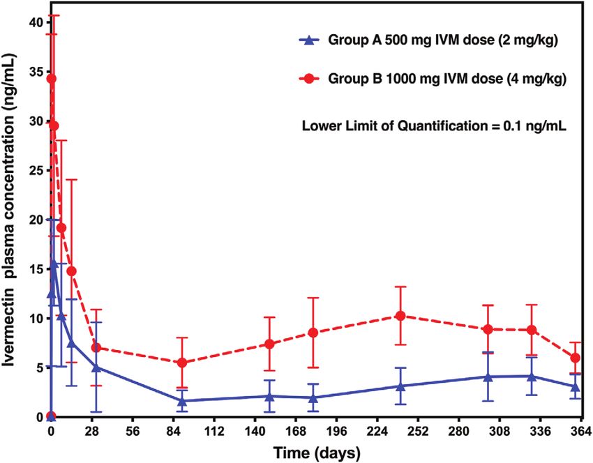
were re-examined until a consensus was reached. Figure 1. Plasma concentration-time profiles of IVM following
subcutaneous injection of a long-acting formulation in cattle (N = 4
The reproductive status of the adult worms was assessed by per group). LLOQ = lower limit of quantification.
the presence of oocytes and of embryos (morulas, coiled mf,
and stretched mf). Live embryos were distinguished from
degenerating ones [14]. Worms with uteri containing live Table 1. Mean pharmacokinetic (PK) parameters in the two groups
embryos of any stage were considered fertile. of animals treated with IVM.
PK parameters Value Treatment group
Results 500 mg 1000 mg
(1.5–2 mg/kg) (3–4 mg/kg)
Safety
Cmax (ng/mL) Mean 16.3 36.2
After injection of the ~5.8 mL of liquid (5.8 mL 2 for SD 5.4 6.9
animals treated with 1000 mg IVM), a small bump (diameter: CV (%) 33.2 19.1
Tmax (days) Median 2.00 0.25
~1 cm) could be palpated at the injection site. No side effects
Clast (ng/mL) Mean 3.10 6.02
were recorded in any of the cows during the follow-up period. SD 1.23 1.55
AUC0–tlast (ng day/mL) Mean 1239 3116
SD 636 581
Pharmacokinetics
Following the subcutaneous injection of the long-acting for- Cmax = maximum observed plasma concentration; Tmax = time of
maximum observed plasma concentration; Clast = last measurable
mulation of IVM in cattle, the obtained plasma-concentration
concentration (above the quantification limit); AUC0–tlast = area
time profiles were characterized by rapid absorption of the drug under the concentration-time curve to the last measurable concentra-
associated with a peak plasma concentration (Cmax) followed by tion; SD = standard deviation; CV = coefficient of variation.
sustained plasma concentrations for at least one year (Fig. 1).
Mean Cmax, Tmax, Clast and AUC0–tlast are presented in
Table 1. As expected, there was a dose-dependent increase in 471.6 (SD = 540.1), 296.5 (SD = 188.2), and 263.8
the mean Cmax and mean AUC0–tlast, the 3–4 mg/kg dose lead- (SD = 196.2) mf/g in cows of groups A, B, and C, respectively.
ing to a 2.2 times higher mean Cmax and a 2.5 times higher The pre-treatment non-ochengi MFDs were 332.3
mean AUC0–tlast compared to the 1.5–2 mg/kg dose group. (SD = 386.9), 230.9 (SD = 193.9), and 17.3 (SD = 2.6) in
Consequently, steady IVM plasma concentrations were main- groups A, B, and C, respectively.
tained in the range 5.52–10.28 ng/mL between days 90 and The O. ochengi mean MFD increased gradually from D0 to
365 for the 3–4 mg/kg dose and 1.65–4.15 ng/mL for the M24 in animals in group C: 324.5, 850.4, and 1399.0 mf/g at
1.5–2 mg/kg dose in the same period. M6, M12, and M24 (SD = 50.5, 532.9, and 1168.3), respec-
tively. The non-ochengi MFDs in this control group fluctuated
Microfilarial densities between 33.7 mf/g and 56.7 mf/g.
At M6, no mf (O. ochengi or non-ochengi) was found in the
The individual and mean MFDs for O. ochengi are shown skin samples taken from the cows treated with IVM. At M12,
in Table 2 and those for non-ochengi Onchocerca are presented only one animal treated with IVM was found positive for
in Table 3. At D0, one of the cows of group B (cow number 3) O. ochengi, with only one mf seen in the incubation liquid
did not present O. ochengi mf. After excluding this animal, (MFD: 0.9 mf/g). The cow was the one that had the highest
the mean O. ochengi MFDs were 367.0 mf/g in the nine pre-treatment MFD and belonged to group A (thus treated with
remaining cows (standard deviation [SD] = 398.6), and the “low” dose of IVM). At M24, none of the cows treated withM. Boussinesq et al.: Parasite 2020, 27, 36 5
Table 2. Onchocerca ochengi MFD per gram of skin in each animal and each treatment group at D0, M6, M12, and M24. Post-treatment-
positive results are shown in bold.
Groupa ID D0 M6 M12 M24
W No. Oo MFD Oo W No. Oo MFD Oo W No. Oo MFD Oo W No. Oo MFD Oo
A 4 0.73 15 20.5 0.18 0 0 1.90 0 0 0.92 0 0
A 9 0.34 177 520.6 0.31 0 0 1.00 0 0 0.70 7 10.0
A 28 0.25 334 1336.0 0.24 0 0 1.10 1 0.9 0.87 0 0
A 294 0.22 2 9.1 0.99 0 0 1.30 0 0 1.47 0 0
Meanb 471.6 0 0.2 2.5
B 3 0.30 0 0 0.44 0 0 1.10 0 0 0.32 0 0
B 92 0.18 6 33.3 0.30 0 0 1.10 0 0 0.86 0 0
B 147 0.62 287 462.9 0.47 0 0 0.30 0 0 1.36 0 0
B 255 0.44 173 393.2 0.16 0 0 1.50 0 0 0.13 0 0
Meanb 296.5 0 0 0
C 254 0.45 207 460.0 0.32 120 375.0 0.90 1110 1233.3 0.52 120 230.8
C 256 0.37 25 67.6 0.50 137 274.0 0.40 187 467.5 0.52 1335 2567.3
Meanb 263.8 324.5 850.4 1399.0
Abbreviations: ID = animal identity number; W = weight of skin specimen (in grams); No. Oo = total number of microfilariae of Onchocerca
ochengi having emerged from the skin specimen; MFD Oo = O. ochengi microfilarial density per gram of skin.
a
A = 500 mg IVM; B = 1000 mg IVM; C = Control (vehicle only).
b
Arithmetic mean of the individual Onchocerca ochengi microfilarial densities in the group (in group B, the mean was calculated after
excluding animal #3, which did not present O. ochengi microfilariae before treatment).
Table 3. Non-O. ochengi (O. gutturosa and O. dukei) MFD per gram of skin in each animal and each treatment group at D0, M6, M12, and
M24. Post-treatment-positive results are shown in bold.
Groupa ID D0 M6 M12 M24
W No. Ogd MFD Ogd W No. Ogd MFD Ogd W No. Ogd MFD Ogd W No. Ogd MFD Ogd
A 4 0.73 2 2.7 0.18 0 0 1.90 0 0 0.92 0 0
A 9 0.34 90 264.7 0.31 0 0 1.00 0 0 0.70 0 0
A 28 0.25 20 80.0 0.24 0 0 1.10 0 0 0.87 0 0
A 294 0.22 216 981.8 0.99 0 0 1.30 0 0 1.47 0 0
Meanb 332.3 0 0 0
B 3 0.30 11 36.7 0.44 0 0 1.10 0 0 0.32 0 0
B 92 0.18 92 511.1 0.30 0 0 1.10 0 0 0.86 0 0
B 147 0.62 40 64.5 0.47 0 0 0.30 0 0 1.36 0 0
B 255 0.44 137 311.4 0.16 0 0 1.50 0 0 0.13 0 0
Meanb 230.9 0 0 0
C 254 0.45 9 20.0 0.32 21 65.6 0.90 0 0 0.52 59 113.5
C 256 0.37 5 14.7 0.50 7 14.0 0.40 27 67.5 0.52 0 0
Meanb 17.3 39.8 33.7 56.7
Abbreviations: ID = animal identity number; W = weight of skin specimen (in grams); No. Ogd = total number of microfilariae of Onchocerca
gutturosa or O. dukei having emerged from the skin specimen; MFD Ogd = O. gutturosa + O. dukei microfilarial density per gram of skin.
a
A = 500 mg IVM; B = 1000 mg IVM; C = Control (vehicle only).
b
Arithmetic mean of the individual Onchocerca ochengi microfilarial densities in the group.
IVM showed non-ochengi skin mf but, again, one cow in group were thus fertile or potentially fertile (results in Table 4). Simi-
A showed O. ochengi mf (MFD: 10.0 mf/g). This cow was the lar percentages (29.0 and 74.2%, respectively) were obtained by
one that had the second highest pre-treatment MFD: 520.6 mf/g. combining all the untreated female worms (thus including the
The cow found positive at M12 was negative at M24. worms collected at M6 and M12 from animals in group C).
On M6 and M12, none of the female worms collected from
Reproductive status and viability of adult worms the treated groups (N = 11 on M6 and 13 on M12) contained
live embryos in their uteri. The proportion of potentially fertile
Sixteen nodules containing 24 female worms were collected females (shedding oocytes), which was fairly high on M6 in the
on D0, just before treatment. Six of the females (25%) con- treated groups (90.9%), decreased to 38.5% at M12.
tained live embryos in their uteri and were thus considered During the course of this study, no dead female worms
fertile, and 19 (79.2%) contained live embryos or oocytes and were observed in the treated groups of cows at M6 and M126 M. Boussinesq et al.: Parasite 2020, 27, 36
Table 4. Results of the histological examination of nodules.
Time point Treatment No. No. female No. No. shedding No. No. % fertile % worms fertile
group(s)a nodules worms fertile oocytes empty dead females or shedding oocytes
D0 A 6 12 1 8 2 1 8.3 75.0
B 6 7 2 3 1 1 28.6 71.4
C 5 5 3 2 0 0 60.0 100
A+B+C 16 24 6 13 3 2 25.0 79.2
M6 A 7 6 0 5 1 0 0 83.3
B 5 5 0 5 0 0 0 100
C 3 1 1 0 0 0 100 100
M12 A 8 8 0 3 5 0 0 37.5
B 7 5 0 2 3 0 0 40.0
C 5 6 2 1 1 2 33.3 50.0
M6 A+B 12 11 0 10 1 0 0 90.9
M12 A+B 15 13 0 5 8 0 0 38.5
All naïve 24 31 9 14 4 4 29.0 74.2
wormsb
a
A = 500 mg IVM; B = 1000 mg IVM; C = control (vehicle only).
b
All worms (groups A, B, and C) collected at D0 and worms in group C collected at M6 and M12.
(total number of females observed at these time-points: N = 11 results would have probably been similar by examining more
and N = 13, respectively). Conversely, 4 of the 31 untreated than one biopsy.
female worms (worms observed at D0 in nodules collected Interestingly, in cows in group C, the MFD increased gra-
from the three groups, plus worms observed at M6 and M12 dually from D0 to M24, both for O. ochengi (from 264 mf/g
in nodules from group C) were dead (12.9%). These results to 1399 mf/g) and non-O. ochengi (from 17.3 mf/g to
suggest that the subcutaneous injectable long-acting formula- 56.7 mf/g). As the REFOTDE field research station is located
tion of IVM did not have a detectable macrofilaricidal effect in an area where there is probably no transmission of bovine
on the adult worms. Onchocerca spp., this increase might be due to the fact that
Male worms were not counted because their numbers were pre-adult or young adult stages which were present at D0 devel-
very low in the examined histologic sections. In addition, some oped during the following two years to adult worms producing
sections were of sub-optimal quality and enabled only assess- mf. This increase in the MFD in group C makes the persistent
ment of female worms. absence of mf for two years in most of the cows in groups A
and B even more remarkable.
The possibility that the decrease in the MFD could be due,
Discussion at last partly, to other treatments received by the animals before
their departure from the Adamaoua region has to be considered.
Wahl et al. [44] assessed the anatomic distribution of Even though little quantitative information is available on the
O. ochengi mf in the hide of eight cows infected with this para- veterinary drug market in this area, it is known that levamisole,
site. At the sites of highest concentration (near the umbilicus albendazole and IVM are widely used by cattle herders to treat
and in the inguinal region) the MFD recorded after 4-h incuba- intestinal nematode infections in their animals [20]. Levamisole
tion in RPMI was 221 mf/g. As the MFD increases by 1.5–2.0 has no significant effect on the microfilariae and macrofilariae
when the incubation time increases from 4 h to 24 h [44], this of O. volvulus [3], and this is probably also the case for
MFD was similar to that recorded during the present study O. ochengi. A single dose of albendazole (400 mg) has little
(367.0 mf/g). effect on the O. volvulus MFD and adult-worm reproductive
Our results show that the in-situ forming depot containing activity [5], but treatment with 800 mg daily for three or seven
IVM used in this study maintained the Onchocerca sp. mf at days, or with 1200 mg daily for three days leads to a gradual
extremely low levels for two years. None of the cows treated decrease in the MFDs, which are reduced by 24–66% one year
with IVM presented skin mf at M6, and only one was mf- after treatment [4]; the latter regimens have no macrofilaricidal
positive at M12 (with 0.9 mf/g), and another at M24 (with effect, and the effect on the MFD is due to an embryotoxic
10.0 mf/g). Both the cows with post-treatment-positive biopsies effect (i.e., preventing the embryos from developing in the uteri
had been treated with the low dose (500 mg) of IVM. These to the stretched mf stage). Regarding IVM, it is known that
results were obtained by examining a single fairly large biopsy O. volvulus MFDs are reduced by 99% of pre-treatment levels
(180–730 mg), and not smaller biopsies (mean weight: 54 mg) 1–2 months after treatment, and then re-increase progressively
taken from three different sites, as done in another study [36]. [8]. As the dynamics are probably similar for O. ochengi
By doing so (mainly to limit the time the animals were held [36], if the drug had been given just before the animals’ depar-
down in an uncomfortable position), we could not account for ture from the Adamaoua region, then the O. ochengi MFD
variation in MFD in the skin [44], but given the considerable would have been close to their nadir when the cows underwent
decrease in the MFD observed in the IVM-treated animals, the pre-treatment biopsy six weeks later. As most of the studyM. Boussinesq et al.: Parasite 2020, 27, 36 7
animals showed significant O. ochengi or non-ochengi MFD at are used to prevent canine infection with D. immitis [34] but,
that time, we can assume that they had not been treated with given the half-life of the drug, oral treatment with MOX would
IVM recently; and should this be the case, then such treatment probably need to be repeated every 2–3 months to have any
could not explain the subsequent decrease in the MFD. These prophylactic effect against O. volvulus. In this case, an IVM-
considerations, together with the fact that the substantial containing subcutaneous depot might be advantageous. The
decrease in the MFD was seen only in those cows that received prophylactic effect of an injectable long-acting formulation of
IVM, lead us to believe that any treatment received by the ani- IVM on O. ochengi could be tested using the same protocol
mals before the study had only a minimal influence, if any, on as that used for IVM [42], i.e. by comparing the incidence of
the results observed. infection in two groups of calves living in an area where the
The prolonged effect of the formulation on the MFD could parasite is transmitted, one treated with the IVM formulation,
be due to an embryotoxic (see above) and/or an embryostatic and one receiving only the vehicle.
effect (preventing the release of mf from the adult female In addition, macrocyclic lactones like IVM and MOX are
worms), and/or a persisting microfilaricidal effect (destruction also effective against soil-transmitted helminths (STH) and
of the mf after their release from the females’ uteri), and/or a ectoparasites such as scabies and lice [7]. A yearly single sub-
macrofilaricidal effect (killing of the adult worms). As the main cutaneous injection of a long-acting formulation of IVM might
objective of the study was to investigate the effects of the for- be as efficient to prevent clinical manifestations of STH as
mulation on the adult worms, we did not collect skin samples two- or three-monthly doses of MOX. Regarding scabies, in
within the first weeks following treatment to evaluate the (prob- vitro assays suggest that the concentration of MOX required
able) microfilaricidal effect. The histologic examination of to kill the mites might be lower than that of IVM [33], and trials
nodules collected at M6 and M12 provided information on using a porcine model suggest that a single oral dose of MOX is
the effects of the formulation on the adult worms’ viability more effective than two consecutive IVM doses [10]. Collateral
and reproductive status. This showed that all the female worms benefits of a sustained-release formulation of IVM would also
collected at M6 and M12 from the cows treated with IVM were include an effect on the longevity of mosquitoes and other
alive, showing that the formulation had no macrofilaricidal insects biting treated people. Studies are being conducted to
effect. These results are similar to those obtained, also using develop long-acting formulations of IVM that could help
the bovine-O. ochengi model, with repeated monthly doses of decrease the density of Anopheles sp. and thus the transmission
subcutaneous IVM at 500 lg/kg [12]. However, the females of malaria, and an oral ultra-long-acting drug delivery system
from animals treated with IVM did not contain live embryos containing IVM was developed to reach this objective [9].
in their uteri, demonstrating that the treatment had a strong A subcutaneous IVM-releasing depot could have the same
effect on the parasite’s fertility. In addition, the fact that the pro- effect, with the significant advantage of showing sustained
portion of worms shedding oocytes decreased markedly release over up to a year. In addition, when the malaria vectors
between M6 and M12 suggests that permanent exposure to are zoophagic, treatment of cattle could play a significant role
IVM for one year might sterilize the worms. No nodules were toward reducing vector density, and thus malaria transmission
collected at M24, but the fact that one (and only one) cow trea- [16]. Conversely, repeated doses of MOX might well have little
ted with IVM showed skin mf at that time-point suggests that impact on malaria transmission because MOX seems to be less
the condition of the worms at M24 was probably similar to that active than IVM on malaria vectors [13, 21].
observed at M12. The fact that the long-acting formulation of IVM used as
Given these results, one may wonder what role the in-situ part of this study does not seem to be macrofilaricidal for
forming depot evaluated as part of this study could play to O. volvulus is disappointing. However, it seems to sterilize
accelerate the elimination of human onchocerciasis, and thus the female worms, and this, added to other effects against
whether it would be worth assessing this formulation in phase soil-transmitted helminths and some ectoparasites and vectors
1 clinical trials. The long-term effect of the IVM-releasing of serious diseases like malaria, makes the concept of a long-
depot on O. ochengi MFD was remarkable and a comparable acting formulation of IVM a potentially highly valuable alterna-
effect would probably be obtained with O. volvulus. However, tive to the existing methods, including the use of oral IVM
a similar effect on the MFD, and thus on the transmission of (and/or MOX) tablets.
O. volvulus, could be obtained with annual oral treatment with Of course, any decision regarding treatment with long-
moxidectin (MOX), a drug whose plasma half-life is 20–43 acting formulations has to be taken considering possible risks
days, i.e. much longer than that of IVM (12–56 h) [35]. The associated with long-term exposure to these drugs. For IVM
question to be answered is “what would be the advantage of long-acting formulations, one of the risks is the possible
treating with an injectable long-acting formulation of IVM, accelerated selection of IVM-resistant parasites, including
instead of annual oral doses of MOX?” As IVM seems to have Onchocerca sp. or intestinal nematodes [39]. Studies have sug-
a prophylactic effect on Onchocerca sp. [40, 42], i.e. prevents gested that the embryostatic effect of IVM against O. volvulus
the development of the parasite up to the adult stage, a sus- could be reduced in populations treated repeatedly with IVM,
tained-release formulation might protect people from new infec- but this phenomenon might not be due to genetic selection,
tions for many months. A solid implant containing IVM was but to other processes that remain to be clarified [19]. Regard-
shown to be effective in preventing experimental infection of ing intestinal nematodes, the risk of resistance is certainly
dogs with Dirofilaria immitis larvae [18]. The prophylactic higher than for filariae, but it could be prevented by treating
effect of MOX on Onchocerca sp. is unknown. Injectable the host simultaneously with another anthelmintic such as a
long-acting formulations of MOX (ProHeart 6 and ProHeart 12) benzimidazole. The second point to consider is the management8 M. Boussinesq et al.: Parasite 2020, 27, 36
of drug-drug interactions (DDIs) in subjects already treated We thank the REFOTDE personnel for their help in the field, as well
with another drug, or who have to start treatment with another as Dr. Elizabeth Hene, veterinarian, and Lucy Enow and Emilia
drug after injection of the IVM long-acting formulation. Ayompe, veterinary assistants, for having performed the skin snips
A review of the interactions of IVM (and other macrocyclic and the nodulectomies of the animals. We thank the personnel of
the Echevarne Laboratory, Barcelona, Spain, for having performed
lactones) with ATP-binding cassette transporters suggests that
the pharmacokinetic analyses.
co-administration of IVM with drugs such as the antifungal
drug ketoconazole, the antihypertensive, and antiarrhythmic
drug verapamil, or the anti-diarrheal drug loperamide can
increase the IVM AUC by 2-fold [31]. In addition, in vitro or References
animal model studies suggest possible DDIs between IVM
and antibiotics or antiretroviral drugs, and further investigations 1. An G, Murry DJ, Gajurel K, Bach T, Deye G, Stebounova LV,
should be conducted to investigate the possibility of in vivo Codd EE, Horton J, Gonzalez AE, Garcia HH, Ince D,
interactions in humans [28]. This being said, one must recall Hodgson-Zingman D, Nomicos EYH, Conrad T, Kennedy J,
here that the depot formed by the IVM-long acting formulation Jones W, Gilman RH, Winokur P. 2019. Pharmacokinetics,
safety, and tolerability of oxfendazole in healthy volunteers: a
used as part of this study can be easily removed if necessary, to randomized, placebo-controlled first-in-human single-dose
prevent adverse effects due to DDIs. Escalation Study. Antimicrobial Agents and Chemotherapy,
Presently, the major indications for subcutaneous implanta- 63, e02255-18.
ble devices or injectable long-acting formulations in humans 2. Assana E, Doba E, Awah-Ndukum J, Soh GB, Mohamadou A,
include contraception (implants containing levonorgestrel or Mebanga AS, Zoli AP. 2018. Formule de barymétrie pour
etonogestrel or in-situ formed depots containing medroxypro- l’estimation du poids chez les zébus Goudali au Cameroun.
gesterone acetate (MPA)), treatment of schizophrenia (in-situ Bulletin of Animal Health and Production in Africa, 66, 469–480.
formed depot containing risperidone), treatment of prostate can- 3. Awadzi K, Schulz-Key H, Howells RE, Haddock DR, Gilles
cer (implants or depot containing goserelin, leuprolide, or his- HM. 1982. The chemotherapy of onchocerciasis. VIII. Leva-
misole and its combination with the benzimidazoles. Annals of
trelin), and treatment of opioid abuse (in-situ formed depot Tropical Medicine and Parasitology, 76, 459–473.
containing buprenorphine) [38]. SayanaÒ Press, a formulation 4. Awadzi K, Hero M, Opoku O, Büttner DW, Gilles HM. 1991.
containing 104 mg MPA in a 0.65 mL suspension and which The chemotherapy of onchocerciasis. XV. Studies with
can be injected subcutaneously by trained community health albendazole. Tropical Medicine and Parasitology, 42, 356–360.
workers or self-injected, is a very popular family planning 5. Awadzi K, Edwards G, Duke BO, Opoku NO, Attah SK, Addy
method in Africa [17]. More than one million doses have been ET, Ardrey AE, Quartey BT. 2003. The co-administration of
used so far. It would certainly be useful to conduct socio- ivermectin and albendazole – safety, pharmacokinetics and
anthropologic studies in population where onchocerciasis and efficacy against Onchocerca volvulus. Annals of Tropical
Medicine and Parasitology, 97, 165–178.
malaria are endemic to evaluate the acceptability of a subcuta-
6. Bain O. 1981. Le genre Onchocerca : hypothèses sur son
neous injection of a long-acting formulation of IVM that is fully
évolution et clé dichotomique des espèces. Annales de
bioresorbable and would therefore not require depot removal Parasitologie Humaine et Comparée, 56, 503–526.
upon completion of the release period. 7. Barda B, Ame SM, Ali SM, Albonico M, Puchkov M,
Huwyler J, Hattendorf J, Keiser J. 2018. Efficacy and
tolerability of moxidectin alone and in co-administration with
Conflicts of interest albendazole and tribendimidine versus albendazole plus oxantel
pamoate against Trichuris trichiura infections: a randomised,
The patent related to the formulation used during this study non-inferiority, single-blind trial. Lancet Infectious Diseases,
belongs to MedinCell S.A. There is no conflict of interest 18, 864–873.
between the co-authors and present or past affiliation with 8. Basáñez MG, Pion SD, Boakes E, Filipe JA, Churcher TS,
Boussinesq M. 2008. Effect of single-dose ivermectin on
MedinCell and the co-authors affiliated at the Institut de
Onchocerca volvulus: a systematic review and meta-analysis.
Recherche pour le Développement (IRD) and the Research Lancet Infectious Diseases, 8, 310–322.
Foundation for Tropical Diseases and the Environment 9. Bellinger AM, Jafari M, Grant TM, Zhang S, Slater HC, Wenger
(REFODTE). Co-authors affiliated with IRD or REFODTE EA, Mo S, Lee YL, Mazdiyasni H, Kogan L, Barman R,
have no specific interest (i.e., shares) or commercial relationship Cleveland C, Booth L, Bensel T, Minahan D, Hurowitz HM, Tai
(i.e., consulting) with MedinCell. In the event of a commercial T, Daily J, Nikolic B, Wood L, Eckhoff PA, Langer R, Traverso
development of the long-acting formulation of IVM described G. 2016. Oral, ultra-long-lasting drug delivery: application
in the present publication, MedinCell would benefit from the toward malaria elimination goals. Science Translational Medi-
cine, 8, 365ra157.
outcomes of the present study. However, the co-authors with
10. Bernigaud C, Fang F, Fischer K, Lespine A, Aho LS,
present or past affiliation at MedinCell did not contribute to Dreau D, Kelly A, Sutra JF, Moreau F, Lilin T, Botterel F,
the examination of the skin samples or the onchocercal nodules Guillot J, Chosidow O. 2016. Preclinical study of single-dose
collected as part of this study, nor to the data analysis and inter- moxidectin, a new oral treatment for scabies: efficacy, safety,
pretation of results. and pharmacokinetics compared to two-dose ivermectin in a
porcine model. PLoS Neglected Tropical Diseases, 10,
Acknowledgements. This study was co-funded by the Institut de e0005030.
Recherche pour le Développement (IRD, Marseille, France), 11. Boussinesq M, Fobi G, Kuesel AC. 2018. Alternative treatment
MedinCell (Jacou, France) and the Research Foundation for Tropical strategies to accelerate the elimination of onchocerciasis.
Diseases and the Environment (REFOTDE, Buea, Cameroon). International Health, 10(Suppl. 1), i40–i48.M. Boussinesq et al.: Parasite 2020, 27, 36 9
12. Bronsvoort BM, Renz A, Tchakouté V, Tanya VN, Ekale D, 26. Guzzo CA, Furtek CI, Porras AG, Chen C, Tipping R,
Trees AJ. 2005. Repeated high doses of avermectins cause Clineschmidt CM, Sciberras DG, Hsieh JY, Lasseter KC.
prolonged sterilisation, but do not kill, Onchocerca ochengi 2002. Safety, tolerability, and pharmacokinetics of escalating
adult worms in African cattle. Filaria Journal, 4, 8. high doses of ivermectin in healthy adult subjects. Journal of
13. Butters MP, Kobylinski KC, Deus KM, da Silva IM, Gray M, Clinical Pharmacology, 42, 1122–1133.
Sylla M, Foy BD. 2012. Comparative evaluation of systemic 27. Hübner MP, Martin C, Specht S, Koschel M, Dubben B,
drugs for their effects against Anopheles gambiae. Acta Tropica, Frohberger SJ, Ehrens A, Fendler M, Struever D, Vallarino-
121, 34–43. Lhermitte N, Gokool S, Townson S, Hoerauf A, Scandale I.
14. Büttner DW, Albiez EJ, von Essen J, Erichsen J. 1988. 2018. Oxfendazole treatment has a macrofilaricidal efficacy
Histological examination of adult Onchocerca volvulus and against the filarial nematode Litomosoides sigmodontis in vivo
comparison with the collagenase technique. Tropical Medicine and inhibits Onchocerca gutturosa adult worm motility in
and Parasitology, 39(Suppl. 4), 390–417. vitro. American Journal of Tropical Medicine and Hygiene,
15. Cady SM, Cheifetz PM, Galeska I. 2013. Veterinary long-acting 99(Suppl. 4), 656–657.
injections and implants, in Long Acting Animal Health Drug 28. Kigen G, Edwards G. 2017. Drug-transporter mediated interac-
Products, Rathborne MJ, McDowell A, Editors. Springer: tions between anthelminthic and antiretroviral drugs across the
Boston, MA. p. 271–294. Caco-2 cell monolayers. BMC Pharmacology and Toxicology,
16. Chaccour CJ, Ngha’bi K, Abizanda G, Irigoyen Barrio A, 18, 20.
Aldaz A, Okumu F, Slater H, Del Pozo JL, Killeen G. 2018. 29. Kulke D, Townson S, Bloemker D, Frohberger S, Specht S,
Targeting cattle for malaria elimination: marked reduction of Scandale I, Glenschek-Sieberth M, Harder A, Hoerauf A,
Anopheles arabiensis survival for over six months using a slow- Hübner MP. 2017. Comparison of the in vitro susceptibility to
release ivermectin implant formulation. Parasites and Vectors, emodepside of microfilariae, third stage larvae and adult worms
11, 287. of related filarial nematodes. American Journal of Tropical
17. Cover J, Blanton E, Ndiaye D, Walugembe F, Lamontagne DS. Medicine and Hygiene, 97(Suppl. 5), 563.
2014. Operational assessments of SayanaÒ Press provision in 30. Lachau-Durand S, Lammens L, van der Leede BJ, Van Gompel J,
Senegal and Uganda. Contraception, 89, 374–378. Bailey G, Engelen M, Lampo A. 2019. Preclinical toxicity and
18. Cunningham CP, Brown JM, Jacobson GA, Brandon MR, pharmacokinetics of a new orally bioavailable flubendazole
Martinod SR. 2006. Evaluation of a covered-rod silicone formulation and the impact for clinical trials and risk/benefit to
implant containing ivermectin for long-term prevention of patients. PLoS Neglected Tropical Diseases, 13, e0007026.
heartworm infection in dogs. American Journal of Veterinary 31. Lespine A, Alvinerie M, Vercruysse J, Prichard RK, Geldhof P.
Research, 67, 1564–1569. 2008. ABC transporter modulation: a strategy to enhance the
19. Doyle SR, Bourguinat C, Nana-Djeunga HC, Kengne-Ouafo activity of macrocyclic lactone anthelmintics. Trends in
JA, Pion SDS, Bopda J, Kamgno J, Wanji S, Che H, Kuesel Parasitology, 24, 293–298.
AC, Walker M, Basáñez MG, Boakye DA, Osei-Atweneboana 32. Makepeace BL, Tanya VN. 2016. 25 years of the Onchocerca
MY, Boussinesq M, Prichard RK, Grant WN. 2017. Genome- ochengi model. Trends in Parasitology, 32, 966–978.
wide analysis of ivermectin response by Onchocerca volvulus 33. Mounsey KE, Walton SF, Innes A, Cash-Deans S, McCarthy JS.
reveals that genetic drift and soft selective sweeps contribute to 2017. In vitro efficacy of moxidectin versus ivermectin against
loss of drug sensitivity. PLoS Neglected Tropical Diseases, 11, Sarcoptes scabiei. Antimicrobial Agents and Chemotherapy, 61,
e0005816. e00381-17.
20. Ebene Njongui J, Onyali Ikechuku O, Mingoas JP, Mfopit 34. Nolan TJ, Lok JB. 2012. Macrocyclic lactones in the treatment
Mouliom Y, Aboubakar Dandjouma AK, Manchang TK, and control of parasitism in small companion animals. Current
Toukala JP, Akuro A, Nwosu CO. 2016. Management of cattle Pharmaceutical Biotechnology, 13, 1078–1094.
parasitism and use of anthelmintics in mixed farming systems in 35. Opoku NO, Bakajika DK, Kanza EM, Howard H, Mambandu
the Vina Division, Cameroon. International Journal of Livestock GL, Nyathirombo A, Nigo MM, Kasonia K, Masembe SL,
Research, 6, 59–72. Mumbere M, Kataliko K, Larbelee JP, Kpawor M, Bolay KM,
21. Fritz ML, Walker ED, Miller JR. 2012. Lethal and sublethal Bolay F, Asare S, Attah SK, Olipoh G, Vaillant M, Halleux
effects of avermectin/milbemycin parasiticides on the African CM, Kuesel AC. 2018. Single dose moxidectin versus
malaria vector, Anopheles arabiensis. Journal of Medical ivermectin for Onchocerca volvulus infection in Ghana, Liberia,
Entomology, 49, 326–331. and the Democratic Republic of the Congo: a randomised,
22. Gardon J, Boussinesq M, Kamgno J, Gardon-Wendel N, controlled, double-blind phase 3 trial. Lancet, 392, 1207–1216.
Demanga-Ngangue Duke BO. 2002. Effects of standard and 36. Renz A, Trees AJ, Achu-Kwi D, Edwards G, Wahl G. 1995.
high doses of ivermectin on adult worms of Onchocerca Evaluation of suramin, ivermectin and CGP 20376 in a new
volvulus: a randomised controlled trial. Lancet, 360, 203–210. macrofilaricidal drug screen, Onchocerca ochengi in African
23. Gaudriault G, Inventor. 2011. Biodegradable drug delivery cattle. Tropical Medicine and Parasitology, 46, 31–37.
compositions. US Patent 9,023,897 B2. 37. Roberge C, Cros JM, Serindoux J, Cagnon ME, Samuel R,
24. Gillon JYA, van den Berg F, Dequatre Cheeseman K, Vrlinic T, Berto P, Rech A, Richard J, Lopez-Noriega A. 2020.
Hopchet N, Delhomme S, Peña Rossi C, Monnot F, Strub- BEPOÒ: bioresorbable diblock mPEG-PDLLA and triblock
Wourgaft N, Rodriguez ML, Don R. 2018. A single-center, PDLLA-PEG-PDLLA based in situ forming depots with
first-in-human, randomized, double-blind, placebo-controlled, flexible drug delivery kinetics modulation. Journal of Con-
parallel-group study to investigate the safety, tolerability and trolled Release, 319, 416–427.
pharmacokinetics of escalading doses of emodepside (BAY44- 38. Stewart S, Domínguez-Robles J, Donnelly R, Larrañeta E. 2018.
4400) in healthy male subjects. American Journal of Tropical Implantable polymeric drug delivery devices: classification,
Medicine and Hygiene, 99(Suppl. 4), 168. manufacture, materials, and clinical applications. Polymers, 10,
25. Gonzalez AE, Codd EE, Horton J, Garcia HH, Gilman RH. 1379.
2019. Oxfendazole: a promising agent for the treatment and 39. Sutherland IA, Leathwick DM. 2011. Anthelmintic resistance in
control of helminth infections in humans. Expert Review of nematode parasites of cattle: a global issue? Trends in
Anti-Infective Therapy, 17, 51–56. Parasitology, 27, 176–181.10 M. Boussinesq et al.: Parasite 2020, 27, 36
40. Taylor HR, Trpis M, Cupp EW, Brotman B, Newland HS, A that has potent anti-Wolbachia and anti-filarial activity. PLoS
Soboslay PT, Greene BM. 1988. Ivermectin prophylaxis against Neglected Tropical Diseases, 13, e0007159.
experimental Onchocerca volvulus infection in chimpanzees. 44. Wahl G, Achu-Kwi MD, Mbah D, Dawa O, Renz A. 1994.
American Journal of Tropical Medicine and Hygiene, 39, 86–90. Bovine onchocercosis in North Cameroon. Veterinary Para-
41. Taylor MJ, von Geldern TW, Ford L, Hübner MP, Marsh K, sitology, 52, 297–311.
Johnston KL, Sjoberg HT, Specht S, Pionnier N, Tyrer HE, 45. Walker M, Specht S, Churcher TS, Hoerauf A, Taylor MJ,
Clare RH, Cook DAN, Murphy E, Steven A, Archer J, Basáñez MG. 2015. Therapeutic efficacy and macrofilaricidal
Bloemker D, Lenz F, Koschel M, Ehrens A, Metuge HM, activity of doxycycline for the treatment of river blindness.
Chunda VC, Ndongmo Chounna PW, Njouendou AJ, Fombad Clinical Infectious Diseases, 60, 1199–1207.
FF, Carr R, Morton HE, Aljayyoussi G, Hoerauf A, Wanji S, 46. Walker M, Pion SDS, Fang H, Gardon J, Kamgno J, Basáñez
Kempf DJ, Turner JD, Ward SA. 2019. Preclinical development MG, Boussinesq M. 2017. Macrofilaricidal efficacy of repeated
of an oral anti-Wolbachia macrolide drug for the treatment of doses of ivermectin for the treatment of river blindness. Clinical
lymphatic filariasis and onchocerciasis. Science Translational Infectious Diseases, 65, 2026–2034.
Medicine, 11, eaau2086. 47. World Health Organization. 2012. Accelerating work to over-
42. Tchakouté VL, Bronsvoort M, Tanya V, Renz A, Trees AJ. come the global impact of neglected tropical diseases: a
1999. Chemoprophylaxis of Onchocerca infections: in a roadmap for implementation: executive summary. Geneva:
controlled, prospective study ivermectin prevents calves becom- World Health Organization. WHO/HTM/NTD/2012.1.
ing infected with O. ochengi. Parasitology, 118(Pt 2), 195–199. 48. Zahner H, Schares G. 1993. Experimental chemotherapy of
43. von Geldern TW, Morton HE, Clark RF, Brown BS, Johnston filariasis: comparative evaluation of the efficacy of filaricidal
KL, Ford L, Specht S, Carr RA, Stolarik DF, Ma J, Rieser MJ, compounds in Mastomys coucha infected with Litomosoides
Struever D, Frohberger SJ, Koschel M, Ehrens A, Turner JD, carinii, Acanthocheilonema viteae, Brugia malayi and
Hübner MP, Hoerauf A, Taylor MJ, Ward SA, Marsh K, Kempf B. pahangi. Acta Tropica, 52, 221–266.
DJ. 2019. Discovery of ABBV-4083, a novel analog of Tylosin
Cite this article as: Boussinesq M, Enyong P, Chounna-Ndongmo P, Njouendou A-J, Pion SD, Rech A, Roberge C, Gaudriault G &
Wanji S. 2020. Effects of an injectable long-acting formulation of ivermectin on Onchocerca ochengi in zebu cattle. Parasite 27, 36.
An international open-access, peer-reviewed, online journal publishing high quality papers
on all aspects of human and animal parasitology
Reviews, articles and short notes may be submitted. Fields include, but are not limited to: general, medical and veterinary parasitology;
morphology, including ultrastructure; parasite systematics, including entomology, acarology, helminthology and protistology, and molecular
analyses; molecular biology and biochemistry; immunology of parasitic diseases; host-parasite relationships; ecology and life history of
parasites; epidemiology; therapeutics; new diagnostic tools.
All papers in Parasite are published in English. Manuscripts should have a broad interest and must not have been published or submitted
elsewhere. No limit is imposed on the length of manuscripts.
Parasite (open-access) continues Parasite (print and online editions, 1994-2012) and Annales de Parasitologie Humaine et Comparée
(1923-1993) and is the official journal of the Société Française de Parasitologie.
Editor-in-Chief: Submit your manuscript at
Jean-Lou Justine, Paris http://parasite.edmgr.com/You can also read