DHA exhibits synergistic therapeutic efficacy with cisplatin to induce ferroptosis in pancreatic ductal adenocarcinoma via modulation of iron ...
←
→
Page content transcription
If your browser does not render page correctly, please read the page content below
www.nature.com/cddis

ARTICLE                 OPEN

DHA exhibits synergistic therapeutic efficacy with cisplatin to
induce ferroptosis in pancreatic ductal adenocarcinoma via
modulation of iron metabolism
Jing Du 1,7, Xu Wang1,7, Yanchun Li2,7, Xueying Ren1, Yi Zhou1, Wanye Hu3, Chaoting Zhou4, Qiangan Jing4, Chen Yang1,
                                                         ✉                      ✉                      ✉
Luyang Wang1, Huanjuan Li1, Lijuan Fang5, Yonglie Zhou1 , Xiangmin Tong 1,3,4,6 and Ying Wang 3,4,6

© The Author(s) 2021

    Pancreatic ductal adenocarcinoma (PDAC) is an extremely lethal cancer with limited treatment options. Cisplatin (DDP) is used as a
    mainstay of chemotherapeutic agents in combination with other drugs or radiotherapy for PDAC therapy. However, DDP exhibits
    severe side-effects that can lead to discontinuation of therapy, and the acquired drug resistance of tumor cells presents serious
    clinical obstacles. Therefore, it is imperative to develop a more effective and less toxic therapeutic strategy. We and others have
    previously discovered that dihydroartemisinin (DHA) represents a safe and promising therapeutic agent to preferentially induce
    cancer cell ferroptosis. In the present study, we find that DHA could intensively strengthen the cytotoxicity of DDP and significantly
    reduce its effective concentrations both in vitro and in vivo. Combination of DHA and DDP synergistically inhibits the proliferation
    and induces DNA damage of PDAC cells. Mechanically, the combinative treatment impairs mitochondrial homeostasis,
    characterized by destroyed mitochondrial morphology, decreased respiratory capacity, reduced ATP production, and accumulated
    mitochondria-derived ROS. Further studies show that ferroptosis contributes to the cytotoxic effects in PDAC cells under the
    challenge of DHA and DDP, together with catastrophic accumulation of free iron and unrestricted lipid peroxidation. Moreover,
    pharmacologic depleting of the free iron reservoir or reconstituted expression of FTH contributes to the tolerance of DHA/DDP-
    induced ferroptosis, while iron addition accelerates the ferroptotic cell death. In summary, these results provide experimental
    evidence that DHA acts synergistically with DDP and renders PDAC cells vulnerable to ferroptosis, which may act as a promising
    therapeutic strategy.

    Cell Death and Disease (2021)12:705 ; https://doi.org/10.1038/s41419-021-03996-y

INTRODUCTION                                                                           6 months [4], the 5-year survival rate for pancreatic cancer patients
Pancreatic ductal adenocarcinoma (PDAC) is the most common                             remains less than 10% [5]. Therefore, it is imperative to develop
type of pancreatic cancer which is an extremely lethal cancer with                     more effective and less toxic therapies that sensitize cancer cells
poor prognosis and high recurrence rate. PDAC often harbors the                        to chemotherapy agents. Ferroptosis, a new mode of regulated
universal mutations in the proto-oncogene K-RAS (>90% pre-                             cell death (RCD), is more prone to occur in Ras mutant cancer cells,
valence in pancreatic cancer), which persistently accelerates and                      which might open up a new strategy to solve this problem [6].
activates various oncogenic events (e.g., uncontrolled prolifera-                         Cisplatin (DDP), an effective platinum-based chemotherapeutic
tion, sustained angiogenesis, metastasis, or invasion), thus leading                   agent, has been used to treat various types of solid tumors,
to metabolic reprogramming and resistance to cell death [1, 2].                        including lung, breast, esophageal, ovarian, and pancreatic
Most patients with pancreatic cancer were diagnosed at a late                          cancers [7, 8]. The inhibition of proliferation through DNA damage
stage even with distant metastasis and died within several                             in rapidly dividing cells is the main anticancer mechanism of DDP.
months. Although the diagnosis and treatment of pancreatic                             Other mechanisms of DDP-induced cytotoxicity are involved in
cancer have achieved great progress, the outcomes of patients are                      impairing glycolysis, mitochondrial dysfunction, and accumulation
still not satisfactory, especially in those patients with K-Ras                        of reactive oxygen species (ROS) [9]. However, DDP exhibits severe
oncogenic mutant [3]. The survival benefits of standard che-                            side-effects that can lead to discontinuation of therapy and
motherapies are still limited with a median survival of fewer than                     acquired drug resistance, which may contribute to the treatment

1
 Laboratory Medicine Center, Department of Laboratory Medicine, Zhejiang Provincial People’s Hospital, Affiliated People’s Hospital, Hangzhou Medical College, Hangzhou,
Zhejiang 310014, China. 2Department of Central Laboratory, Affiliated Hangzhou first people’s Hospital, Zhejiang University School of Medicine, Hangzhou, Zhejiang 310006,
China. 3Bengbu Medical College, Bengbu, Anhui 233000, China. 4Zhejiang University of Technology, Hangzhou, Zhejiang 310014, China. 5Department of Laboratory Medicine,
Hangzhou Ninth People’s Hospital, Hangzhou, Zhejiang 310014, China. 6Phase I Clinical Research Center, Zhejiang Provincial People’s Hospital, Affiliated People’s Hospital,
Hangzhou Medical College, Hangzhou, Zhejiang 310014, China. 7These authors contributed equally: Jing Du, Xu Wang, Yanchun Li. ✉email: lab_zyl@126.com ;
tongxiangmin@163.com; nancywangying@163.com
Edited by M. Agostini

Received: 4 March 2021 Revised: 29 June 2021 Accepted: 6 July 2021

Official journal of CDDpress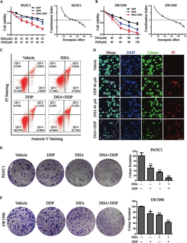
J. Du et al.
                  2
                      failure in pancreatic cancer [10]. Currently, the depletion of               culture plate (NEST Biotechnology) at a density of 1.5 × 10⁴ cells per well in
                      glutathione caused by DDP and the inactivation of glutathione                100 μL medium overnight. Then, different concentrations of DHA and DDP
                      peroxidase were found to play a vital role in its underlying                 (20–45 µM for PANC1 or 40–120 µM for SW1990) were added and
                      mechanism [11, 12], suggesting that combination chemotherapy                 incubated for 24 h, respectively. For the rescue assay, DHA and DDP were
                      based on ferroptosis could be developed to strengthen the                    co-treated with a range of pharmacological inhibitors of specific cell death
                                                                                                   pathways including DFO (100 µM), ferrostatin-1 (fer-1, 1 µM), GSH (0.5 mM),
                      therapeutic effect and reduce the cytotoxicity of DDP.                       Necrosulfonamide (0.5 µM), Z-VAD-FMK (5 µM), XL019 (2.5 µM), JSH-23
                         Due to the high cost and long time needed to develop a new                (10 µM), SB203580 (10 µM), SP600125 (10 µM), and SCH772984 (10 µM) for
                      therapeutic drug, drug-repurposing can be a faster and less costly           24 h. After treatment, 10 μL CCK-8 was added to each well, and incubation
                      alternative approach to the development of new drugs [13].                   continued for 2 h at 37 °C. The absorbance was measured at 450 nm on the
                      Artemisinin (ART), an extract derived from the Chinese plant                 microplate reader. The effect of the drug combination was determined
                      Artemisia annua [14], has been widely used as an antimalarial drug           based on the combination index (CI) computed by CompuSyn software. CI
                      with high safety and efficiency. In addition to the advantages of             values below 1 indicate synergistic effects, whereas CI values above
                      antimalarial effect, ART and its derivatives, including artesunate,          1 suggest an antagonism between the drugs, CI values in the 0.9–1.10
                                                                                                   range mainly indicate additive effects.
                      dihydroartemisinin (DHA), and artemether, have been reported to
                      exhibit strong antiviral, antibacterial, and anticancer activities
                      [15, 16]. DHA, the main active metabolite of ART, has been reported          Live/Dead staining assay
                      to be capable of inhibiting cancer growth in lymphocytic leukemia,           PANC-1 cells were seeded in 6-well plates (2 × 10⁵/well) and cultured for
                      breast cancer, cervical cancer, and liver cancer with its active             24 h. DHA (40 µM) and DDP (40 µM) were added to the indicated culture
                      endoperoxide bridge (R-O-O-R0) [17]. Recent studies have demon-              plate wells for 24 h. Then, cells were harvested and subjected to Calcein-
                                                                                                   AM/PI staining kit (Beyotime, Shanghai, China) at 37 °C for 15 min. Nuclei
                      strated that DHA induced cell death via suppressing JAK2/STAT3,              were counterstained with DAPI (10 μg/mL) for 5 min. Fluorescence
                      JNK1/2, NF-κB, and p38 MAPK signaling pathways [18–20]. Our                  microscope (Nikon) was employed to take photos; PI stained for the dead
                      previous study revealed that DHA represents a promising ther-                cells, and Calcein AM stained for the live cells. Treated cells were also
                      apeutic agent to preferentially target AML cells and induced                 incubating with 5 μL Annexin V-FITC and 10 μL PI for 5 min at room
                      ferroptosis through degradation of ferritin [21]. Considering this           temperature in the dark according to the manufacturer’s instructions
                      fact, we are wondering whether DHA could act synergistic effects             (MultiSciences, Hangzhou, China). Single-cell suspensions were subjected
1234567890();,:

                      with cisplatin through inducing pancreatic cancer cells ferroptosis.         to flow cytometry (ACEA NovoCyte, USA) and the percentage of Live/Dead
                         In this work, we examined the synergistic effect of DHA and DDP,          cells was quantified.
                      and found DHA could intensively strengthen the cytotoxicity effect
                      of DDP by inhibition of cell proliferation and migration, impairing          Colony formation assays
                      mitochondrial functions, thus eventually resulting in ferroptotic cell       PANC-1 and SW1990 cells (1 × 10³/well) were seeded in 12-well plates and
                      death. Mechanistically, we identified that the synergistic effect of          cultured for 24 h. The medium was then replaced with complete culture
                      DHA and DDP is associated with the degradation of GPX4 and the               medium containing DHA (5 µM for PANC1 or 10 µM for SW1990) and DDP
                      accumulation of free iron. Pharmacological or genetic blocking of            (5 µM for PANC1 or 10 µM for SW1990), or a combination of the two
                                                                                                   compounds for an additional 7 days. The colonies were fixed with 4%
                      the free iron accumulation could largely block the synergistic effect        paraformaldehyde after washing with PBS, stained with 0.1% crystal violet
                      of DHA and DDP both in vitro and in vivo. Of note, we firstly present         for 30 min, and then the colonies were imaged and counted by ImageJ
                      the detailed experiments to uncover the mechanism of the                     software.
                      synergistic cytotoxicity of DHA and DDP in treating pancreatic
                      cancer cells and present a prospective therapeutic strategy through
                                                                                                   Edu incorporation assay
                      ferroptosis.                                                                 The 5-ethynyl-2-deoxyuridine (EdU) incorporation assay was carried out to
                                                                                                   test the cell proliferation capacity of PANC1 cells treated with mono DHA
                                                                                                   and DDP or the combination according to the manufacturer’s instructions
                      METHODS                                                                      (Beyotime, Shanghai, China). Images were viewed and captured under a
                      Cell lines                                                                   confocal microscope.
                      The pancreatic cancers cell lines PANC1 and SW1990 were preserved and
                      passaged by our laboratory and maintained in DMEM medium (Hyclone,
                      Logan, UT, USA) containing 10% fetal bovine serum (Gibco, Grand Island,      Cell cycle analysis
                      USA), supplementary with 100 U/mL penicillin and 100 μg/mL streptomy-        Cell cycle distribution was examined by flow cytometry. PANC1 cells were
                      cin at 37 °C in an atmosphere of 5% CO2.                                     cultured in the serum deprivation medium for 24 h to synchronize the cell
                                                                                                   cycle. Following the corresponding treatments, cell cycle distribution was
                                                                                                   detected by Cell Cycle Staining Kit (MultiSciences, Hangzhou, China)
                      Reagents and antibodies                                                      according to the manufacturer’s protocol. Briefly, PANC1 cells were
                      The antibodies to Ferritin Heavy Chain (ab65080), GPX4 (ab125066), to        harvested and fixed with 70% cold ethanol. After fixation, the PI staining
                      NRF2 (ab62352), to MDA (ab27642), to GAPDH (ab181602), and to γ-H2AX         solution with RNase A was added and incubated in dark at 37 °C for 30 min.
                      (ab81299) were obtained from Abcam (Cambridge, MA). The antibody to          Stained samples were tested by flow cytometry (ACEA NovoCyte, USA).
                      TFR (sc-32272) was obtained from Santa Cruz (Dallas, USA). The antibodies
                      to IRP2 (23829-1-AP), to FSP1 (20886-1-AP), and to DRP1 (12186-1-AP) were
                      obtained from Proteintech (Wuhan, China). The antibody to XCT (12691)
                                                                                                   Confocal microscopy assay
                                                                                                   The cells were seeded in a chamber confocal dish. After treatment for 12 h,
                      was obtained from Cell Signaling Technology (Danvers, MA). The antibody
                      to 4-HNE (MAB3249) was obtained from Novus Biologicals (Littleton, CO,       cells were incubated with C11-BODIPY(5 μM) or MitoTracker (100 nM) or
                      USA). The antibody to NCOA4 (203674-T08) was obtained from Sino              RPA (4 μM) in the dark for 30 min, nuclei were counterstained with DAPI
                      Biological (Beijing, China). CCK-8 Assay Kit was obtained from Meilunbio     (10 μg/mL). Then cells were washed three times with PBS and photo-
                      (Dalian, China), Ferrostatin-1, Z-VAD-FMK, and Necrosuifonamide were         graphed under a confocal microscope (Leica, Germany).
                      obtained from Selleck Chemicals (Houston, TX). C11-BODIPY (581/591),
                      MitoTracker were obtained from Thermo Fisher Scientific (Waltham, MA).        Oxygen consumption rate (OCR) determination
                      Desferrioxamine (DFO) and 2ʹ,7ʹ-dichlorofluore scindiacetate (DCF-DA)         The oxygen consumption rate (OCR) was determined by a Seahorse XFe24
                      were purchased from Sigma-Aldrich (St. Louis, USA).                          Bioanalyzer (Seahorse Bioscience). On the first day, PANC1 cells were placed
                                                                                                   in a XFe24 Seahorse Cell Culture Microplate (Seahorse Bioscience) at a
                      Cell viability assay                                                         density of 3 × 10⁴ cells/well. Meanwhile, the XFe24 sensor cartridges were
                      The cytotoxicity of DHA and DDP in pancreatic cancer cells was evaluated     hydrated. On the following day, the cells were treated for 8 h with DHA and
                      by the CCK8 assay Kit. PANC1 and SW1990 cells were seeded in a 96-well       DDP using aforementioned concentrations, then the cell media was changed
                                                                                                   to basic Seahorse DMEM containing 10 mM glucose, 2 mM glutamine, and

                                                                                                                          Cell Death and Disease (2021)12:705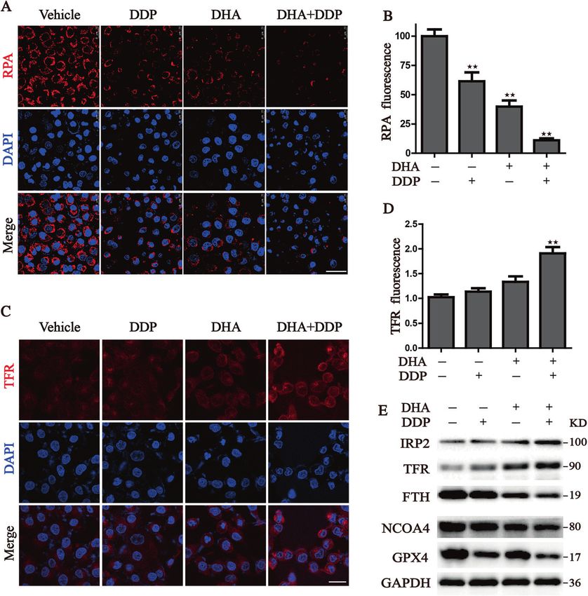
J. Du et al.
                                                                                                                                                            3
1 mM sodium pyruvate, plates were then incubated at 37 °C in a CO2-free        dewaxing, dehydration, and antigen retrieval. After being washed with PBS
incubator for 1 h before placing in the analyzer; OCR was measured with        three times, the slides were treated with 3% hydrogen peroxide for 15 min
sequential injection of 1.5 μM oligomycin, 1.5 μM carbonyl cyanide-            for blocking the endogenous peroxidase activity, then blocked with 5% BSA
chlorophenylhydrazone (CCCP), and 0.5 μM Antimycin A/Rotenone.                 for 15 min at room temperature. Subsequently, anti-TFR antibody (1:50) and
                                                                               anti-Ki67 antibody (1:100) were used for incubation at 4 °C overnight. The
                                                                               streptavidin peroxidase method was used for signal detection and then
Determination of ROS production                                                stained by diaminobenzidine (DAB) and counterstained with hematoxylin,
Cellular ROS level and mitochondria-derived superoxide were measured           the sections were observed and photographed under the light microscope.
using 2ʹ,7ʹ-dichlorofluore scindiacetate (DCF-DA) and mitoSOX probe,
respectively. Following indicated treatments, PANC1 and SW1990 cells
were washed with PBS and stained with DCF-DA (4 μM) or mitoSOX (3 μM)          Statistical analysis
in the dark at 37 °C for 30 min. Cells were then washed with PBS, and the      All statistical calculations were performed using GraphPad Prism (version
fluorescence intensity was measured by flow cytometry.                           7.0). Results are represented as mean ± SD. The differences between the
                                                                               two groups were performed by the Student’s t-test. Comparisons among
                                                                               multiple groups were analyzed by one-way ANOVA. Statistical significance
Immunofluorescence                                                              was defined at ★P < 0.05, ★★P < 0.01, compared to the corresponding
Cells were grown on glass coverslips placed in the 24-well culture plate.      control.
After designated treatments, cells were followed by fixation with 4%
formaldehyde and permeabilization with 1% Triton X-100. Fixed cells were
washed with PBS and blocked in 5% BSA, and then incubated with primary
antibodies (Rabbit anti-MDA, Mouse anti-4HNE, Mouse anti-TFR, Rabbit           RESULTS
anti-γ-H2AX) at 4 °C overnight. After washing twice with PBS, Alexa Fluor      DHA exerts a synergistic effect with DDP, and significantly
488-labeled anti-rabbit IgG and Alexa Fluor 594-labeled anti-mouse             reduces its effective concentrations
antibody (Thermo, Waltham, MA) were added on the glass coverslips for          Sustained DDP treatments for pancreatic cancer cells are
1 h, nucleus was stained with DAPI. Subsequently, monolayer cell images        ineffective and may lead to high drug resistance. Considering
were observed and recorded under a laser scanning confocal microscope.         the previous study mentioning that DHA exerts a high capacity in
                                                                               cancer treatment [21], we wondered whether DHA combination
Lentiviral packaging and transduction                                          with DDP could effectively optimize its antitumor activity. Firstly,
Full-length FTH was ordered from Sino Biological (Beijing, China) and          we treated human pancreatic cancer cell lines (PANC1 and
amplified by PCR (AP131-11, TransGen Biotech), then subcloned into pLVX-        SW1990) with DHA and DDP, and tested the cytotoxicity against
IRES-Neo lentivirus vector (Takara, Dalian, China) by ClonFast Seamless        the two K-Ras mutant PDAC cell lines through CCK-8 assay. The
Cloning kit (Obio, Nanjing, China). The recombinant lentiviral plasmid was     combination index was calculated by Compusyn software. The
verified by sequencing. The recombinant lentiviral plasmid was co-
transfected with pMD2.G, pSPAX2 into 293T cells to produce recombinant         results showed that DHA and DDP each inhibited the growth of
lentiviral. After 72 h of transfection, the supernatants were collected,       pancreatic cancer cells in a dose-dependent manner. In addition,
centrifuged, and filtered through a 0.4 μm filter. To generate stably            they exerted a synergistic effect with the increase of the
transfected cell lines, PANC1 cells (8 × 10⁴) were seeded in 24-well plates.   concentrations, suggesting that DHA may be used as novel drug
After 12 h, the culture medium was removed, and cells were transfected         candidates to enhance the efficacy of DDP in pancreatic cancer
with the corresponding lentivirus. After 2 days of transfection, 1 mg/mL       treatment (Fig. 1A, B). The synergetic effect was obtained when
G418 was added for selection for 7 days. Then the stable cells were            40 µM DDP was combined with 40 µM DHA for PANC1, and 60 µM
maintained in 0.2 mg/mL G418. The transduction efficiency was evaluated         DDP was combined with 60 µM DHA for SW1990. We utilized the
by western blot analysis.                                                      Annexin V-FITC/PI assay and Calcein-AM/PI staining to further
                                                                               clarify the synergetic effect of DHA and DDP. Both of the
Western blotting                                                               experiments demonstrated that the combination of DHA and DDP
Following treatment, the cells were harvested and lysed in RIPA buffer on      synergistically increased the amounts of dead cells and decreased
ice for 10 min. The protein concentration was quantified using BCA Protein      the amounts of viable cells (Fig. 1C, D). In addition, we examined
Assay Kit. Subsequently, equal amounts of protein were separated by 10%
                                                                               the combined effect of DHA and DDP on the capacity of long-term
SDS-PAGE and transferred to PVDF membranes. The membranes were
blocked with 5% non-fat milk for 1 h at room temperature and incubated
                                                                               cell viability by the clonogenic survival assay in PANC1 and
with the primary antibodies at 4 °C overnight. After being washed three        SW1990 cells. As a result, clone clusters of the two pancreatic
times with TBST, the membranes were incubated with the secondary               cancer cell lines exposed to the combinative treatment were less
antibodies at room temperature for 1 h and washed again. The blots were        and smaller compared with either agent alone after culture for
visualized using an ECL-Plus chemiluminescence detection kit.                  7 days (Fig. 1E, F). Collectively, these results suggested that DHA
                                                                               remarkably strengthens the anticancer effect of DDP.
In vivo tumor model
Six-week-old female BALB/c nude were purchased from Jiangsu Jizui Yao          Combination of DHA and DDP inhibits the proliferation and
Kang Biological Technology Co. LTD. PANC1 cells were harvested and             induces DNA damage in pancreatic cancer cells
washed with cold PBS three times, then the cell density was adjusted to        Next, we wonder whether the combination treatment could
2 × 10⁷/mL in cold DMEM. All animals were injected subcutaneously with         suppress pancreatic cancer cell proliferation. As expected, either
2 × 10⁶ cells into the right dorsal flank. Once the tumors reached              DHA or DDP treatment moderately inhibited EdU incorporation
80–100 mm3, mice were randomly allocated into groups and treated with          into cell nuclei, while DHA co-administration with DDP caused a
vehicle, DHA (50 mg/kg), DDP (5 mg/kg), DHA plus DDP, and the                  remarkable reduction in the percentage of EdU+ cells, suggesting
combination plus DFO (100 mg/kg), respectively.
                                                                               that DHA in combination with DDP significantly disrupted
                                                                               pancreatic cancer growth (Fig. 2A, B). We have previously
Hematoxylin-eosin (H&E) staining                                               demonstrated that DHA suppresses the proliferation of AML cells
Tumors dissected from mice were fixed in 4% paraformaldehyde,                   by arresting the cell cycle [21]. To further investigate the
embedded into paraffin. The embedded samples were cut to 4 μm
                                                                               mechanism of growth inhibition, we performed flow cytometry
thickness, deparaffinized and stained with H&E (Sigma) routinely. Stained
sections were viewed and photographed under a microscope.                      to analyze the cell cycle distribution. As shown in Fig. 2C, D,
                                                                               significant changes in the distribution of cell cycle were found in
                                                                               PANC1 cells under the treatment of DHA and DDP, with a
Immunohistochemistry (IHC)                                                     dramatically increased proportion of G2/M phase and decreased
Tissues were fixed with 4% paraformaldehyde and embedded in paraffin. The        proportion of G0/G1. As the primary anticancer mechanism of
embedded block tissues were cut into 4 μm sections and followed by
                                                                               DDP is an interaction with DNA to form intrastrand crosslink

Cell Death and Disease (2021)12:705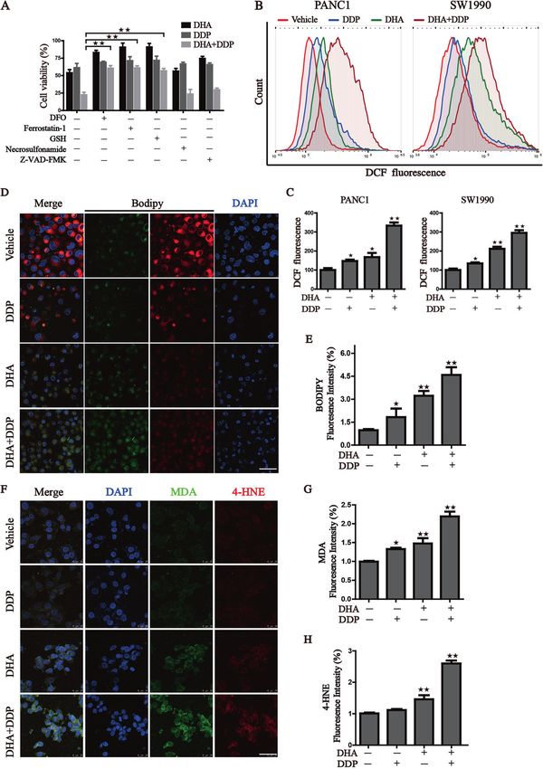
J. Du et al.
4

    Fig. 1 Synergistic antitumor effect of DHA in combination with DDP. A, B Pancreatic cancer cells were subjected to different concentrations
    of DHA and DDP treatment for 24 h and cell viability was detected by CCK-8 assay. Combination index (CI) values were calculated by
    Compusyn software. C PANC1 cells were subjected to 40 µM DHA or/and DDP treatment for 24 h and followed by Annexin V-FITC/PI assay. D
    PANC1 cells were pretreated with 40 µM DHA or/and DDP for 24 h and subjected to the Calcein-AM/PI staining (Calcein AM: live cells, PI: dead
    cells). Scale bar: 100 µm. E, F The colony formation assay of PANC1 and SW1990 cells were performed under mono or combination treatment
    of DHA (5 µM for PANC1 or 10 µM for SW1990) and DDP (5 µM for PANC1 or 10 µM for SW1990) for 7 days. The representative images and the
    corresponding quantitative histograms from three independent experiments are shown (values represented mean ± SD. ★P < 0.05, ★★P < 0.01
    versus control).

                                                                                                 Cell Death and Disease (2021)12:705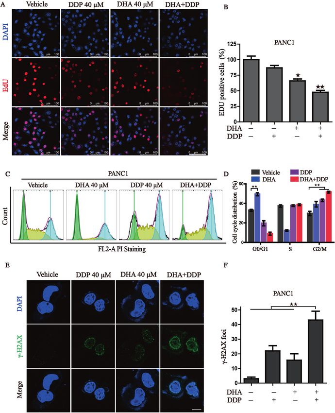
J. Du et al.
                                                                                                                                              5

Fig. 2 Combinative treatment of DHA and DDP inhibits proliferation and induces DNA damage in pancreatic cancer cells. A, B Cell
proliferation of PANC1 was detected by 5-ethynyl-2′-deoxyuridine (EdU) incorporation assay under mono or combination treatment of DHA
and DDP for 12 h. Representative images and statistical histograms are shown. Scale bar: 100 µm. C, D PANC1 cells were treated with mono or
combination of DHA and DDP for 12 h, cell cycle distribution was determined by flow cytometry. E, F PANC1 cells were treated with mono or
combination of DHA and DDP for 12 h and stained with γ-H2AX antibody. DAPI was used for nucleus staining. Images were acquired with
confocal laser scanning microscopy. Quantification of γ-H2AX immunofluorescence was shown on the right. Scale bar: 10 µm. ★P < 0.05, ★★P
< 0.01 versus control.

adducts, resulting in elevated DNA damage and cell cycle arrest.       damage, characterized by the dramatically increased amounts of
We further detected the expression of γ-H2AX, a marker in the          γ-H2AX foci (Fig. 2E, F). Taken together, our data support a
early stage of DNA damage, by immunofluorescence. Results               synergistic role of DHA and DDP in repressing the proliferation
showed that DHA sensitizes PANC1 cells to DDP-induced DNA              and inducing DNA damage of pancreatic cancer cells.

Cell Death and Disease (2021)12:705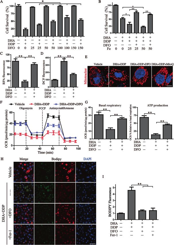
J. Du et al.
6

    Fig. 3 DHA co-treatment with DDP synergistically impairs mitochondrial homeostasis. A, B Observation of the changes in mitochondrial
    morphology. The treated PANC1 cells were stained with MitoTracker probe (100 nM) and DAPI (10 μg/mL) and then photographed by confocal
    laser microscope. Scale bars: 10 μm. C, D Mitochondrial oxygen consumption rate (OCR) was carried out with a Seahorse analyzer after the
    sequential addition of oligomycin, FCCP, and Antimycin A/ Rotenone. The OCR values of maximal respiration, ATP and non-mitochondrial
    oxygen consumption were normalized to the basal respiration. E, F Flow cytometry was performed to measure mitochondrial ROS by
    mitoSOX probe (3 μM) (values represented mean ± SD. ★P < 0.05, ★★P < 0.01 versus control).

    Combined treatment of DHA and DDP synergistically impairs              focused on mitochondrial morphology, which is an upstream
    mitochondrial homeostasis                                              event of mitochondrial dysfunction. Following the administration
    Given the links between DHA and mitochondrial dysfunction, we          of DHA/DDP, the mitochondria became disorganized, smaller, and
    then tested whether DHA combined with DDP would amplify                networks collapsed around the perinuclear region (Fig. 3A), which
    mitochondrial dysfunction in pancreatic cancer cells. We first          is more serious than the mono-treatment group. Statistical

                                                                                              Cell Death and Disease (2021)12:705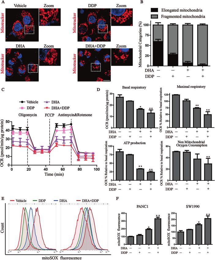
J. Du et al.
                                                                                                                                                7
analysis revealed that administration of DHA/DDP decreased the         IRP2, FTH, and FTL were regulated by DHA, and the iron-starvation
proportion of elongated mitochondria, and induced a pronounced         effect was further enhanced under DDP co-treatment in
increase of fragmented mitochondria (Fig. 3B). Once mitochon-          PANC1 cells. Besides, DDP synergistic with DHA to induce the
drial dysfunction occurs, mitochondrial oxidative phosphorylation      degradation of GPX4 and NCOA4, an autophagy cargo receptor
process may be affected. We then explored the mitochondrial            that binds ferritin for degradation and releases free iron (Fig. 5E).
respiratory capacity through Seahorse XFe24 Extracellular Flux         Collectively, our results demonstrate that combinative treatment
Analyzer (Fig. 3C). The OCR, an indicator of mitochondrial             triggered an evident accumulation of free iron pool, resulting in
respiration, represented that DHA acted synergistically with DDP       the initiation of ferroptosis.
to suppress basal respiratory, maximal respiratory, non-
mitochondrial oxygen consumption and thus lead to the                  Pharmacologic depletion of the free iron reservoir attenuates
reduction of ATP production (Fig. 3D). We also detected                the DHA/DDP-induced ferroptosis
mitochondrial ROS production, which originates from the electron       Due to the significantly activated iron-starvation stress, we, there-
leakage of the electron transport chain and results in cellular        fore, raise the hypothesis that the synergistic cytotoxicity of DHA/
oxidative stress. The results determined by MitoSOX probe              DDP may result from free iron accumulation. Firstly, iron chelator
staining validated that DHA reinforced the mitochondrial ROS           DFO was utilized to verify this hypothesis that DFO inhibited the
production under the challenge of DDP (Fig. 3E, F). These results      synergistic cytotoxicity of DHA/DDP-induced ferroptotic cell death,
demonstrate that the combinative treatment destroys mitochon-          as evidenced by the following: (i) DFO mitigated DHA/DDP-induced
drial homeostasis and facilitates the accumulation of                  ferroptosis in a concentration-dependent manner (Fig. 6A), (ii) Fe2+
mitochondria-derived ROS.                                              addition accelerated DHA/DDP-induced ferroptotic cell death which
                                                                       was able to be blocked by DFO treatment (Fig. 6B), and (iii) DFO also
Ferroptosis contributes to the cytotoxic effects in PDAC cells         exhibited a strong effect on the alleviation of free iron accumulation
under the treatment with DHA and DDP                                   and cellular ROS peroxidation (Fig. 6C, D). Moreover, we confirmed
To further characterize the basis of cell death induced by DHA/        that DFO could ameliorate the mitochondrial morphogenesis
DDP, we treated PDAC cells with mono- or combination treatment         change as effectively as mitoQ (Fig. 6E). Notably, DFO attenuated
of DHA and DDP in the absence or presence of several cell death        the mitochondrial dysfunction resulting from DHA/DDP treatment
inhibitors, including deferoxamine (DFO, iron-chelating agent),        and rescued the mitochondrial respiration and ATP production
ferrostatin-1 (fer-1, ferroptosis inhibitor), Z-VAD-FMK (apoptosis     (Fig. 6F, G), as evidence by the detection of oxygen consumption.
inhibitor), Necrosulfonamide (necroptosis inhibitor) and GSH. Iron     These data indicated that free iron accumulation resulting from
chelating agent, ferroptosis inhibitor and GSH significantly            DHA/DDP treatment acts upstream of mitochondrial dysfunction.
alleviate the decline of cell viability challenge by DHA and DDP       Lastly, the production of lipid peroxides was monitored by the
(Fig. 4A). Western blot results also verified that pyroptosis and       staining of Bodipy C11 probe, images showed that DFO ameliorated
necroptosis may not participate in the cell death induced by DHA       the production of lipid ROS, as effective as Fer-1 (Fig. 6H, I).
and DPP (Fig. S1). Recent studies have demonstrated that DHA           Collectively, these data indicate that pharmacological chelation free
induced cell death via suppressing JAK2/STAT3, NF-κB, and MAPK         iron rescues DHA/DDP-mediated ferroptotic cell death and mito-
signaling pathways. Then, XL019 (JAK2 inhibitor), JSH-23 (NF-κB        chondrial dysfunction.
inhibitor), SB203580 (p38 inhibitor), SP600125 (JNK inhibitor),
SCH772984 (ERK inhibitor) were incubated with DHA and DPP.             Reconstituted expression of FTH contributes to the tolerance
Inhibition of NF-κB, JAK2, and ERK could further strengthen the        of DHA/DDP-induced ferroptosis
cytotoxicity effect of DHA/DPP (Fig. S2), demonstrating that not       Ferritin heavy chain (FTH) not only harbors ferroxidase activity
these pathways but ferroptosis contribute to the main cytotoxic        which could catalytically oxidize Fe2+ to inactive Fe3+, but also
effect induced by DHA and DDP. The generation of lipid peroxides       plays a vital role in maintaining iron homeostasis by storing iron in
and accumulation of free iron are two major hallmarks of               a soluble, non-toxic form for its detoxification. Previously, studies
ferroptosis. Therefore, we quantified the oxidative stress damage       have shown that free iron released from lysosomal degradation of
by the following assays: (i) cellular total ROS generation indicated   ferritin plays a critical role in ferroptosis [21, 23, 24]. Given that
by DCF-DA probe (Fig.4B, C), (ii) lipid ROS measured by BODIPY         synergistic cytotoxicity of DHA/DDP resulted from FTH degrada-
C11 fluoroprobe (Fig. 4D, E), and (iii) secondary products of lipid     tion, a process that gives rise to the free iron accumulation. We
peroxidation detected by immunofluorescence staining of MDA             then discovered whether genetically enforced expression of FTH
and 4-HNE (Fig. 4F–H). These results indicated that combinative        could abolish the synergistic cytotoxicity of DHA/DDP. Firstly, the
treatment triggered an evident increase in cellular ROS production     FTH enforced expression PANC1 cells were constructed and
and lipid peroxide generation.                                         transfection efficiency was verified by western blot (Fig. S3). Then,
   To check whether the increase of lipid peroxidation is due to       the FTH-PANC1 cells were subjected to different concentrations of
the increase of free iron pool. We examined the changes in iron        DHA and/or DDP. Cell viability assay indicated that FTH efficiently
metabolism in PDAC cells treated with DDP and DHA. We first             relieved the cytotoxicity of DHA/DDP (Fig. 7A). Furthermore, the
utilized the selective yield fluorescence probe RPA to monitor the      results from western blot assay manifested that this cytoprotec-
free iron level. The RPA staining images captured by confocal          tion effect mainly originates from the lesser degradation of FTH,
microscopy reflected that DDP combined with DHA significantly            but not from modulating other ferroptosis regulators, such as
resulted in the accumulation of labile iron level manifested by the    GPX4 or XCT (Fig. 7B). FTH reconstituted PANC1 cells also
decreased fluorescence of RPA (Fig. 5A, B). TFR (Transferrin            exhibited decreased intracellular labile iron pool, reduced cellular
receptor) which imports iron from the extracellular environment        ROS lever and diminished lipid peroxides generation when co-
into cells, is a specific ferroptosis marker and can be used to label   treated with DHA and DDP (Fig. 7C–G). Accordingly, we come to a
cells undergoing ferroptosis [22]. We then detected the TFR            conclusion that reconstituted expression of FTH is able to abolish
expression via immunofluorescence staining (Fig. 5C, D). Consis-        synthetic cytotoxicity of DHA/DDP through chelating intracellular
tent with RPA staining, accumulated TFR is located on the cell         labile iron pool.
membrane under treatment with DHA and DDP, leading to the
dramatic accumulation of unchelatable iron. We further investi-        The combination of DHA/DDP treatment inhibits the growth
gated the expression of key proteins associated with iron              of xenografts in vivo
homeostasis in pancreatic cancer cells. Similar results were           In order to study the in vivo therapeutic potential of DHA/DDP
observed that proteins related to iron metabolisms, such as            combination, we established the subcutaneous tumor model

Cell Death and Disease (2021)12:705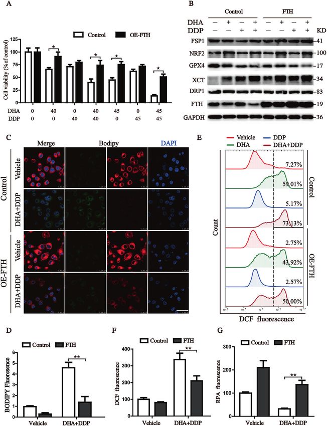
J. Du et al.
8
Fig. 4 Ferroptosis contributed to the cytotoxic effects in PDAC cells under the treatment with DHA and DDP. A Cell viability was detected
by CCK8 assay under treatment of DHA and DDP (45 μM) in the presence or absence of several cell death inhibitors. B, C Flow cytometry was
performed to measure cellular ROS, and quantitation of fluorescence intensities was shown. D, E Representative images of BODIPY staining
were photographed by confocal laser microscope with the designed treatment. Scale bars: 50 μm. Statistical results of the fluorescence
intensities were shown on the right. F–H Immunofluorescence staining of MDA and 4-HNE were captured by confocal laser microscope with
the designed treatment. Scale bars: 50 μm. Statistical results of the fluorescence intensities were shown on the right (values represented mean
± SD. ★P < 0.05, ★★P < 0.01 versus control).
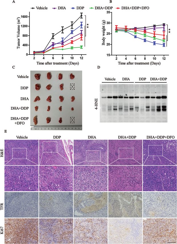
bearing PANC1 cells and randomly divided them into five groups pronounced in DHA/DDP combination-treated group compared to
with indicated treatment. Mirroring our in vitro data, the in vivo DDP or DHA mono-treated group (Fig. 8A, C). Modulating iron
data further demonstrated that administration of DHA or DDP homeostasis through pharmacological treatment of iron chelator
suppressed tumor growth and the antitumor efficacy was more DFO could weaken the effect of DHA/DDP in vivo. It is well known
Cell Death and Disease (2021)12:705J. Du et al.
9
Fig. 5 Iron accumulation in pancreatic cancer cells induced by the combination treatment of DHA and DDP. A, B Intracellular Fe2+ was
measured by the staining of RPA and photographed by the confocal microscope after indicated treatment. Scale bars: 50 μm. C, D Detection of
cell surface TFR levels by immunofluorescence staining of PANC1 cells after indicated treatment. E The expression of iron metabolism proteins
in DHA- and DDP-treated cells, which was determined by western blot assay (values represented mean ± SD. ★P < 0.05, ★★P < 0.01 versus
control).
that DDP exhibits severe side-effects, we also focus on animal DISCUSSION
weight as an indicator of drug toxicity. As shown in Fig. 8B, DDP- Distinct lethal subroutines are designed for selectively targeting
treated animals exhibited a significant loss of weight, while cancer cells via diverse regulated cell death (RCD) processes,
administration of DHA did not exhibit obvious toxicity with little including apoptosis, ferroptosis, pyroptosis, necroptosis, etc. Each
loss of mice’s body weight. Unexpectedly, DDP co-treatment with of the RCD processes is modulated by unique signal transduction
DHA not only reduced the tumor growth but also partly alleviated pathways and differentially affect tumor response to treatment.
the toxicity of DDP, suggesting higher safety for clinical use. The The most extensively studied type of RCD is apoptosis, whose
immunoblotting assay of lysates from tumor tissues also verified activation relies on the cleavage of intracellular proteases’ caspase.
the in vitro observations that DHA co-administration with DDP However, the clinical application of the therapeutic approach
synergistically induced lipid peroxidation locally, with the through inducing apoptosis in oncology remains an insurmoun-
dramatic increasing of 4HNE (Fig. 8D). In addition, the IHC assay table challenge for the high resistance rate [25]. Thus, targeting
displayed that DHA acted synergistically with DDP to increase the non-apoptotic RCD processes may provide an alternative strategy
area of necrosis, augment the expression of TFR, and decrease the for suppressing tumor growth.
Ki67 staining. Whereas, administration of DFO could significantly Ferroptosis is a non-apoptotic, RCD first observed in cancer cells
reverse these phenotypes by suppressing ferroptosis (Fig. 8E). Our with oncogenic Ras mutation in 2012 [26], which is characterized
findings show that the combination of DHA with DDP is not only as a catastrophic accumulation of free iron and unrestricted lipid
tolerable but also beneficial, which may act as a promising peroxidation. Several signal transduction pathways including iron
therapeutic agent for treating PDAC. metabolism, GSH-GPX4, and FSP1-COQ10 constitute the core
Cell Death and Disease (2021)12:705J. Du et al.
10
Fig. 6 Pharmacologic depleting of the free iron reservoir attenuates the DHA/DDP-induced ferroptosis. A PANC1 cells were treated with
DHA/DDP (45 μM) in the presence or absence of DFO. B PANC1 cells were treated with DHA/DDP (30 μM) in the presence or absence of DFO
and Fe2+, cell survival was detected by CCK8. C, D After indicated treatment, PANC1 cells were loaded with RPA or DCF-DA probe for 30 min,
and fluorescence intensities were detected by microplate spectrophotometer and normalized to the corresponding cell number. E
MitoTracker Red labeled pancreatic cancer cells were subjected to the confocal microscope for observing the changes of mitochondrial
morphology after the indicated treatment. Scale bars: 10 μm. F, G OCR of PANC1 cells was carried out with a Seahorse analyzer after the
addition of oligomycin, FCCP, and Antimycin A/Rotenone. The basal respiration and ATP production were calculated on the right. H, I To assess
lipid ROS production, pancreatic cancer cells were treated with DHA and DDP with or without DFO, fer-1. Treated cells were loaded with
BODIPY C11 probe for 30 min followed by confocal laser microscope. Scale bars: 50 μm. Statistical results of the fluorescence intensities were
shown on the right (values represented mean ± SD. ★P < 0.05, ★★P < 0.01 versus control).
Cell Death and Disease (2021)12:705J. Du et al.
11
Fig. 7 Reconstituted expression of FTH contributes to the tolerance of DHA/DDP-induced ferroptosis. A PANC1 cells were treated with
various concentrations of DHA and DDP for 24 h, followed by CCK8 assay. B Proteins isolated from the treated cells were assayed by western
blot to detect the expression of ferroptosis-related proteins. C, D Indicated cells were stained with BODIPY C11 probe for the assessment of
lipid ROS production and observed under the confocal laser microscope. Scale bars: 50 μm. Cells with indicated treatment were stained with
RPA and DCF-DA probe for 30 min and followed by flow cytometry to assess intracellular Fe2+ and ROS level (E–G) (values represented mean
± SD. ★P < 0.05, ★★P < 0.01 versus control).
molecular mechanism of ferroptosis. Increasing evidence has selectively targeted by erastin through inducing ferroptosis
revealed that inducing ferroptosis could act as a promising [28, 29]. Activation of the RAS/RAF-MEK/ERK signaling pathway
therapeutic strategy and eliminate the resistance of cancer cells to is required for erastin-induced cell death, while restraining RAS or
drug-induced apoptosis [27]. The mutated RAS family genes are its downstream signaling molecules reverses the lethality against
the most common oncogenes in pancreatic cancer that could be cancer cells [30, 31].
Cell Death and Disease (2021)12:705J. Du et al.
12
Fig. 8 The combination of DHA and DDP inhibits the growth of xenograft in vivo. A, B Xenografts were established in mice and treated
with vehicle, DHA, DDP, DHA/DDP, and DHA/DDP plus DFO, respectively. The tumor volume and body weight were measured every 2 days. C
The dissected xenografts were photographed at the end of the experiment. D Western blots analysis for 4-HNE on lysates isolated from
xenografts in different groups. E H&E staining and IHC analysis for TFR, Ki67 in indicated tumor specimens. Scale bars: 200 μm (values
represented as mean ± SD. ★★P < 0.01 versus control).
Previous studies have demonstrated that DHA and cisplatin and suppressed the proliferation of pancreatic cancer cells. Further
exert synergistic anti-angiogenic and anti-tumor effects through experiments revealed that the cell death could be blocked by
inducing apoptosis [32, 33]. In this study, we found DHA could ferroptosis inhibitor, iron chelator, and lipid ROS scavenger, rather
effectively optimize its antitumor activity of DDP, and significantly than apoptosis or necroptosis inhibitors. Importantly, accumula-
reduced its effective concentrations. In addition, combination of tion of lipid peroxides detected by fluorescence staining of MDA,
DHA and DDP dramatically impaired mitochondrial homeostasis 4-HNE, or BODIPY C11 was observed prior to the onset of cell
Cell Death and Disease (2021)12:705J. Du et al.
13
death, which could be rescued by pharmacological chelation free cells and exhibit the synergistic effect through increasing the
iron or genetically enforced expression of FTH. These results, intracellular free iron. Therefore, our study provides evidence that
consistent with a previous study, manifested that it is a promising DHA may be a promising adjuvant to improve the cisplatin-based
strategy to induce ferroptosis in PDAC [34]. Collectively, our data treatment of patients with pancreatic cancer. We also provide a
together with other preclinical findings support the notion that framework for further understanding and targeting of ferroptosis
induction of ferroptosis might constitute a suitable strategy in cancer therapy.
against PDAC tumors.
Iron is an essential micronutrient and enables the function of
vital enzymes [35]. Available amount of free iron is essential for CONCLUSION
the process of electron transport, cellular respiration, cell This study presents strong evidence that DHA could intensively
proliferation and differentiation, and gene expression regulation strengthen the cytotoxicity effect of DDP and significantly reduce
in cancer cells. However, overload of labile iron pool is its effective concentrations both in vitro and in vivo. In particular,
biochemically dangerous with the high capacity to promote the ferroptosis contributes to the main cytotoxic effects in PDAC cells
formation of ROS via the Fenton reaction, leading to severe under the challenge of DHA and DDP. Our results provide
damage to the main cellular biomolecules. As reported, free iron experimental evidence that DHA acts synergistically with DDP and
levels can be used as a criterion to determine which type of cancer renders PDAC cells vulnerable to ferroptosis, which may act as a
is more prone to benefit from ferroptosis-promoting therapies promising therapeutic strategy.
[36]. Iron-rich tumors (such as PDAC [37], HCC [38], breast cancer
[39], and NSCLC [40]) could be more suitable to therapies of
inducing ferroptosis, and more sensitive to agents that promote DATA AVAILABILITY
ferroptosis. Additionally, subsequent studies have identified that All data generated during this study are included either in this article or in the
mutant RAS signaling enriches the cellular iron reservoir via supplementary information files.
transcriptional regulation of iron metabolism genes [30]. In the
present study, we have provided evidence to support a means to
induce ferroptosis in PDAC via modulation of iron metabolism. REFERENCES
First, DDP combined with DHA significantly accelerated the 1. Hingorani SR, Petricoin EF, Maitra A, Rajapakse V, King C, Jacobetz MA, et al.
Preinvasive and invasive ductal pancreatic cancer and its early detection in the
accumulation of labile free iron reflected by the decreased
mouse. Cancer Cell. 2003;4:437–50.
fluorescence of RPA, and increased expression of TFR, which 2. Bryant KL, Mancias JD, Kimmelman AC, Der CJ. KRAS: feeding pancreatic cancer
subsequently results in lipid peroxidation. Second, DFO mitigated proliferation. Trends Biochem Sci. 2014;39:91–100.
DHA/DDP-induced ferroptosis in a concentration-dependent 3. Buscail L, Bournet B, Cordelier P. Role of oncogenic KRAS in the diagnosis,
manner, while Fe2+ addition accelerated DHA/DDP-induced prognosis and treatment of pancreatic cancer. Nat Rev Gastroenterol Hepatol.
ferroptotic cell death which was able to be blocked by DFO 2020;17:153–68.
treatment. Third, reconstituted expression of FTH is able to abolish 4. Tsai LH, Hsu KW, Chiang CM, Yang HJ, Liu YH, Yang SF, et al. Targeting interleukin-
synergistic cytotoxicity of DHA/DDP through chelation intracellular 17 receptor B enhances gemcitabine sensitivity through downregulation of
transitional iron pool. Lastly, pharmacological treatment of iron mucins in pancreatic cancer. Sci Rep. 2020;10:17817.
5. Siegel RL, Miller KD, Jemal A. Cancer statistics, 2019. CA Cancer J Clin.
chelator DFO could weaken the effect of DHA/DDP on ferroptosis
2019;69:7–34.
in vivo. Our data show that the combination of DHA with DDP is 6. Bebber CM, Müller F, Prieto Clemente L, Weber J, von Karstedt S. Ferroptosis in
not only tolerable but could also be beneficial through inducing cancer cell biology. Cancers 2020;12:164.
ferroptosis. 7. Ferreira JA, Peixoto A, Neves M, Gaiteiro C, Reis CA, Assaraf YG, et al. Mechanisms
DDP is still used as a mainstay of chemotherapeutic agent in of cisplatin resistance and targeting of cancer stem cells: adding glycosylation to
combination with other drugs or radiotherapy for pancreatic the equation. Drug Resist Updat. 2016;24:34–54.
cancer therapy [41]. However, DDP is commonly associated with 8. Osanto S, Bukman A, Van Hoek F, Sterk PJ, De Laat JA, Hermans J. Long-term
acquired drug resistance and high toxicity in clinical settings. effects of chemotherapy in patients with testicular cancer. J Clin Oncol.
Previous research regards that DDP leads to DNA injury and 1992;10:574–9.
9. Choi YM, Kim HK, Shim W, Anwar MA, Kwon JW, Kwon HK, et al. Mechanism of
ultimately induces apoptosis [42]. Recent studies have discovered
cisplatin-induced cytotoxicity is correlated to impaired metabolism due to
that DDP could induce GSH depletion and GPX4 inactivation, mitochondrial ROS generation. PLoS ONE. 2015;10:e0135083.
which emerges as an inducer of ferroptosis [12, 43]. Similarly, DDP- 10. de Oliveira G, Freire PP, Cury SS, de Moraes D, Oliveira JS, Dal-Pai-Silva M, et al. An
resistant or platinum-tolerant cancer cells were also shown to integrated meta-analysis of secretome and proteome identify potential bio-
exhibit increased vulnerability to ferroptosis [44]. In our present markers of pancreatic ductal adenocarcinoma. Cancers 2020;12:716.
work, we observed that DDP acts synergistically with DHA to 11. Liu Q, Wang K. The induction of ferroptosis by impairing STAT3/Nrf2/
induce oxidative stress which originated from impaired mitochon- GPx4 signaling enhances the sensitivity of osteosarcoma cells to cisplatin. Cell
drial homeostasis. Mechanically, DHA acts synergistically with DDP Biol Int. 2019;43:1245–56.
to suppress mitochondrial oxidative phosphorylation process 12. Guo J, Xu B, Han Q, Zhou H, Xia Y, Gong C, et al. Ferroptosis: a novel anti-tumor
action for cisplatin. Cancer Res Treat. 2017;50:445–60.
through inhibiting basal respiratory, maximal respiratory, non-
13. Frantzi M, Latosinska A, Mokou M, Mischak H, Vlahou A. Drug repurposing in
mitochondrial oxygen consumption, thus leading to the reduction oncology. Lancet Oncol. 2020;21:e543.
of ATP production and enhancement of mitochondrial ROS 14. Miller LH, Su X. Artemisinin: discovery from the Chinese herbal garden. Cell
generation. Iron depletion rescued the mitochondrial respiration 2011;146:855–8.
and ATP production under the challenge of DHA/DDP, which 15. Flobinus A, Taudon N, Desbordes M, Labrosse B, Simon F, Mazeron MC, et al.
indicated that free iron accumulation originating from DHA/DDP Stability and antiviral activity against human cytomegalovirus of artemisinin
treatment acts upstream of mitochondrial dysfunction. In addition, derivatives. J Antimicrob Chemother. 2014;69:34–40.
we found that co-treatment of DHA with DDP synergistically 16. Chen HH, Zhou HJ, Fang X. Inhibition of human cancer cell line growth and
decreased the expression of GPX4. This may be another human umbilical vein endothelial cell angiogenesis by artemisinin derivatives
in vitro. Pharmacol Res. 2003;48:231–6.
mechanism involved in the activation of ferroptotic cell death.
17. Luan S, Zhong H, Zhao X, Yang J, Jing Y, Liu D, et al. Synthesis, anticancer
Furthermore, several completed clinical trials (NCT00764036, evaluation and pharmacokinetic study of novel 10-O-phenyl ethers of dihy-
NCT02353026) and ongoing clinical trials (NCT02633098 and droartemisinin. Eur J Med Chem. 2017;141:584–95.
NCT03093129) have shown the efficiency and great tolerance of 18. Hu W, Chen SS, Zhang JL, Lou XE, Zhou HJ. Dihydroartemisinin induces autop-
artemisinins in patients with solid tumors. In addition to inducing hagy by suppressing NF-κB activation. Cancer Lett. 2014;343:239–48.
apoptosis, both DHA and DDP can trigger ferroptosis in cancer
Cell Death and Disease (2021)12:705J. Du et al.
14
19. Yan X, Li P, Zhan Y, Qi M, Liu J, An Z, et al. Dihydroartemisinin suppresses 43. Zhang X, Sui S, Wang L, Li H, Zhang L, Xu S, et al. Inhibition of tumor propellant
STAT3 signaling and Mcl-1 and Survivin expression to potentiate ABT-263- glutathione peroxidase 4 induces ferroptosis in cancer cells and enhances
induced apoptosis in non-small cell lung cancer cells harboring EGFR or RAS anticancer effect of cisplatin. J Cell Physiol. 2020;235:3425–37.
mutation. Biochem Pharmacol. 2018;150:72–85. 44. Wang B, Hou D, Liu Q, Wu T, Guo H, Zhang X, et al. Artesunate sensitizes ovarian
20. Beccafico S, Morozzi G, Marchetti MC, Riccardi C, Sidoni A, Donato R, et al. cancer cells to cisplatin by downregulating RAD51. Cancer Biol. Ther.
Artesunate induces ROS- and p38 MAPK-mediated apoptosis and counteracts 2015;16:1548–56.
tumor growth in vivo in embryonal rhabdomyosarcoma cells. Carcinogenesis
2015;36:1071–83.
21. Du J, Wang T, Li Y, Zhou Y, Wang X, Yu X, et al. DHA inhibits proliferation and AUTHOR CONTRIBUTIONS
induces ferroptosis of leukemia cells through autophagy dependent degradation J.D., X.T., and Y.W. conceived and designed the experiments. X.W., Y.L., X.R., Q.J., C.Y.,
of ferritin. Free Radic Bio Med. 2018;131:356–69. and Yi Zhou performed the experiments. W.H., C.Z., and L.W. analyzed the data. H.L.
22. Feng H, Schorpp K, Jin J, Yozwiak CE, Hoffstrom BG, Decker AM, et al. Transferrin and L.F. contributed with material and data sharing. J.D. and X.W. wrote the paper.
receptor is a specific ferroptosis marker. Cell Rep. 2020;30:3411–23. Yo. Zhou, X.T., and Y.W. revised and finalized the manuscript. All authors read and
23. Yang ND, Tan SH, Ng S, Shi Y, Zhou J, Tan KS, et al. Artesunate induces cell death approved the final manuscript.
in human cancer cells via enhancing lysosomal function and lysosomal degra-
dation of ferritin. J Biol Chem. 2014;289:33425–41.
24. Chen GQ, Benthani FA, Wu J, Liang D, Bian ZX, Jiang X. Artemisinin compounds
sensitize cancer cells to ferroptosis by regulating iron homeostasis. Cell Death FUNDING
Differ. 2020;27:242–54. This research was supported by National Science and Technology Major Project for
25. Carneiro Benedito A, El-Deiry Wafik S. Targeting apoptosis in cancer therapy. Nat New Drug (No. 2017ZX301033), Zhejiang Public Welfare Technology Application
Rev Clin Oncol. 2020;17:395–417. Research Project (Grant Nos.LGF19H080006, LGF21H010008, LGF20H080005), Med-
26. Dixon SJ, Lemberg KM, Lamprecht MR, Skouta R, Zaitsev EM, Gleason CE, et al. ical and Health Science and Technology Project of Zhejiang Province (Nos.
Ferroptosis: an iron-dependent form of nonapoptotic cell death. Cell 2019RC014, 2019RC115, 2021KY842, 2021KY483, 2021KY077). Outstanding Youth
2012;149:1060–72. Foundation of Zhejiang Provincial People’s Hospital (No. ZRY2020B001).
27. Greco G, Catanzaro E, Fimognari C. Natural products as inducers of non-canonical
cell death: a weapon against cancer. Cancers 2021;13:304.
28. Ryan Meagan B, Corcoran Ryan B. Therapeutic strategies to target RAS-mutant COMPETING INTERESTS
cancers. Nat Rev Clin Oncol. 2018;15:709–20. The authors declare no competing interests.
29. Sonam D, Lessnick Stephen L, Hahn William C, Stockwell Brent R. Identification of
genotype-selective antitumor agents using synthetic lethal chemical screening in
ETHICS APPROVAL
engineered human tumor cells. Cancer Cell. 2003;3:285–96.
Animal experiments were performed in strict adherence with the relevant guidelines
30. Yang WS, Stockwell BR. Synthetic lethal screening identifies compounds acti-
and regulations of the Animal Care and Use Committee of the Zhejiang Provincial
vating iron-dependent, nonapoptotic cell death in oncogenic-RAS-harboring
People’s Hospital and approved by the animal ethics committee of the Zhejiang
cancer cells. Chem Biol. 2008;15:234–45.
Provincial People’s Hospital.
31. Yagoda N, von Rechenberg M, Zaganjor E, Bauer AJ, Yang WS, Fridman DJ, et al.
RAS-RAF-MEK-dependent oxidative cell death involving voltage-dependent
anion channels. Nature 2007;447:864–8.
32. Zhang JL, Wang Z, Hu W, Chen SS, Lou XE, Zhou HJ. DHA regulates angiogenesis
ADDITIONAL INFORMATION
and improves the efficiency of CDDP for the treatment of lung carcinoma. Supplementary information The online version contains supplementary material
Microvasc Res. 2013;87:14–24. available at https://doi.org/10.1038/s41419-021-03996-y.
33. Li Q, Ni W, Deng Z, Liu M, She L, Xie Q. Targeting nasopharyngeal carcinoma by
artesunate through inhibiting Akt/mTOR and inducing oxidative stress. Fundam Correspondence and requests for materials should be addressed to Y.Z., X.T. or Y.W.
Clin Pharmacol. 2017;31:301–10.
34. Badgley Michael A, Kremer Daniel M, Carlo Maurer H, Delgiorno Kathleen E, Lee Reprints and permission information is available at http://www.nature.com/
H-J, Purohit V, et al. Cysteine depletion induces pancreatic tumor ferroptosis in reprints
mice. Science 2020;368:85–89.
35. Torti SV, Torti FM. Iron and cancer: more ore to be mined. Nat Rev Cancer. Publisher’s note Springer Nature remains neutral with regard to jurisdictional claims
2013;13:342–55. in published maps and institutional affiliations.
36. Tang D, Chen X, Kang R, Kroemer G. Ferroptosis: molecular mechanisms and
health implications. Cell Res. 2021;31:107–25. Consent for publication Not applicable
37. Zhu S, Zhang Q, Sun X, Zeh Herbert J, Lotze Michael T, Kang R, et al. HSPA5
regulates ferroptotic cell death in cancer cells. Cancer Res. 2017;77:2064–77.
38. Sun X, Ou Z, Chen R, Niu X, Chen D, Kang R, et al. Activation of the p62-Keap1- Open Access This article is licensed under a Creative Commons
NRF2 pathway protects against ferroptosis in hepatocellular carcinoma cells. Attribution 4.0 International License, which permits use, sharing,
Hepatology 2016;63:173–84. adaptation, distribution and reproduction in any medium or format, as long as you give
39. Doll S, Proneth B, Tyurina YY, Panzilius E, Kobayashi S, Ingold I, et al. ACSL4 appropriate credit to the original author(s) and the source, provide a link to the Creative
dictates ferroptosis sensitivity by shaping cellular lipid composition. Nat Chem Commons license, and indicate if changes were made. The images or other third party
Biol. 2017;13:91–98. material in this article are included in the article’s Creative Commons license, unless
40. Poursaitidis I, Wang X, Crighton T, Labuschagne C, Mason D, Cramer SL, et al. indicated otherwise in a credit line to the material. If material is not included in the
Oncogene-selective sensitivity to synchronous cell death following modulation of article’s Creative Commons license and your intended use is not permitted by statutory
the amino acid nutrient cystine. Cell Rep. 2017;18:2547–56. regulation or exceeds the permitted use, you will need to obtain permission directly
41. Yu D, Gu J, Chen Y, Kang W, Wang X, Wu H. Current strategies to combat from the copyright holder. To view a copy of this license, visit http://creativecommons.
cisplatin-induced ototoxicity. Front Pharmacol. 2020;11:999. org/licenses/by/4.0/.
42. Ghosh S. Cisplatin: the first metal based anticancer drug. Bioorg Chem.
2019;88:102925.
© The Author(s) 2021
Cell Death and Disease (2021)12:705You can also read


