Cytokine Release Syndrome Associated with T-Cell-Based Therapies for Hematological Malignancies: Pathophysiology, Clinical Presentation, and ...
←
→
Page content transcription
If your browser does not render page correctly, please read the page content below
International Journal of
               Molecular Sciences

Review
Cytokine Release Syndrome Associated with T-Cell-Based
Therapies for Hematological Malignancies: Pathophysiology,
Clinical Presentation, and Treatment
Maria Cosenza * , Stefano Sacchi and Samantha Pozzi

                                          Department of Medical and Surgical Sciences, University of Modena and Reggio Emilia, 41124 Modena, Italy;
                                          stefano.sacchi@unimore.it (S.S.); samantha.pozzi@unimore.it (S.P.)
                                          * Correspondence: maria.cosenza@unimore.it; Tel.: +39-059-4222719

                                          Abstract: Cytokines are a broad group of small regulatory proteins with many biological functions
                                          involved in regulating the hematopoietic and immune systems. However, in pathological conditions,
                                          hyperactivation of the cytokine network constitutes the fundamental event in cytokine release
                                          syndrome (CRS). During the last few decades, the development of therapeutic monoclonal antibodies
                                          and T-cell therapies has rapidly evolved, and CRS can be a serious adverse event related to these
                                          treatments. CRS is a set of toxic adverse events that can be observed during infection or following
                                          the administration of antibodies for therapeutic purposes and, more recently, during T-cell-engaging
                                          therapies. CRS is triggered by on-target effects induced by binding of chimeric antigen receptor (CAR)
                                          T cells or bispecific antibody to its antigen and by subsequent activation of bystander immune and

                                   non-immune cells. CRS is associated with high circulating concentrations of several pro-inflammatory
                                          cytokines, including interleukins, interferons, tumor necrosis factors, colony-stimulating factors,
Citation: Cosenza, M.; Sacchi, S.;
                                          and transforming growth factors. Recently, considerable developments have been achieved with
Pozzi, S. Cytokine Release Syndrome
                                          regard to preventing and controlling CRS, but it remains an unmet clinical need. This review
Associated with T-Cell-Based
Therapies for Hematological
                                          comprehensively summarizes the pathophysiology, clinical presentation, and treatment of CRS
Malignancies: Pathophysiology,            caused by T-cell-engaging therapies utilized in the treatment of hematological malignancies.
Clinical Presentation, and Treatment.
Int. J. Mol. Sci. 2021, 22, 7652.         Keywords: cytokine release syndrome; CAR T cell therapy; monoclonal antibodies; hematologi-
https://doi.org/10.3390/ijms22147652      cal malignancies

Academic Editor: Antonio
F. Campese
                                          1. Introduction
Received: 9 July 2021
                                                Cytokines are small molecular messengers produced by a wide variety of immune and
Accepted: 15 July 2021
                                          non-immune cells [1–4] that act as mediators and modulators within microenvironments.
Published: 17 July 2021
                                          Cytokines regulate immunological responses, hematopoietic development, and cell to cell
                                          communication, as well as host responses to infectious agents, inflammatory stimuli, and
Publisher’s Note: MDPI stays neutral
                                          drugs, modulating their effects [4–6]. Cytokines with important roles in the hematopoietic
with regard to jurisdictional claims in
                                          and immune systems may be classified based on their structure or function as interleukins
published maps and institutional affil-
iations.
                                          (ILs), interferons (IFNs), tumor necrosis factors (TNFs), colony-stimulating factors (CSFs),
                                          and transforming growth factors (TGFs) [7]. Under physiological conditions, the secretion
                                          of cytokines is highly regulated, and the excess production of one cytokine is antagonized
                                          by the production of others with opposing functions through a counter-regulatory home-
                                          ostatic mechanism. Cytokines can be pleiotropic, with different effects on diverse cell
Copyright: © 2021 by the authors.
                                          types, and can act synergistically. They form complex interactive networks with potential
Licensee MDPI, Basel, Switzerland.
                                          autocrine, paracrine, and endocrine functions [4,8,9]. Cytokine release syndrome (CRS) is a
This article is an open access article
                                          systemic inflammatory response that can be triggered by a variety of factors, such as infec-
distributed under the terms and
conditions of the Creative Commons
                                          tion and certain medications, including monoclonal antibodies. CRS has been described
Attribution (CC BY) license (https://
                                          after the infusion of several antibody-based therapies, including rituximab [10,11], obinu-
creativecommons.org/licenses/by/          tuzumab [12], alemtuzumab [13], brentuximab [14], dacetuzumab [15], and nivolumab [16].
4.0/).

Int. J. Mol. Sci. 2021, 22, 7652. https://doi.org/10.3390/ijms22147652                                         https://www.mdpi.com/journal/ijms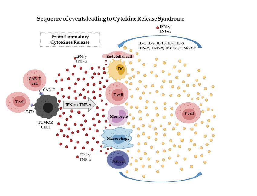
Int. J. Mol. Sci. 2021, 22, 7652                                                                                            2 of 14

                                   Severe viral infections, such as influenza and SARS-CoV-2 (COVID-19) [17], can also trigger
                                   CRS through massive immune and non-immune cell stimulation.
                                         Several efforts have been made to identify new therapeutic strategies for treating
                                   hematological malignancies, and some immunotherapy approaches have been tested to
                                   fortify the immune system of the patient against tumors. In the last few years, immune
                                   checkpoint inhibition and T-cell-engaging therapies, such as bispecific T-cell-engaging
                                   (BiTE) single-chain antibody constructs and chimeric antigen receptor (CAR) T cells, have
                                   opened up a new frontier in cancer immunotherapy [18–20]. However, one of the most
                                   important serious adverse effects of these therapies is CRS.
                                         CRS is characterized by hypersecretion of pro-inflammatory cytokines, including IL-6,
                                   IL-1, IL-5, IL-10, IFN-γ, TNF, and TGFs by B and T lymphocytes and natural killer (NK) cells.
                                   The CRS may be further enhanced by numerous cellular interactions with bystander cells,
                                   such as endothelial cells, monocyte/macrophages, and dendritic cells, further increasing
                                   cytokine hypersecretion, aggravating symptoms, and inducing various grades of organ
                                   damage [21]. CRS symptoms may occur immediately after the administration of T-cell-
                                   engaging therapies or may be delayed until days or weeks after treatment. CRS can
                                   manifest as mild, with flu-like symptoms, including fever, nausea, and chills, or may be life-
                                   threatening and severe with shock and respiratory compromise, leading to multi-system
                                   organ failure and even death [10,22].
                                         This review comprehensively summarizes the biological and clinical aspects of the CRS
                                   triggered by T-cell-engaging therapies used in the treatment of hematological malignancies.

                                   2. Pathophysiology
                                         The pathophysiology of CRS has been associated with invasive pathogens and ther-
                                   apeutic infusions of several monoclonal antibodies [10,12–15,23] (Table 1). CRS can also
                                   develop in association with severe viral infections, including COVID-19, which is caused by
                                   SARS-CoV-2 [24,25]. Recently, with the success of the newer T-cell-engaging immunothera-
                                   peutic agents, such as BiTE constructs and CAR T cells, in hematological malignancies [26],
                                   the interest in CRS has grown, as this is a major serious adverse event of these treatments.
                                   The immunotherapeutic strategies have been carried forward into clinical applications and
                                   shown impressive therapeutic activity in several hematological malignancies, including
                                   acute lymphoblastic B cell leukemia (B-ALL), chronic lymphocytic leukemia (CLL), and
                                   diffuse large B cell lymphoma (DLBCL) [27]. CRS describes an exaggerated systemic im-
                                   mune response involving the release of more than 150 inflammatory mediators, cytokines,
                                   chemokines, oxygen radicals, and complement factors (Table 2) [28]. CRS is due to on-
                                   target effects induced by the binding of CAR T cells or BiTE antibody to its antigen on the
                                   surface of target cells and subsequent activation of bystander immune and non-immune
                                   cells, such as monocytes/macrophages, dendritic cells, and endothelial cells. Activation of
                                   these cells results in the massive release of several cytokines, initiating a cascade of events
                                   that overwhelms counter-regulatory homeostatic mechanisms, leading to CRS [29,30]. As
                                   clearly illustrated in Figure 1, T-cell engaging therapies target tumor cells and induce the
                                   release of cytokines as IFN-γ or TNF-α, which lead to the activation of bystander immune
                                   and non-immune cells as monocytes/macrophages, dendritic cells, NK and T-cell, and
                                   endothelial cells. These cells further release proinflammatory cytokines triggering a cascade
                                   reaction. Macrophages and endothelial cells produce large amounts of IL-6 which in turn
                                   activates T cells and other immune cells leading to a cytokine storm (Figure 1). However,
                                   the pathophysiology of CRS is still poorly understood.
Int. J. Mol. Sci. 2021, 22, 7652 3 of 14
Table 1. Monoclonal antibodies associated with cytokine release syndrome (CRS) in hematologi-
cal malignancies.
Antibody Antigen Class Reference
TGN1412 CD28 Human IgG4 [31]
Alemtuzumab CD52 Human IgG2 [13]
Rituximab CD20 Murine-human chimeric IgG1 [10,11]
Obinutuzumab CD20 Human IgG1 [12]
Brentuximab CD30 Human IgG1 [14]
Dacetuzumab CD40 Human IgG1 [15]
Table 2. Soluble mediators over-expressed in cytokine release syndrome.
Main Cell Source Cytokines Type and Function Reference
Proinflammatory alarming cytokine;
Macrophages, epithelial cells IL-1 pyrogenic function, macrophage, and [32,33]
Th17 cell activation
Effector T-cell and regulatory T-cell
T cells IL-2 [33,34]
growth factor
Proinflammatory cytokine; pyrogenic
Monocyte/macrophages, T cells, function, increased antibody production,
endothelial cells, mesenchymal IL-6 growth and differentiation of [33,35]
cells, osteoblasts hematopoietic stem cells, induction of
acute-phase reactants
Anti-inflammatory cytokine, inhibition of
Regulatory T cells, T cells IL-10 [33,36]
Th1 cells, and cytokine release
Chemokines
Macrophages, epithelial cells IL-8 (CXCL8) Recruitment of neutrophils [33,37]
Interferon-inducible chemokine:
Monocyte, endothelial cells,
IP-10 (CXCL10) recruitment of Th1 cells, NK cells, [38]
keratinocytes
plasmacytoid dendritic cells
Macrophages, dendritic cells, Recruitment of Th2 cells, monocyte,
MCP-1 (CCL2) [39]
cardiac myocytes dendritic cells, basophils
Recruitment of macrophages, Th1 cells,
Monocyte, neutrophils, dendritic
MIP-1α (CCL3) NK cells, eosinophils, dendritic cells, [40,41]
cells, NK cells, mast cells
pyrogenic function
Macrophages, neutrophils, Recruitment of macrophages, Th1 cells,
MIP-1β (CCL4) [40,41]
endothelium NK cells, dendritic cells
Growth Factors
Th1 cells, CTLs, group 1 innate Proinflammatory cytokine, activation of
IFN-γ [42]
lymphoid cells, and NK cells macrophages
Macrophages, T cells, NK cells, Increasing vascular permeability,
TNF-α [43]
mast cells pyrogenic function
Th17 cells GM-CSF Proinflammatory cytokine [44,45]
Endothelium and macrophages CSF Growth and differentiation of neutrophils [44]
Plasma Protein
Monomeric CRP increases IL-8 and
Hepatocytes CRP MCP-1 secretion, IL-6 increases CRP [46]
expression
Ubiquitous Ferritin Primary site of iron storage in cells [47]
IL: interleukin; IP-10: interferon—inducible protein 10; MCP-1: monocyte chemoattractant protein; MIP: macrophage inflammatory protein
1α; IFN-γ: interferon-gamma; TNF-α: tumor necrosis factor-alpha; GM-CSF: granulocyte colony-stimulating factor; CSF: colony-stimulating
factor; CRP: C-reactive protein; Th cell: T helper cell; CTLs: cytotoxic T lymphocytes; NK cell: natural killer cell.Ubiquitous Ferritin Primary site of iron storage in cells [47]
IL: interleukin; IP-10: interferon—inducible protein 10; MCP-1: monocyte chemoattractant protein; MIP: macrophage in-
flammatory protein 1α; IFN-γ: interferon-gamma; TNF-α: tumor necrosis factor-alpha; GM-CSF: granulocyte colony-stim-
ulating factor; CSF: colony-stimulating factor; CRP: C-reactive protein; Th cell: T helper cell; CTLs: cytotoxic T lympho-
cytes;
Int. NK
J. Mol. Sci.cell:
2021,natural
22, 7652 killer cell. 4 of 14
Figure 1. CAR T cells target tumor cells and induce the release of cytokines as IFN-γ or TNF-α,
Figure lead
which to theTactivation
1. CAR of bystander
cells target immune
tumor cells and and non-immune
induce cells of
the release as monocytes/macrophages,
cytokines as IFN-γ or TNF-α,
dendritic cells, NK and T-cell, and endothelial cells.
which lead to the activation of bystander immune and non-immune These cells further release
cellsproinflammatory
as monocytes/macro-
cytokines triggering a cascade reaction. Macrophages and endothelial cells produce
phages, dendritic cells, NK and T-cell, and endothelial cells. These cells further release large amountsproinflam-
of
IL-6 which in turn activates T cells and other immune cells leading to a cytokine storm.
matory cytokines triggering a cascade reaction. Macrophages and endothelial cells produce large BiTe: bispecific
T-cell engager;
amounts of IL-6CAR:
which chimeric
in turnantigen receptor;
activates T cellsIFN-γ: interferon-gamma;
and other immune cellsTNF-α:
leadingtumor necrosis storm.
to a cytokine
factor-alpha; IL: interleukin; GM-CSF: granulocyte colony-stimulating factor; MCP-1:
BiTe: bispecific T-cell engager; CAR: chimeric antigen receptor; IFN-γ: interferon-gamma; TNF-α: monocyte
chemoattractant
tumor protein; NK cell:
necrosis factor-alpha; natural killer GM-CSF:
IL: interleukin; cell; DC: dendritic cell. colony-stimulating factor; MCP-
granulocyte
1:2.1.
monocyte
The Keychemoattractant
Role of IL-6 protein; NK cell: natural killer cell; DC: dendritic cell.
Physiologically, cytokines play a key role in coordinating effector cells of the immune
2.1. The Key Role of IL-6
system and providing regulatory signals that direct, amplify, and resolve the immune
Physiologically,
response. Cytokines havecytokines
short play a keywhich
half-lives, role innormally
coordinating effector
prevents themcells
fromofhaving
the immune
effects and
system outside the lymphoid
providing regulatorytissuesignals
and sites
thatofdirect,
inflammation.
amplify, InandCRS, immune
resolve over-
the immune re-
activation occurs as a result of perceived danger, resulting in excessive activation
sponse. Cytokines have short half-lives, which normally prevents them from having ef- of effector
immune
fects cellsthe
outside andlymphoid
prolongedtissue
immune and activation.
sites of inflammation. In CRS, immune over-activa-
The overabundance of cytokines causes clinically significant collateral damage. The
tion occurs as a result of perceived danger, resulting in excessive activation of effector
cytokines involved in the pathophysiology of CRS include IFN-γ, TNF, IL-6, and IL-10,
immune cells and prolonged immune activation.
which are consistently found to be elevated in the serum of patients with CRS [22,48]
The1).
(Figure overabundance
IL-6 seems to play of cytokines
a key role incauses clinically significant
CRS pathophysiology, collateral
as highly elevateddamage.
IL-6 The
cytokines involved in the pathophysiology of CRS include IFN-γ, TNF,
levels are found in almost all patients with CRS [10,35]. IL-6 is a pleiotropic cytokine that IL-6, and IL-10,
which areboth
exhibits consistently found to
anti-inflammatory bepro-inflammatory
and elevated in the serum of patients
characteristics. IL-6with CRSdiverse
can play [22,48] (Fig-
roles
ure 1).in different
IL-6 seemsphases
to play ofainflammation,
key role in CRS promoting damaging reactions
pathophysiology, as highly within the tissue,
elevated IL-6 levels
are found in almost all patients with CRS [10,35]. IL-6 is a pleiotropic cytokine thatinexhibits
and then contributing to resolving the inflammation, and finally helping to repair tissue
the late
both stages [49]. IL-6 isand
anti-inflammatory secreted by T lymphocytes,
pro-inflammatory monocytes/macrophages,
characteristics. IL-6 can play dendritic
diverse roles
cells, mesenchymal cells, and osteoblasts and is present in processes, such as neutrophil
in different phases of inflammation, promoting damaging reactions within the tissue, and
migration, the acute phase response, angiogenesis, B-cell differentiation, and antibody
then contributing to resolving the inflammation, and finally helping to repair tissue in the
generation [50] (Figure 2A). IL-6 exerts its biological functions via two major pathways:
“classical signaling” and “trans-signaling pathways.” In “classical signaling” pathways,
IL-6 activates cells by binding to the IL-6 receptor (IL6R), which leads to dimerization
of the membrane protein gp130 [50] and intracellular tyrosine kinases, especially Janus
kinase (JAK) 1 and JAK2, which then activate transcription factors signal transducer and
activator of transcription (STAT) 1 and STAT3 [51,52]. Other cascades that can be activated
by IL-6 include the SHP-2/ERK MAPK, PI3K-AKT-mTORC1, and SRC-YAP-NOTCH
pathways [53–55] (Figure 2B). IL-6 can bind cells that express IL6R and signal transducer“classical signaling” and “trans-signaling pathways.” In “classical signaling” pathways,
IL-6 activates cells by binding to the IL-6 receptor (IL6R), which leads to dimerization of
the membrane protein gp130 [50] and intracellular tyrosine kinases, especially Janus ki-
nase (JAK) 1 and JAK2, which then activate transcription factors signal transducer and
Int. J. Mol. Sci. 2021, 22, 7652 activator of transcription (STAT) 1 and STAT3 [51,52]. Other cascades that can be activated 5 of 14
by IL-6 include the SHP-2/ERK MAPK, PI3K-AKT-mTORC1, and SRC-YAP-NOTCH
pathways [53–55] (Figure 2B). IL-6 can bind cells that express IL6R and signal transducer
membrane protein gp130 on their surfaces. IL6R is also found in its soluble form in body
membrane protein
fluids (sIL6R). Whengp130 on their to
IL6 attaches surfaces. IL6Rstarts
sIL6R and is also
thefound in itscascade
signaling soluble by
form in body
binding to
fluids (sIL6R). When IL6 attaches to sIL6R and starts the signaling cascade
cells that express gp130 alone (without IL6R on their surface), it is known as “trans-sig- by binding
to cells that
naling” [56]. express
The modegp130 alone (without
of signaling IL6R
influences theon their“classical
result: surface),IL-6
it issignaling”
known asreduces
“trans-
signaling” [56]. The mode of signaling influences the result: “classical
inflammation, whereas “IL-6 trans-signaling” promotes T-cell migration, decreases apop-IL-6 signaling”
reduces inflammation,
tosis, enhances whereasand
cytotoxicity, “IL-6suppresses
trans-signaling” promotes
regulatory T-cell
T-cell migration, decreases
differentiation [57–59].
apoptosis, enhances cytotoxicity, and suppresses regulatory T-cell differentiation [57–59].
“Classical IL-6 signaling” does not seem to affect CAR T cell efficacy [60], but the impact
“Classical IL-6 signaling” does not seem to affect CAR T cell efficacy [60], but the impact of
of IL-6 trans-signaling on CAR T cells has not been described.
IL-6 trans-signaling on CAR T cells has not been described.
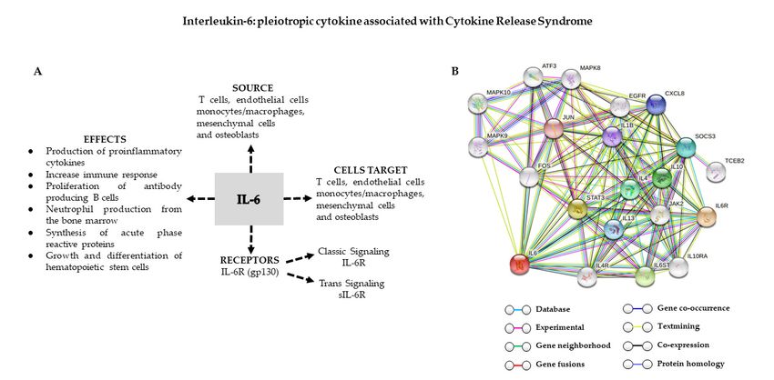
Figure 2. (A)
Figure 2. (A)Source
Sourceand
andbiological
biologicalfunctions
functionsofofIL-6.
IL-6.(B)
(B)Genetic
Geneticinteraction
interactionnetwork
network(String:
(String:https://string-db.org)
https://string-db.org) that
that
evaluates
evaluates pathways and visualizes the connection among target genes according to the literature search.IL6R:
pathways and visualizes the connection among target genes according to the literature search. IL6R:interleukin-6
interleukin-
receptor
6 receptorsubunit
subunitalpha;
alpha;STAT3:
STAT3:signal
signaltransducer
transducerand
andactivator
activatorofoftranscription
transcription3;3;IL6ST:
IL6ST:interleukin-6
interleukin-6receptor
receptor subunit
subunit
beta;
beta; IL10: interleukin-10;
interleukin-10; IL4:
IL4:interleukin-4;
interleukin-4;SOCS3:
SOCS3:suppressor
suppressor ofof cytokine
cytokine signaling
signaling 3; 3; IL13:
IL13: interleukin-13;
interleukin-13; CXCL8:
CXCL8: in-
terleukin-8; IL1B:
interleukin-8; IL1B:interleukin-1
interleukin-1beta;
beta;JUN:
JUN:transcription
transcriptionfactor
factorAP-1.
AP-1.
2.2. The Roles of Other Cytokines
IL-6 determines the release of other cytokines, such as IFN-γ and TNF-α, by lympho-
cytes, monocytes, and neutrophils (Figures 1 and 2A). The release release ofof TNF-α
TNF-α andand IFN-γ
IFN-γ
within 1–2 h is followed
followed by an increase
increase inin IL-6
IL-6 and
and IL-10,
IL-10, and
and inin some
some cases,
cases, of
of IL-8
IL-8 and
and
IL-2. IFN-γ
IFN-γgenerated
generatedby byT Tcells
cells and
and NKNK cells,
cells, or byor by
thethe tumor
tumor cellscells themselves,
themselves, is a
is a pro-
pro-inflammatory cytokine
inflammatory cytokine that activates
that activates otherother
immune immune cells,as
cells, such such as macrophages
macrophages [61],
[61], which
which produce excessive quantities of additional cytokines [62]. Moreover,
produce excessive quantities of additional cytokines [62]. Moreover, IFN-γ triggers mac-IFN-γ triggers
macrophage activation,
rophage activation, leading
leading to theto secretion
the secretion of host
of host cytokines,
cytokines, including
including IL-6,IL-6, TNF-α,
TNF-α, and
and IL-10 [22], which could further intensify the CRS. IL-10 fails to control this process,
though it suppresses cellular immunity. Other cytokines have also been found to be ele-
vated during the course of CRS, including IL-1, IL-2, IL-8, IL-5, monocyte chemoattractant
protein 1 (MCP-1), and granulocyte-macrophage colony-stimulating factor (GM-CSF), driv-
ing some of its manifestations (Figure 1 and Table 2) [22,48,63–68]. IL-1 has one of the
simplest signaling mechanisms in the innate immune system. It is capable of sensing an
infection and triggering an inflammatory response [69]. IL-1 is released from activated
macrophages and monocytes, further stimulating the release of IL-6 and inducing nitric
oxide synthetase [48]. Endothelial cell activation also plays a role in CRS. Ang-2, a typical
marker of endothelial cell activation, is a hallmark of severe CRS, showing that the en-Int. J. Mol. Sci. 2021, 22, 7652 6 of 14
dothelium plays a key role in the pathophysiology of CRS by amplifying the inflammatory
response [67].
2.3. Biomarkers
Various studies indicate a correlation between the severity of the CRS and tumor
burden, C-reactive protein (CRP) and ferritin levels, and cytokine levels [29,63,70,71]
(Table 2). However, whether the cytokines can act as prognostic factors, especially in
patients with CRS, in whom cytokine concentrations may already be high, and in patients
with malignancies, is not clear. Comparing the increased cytokine level to the baseline
value while simultaneously considering some specific markers and following a series
of measurements rather than the amount of a single cytokine has been suggested [72].
Furthermore, the serum cytokine measurement is usually not quickly available in most
hospitals. In response to IL-6, the liver produces CRP, and this concentration is easily
measured by a rapid and inexpensive assay available in the majority of hospitals. Thus,
CRP measurements are widely utilized as a surrogate marker of IL-6 bioactivity [73,74].
However, CRP is not specific to CRS and cannot be used to distinguish inflammation due
to infectious or non-infectious disease [75]. An excessive increase in ferritin has also been
reported in several patients after CAR T-cell infusion, supporting a relationship between
CRS and macrophage activation syndrome (MAS)/hemophagocytic lymphohistiocytosis
(HLH). However, in the same experience, ferritin did not show utility in predicting the
severity of CRS [29].
3. Clinical Manifestations
The incidence of CRS varies with the type of immunotherapy, and it is more frequently
observed during CAR T-cell therapy than with bispecific antibody blinatumomab infusion.
During T-cell therapies, CRS occurs early during the course of treatment [76]. CRS related
to blinatumomab therapy usually occurs during the first cycle of therapy, and typically
upon starting the infusion. CRS manifests with a wide variety of signs and symptoms of
varying severity. The most important and frequent symptoms are summarized in Figure 3.
Fever is usually present, and several other symptoms can mimic infection. The patient’s
body temperature quite commonly exceeds 40.0 ◦ C. For this reason, the possibility of
infection must be ruled out after appropriate cultures, particularly if the patient is neu-
tropenic. Other constitutional symptoms, such as myalgia and arthralgia, may be present.
Any organ can be affected during CRS, and the patient can develop nausea, vomiting,
skin rash, hemodynamic instability, and capillary leak syndrome with hypotension and
tachycardia, disseminated intravascular coagulation, and neurological toxicity [67]. Neu-
rological toxicity may occur together with other symptoms of CRS or when the other
symptoms are disappearing. Neurological toxicity includes headache, confusion, delirium,
aphasia, tremor, and seizures. Patients with severe neurotoxicity show signs of endothelial
activation, including disseminated intravascular coagulation and increased blood-brain
barrier (BBB) permeability, which may allow the entry of high concentrations of systemic
cytokines, particularly IFN-γ, inducing brain vascular pericyte stress and the consequent
secretion of endothelium-activating cytokines. Adverse neurological events can be fatal
in rare cases [77]. Although usually reversible, severe cardiac dysfunction is of particular
concern. The pathophysiology of acute cardiac toxicity is not clear but it appears similar to
that cardiomyopathy is associated with sepsis [78].Int. J. Mol. Sci. 2021, 22, x FOR PEER REVIEW 7 of 14
can be fatal in rare cases [77]. Although usually reversible, severe cardiac dysfunction is
Int. J. Mol. Sci. 2021, 22, 7652 of particular concern. The pathophysiology of acute cardiac toxicity is not clear but 7itofap-
14
pears similar to that cardiomyopathy is associated with sepsis [78].
Figure 3. Pathophysiology of signs and symptoms of CRS. IFN-γ: interferon-gamma; TNF-α: tumor necrosis factor-alpha;
Figure
IL: 3. Pathophysiology
interleukin; of signs and symptoms
CSF: colony-stimulating of monocyte
factor; MCP: CRS. IFN-γ: interferon-gamma;
chemoattractant TNF-α: tumor necrosis factor-alpha;
protein.
IL: interleukin; CSF: colony-stimulating factor; MCP: monocyte chemoattractant protein.
Disseminated intravascular coagulation can occur in patients with CRS, especially
Disseminated
in those intravascular
who develop grade ≥4. Consumptive
a CRS of coagulation can occur in patients with CRS,
coagulopathy especially
manifests in
with
those who develop athe
thrombocytopenia, CRS of gradeof≥4.
elevation Consumptive
D-dimer, coagulopathy
prolongation manifests
of prothrombin with
time throm-
(PT) and
bocytopenia,
partial the elevation
thromboplastin of D-dimer,
time prolongation of prothrombin
(PTT), hyperfibrinogenemia, time (PT)
and elevation and partial
of endothelial
activation markers
thromboplastin time[67].
(PTT),CRS symptoms may also
hyperfibrinogenemia, and mimic MAS/HLH
elevation [79,80],activation
of endothelial and the
pathophysiology
markers [67]. CRSof the syndromes
symptoms may
may also overlap
mimic [81].
MAS/HLH [79,80], and the pathophysiology
of the syndromes may overlap [81].
4. Management of CRS
The optimalof
4. Management clinical
CRS management of CRS is still not well-defined since T-cell-engaging
therapies
The optimal clinicalintroduced
were recently management intoofclinical
CRS is practice; thus, severalsince
still not well-defined questions are still
T-cell-engag-
unanswered. A reduced incidence of severe CRS has been obtained with cytoreduction,
ing therapies were recently introduced into clinical practice; thus, several questions are
doseunanswered.
still adjustment, and premedication
A reduced with
incidence corticosteroids
of severe CRS has[82].been obtained with cytoreduc-
In patients receiving T-cell-engaging therapies,
tion, dose adjustment, and premedication with corticosteroids the approaches
[82]. to prevention and
treatment of CRS may differ substantially. Blinatumomab
In patients receiving T-cell-engaging therapies, the approaches has a short half-life (~2and
to prevention h),
and CRS symptoms may resolve quickly by interrupting therapy and starting supportive
treatment of CRS may differ substantially. Blinatumomab has a short half-life (~2 h), and
care with or without additional interventions. However, BiTE constructs can be given
CRS symptoms may resolve quickly by interrupting therapy and starting supportive care
repeatedly, whereas CAR T cells are usually administered once, but the side effects after
with or without additional interventions. However, BiTE constructs can be given repeat-
CAR T infusion are difficult to reverse because the infused cells can persist for prolonged
edly, whereas CAR T cells are usually administered once, but the side effects after CAR T
periods. Therefore, the management of CRS can be diverse between the two therapeutic
infusion are difficult to reverse because the infused cells can persist for prolonged periods.
modalities. Current treatments are based on the severity of the side effects [29,83] using
Therefore, the management of CRS can be diverse between the two therapeutic modali-
the grading scheme developed by Lee et al. [29] (Table 3 and Figure 3).
ties. Current treatments are based on the severity of the side effects [29,83] using the grad-
Efficient management of patients requires very strict collaboration between several
ing scheme developed by Lee et al. [29] (Table 3 and Figure 3).
specialties, such as hematology, neurology, and radiology. Sometimes intensive care unit
(ICU) referral should be considered so that mechanical ventilation can be offered when
necessary. Patients with grade 1 and 2 toxicity experiencing fever, constitutional symptoms,
and moderate hypotension are treated symptomatically with antipyretics and antibiotics for
fever, with fluids and low dose vasopressors for hypotension and oxygen supplementation
when saturation drops.
After CAR T-cell therapy, fever usually precedes CRS. Therefore, patients who develop
persistent fever should be frequently evaluated for signs and symptoms of CRS. If an
infection cannot be ruled out, empiric antibiotic therapy should be started.Int. J. Mol. Sci. 2021, 22, 7652 8 of 14
Table 3. Cytokine release syndrome (CRS) grading system (Lee et al. [29] partially modify).
Toxicity Grade
Symptoms are not life threatening and require symptomatic treatment only, eg, fever, nausea, Grade 1
fatigue, headache, myalgia, malaise
Symptoms require and respond to moderate intervention. Oxygen requirement < 40 % or Grade 2
hypotension responsive to fluids or low dose of one vasopressor or grade 2 organ toxicity
Symptoms require and respond to aggressive intervention. Oxygen requirement ≥ 40 % or Grade 3
hypotension requiring high dose or multiple vasopressor or grade 3 organ toxicity or grade
4 transaminitis
Life-threatening symptoms. Requirement for ventilator support or grade 4 organ toxicity Grade 4
(excluding transaminitis)
Grades 2–4 refer to CTCAE v4.0 grading [21].
For severe cases of CRS, the patient should be admitted to the ICU for close monitoring.
In addition to aggressive supportive care, steroids and repeated doses of Il-6 inhibitor
need to be administered when no improvement is observed. In some resistant cases, other
anti-inflammatory agents need to be administered, but clinical experience in this setting is
undeveloped [29,80,84,85].
5. Treatment of CRS
The therapy for CRS is not yet well defined but is based on the use of steroids
and inhibitors of IL-6 activity, as well as IL-1, IFN-γ, TNF-α, and IL-2 inhibitors for
unresponsive patients.
5.1. Steroids
Clinical experience shows that steroids are an effective treatment for suppressing the
excessive inflammatory response and CRS [86]. Opinions differ on the timing and dosing
of corticosteroids. Some choose to use corticosteroids as a first-line agent, whereas others
do not [29]. However, corticosteroids have generalized effects on the immune system and
may also inhibit the anti-tumor efficacy and affect the amplification and persistence of CAR
T cells in vivo [71]. Thus, steroids should generally be avoided as first-line treatment but
used in resistant patients with severe CRS and given at high doses when it is necessary
to ablate CAR T cells. Furthermore, steroids are recommended in patients with adverse
neurological effects.
5.2. IL-6 Activity Inhibitors
Tocilizumab is a humanized monoclonal antibody against both the soluble IL-6 and
membrane-bound IL-6 receptors, inhibiting both classical and trans-IL-6 signaling. After
multiple trials demonstrated its efficacy [76,87], it was approved by the FDA in 2017 as the
first approach for the treatment of CRS-related toxicities following CAR T-cell infusion.
Tocilizumab controls CRS without significant loss of CAR T-cell activity. Improvements
occur within a few hours after drug infusion, reducing adjuvant therapy. Advantageous
effects of a single injection in patients with CRS induced by CAR T-cell therapy strongly
suggest that IL-6 blockade may constitute a new therapeutic approach for an acute, severe,
systemic inflammatory response such as CRS. Its most used dosage for CRS is 12 mg/kg
for patients weighingInt. J. Mol. Sci. 2021, 22, 7652 9 of 14
whereas others wonder if it is possible to use this drug prophylactically in order to avoid
or reduce the symptoms of CRS [22].
Another monoclonal antibody that blocks IL-6 signaling is siltuximab, which prevents
the activation of immune effector cells through either the trans or classical mechanisms [50].
Siltuximab has a higher affinity for IL-6 than tocilizumab has for IL6R, making it an
attractive tool in managing CRS. The use of siltuximab is encouraged in patients that do
not respond to tocilizumab and corticosteroids.
5.3. IL-1, IFN-γ, TNF-α, and IL-2 Inhibitors
IL-6/IL6R blockade alone cannot alleviate CRS symptoms completely and would be
insufficient in treating severe CRS in patients undergoing CAR T-cell therapy. For patients
who become critically ill and do not respond to IL-6, directed therapy targeting IL-1, IFN-γ,
TNF-α, or sIL-2 could ameliorate their symptoms. This has led to the use of other cytokine
inhibitors, including TNF-α and IL1R inhibitors [29,56,70,80]. Etanercept and infliximab
both target TNF-α, which is known to be elevated in CRS, and have been used to treat
severe CRS, with varying results [29,80]. Anakinra, a recombinant and slightly altered form
of the IL1R antagonist, may be helpful in a subset of patients with increased IL-1α, but this
cytokine is also not consistently elevated in patients with severe CRS [70]. Although IFN-γ
is consistently elevated very early in severe CRS, it is not thought to be an ideal target due
to its role in T-cell proliferation [90].
Finally, other research groups have thought of inserting a “suicidal” gene into the
genetic construct used to arm T lymphocytes, a “command” that, when activated from the
outside, drives the lymphocytes to commit suicide, self-limiting activity when the CRS
becomes life-threatening [76].
6. Conclusions
The introduction of CAR T-cell therapy into clinical practice is revolutionizing the
treatment of numerous hematological malignancies. This treatment is able to induce
prolonged remission in patients in a very advanced stage of the disease and for whom
the most common therapeutic options have been exhausted. However, it is essential
that life-threatening toxicities, such as CRS, can be managed in an optimal and effective
way. Therefore, the goal is to prevent or effectively treat CRS without diminishing the
antitumor efficacy. The BiTE construct blinatumomab, a prophylactic procedure including
cytoreduction, premedication with corticosteroids, and dose adjustment, appears to be able
to reduce the incidence of severe CRS [82]. Considering the type of CAR T cells currently
available, the most used sequence of agents to control severe CRS include tocilizumab and
high-dose corticosteroids. When these treatments fail, other cytokine inhibitors, such as
TNF-α or IL1R inhibitors, are often used. Isolated and severe neurotoxicity is usually, at
least initially, treated with corticosteroids rather than tocilizumab.
In this review, we have summarized the pathophysiology, symptoms, and manage-
ment of CRS associated with T-cell-based therapies utilized in the treatment of hema-
tological malignancies. Although several grading scales and very effective treatment
algorithms have been proposed, we would like to emphasize that optimal management
of patients is based on the close collaboration of a multidisciplinary team that includes
hematologists, neurologists, radiologists, and intensive care specialists, so that mechanical
ventilation can be offered if necessary. Until new knowledge on the pathophysiology of
CRS allows the use of new and more effective treatments, we think that early intervention
by a multidisciplinary team and the use of tocilizumab and corticosteroids remains the best
management strategy.
7. Future Prospect
In the near future, every effort will be made to balance treatment toxicity and efficacy
and to prevent or reduce the symptoms of CRS after infusion of T-cell-based therapies in
hematological patients. It will be necessary to act both on the aspects of patient care and inInt. J. Mol. Sci. 2021, 22, 7652 10 of 14
the field of research on that intricate network of different cell types, which constitutes the
immune system. These goals can be achieved:
(a) Developing even greater cooperation between experts from various fields such as
onco-hematology, neuroscience, immunology virology, and ICU: the multidisciplinary
approach is an essential requirement for an adequate treatment of patients with CRS.
(b) Improving specificity and further unlocking the potential of immunotherapy.
A typical example is the recently developed split, universal, and programmable
(SUPRA) CAR system which is composed of an antigen-binding portion, and a universal
signal transduction receptor. The system seems able to improve the specificity and control-
lability [91] of the immune cell engineering strategy. Thus, the SUPRA CAR system has the
potential to reduce CRS without reducing the antitumor response [92].
Author Contributions: M.C. and S.S. performed the literature search and wrote the manuscript. S.P.
critically reviewed the manuscript for intellectual content. All authors have read and agreed to the
published version of the manuscript.
Funding: This research received no external funding.
Institutional Review Board Statement: Not applicable.
Informed Consent Statement: Not applicable.
Data Availability Statement: Not applicable.
Acknowledgments: We thank the “Associazione Mantovana per la Ricerca sul Cancro”, Italy for
technical support.
Conflicts of Interest: The authors declare no conflict of interest.
Abbreviations
(T-PLL) T-cell prolymphocytic leukemia
AKT serine/threonine-specific protein kinase
ALL acute lymphoblastic leukemia
AML acute myeloid leukemia
BiTE bispecific T-cell engaging
B-NHL B-cell non-Hodgkin lymphoma
CAR chimeric antigen receptor
CLL chronic lymphocytic leukemia
COVID 19 coronavirus disease 2019
CRES CAR T-cell-related encephalopathy syndrome
CRP C-reactive protein
CRS cytokine release syndrome
CSF colony-stimulating factor
DLBCL diffuse Large B-cell Lymphoma
FDA Food and Drug Administration
GCSF granulocyte colony-stimulating factor
HGF hepatocyte growth factor
HLH haemophagocytic lymphohistiocytosis
HSCT hematopoietic stem cell transplantation
ICU intensive care unit
IFN interferon
IL interleukin
IL6R IL6 Receptor
IP-10 interferon–inducible protein 10
JAK Janus associated kinase
mAbs monoclonal antibodiesInt. J. Mol. Sci. 2021, 22, 7652 11 of 14
MAPK mitogen-activated protein kinase
MAS macrophage activation syndrome
MCP monocyte chemoattractant protein
MIP macrophage inflammatory protein 1α
MSC mesenchymal stem cells
NHL non-Hodgkin lymphoma
NK natural killer
PT prothrombin time
PTT partial thromboplastin time
SARS-CoV-2 severe acute respiratory syndrome coronavirus 2
sIL6R soluble IL6 receptor
SS Sézary syndrome
STAT signal transducer and activator of transcription
TGF transforming growth factors
TNF tumor necrosis factors
References
1. Holtmann, H.; Resch, K. Cytokines. Naturwissenschaften 1995, 82, 178–187. [CrossRef]
2. Borish, L.C.; Steinke, J.W. 2. Cytokines and chemokines. J. Allergy Clin. Immunol. 2003, 111, S460–S475. [CrossRef] [PubMed]
3. Dinarello, C.A. Historical insights into cytokines. Eur. J. Immunol. 2007, 37 (Suppl. 1), S34–S45. [CrossRef]
4. Ramani, T.; Auletta, C.S.; Weinstock, D.; Mounho-Zamora, B.; Ryan, P.C.; Salcedo, T.W.; Bannish, G. Cytokines: The Good, the
Bad, and the Deadly. Int. J. Toxicol. 2015, 34, 355–365. [CrossRef]
5. Lefkowitz, D.L.; Lefkowitz, S.S. Macrophage-neutrophil interaction: A paradigm for chronic inflammation revisited. Immunol.
Cell Biol. 2001, 79, 502–506. [CrossRef]
6. Stenken, J.A.; Poschenrieder, A.J. Bioanalytical chemistry of cytokines—A review. Anal. Chim. Acta 2015, 853, 95–115. [CrossRef]
7. Kato, H.; Kinoshita, T.; Suzuki, S.; Nagasaka, T.; Hatano, S.; Murate, T.; Saito, H.; Hotta, T. Production and effects of interleukin-6
and other cytokines in patients with non-Hodgkin’s lymphoma. Leuk. Lymphoma 1998, 29, 71–79. [CrossRef] [PubMed]
8. Balkwill, F.R.; Burke, F. The cytokine network. Immunol. Today 1989, 10, 299–304. [CrossRef]
9. Schmitz, M.L.; Weber, A.; Roxlau, T.; Gaestel, M.; Kracht, M. Signal integration, crosstalk mechanisms and networks in the
function of inflammatory cytokines. Biochim. Biophys. Acta 2011, 1813, 2165–2175. [CrossRef] [PubMed]
10. Winkler, U.; Jensen, M.; Manzke, O.; Schulz, H.; Diehl, V.; Engert, A. Cytokine-release syndrome in patients with B-cell
chronic lymphocytic leukemia and high lymphocyte counts after treatment with an anti-CD20 monoclonal antibody (rituximab,
IDEC-C2B8). Blood 1999, 94, 2217–2224. [CrossRef] [PubMed]
11. Agarwal, A.; Vieira, C.A.; Book, B.K.; Sidner, R.A.; Fineberg, N.S.; Pescovitz, M.D. Rituximab, anti-CD20, induces in vivo cytokine
release but does not impair ex vivo T-cell responses. Am. J. Transplant. 2004, 4, 1357–1360. [CrossRef] [PubMed]
12. Freeman, C.L.; Morschhauser, F.; Sehn, L.; Dixon, M.; Houghton, R.; Lamy, T.; Fingerle-Rowson, G.; Wassner-Fritsch, E.;
Gribben, J.G.; Hallek, M.; et al. Cytokine release in patients with CLL treated with obinutuzumab and possible relationship with
infusion-related reactions. Blood 2015, 126, 2646–2649. [CrossRef]
13. Wing, M.G.; Moreau, T.; Greenwood, J.; Smith, R.M.; Hale, G.; Isaacs, J.; Waldmann, H.; Lachmann, P.J.; Compston, A. Mechanism
of first-dose cytokine-release syndrome by CAMPATH 1-H: Involvement of CD16 (FcgammaRIII) and CD11a/CD18 (LFA-1) on
NK cells. J. Clin. Investig. 1996, 98, 2819–2826. [CrossRef] [PubMed]
14. Alig, S.K.; Dreyling, M.; Seppi, B.; Aulinger, B.; Witkowski, L.; Rieger, C.T. Severe cytokine release syndrome after the first dose of
Brentuximab Vedotin in a patient with relapsed systemic anaplastic large cell lymphoma (sALCL): A case report and review of
literature. Eur. J. Haematol. 2015, 94, 554–557. [CrossRef]
15. De Vos, S.; Forero-Torres, A.; Ansell, S.M.; Kahl, B.; Cheson, B.D.; Bartlett, N.L.; Furman, R.R.; Winter, J.N.; Kaplan, H.;
Timmerman, J.; et al. A phase II study of dacetuzumab (SGN-40) in patients with relapsed diffuse large B-cell lymphoma (DLBCL)
and correlative analyses of patient-specific factors. J. Hematol. Oncol. 2014, 7. [CrossRef]
16. Rotz, S.J.; Leino, D.; Szabo, S.; Mangino, J.L.; Turpin, B.K.; Pressey, J.G. Severe cytokine release syndrome in a patient receiving
PD-1-directed therapy. Pediatr. Blood Cancer 2017, 64. [CrossRef]
17. Moore, J.B.; June, C.H. Cytokine release syndrome in severe COVID-19. Science 2020, 368, 473–474. [CrossRef]
18. June, C.H.; O’Connor, R.S.; Kawalekar, O.U.; Ghassemi, S.; Milone, M.C. CAR T cell immunotherapy for human cancer. Science
2018, 359, 1361–1365. [CrossRef] [PubMed]
19. Sadelain, M.; Rivière, I.; Riddell, S. Therapeutic T cell engineering. Nature 2017, 545, 423–431. [CrossRef] [PubMed]
20. Lim, W.A.; June, C.H. The Principles of Engineering Immune Cells to Treat Cancer. Cell 2017, 168, 724–740. [CrossRef]
21. Common Terminology Criteria for Adverse Events (CTCAE); National Cancer Institute: Bethesda, MD, USA, 2017; p. 155.
22. Wang, Z.; Han, W. Biomarkers of cytokine release syndrome and neurotoxicity related to CAR-T cell therapy. Biomark. Res. 2018,
6, 4. [CrossRef] [PubMed]Int. J. Mol. Sci. 2021, 22, 7652 12 of 14
23. Vessillier, S.; Eastwood, D.; Fox, B.; Sathish, J.; Sethu, S.; Dougall, T.; Thorpe, S.J.; Thorpe, R.; Stebbings, R. Cytokine release assays
for the prediction of therapeutic mAb safety in first-in man trials—Whole blood cytokine release assays are poorly predictive for
TGN1412 cytokine storm. J. Immunol. Methods 2015, 424, 43–52. [CrossRef] [PubMed]
24. Pedersen, S.F.; Ho, Y.-C. SARS-CoV-2: A storm is raging. J. Clin. Investig. 2020, 130, 2202–2205. [CrossRef]
25. Mehta, P.; McAuley, D.F.; Brown, M.; Sanchez, E.; Tattersall, R.S.; Manson, J.J.; HLH Across Speciality Collaboration UK.
COVID-19: Consider cytokine storm syndromes and immunosuppression. Lancet 2020, 395, 1033–1034. [CrossRef]
26. Frey, N.; Porter, D. Cytokine Release Syndrome with Chimeric Antigen Receptor T Cell Therapy. Biol. Blood Marrow Transplant.
2019, 25, e123–e127. [CrossRef] [PubMed]
27. Shimabukuro-Vornhagen, A.; Gödel, P.; Subklewe, M.; Stemmler, H.J.; Schlößer, H.A.; Schlaak, M.; Kochanek, M.; Böll, B.; von
Bergwelt-Baildon, M.S. Cytokine release syndrome. J. Immunother. Cancer 2018, 6, 56. [CrossRef] [PubMed]
28. Panelli, M.C.; White, R.; Foster, M.; Martin, B.; Wang, E.; Smith, K.; Marincola, F.M. Forecasting the cytokine storm following
systemic interleukin (IL)-2 administration. J. Transl. Med. 2004, 2, 17. [CrossRef]
29. Lee, D.W.; Gardner, R.; Porter, D.L.; Louis, C.U.; Ahmed, N.; Jensen, M.; Grupp, S.A.; Mackall, C.L. Current concepts in the
diagnosis and management of cytokine release syndrome. Blood 2014, 124, 188–195. [CrossRef]
30. Liu, Q.; Zhou, Y.; Yang, Z. The cytokine storm of severe influenza and development of immunomodulatory therapy. Cell Mol.
Immunol. 2016, 13, 3–10. [CrossRef]
31. Farzaneh, L.; Kasahara, N.; Farzaneh, F. The strange case of TGN1412. Cancer Immunol. Immunother. 2007, 56, 129–134. [CrossRef]
32. Kaneko, N.; Kurata, M.; Yamamoto, T.; Morikawa, S.; Masumoto, J. The role of interleukin-1 in general pathology. Inflamm. Regen.
2019, 39, 12. [CrossRef] [PubMed]
33. Justiz Vaillant, A.A.; Qurie, A. Interleukin. In StatPearls; StatPearls Publishing: Treasure Island, FL, USA, 2021.
34. Ross, S.H.; Cantrell, D.A. Signaling and Function of Interleukin-2 in T Lymphocytes. Annu. Rev. Immunol. 2018, 36, 411–433.
[CrossRef] [PubMed]
35. Tanaka, T.; Narazaki, M.; Kishimoto, T. Interleukin (IL-6) Immunotherapy. Cold Spring Harb. Perspect. Biol. 2018, 10, a028456.
[CrossRef] [PubMed]
36. Iyer, S.S.; Cheng, G. Role of Interleukin 10 Transcriptional Regulation in Inflammation and Autoimmune Disease. Crit. Rev.
Immunol 2012, 32, 23–63. [CrossRef] [PubMed]
37. Bickel, M. The role of interleukin-8 in inflammation and mechanisms of regulation. J. Periodontol. 1993, 64, 456–460.
38. Liu, M.; Guo, S.; Hibbert, J.M.; Jain, V.; Singh, N.; Wilson, N.O.; Stiles, J.K. CXCL10/IP-10 in infectious diseases pathogenesis and
potential therapeutic implications. Cytokine Growth Factor Rev. 2011, 22, 121–130. [CrossRef]
39. Deshmane, S.L.; Kremlev, S.; Amini, S.; Sawaya, B.E. Monocyte Chemoattractant Protein-1 (MCP-1): An Overview. J. Interferon
Cytokine Res. 2009, 29, 313–326. [CrossRef] [PubMed]
40. Bhavsar, I.; Miller, C.S.; Al-Sabbagh, M. Macrophage Inflammatory Protein-1 Alpha (MIP-1 alpha)/CCL3: As a Biomarker. In
General Methods in Biomarker Research and Their Applications; Springer: Dordrecht, The Netherlands, 2015; pp. 223–249. [CrossRef]
41. Fahey, T.J.; Tracey, K.J.; Tekamp-Olson, P.; Cousens, L.S.; Jones, W.G.; Shires, G.T.; Cerami, A.; Sherry, B. Macrophage inflammatory
protein 1 modulates macrophage function. J. Immunol. 1992, 148, 2764–2769.
42. Tau, G.; Rothman, P. Biologic functions of the IFN-γ receptors. Allergy 1999, 54, 1233–1251. [CrossRef]
43. Parameswaran, N.; Patial, S. Tumor Necrosis Factor-α Signaling in Macrophages. Crit. Rev. Eukaryot. Gene Expr. 2010, 20, 87–103.
[CrossRef] [PubMed]
44. Ushach, I.; Zlotnik, A. Biological role of granulocyte macrophage colony-stimulating factor (GM-CSF) and macrophage colony-
stimulating factor (M-CSF) on cells of the myeloid lineage. J. Leukoc. Biol. 2016, 100, 481–489. [CrossRef] [PubMed]
45. Shi, Y.; Liu, C.H.; Roberts, A.I.; Das, J.; Xu, G.; Ren, G.; Zhang, Y.; Zhang, L.; Yuan, Z.R.; Tan, H.S.W.; et al. Granulocyte-
macrophage colony-stimulating factor (GM-CSF) and T-cell responses: What we do and don’t know. Cell Res. 2006, 16, 126–133.
[CrossRef] [PubMed]
46. Sproston, N.R.; Ashworth, J.J. Role of C-Reactive Protein at Sites of Inflammation and Infection. Front. Immunol. 2018, 9, 754.
[CrossRef] [PubMed]
47. Knovich, M.A.; Storey, J.A.; Coffman, L.G.; Torti, S.V. Ferritin for the Clinician. Blood Rev. 2009, 23, 95–104. [CrossRef]
48. Norelli, M.; Camisa, B.; Barbiera, G.; Falcone, L.; Purevdorj, A.; Genua, M.; Sanvito, F.; Ponzoni, M.; Doglioni, C.;
Cristofori, P.; et al. Monocyte-derived IL-1 and IL-6 are differentially required for cytokine-release syndrome and neurotoxicity
due to CAR T cells. Nat. Med. 2018, 24, 739–748. [CrossRef] [PubMed]
49. Del Giudice, M.; Gangestad, S.W. Rethinking IL-6 and CRP: Why they are more than inflammatory biomarkers, and why it
matters. Brain Behav. Immun. 2018, 70, 61–75. [CrossRef] [PubMed]
50. Rose-John, S.; Scheller, J.; Elson, G.; Jones, S.A. Interleukin-6 biology is coordinated by membrane-bound and soluble receptors:
Role in inflammation and cancer. J. Leukoc. Biol. 2006, 80, 227–236. [CrossRef]
51. Johnson, D.E.; O’Keefe, R.A.; Grandis, J.R. Targeting the IL-6/JAK/STAT3 signalling axis in cancer. Nat. Rev. Clin. Oncol. 2018, 15,
234–248. [CrossRef]
52. Villarino, A.V.; Kanno, Y.; O’Shea, J.J. Mechanisms and consequences of Jak-STAT signaling in the immune system. Nat. Immunol.
2017, 18, 374–384. [CrossRef]
53. Heinrich, P.C.; Behrmann, I.; Haan, S.; Hermanns, H.M.; Müller-Newen, G.; Schaper, F. Principles of interleukin (IL)-6-type
cytokine signalling and its regulation. Biochem. J. 2003, 374, 1–20. [CrossRef]Int. J. Mol. Sci. 2021, 22, 7652 13 of 14
54. Taniguchi, K.; Wu, L.-W.; Grivennikov, S.I.; de Jong, P.R.; Lian, I.; Yu, F.-X.; Wang, K.; Ho, S.B.; Boland, B.S.; Chang, J.T.; et al. A
gp130-Src-YAP module links inflammation to epithelial regeneration. Nature 2015, 519, 57–62. [CrossRef]
55. Yamada, O.; Ozaki, K.; Akiyama, M.; Kawauchi, K. JAK-STAT and JAK-PI3K-mTORC1 pathways regulate telomerase transcrip-
tionally and posttranslationally in ATL cells. Mol. Cancer Ther. 2012, 11, 1112–1121. [CrossRef]
56. Jones, S.A.; Scheller, J.; Rose-John, S. Therapeutic strategies for the clinical blockade of IL-6/gp130 signaling. J. Clin. Investig.
2011, 121, 3375–3383. [CrossRef]
57. Böttcher, J.P.; Schanz, O.; Garbers, C.; Zaremba, A.; Hegenbarth, S.; Kurts, C.; Beyer, M.; Schultze, J.L.; Kastenmüller, W.; Rose-John,
S.; et al. IL-6 trans-signaling-dependent rapid development of cytotoxic CD8+ T cell function. Cell Rep. 2014, 8, 1318–1327.
[CrossRef]
58. McLoughlin, R.M.; Jenkins, B.J.; Grail, D.; Williams, A.S.; Fielding, C.A.; Parker, C.R.; Ernst, M.; Topley, N.; Jones, S.A. IL-6
trans-signaling via STAT3 directs T cell infiltration in acute inflammation. Proc. Natl. Acad. Sci. USA 2005, 102, 9589–9594.
[CrossRef] [PubMed]
59. Atreya, R.; Mudter, J.; Finotto, S.; Müllberg, J.; Jostock, T.; Wirtz, S.; Schütz, M.; Bartsch, B.; Holtmann, M.; Becker, C.; et al.
Blockade of interleukin 6 trans signaling suppresses T-cell resistance against apoptosis in chronic intestinal inflammation:
Evidence in crohn disease and experimental colitis in vivo. Nat. Med. 2000, 6, 583–588. [CrossRef] [PubMed]
60. Singh, N.; Hofmann, T.J.; Gershenson, Z.; Levine, B.L.; Grupp, S.A.; Teachey, D.T.; Barrett, D.M. Monocyte lineage-derived IL-6
does not affect chimeric antigen receptor T-cell function. Cytotherapy 2017, 19, 867–880. [CrossRef] [PubMed]
61. Matthys, P.; Dillen, C.; Proost, P.; Heremans, H.; Van Damme, J.; Billiau, A. Modification of the anti-CD3-induced cytokine release
syndrome by anti-interferon-gamma or anti-interleukin-6 antibody treatment: Protective effects and biphasic changes in blood
cytokine levels. Eur. J. Immunol. 1993, 23, 2209–2216. [CrossRef] [PubMed]
62. Saha, B.; Jyothi Prasanna, S.; Chandrasekar, B.; Nandi, D. Gene modulation and immunoregulatory roles of interferon gamma.
Cytokine 2010, 50, 1–14. [CrossRef]
63. Maude, S.L.; Frey, N.; Shaw, P.A.; Aplenc, R.; Barrett, D.M.; Bunin, N.J.; Chew, A.; Gonzalez, V.E.; Zheng, Z.; Lacey, S.F.; et al.
Chimeric antigen receptor T cells for sustained remissions in leukemia. N. Engl. J. Med. 2014, 371, 1507–1517. [CrossRef]
64. Klinger, M.; Brandl, C.; Zugmaier, G.; Hijazi, Y.; Bargou, R.C.; Topp, M.S.; Gökbuget, N.; Neumann, S.; Goebeler, M.;
Viardot, A.; et al. Immunopharmacologic response of patients with B-lineage acute lymphoblastic leukemia to continuous
infusion of T cell-engaging CD19/CD3-bispecific BiTE antibody blinatumomab. Blood 2012, 119, 6226–6233. [CrossRef]
65. Hao, Z.; Li, R.; Meng, L.; Han, Z.; Hong, Z. Macrophage, the potential key mediator in CAR-T related CRS. Exp. Hematol. Oncol
2020, 9, 15. [CrossRef]
66. Bastian, D.; Tamburstuen, M.V.; Lyngstadaas, S.P.; Reikerås, O. Systemic and local cytokine kinetics after total hip replacement
surgery. Eur. Surg. Res. 2008, 41, 334–340. [CrossRef] [PubMed]
67. Hay, K.A.; Hanafi, L.-A.; Li, D.; Gust, J.; Liles, W.C.; Wurfel, M.M.; López, J.A.; Chen, J.; Chung, D.; Harju-Baker, S.; et al. Kinetics
and biomarkers of severe cytokine release syndrome after CD19 chimeric antigen receptor-modified T-cell therapy. Blood 2017,
130, 2295–2306. [CrossRef] [PubMed]
68. Nägele, V.; Kratzer, A.; Zugmaier, G.; Holland, C.; Hijazi, Y.; Topp, M.S.; Gökbuget, N.; Baeuerle, P.A.; Kufer, P.; Wolf, A.; et al.
Changes in clinical laboratory parameters and pharmacodynamic markers in response to blinatumomab treatment of patients
with relapsed/refractory ALL. Exp. Hematol. Oncol. 2017, 6, 14. [CrossRef] [PubMed]
69. Orzalli, M.H.; Kagan, J.C. A one-protein signaling pathway in the innate immune system. Sci. Immunol. 2016, 1, eaah6184.
[CrossRef] [PubMed]
70. Teachey, D.T.; Lacey, S.F.; Shaw, P.A.; Melenhorst, J.J.; Maude, S.L.; Frey, N.; Pequignot, E.; Gonzalez, V.E.; Chen, F.;
Finklestein, J.; et al. Identification of Predictive Biomarkers for Cytokine Release Syndrome after Chimeric Antigen Receptor
T-cell Therapy for Acute Lymphoblastic Leukemia. Cancer Discov. 2016, 6, 664–679. [CrossRef]
71. Davila, M.L.; Riviere, I.; Wang, X.; Bartido, S.; Park, J.; Curran, K.; Chung, S.S.; Stefanski, J.; Borquez-Ojeda, O.;
Olszewska, M.; et al. Efficacy and toxicity management of 19-28z CAR T cell therapy in B cell acute lymphoblastic leukemia. Sci.
Transl. Med. 2014, 6, 224. [CrossRef] [PubMed]
72. Yildizhan, E.; Kaynar, L. Cytokine release syndrome. J. Oncol. Sci. 2018, 4, 134–141. [CrossRef]
73. Schultz, D.R.; Arnold, P.I. Properties of four acute phase proteins: C-reactive protein, serum amyloid A protein, alpha 1-acid
glycoprotein, and fibrinogen. Semin. Arthritis Rheum. 1990, 20, 129–147. [CrossRef]
74. Pepys, M.B.; Hirschfield, G.M. C-reactive protein: A critical update. J. Clin. Investig. 2003, 111, 1805–1812. [CrossRef] [PubMed]
75. Arkader, R.; Troster, E.J.; Abellan, D.M.; Lopes, M.R.; Júnior, R.R.; Carcillo, J.A.; Okay, T.S. Procalcitonin and C-reactive protein
kinetics in postoperative pediatric cardiac surgical patients. J. Cardiothorac. Vasc. Anesth. 2004, 18, 160–165. [CrossRef] [PubMed]
76. Maude, S.L.; Laetsch, T.W.; Buechner, J.; Rives, S.; Boyer, M.; Bittencourt, H.; Bader, P.; Verneris, M.R.; Stefanski, H.E.;
Myers, G.D.; et al. Tisagenlecleucel in Children and Young Adults with B-Cell Lymphoblastic Leukemia. N. Engl. J. Med.
2018, 378, 439–448. [CrossRef]
77. Gust, J.; Hay, K.A.; Hanafi, L.-A.; Li, D.; Myerson, D.; Gonzalez-Cuyar, L.F.; Yeung, C.; Liles, W.C.; Wurfel, M.; Lopez, J.A.; et al.
Endothelial Activation and Blood-Brain Barrier Disruption in Neurotoxicity after Adoptive Immunotherapy with CD19 CAR-T
Cells. Cancer Discov. 2017, 7, 1404–1419. [CrossRef] [PubMed]
78. Romero-Bermejo, F.J.; Ruiz-Bailen, M.; Gil-Cebrian, J.; Huertos-Ranchal, M.J. Sepsis-induced cardiomyopathy. Curr. Cardiol. Rev.
2011, 7, 163–183. [CrossRef]You can also read