Metformin: A Candidate Drug for Renal Diseases - MDPI
←
→
Page content transcription
If your browser does not render page correctly, please read the page content below
International Journal of
           Molecular Sciences

Review
Metformin: A Candidate Drug for Renal Diseases
Raphaëlle Corremans , Benjamin A. Vervaet , Patrick C. D’Haese, Ellen Neven and
Anja Verhulst *
 Laboratory of Pathophysiology, Department of Biomedical Sciences, University of Antwerp, 2000 Antwerp,
 Belgium; raphaelle.corremans@uantwerpen.be (R.C.); benjamin.vervaet@uantwerpen.be (B.A.V.);
 patrick.dhaese@uantwerpen.be (P.C.D.); ellen.neven@uantwerpen.be (E.N.)
 * Correspondence: anja.verhulst@uantwerpen.be; Tel.: +32-326-59085

 Received: 27 October 2018; Accepted: 20 December 2018; Published: 21 December 2018

 Abstract: Over the past decades metformin has been the optimal first-line treatment for type 2 diabetes
 mellitus (T2DM). Only in the last few years, it has become increasingly clear that metformin exerts
 benign pleiotropic actions beyond its prescribed use and ongoing investigations focus on a putative
 beneficial impact of metformin on the kidney. Both acute kidney injury (AKI) and chronic kidney
 disease (CKD), two major renal health issues, often result in the need for renal replacement therapy
 (dialysis or transplantation) with a high socio-economic impact for the patients. Unfortunately, to
 date, effective treatment directly targeting the kidney is lacking. Metformin has been shown to
 exert beneficial effects on the kidney in various clinical trials and experimental studies performed
 in divergent rodent models representing different types of renal diseases going from AKI to CKD.
 Despite growing evidence on metformin as a candidate drug for renal diseases, in-depth research is
 imperative to unravel the molecular signaling pathways responsible for metformin’s renoprotective
 actions. This review will discuss the current state-of-the-art literature on clinical and preclinical
 data, and put forward potential cellular mechanisms and molecular pathways by which metformin
 ameliorates AKI/CKD.

 Keywords: metformin; type 2 diabetes mellitus; acute kidney injury; chronic kidney disease; lactic
 acidosis; renoprotection; AMP-activated protein kinase pathway

1. Introduction
      Metformin is widely accepted as the first-line therapy of type 2 diabetes mellitus (T2DM), because
of its capacity to lower blood glucose in association with beneficial effects on plasma lipids, body
weight, and a low incidence of micro- and macrovascular events [1,2]. It is one of the oldest and
most prescribed antidiabetic drugs worldwide [3]. Historically, metformin originates from galegine,
a guanidine derivative found in Galega officinalis, and was used in herbal medicine in medieval
Europe [2,4]. After the discovery of its glucose-lowering properties in the 1920s, metformin has gone
through a turbulent route of being disregarded, forgotten, rediscovered, and repurposed. It was
ultimately Jean Sterne who first reported the use of metformin, introduced as “Glucophage”, to treat
diabetes in 1957 [5,6].
      Metformin acts primarily in the liver, where its uptake is mediated by organic cation transporter 1
(OCT1), by inhibiting hepatic gluconeogenesis and glycogenolysis [1,4]. It enhances peripheral glucose
uptake and utilization, mainly in the muscles, by improving insulin sensitivity. Further, an important
contribution to the antihyperglycemic efficacy of metformin arises from actions within the gut, by
increasing glucagon-like peptide 1 (GLP-1) secretion, and possibly altering the gut microbiome [3,4,7,8].
GLP-1, a glucose-lowering gut incretin hormone, secreted in response to food ingestion, stimulates
glucose-dependent insulin secretion, and inhibits glucagon release from the endocrine pancreas, which

Int. J. Mol. Sci. 2019, 20, 42; doi:10.3390/ijms20010042                        www.mdpi.com/journal/ijms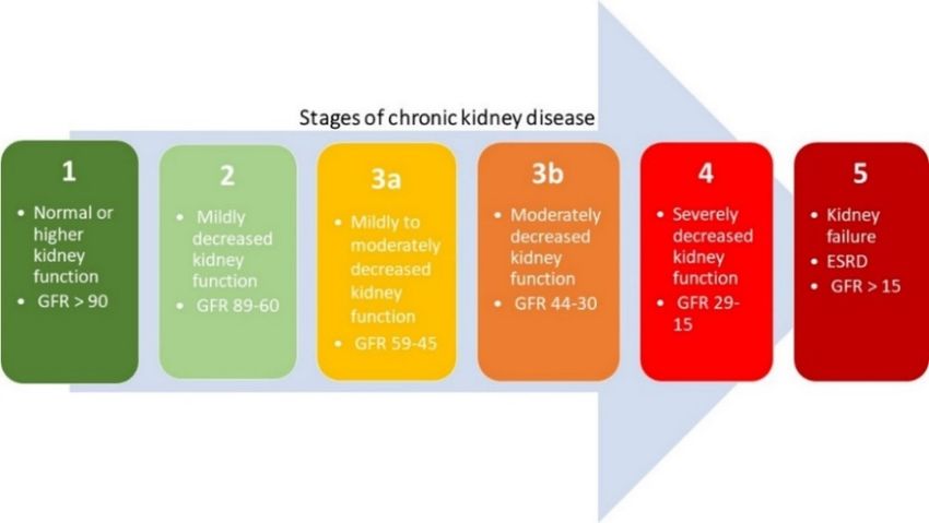
Int. J. Mol. Sci. 2019, 20, 42                                                                      2 of 15

normalizes glycaemia [9]. In addition, GLP-1 slows gastric emptying and, thus, reduces appetite and
food intake, which is essential for postprandial glucose control [9]. Furthermore, metformin interacts
with different gut bacteria, possibly through the regulation of metal homeostasis, which might also
contribute to the glucose-lowering effect of metformin [10]. Metformin is not generally associated with
a risk of hypoglycemia, because it does not stimulate endogenous insulin production, in contrast to
other antidiabetic drugs [3,7].
     In the last decade, the use of metformin in T2DM, to simply lower blood glucose, has switched
to a much more complex picture, due to its benign pleiotropic effects. Metformin has shown
substantial effectiveness in polycystic ovarian syndrome [11], cancer [12], heart and cardiovascular
disorders [13], nonalcoholic fatty liver disease [14], and premature puberty [3,15]. Furthermore,
ongoing investigations focus on its putative beneficial impact on the kidney. Metformin, when
administered during acute kidney injury (AKI) in rats, has been demonstrated to attenuate ensuing
chronic renal impairment [16], whilst pretreatment of healthy kidneys significantly protected the
kidney from subsequent AKI [16–18]. Additionally, metformin has shown a beneficial and strong
protective effect against the progression of chronic kidney disease (CKD) [19]. The aim of this review is
to summarize the current knowledge regarding the renoprotective effect of metformin. We will review
the available clinical and preclinical evidence that points towards a protective effect of metformin
treatment on the development/progression of renal diseases with different underlying etiologies, and
will discuss the cellular mechanisms and potential molecular signaling pathways by which metformin
exerts its beneficial impact on the kidney.

2. Metformin’s Glucose Lowering Effect: Molecular Mechanisms
      Despite its clinical use and detailed investigations for more than 60 years, the molecular
pathways by which metformin lowers blood glucose are still not fully understood [4,7]. A decade
ago, it was generally accepted that metformin reduced hepatic glucose synthesis via activation
of AMP-activated protein kinase (AMPK) through decreases in hepatic energy charge or directly
by its upstream activator liver kinase B1 (LKB1). Hepatic gluconeogenesis is an energy-intensive
process which starts in the mitochondria, where pyruvate enters the gluconeogenic route [20].
Metformin (>1 mM in vitro) is able to inhibit complex 1 of the mitochondrial respiratory chain,
which suppresses ATP production [4,21,22]. However, it needs to be mentioned that the claimed
effect of metformin should be interpreted with caution since several in vitro studies were conducted
with supra-pharmacological concentrations. After oral administration of metformin, a plasma
concentration of 10–40 µM is achieved in humans [23,24]. Subsequently, cytoplasmic ADP/ATP
and AMP/ATP ratio’s will increase and activate AMPK, leading to reduction of gluconeogenic gene
transcription [4,21,25,26]. Interestingly, recent studies report that metformin inhibits gluconeogenesis
through AMPK-independent mechanisms. Foretz et al. [27] demonstrated that metformin (>250 µM
in vitro) inhibited hepatic gluconeogenesis via a decrease in the hepatic energy state in an
AMPK-independent manner. They used genetic ablation of both AMPK catalytic subunits and LKB1,
to show that both AMPK and LKB1 are not required for metformin-induced (50 mg/kg, 150 mg/kg,
300 mg/kg p.o.) decreases in hepatic glucose output in mice. They found that in mice lacking AMPK in
the liver, blood glucose levels were comparable to those in wild-type mice, and that the hypoglycemic
effect of metformin was maintained [27]. Counteracting hepatic glucagon signaling by metformin, as a
novel contributor to its therapeutic actions, was reported by Miller et al. [28]. In this study, metformin
treatment (250 mg/kg/day p.o.) reduced levels of cyclic AMP, as a result of adenylate cyclase inhibition
and, consequently, attenuated glucagon’s ability to promote hepatic glucose production [28]. Later,
Madiraju et al. [29] showed that metformin (50 µM in vitro, single dose 50 mg/kg i.v.) inhibited
mitochondrial glycerophosphate dehydrogenase, resulting in an altered hepatocellular redox state,
ultimately leading to impaired utilization of lactate and glycerol for gluconeogenesis [29]. Recently,
a new mechanism of action was discovered by Hunter et al. [30]. Metformin (250 mg/kg single dose
p.o.) inhibited fructose-1-6-bisphosphatase allosterically, by increasing AMP concentration [30]. These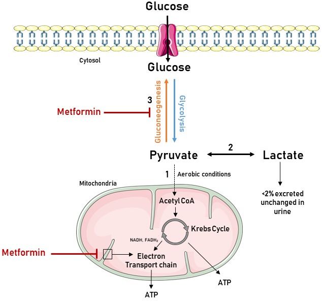
Int. J. Mol. Sci. 2019, 20, 42                                                                       3 of 15

findings clearly demonstrate that the underlying mechanisms responsible for the glucose-lowering
effects of metformin in diabetes may not be explained by any single target or pathway.

3. Acute and Chronic Kidney Disease
     Before reviewing the molecular signaling pathways by which metformin has been shown to
protect the kidneys, we will briefly elaborate on AKI and CKD. AKI is defined as an abrupt and rapid
loss of renal function that occurs rapidly over a few hours or days, and is mainly caused by exposure to
nephrotoxic substances, impaired renal blood flow, obstruction of the urinary tract, or inflammation in
the kidney [31]. CKD represents a progressive loss of renal function over a period of months or years,
often (but not consistently) progressing towards end stage renal disease (ESRD), which inevitably
requires renal replacement therapy, i.e., dialysis or kidney transplantation. However, distinction
between AKI and CKD may be artificial, since both syndromes are closely interconnected [32].

3.1. Acute Kidney Injury
     AKI is an increasingly common complication in, often elderly, patients admitted to hospitals [33].
AKI is defined as an increase in serum creatinine of more than 0.3 mg/dL within 48 h, or an increase
in serum creatinine to 1.5 times baseline, which is known, or presumed, to have occurred within the
prior 7 days, or a urine output of less than 0.5 mL/kg/h for 6 h [34]. Clinically, AKI compromises
three primary etiologies, including prerenal (azotemia), renal (tubular necrosis, interstitial nephritis or
acute glomerulonephritis), and post-renal (acute obstruction to urinary flow) [35,36]. Hypoperfusion,
a complication of major surgery and sepsis, can induce ischemia-reperfusion injury to the kidney
and, together with exposure to nephrotoxins, is the main cause of AKI [37,38]. Patients need to be
managed according to the cause of their renal disease, although the wide variety of injuries that can
occur to the kidney makes the latter challenging. Furthermore, despite the regenerative capacity of the
kidney to restore its function, patients who survive AKI have an increased risk of developing CKD
and ESRD [39].

3.2. Chronic Kidney Disease
      CKD is a worldwide recognized public health issue affecting 3–16% of the world
population [40,41]. CKD is defined as histopathological kidney damage and/or decreased kidney
function, lasting more than 3 months [42]. Kidney damage usually precedes alterations in function [43].
The kidney structure is considered to be affected when one or more markers of kidney damage
are present, such as proteinuria and/or abnormalities detected by histology or non-invasive
imaging [42–44]. The histopathological features of CKD are tubulointerstitial fibrosis, tubular atrophy,
cellular infiltration, and glomerulosclerosis [45]. The most prominent functional parameter of the
kidney is the glomerular filtration rate (GFR), which equals the total amount of fluid filtered through
the glomeruli per unit of time [44,46]. The Kidney Disease Outcomes Quality Initiative (KDOQI) of the
National Kidney Foundation (NKF) divided CKD into five stages, regardless of underlying causes,
which are shown in Figure 1. A decreased estimated glomerular filtration rate (eGFR) of less than
60 mL/min/1.73 m2 is defined by the current international guidelines as CKD [42,47]. When the eGFR
is less than 15 mL/min/1.73 m2 , the final stage of renal failure, at which kidney function is no longer
sufficient to meet the body’s need, is reached [42,44].
      In developed countries, age, diabetes, hypertension, cardiovascular disease and obesity, are the
leading causes of CKD [44]. These traditional CKD risk factors are accompanied by nontraditional
CKD risk factors, such as infections, kidney stones, and exposure to drugs and toxins, which result in
glomerular and tubulointerstitial diseases that contribute to the global burden of CKD [40,44,48].
Int. J. Mol. Sci. 2019, 20, 42 4 of 15
Int. J. Mol. Sci. 2018, 19, x FOR PEER REVIEW 4 of 15
Figure 1. Five
Figure 1. Fivestages
stagesofof chronic
chronic kidney
kidney disease
disease based
based onestimated
on the the estimated glomerular
glomerular filtration
filtration rate
rate (eGFR)
(eGFR) (mL/min per
(mL/min per 1.73 m ). 21.73 m²).
3.3. Treatment
3.3. Treatment
Current treatment
Current treatmentstrategies
strategiesforfor
AKIAKI
are mainly supportive
are mainly with the
supportive correction
with of volume
the correction ofoverload
volume
and biochemical abnormalities as primary goal of treatment [31]. Also, for CKD,
overload and biochemical abnormalities as primary goal of treatment [31]. Also, for CKD, treatment treatment strategies
mainly focus
strategies on controlling
mainly the above-mentioned
focus on controlling risk factors by
the above-mentioned riskadministration of conventional
factors by administration of
medication. However, both the IDNT (Irbesartan Diabetic Nephropathy
conventional medication. However, both the IDNT (Irbesartan Diabetic Nephropathy Trial) and Trial) and RENAAL
(Reduction of Endpointsof
RENAAL (Reduction in Non-insulin-dependent T2DM with the Angiotensin
Endpoints in Non-insulin-dependent T2DM with II Antagonist Losartan)
the Angiotensin II
studies haveLosartan)
Antagonist shown that patients
studies haveprogressively
shown that lose kidney
patients function whilst
progressively losebeing
kidney treated withwhilst
function these
medications
being treated[49,50]. To date,
with these effective
medications treatment
[49,50]. directly
To date, targeting
effective the kidney
treatment directlytotargeting
halt progression
the kidney of
CKD, to attenuate AKI, or expedite recovery, is lacking. In view of the expanding
to halt progression of CKD, to attenuate AKI, or expedite recovery, is lacking. In view of the AKI/CKD population
in our agingAKI/CKD
expanding society, thepopulation
demand forin new
ourtherapies with far-reaching
aging society, the demand clinical
for and
newsocial benefits
therapies is high.
with far-
Given the current experimental and clinical data, metformin is an interesting candidate
reaching clinical and social benefits is high. Given the current experimental and clinical data, that certainly
deserves further
metformin research attention.
is an interesting candidate that certainly deserves further research attention.
4. Lactic
4. Lactic Acidosis
Acidosis during
during Metformin
Metformin Treatment
Treatment in in Renal
Renal Failure:
Failure: A A Manageable
Manageable Contraindication
Contraindication
The historical fear for the development of lactic acidosis has hampered the exploration of
The historical fear for the development of lactic acidosis has hampered the exploration of the
the
clinical use of metformin in conditions of renal impairment. Lactic acidosis is the
clinical use of metformin in conditions of renal impairment. Lactic acidosis is the most common cause most common cause
of metabolic
of metabolic acidosis
acidosis and
and aa well-recognized
well-recognized complication
complication of of biguanide
biguanide therapy
therapy [51,52]. It occurs
[51,52]. It occurs when
when
lactic acid production exceeds lactic acid clearance, and is generally defined as a
lactic acid production exceeds lactic acid clearance, and is generally defined as a decreased blood pH decreased blood pH
(45.0mg/dL,
mg/dL, >5 >5 mmol/L)
mmol/L) [53]. [53]. Lactate,
Lactate, formed
formed
by the reduction of pyruvate, is produced in the gut, liver, and peripheral tissues as a metabolic end
by the reduction of pyruvate, is produced in the gut, liver, and peripheral tissues as a metabolic end
product of anaerobic glycolysis. Under aerobic conditions, pyruvate enters the
product of anaerobic glycolysis. Under aerobic conditions, pyruvate enters the mitochondria, where mitochondria, where its
energy is transferred to ATP via the Krebs cycle and oxidative phosphorylation,
its energy is transferred to ATP via the Krebs cycle and oxidative phosphorylation, respectively. respectively. During
the gluconeogenesis,
During pyruvatepyruvate
the gluconeogenesis, can be converted back to glucose
can be converted back to in glucose
the liverinandthekidney [53].kidney
liver and Metformin
[53].
increases lactate levels by inhibiting complex 1 of the mitochondrial respiratory
Metformin increases lactate levels by inhibiting complex 1 of the mitochondrial respiratory chain, chain, which negatively
impactsnegatively
which (i.e., reduces) the flow
impacts (i.e.,through thethe
reduces) Krebs
flowcycle as shown
through the in Figure
Krebs 2. Consequently,
cycle as shown in metabolic
Figure 2.
degradation of lactic acid by oxidation or gluconeogenesis is reduced and plasma
Consequently, metabolic degradation of lactic acid by oxidation or gluconeogenesis is reduced and lactate concentration
increases
plasma [53,54].
lactate concentration increases [53,54].Int. J. Mol. Sci. 2019, 20, 42 5 of 15
Int. J. Mol. Sci. 2018, 19, x FOR PEER REVIEW 5 of 15
Figure
Figure 2.2. Association
Association between
between the the biochemistry
biochemistry of of lactate
lactate production
production and and metformin
metformin use. use. Glycolysis
Glycolysis
is
is the metabolic pathway that converts glucose into pyruvate in the cytoplasm. Pyruvate is the
the metabolic pathway that converts glucose into pyruvate in the cytoplasm. Pyruvate is the only
only
precursor of lactate. (1) When oxygen is available, pyruvate enters the mitochondria,
precursor of lactate. (1) When oxygen is available, pyruvate enters the mitochondria, and is converted and is converted
to
to Acetyl
AcetylCoA CoAand oxidized
and oxidizedin thein Krebs cycle, delivering
the Krebs nicotinamide
cycle, delivering adenine dinucleotide
nicotinamide (NADH),
adenine dinucleotide
flavin
(NADH), adenine dinucleotide
flavin (FADH2), and
adenine dinucleotide adenosine
(FADH2), andtriphosphate (ATP). NADH
adenosine triphosphate and FADH2
(ATP). NADH feed and
the electron transport chain in the inner mitochondrial membrane to eventually
FADH2 feed the electron transport chain in the inner mitochondrial membrane to eventually generate generate the bulk of
ATP by chemiosmosis. (2) Under anaerobic conditions, pyruvate is reduced to
the bulk of ATP by chemiosmosis. (2) Under anaerobic conditions, pyruvate is reduced to lactate. lactate. However, in the
liver and kidney,
However, lactate
in the liver andcankidney,
be converted
lactate tocanpyruvate again to
be converted bypyruvate
the Cori cycle.
again (3)
by Pyruvate, the first
the Cori cycle. (3)
designated
Pyruvate, the substrate of the gluconeogenic
first designated substratepathway, can be used to generate
of the gluconeogenic pathway,glucose.
can beMetformin inhibits
used to generate
gluconeogenesis,
glucose. Metformin leading to pyruvate
inhibits accumulation
gluconeogenesis, and to
leading subsequent
pyruvate increased lactate
accumulation production.
and subsequent It
also inhibits the electron transport chain, resulting in elevated levels of NADH, a reduced
increased lactate production. It also inhibits the electron transport chain, resulting in elevated levels Krebs cycle
flow and, hence,
of NADH, further
a reduced contributing
Krebs cycle flow to and,
increased
hence,pyruvate levels. Figuretowas
further contributing produced
increased using Servier
pyruvate levels.
Medical
Figure wasArt:produced
https://smart.servier.com/
using Servier Medical (accessed on 25 October 2018).
Art: https://smart.servier.com/ (accessed on 25 October
2018).
In 1994, the U.S. Food and Drug Administration (FDA) approved the therapeutic use of metformin
in T2DM, but contraindicated it in patients with decreased kidney function, as well as in those with
In 1994, the U.S. Food and Drug Administration (FDA) approved the therapeutic use of
liver dysfunction [55,56]. Since metformin is not metabolized, but excreted, unchanged, via urine,
metformin in T2DM, but contraindicated it in patients with decreased kidney function, as well as in
the legitimate concern arose that metformin would accumulate in the circulatory system of patients
those with liver dysfunction [55,56]. Since metformin is not metabolized, but excreted, unchanged,
with impaired kidney function, thereby increasing the risk for lactic acidosis [1]. The incidence of
via urine, the legitimate concern arose that metformin would accumulate in the circulatory system of
“metformin-associated lactic acidosis” (MALA) in T2DM is 3–9 cases per 100,000 patient-years, with a
patients with impaired kidney function, thereby increasing the risk for lactic acidosis [1]. The
mortality
incidenceofof almost 50% [57,58]. Clinical
“metformin-associated lacticstudies exploring
acidosis” (MALA) theineffect
T2DM ofismetformin on the
3–9 cases per incidence
100,000 of
patient-
lactic acidosis are mostly performed in diabetic patients with varying degrees of
years, with a mortality of almost 50% [57,58]. Clinical studies exploring the effect of metformin on renal function and
report conflicting
the incidence results.
of lactic Ekström
acidosis et al. [59]
are mostly revealedinthat
performed use ofpatients
diabetic metformin with(± 1700 mg
varying monotherapy,
degrees of renal
>1700 mgand
function combination therapy)
report conflicting was associated
results. Ekström etwith reduced
al. [59] revealedriskthat
of all-cause mortality,
use of metformin (± acidosis,
1700 mg
serious infection,
monotherapy, or cardiovascular
>1700 mg combination disease in patients
therapy) with impaired
was associated with renal function
reduced risk (eGFR 30 to
of all-cause
45 mL/minacidosis,
per 1.73 m 2 ). Richy et al. [60]
mortality, serious infection, or focused on the risk
cardiovascular of lactic
disease acidosis in
in patients metformin-treated
with impaired renal
T2DM
function (eGFR 30 to 45 mL/min per 1.73 m²). Richy et al. [60] focused on the risk of lacticuse
patients with various degrees of renal failure. They concluded that metformin did not
acidosis in
metformin-treated T2DM patients with various degrees of renal failure. They concluded that
metformin use did not affect the incidence of lactic acidosis in patients with T2DM who had eitherInt. J. Mol. Sci. 2019, 20, 42 6 of 15
affect the incidence of lactic acidosis in patients with T2DM who had either normal, mild, moderate,
or severe renal dysfunction [60]. However, Eppenga et al. [61] based on results of a retrospective
patient database analysis reported that the risk of lactic acidosis or elevated lactate concentrations is
significantly increased in patients with mild to moderate renal insufficiency (2 g [61]. In 2016, the FDA revised their warning
with regard to metformin use in patients with impaired kidney function (eGFR < 60 mL/min/1.73 m2 ),
expanding its use in CKD to those with an eGFR of 30 mL/min/1.73 m2 . The revised guidelines state
that metformin could be used safely in patients with mild to moderate impairment in kidney function,
but remains absolutely contraindicated in patients with severe CKD (eGFR < 30 mL/min/1.73 m2 ) [62].
Interestingly, a recent clinical study of Lalau et al. [63] indicated that metformin treatment also
appeared to be safe and still pharmacologically efficacious in moderate-to-severe CKD, when the dose
is adjusted for the degree of renal failure. More precisely, on the basis of dose-finding study’s, they
selected a chronic dosage regimen of 1500 mg/day for patients with CKD stage 3a, 1000 mg/day for
patients with CKD stage 3b, and 500 mg/day for patients with CKD stage 4 [63].
In conclusion, the historical fear for metformin treatment in patients with renal impairment is
seemingly overemphasized. Provided that the dose is adjusted for renal function and a close follow-up
of the patients is ensured, metformin can be used safely in patients with mild-to-moderate renal failure.
5. Metformin’s Renoprotection: Clinical Evidence
In several clinical studies, metformin has been shown to improve survival of AKI and CKD
patients. In a large cohort of over 25,000 patients with T2DM, Bell et al. [64] provided a reassuring
message of the safety of metformin in patients with or without CKD, as mortality was not adversely
affected by metformin use. Metformin did not increase the incidence of AKI, and survival rates
were higher in patients with AKI previously treated with metformin [64]. In a retrospective cohort
study, Stephen et al. [65] linked Scientific Registry of Transplant Recipients data for all incident
kidney transplants from 2001 until 2012, and national pharmacy claims. They found that survival was
superior for all outcomes for recipients who filled metformin claims compared with those who filled
non-metformin agent claims [65]. In an open cohort study of 469,688 T2DM patients, the relationship
between a range of complications and antidiabetic therapy was analyzed. Severe kidney failure,
including dialysis treatment, kidney transplant, and CKD stage 5, were among the five pre-specified
key outcomes. Compared with non-use, metformin was associated with a significantly decreased risk of
severe kidney failure, whereas sulfonylureas and insulin increased this risk [66]. In a recent systematic
review involving 17 observational studies, metformin use appeared to be associated with reduced
all-cause mortality in patients with CKD, congestive heart failure, and chronic liver disease [67].
Conversely, in a study of 616 patients, Hsu et al. [68] evaluated the effect of continuous metformin
treatment on renal function in patients with T2DM and moderate CKD (eGFR 30–60 mL/min/1.73 m2 ).
They concluded that continuous metformin therapy was associated with a decline in renal function in
patients with T2DM and moderate CKD [68]. However, as this was a retrospective study, the authors
could not exclude putative confounding factors such as life-style, use of angiotensin-converting
enzyme inhibitor/angiotensin receptor blocker, cumulative duration of exposure, and defined daily
dose of metformin.
These findings open perspectives for urgently needed large, long-term prospective randomized
controlled trials that investigate the effect of metformin on renal function.
6. Metformin’s Renoprotection: Experimental Evidence
Due to the fact that metformin has been historically contraindicated in patients with advanced
CKD, because of the lingering concern of life-threatening lactic acidosis, a large body of evidence to
support the renoprotective effect of metformin is provided by preclinical studies. Metformin has been
shown to exert positive effects on the kidney in various experimental studies performed in divergent
rodent models representing different types of renal diseases going from AKI to CKD.Int. J. Mol. Sci. 2019, 20, 42 7 of 15
Satriano et al. [69] studied the effect of metformin on kidney function and structure in 5/6 th
nephrectomized rats, a model of mild-to-moderate CKD, to determine whether changes in AMPK
enzymatic activity correlates with the changes in kidney metabolism and kidney function [69]. Daily
administration of metformin (250 mg/kg/day p.o.) by oral gavage ameliorated kidney fibrosis,
as well as structural and morphologic renal abnormalities. In addition, metformin normalized kidney
function and oxygen consumption, which was also mediated via induction of AMPK [69]. Lee et al. [70]
evaluated the role of the AMPK-acetyl-CoA carboxylase (ACC) pathway in the antifibrotic effect of
metformin in a mouse model with folic acid-induced CKD. Activated AMPK phosphorylates and
inactivates ACC activity, which improves lipid availability for energy generation from fatty acids in
renal tubular cells. By use of ACC knock-in (KI) mice, with inactivating Ser to Ala mutations preventing
ACC phosphorylation, it was observed that metformin’s protective effects were abrogated, as folic
acid induced tubulointerstitial fibrosis, and lipid accumulation were not attenuated. The antifibrotic
effect of metformin is, thus; dependent on its ability to increase phosphorylation of ACC, which in turn
increases fatty acid oxidation in damaged renal tissues [71]. In a rat model of CKD-mineral and bone
disorder, Neven et al. [19] showed that daily metformin treatment (200 mg/kg/day p.o.) was able
to strongly attenuate development of severe CKD, as evidenced by a preserved renal function with
maintained mineral homeostasis, and a significantly reduced renal inflammation, cellular infiltration
and fibrosis [19].
The potential role of metformin in protecting the kidney from a nephrotoxic insult was examined
by Morales et al. [71]. They hypothesized that metformin treatment (100 mg/kg/day p.o.) protected
the kidney from gentamicin-induced toxicity in rats, a model of acute renal failure, through a
mitochondria-dependent pathway. Indeed, endogenous oxidative stress has a major role in the
severity of gentamicin-induced acute renal failure, suggesting that the protective effect, afforded
by metformin, could be mediated, at least in part, by enhanced antioxidant defenses, and by its
ability to prevent gentamicin-induced lipid peroxidation [71]. In a mouse model of unilateral ureteral
obstruction, Cavaglieri et al. [72] showed that metformin treatment (200 mg/kg/day p.o.), initiated
one day before surgery, prevented or slowed down the onset of renal inflammation and fibrosis. This
amelioration was accompanied by increased activity of AMPK [72]. Declèves et al. [18] investigated the
effects of AMPK activity, using agonists of AMPK, among which included metformin (300 mg/kg/day
p.o.), on autophagy and cell stress proteins in a model of kidney ischemia-reperfusion (IR). They
showed that IR led to downregulation of AMPK and autophagy which consequently induced cellular
changes. Additionally, they demonstrated that induction of the AMPK/autophagy axis by metformin
provided beneficial cellular effects which offered a viable strategy in reducing kidney IR injury [18].
Conversely, Lieberthal et al. [73] demonstrated that inhibition of the mammalian target of
rapamycin (mTOR) impaired renal recovery, assessed by GFR, after IR in rats, which appeared
to be due, at least in part, to the inhibition of tubular cell regeneration [73,74]. The mTOR pathway
is one of the downstream signaling pathways regulated by AMPK [75]. Consequently, metformin,
as AMPK activator, is able to inhibit the mTOR pathway and cause a delay in renal regeneration and
repair [73,76]. However, Lieberthal et al. [74] also proved that the regeneration of tubular cells and
recovery of GFR after IR in rats, although delayed by the mTOR inhibitor, rapamycin, eventually
occurred, despite continued administration of the latter inhibitor [74].
7. Metformin’s Renoprotection: Molecular Mechanisms
Knowledge about the molecular mechanism by which metformin appears to prevent or attenuate
AKI and CKD is fragmentary, however, it is clear that metformin protects the kidney via pleiotropic
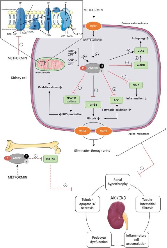
actions against different aspects of the pathophysiology of renal diseases, as presented in Figure 3.Int. J. Mol. Sci. 2019, 20, 42 8 of 15
Int. J. Mol. Sci. 2018, 19, x FOR PEER REVIEW 8 of 15
Figure 3. Potential underlying molecular mechanisms of metformin’s renoprotection. Metformin is
Figure 3. Potential underlying molecular mechanisms of metformin’s renoprotection. Metformin
transported into the renal tubular cells mainly through the plasma membrane transporter OCT2.
is transported into the renal tubular cells mainly through the plasma membrane transporter OCT2.
Inside the renal cell metformin activates AMPK via two separate mechanisms, i.e., the inhibition of
Inside the
therenal cell metformin
mitochondrial activates
respiratory AMPK
chain complex viasubsequent
1, and two separate mechanisms,
increase in AMP/ATPi.e.,
and the inhibition of
ADP/ATP
the mitochondrial
ratio, and/orrespiratory chain complex
the direct activation of AMPK.1,AMPK
and subsequent increase
has pleiotropic in AMP/ATP
downstream and ADP/ATP
signaling pathways
involved
ratio, and/or theindirect
divergent cellularof
activation processes,
AMPK. such
AMPK as autophagy, fatty acid
has pleiotropic oxidation, signaling
downstream inflammation,
pathways
involvedfibrosis, oxidative
in divergent stress, processes,
cellular and reactivesuch
oxygen species (ROS)fatty
as autophagy, in renal
acidcells and FGF23
oxidation, production in fibrosis,
inflammation,
bone cells, which have been shown to protect the kidney against AKI and CKD. Further, MATE1 and
oxidative stress, and reactive oxygen species (ROS) in renal cells and FGF23 production in bone cells,
MATE2 contribute to the renal excretion of metformin. ACC, acetyl-CoA carboxylase; AKI, acute
which have been shown to protect the kidney against AKI and CKD. Further, MATE1 and MATE2
kidney injury; AMPK, AMP-activated protein kinase; CKD, chronic kidney disease; FGF23, fibroblast
contribute to the
growth renal
factor 23;excretion of metformin.
MATE1, multidrug ACC,
and toxin acetyl-CoA
extrusion carboxylase;
1; MATE2, multidrug AKI, acute
and toxin kidney injury;
extrusion
AMPK, AMP-activated protein kinase; CKD, chronic kidney disease; FGF23, fibroblast growth factor 23;
MATE1, multidrug and toxin extrusion 1; MATE2, multidrug and toxin extrusion 2; mTOR, mammalian
target of rapamycin; NADPH, nicotinamide adenine dinucleotide phosphate; NFκB, nuclear factor
kappa B; OCT2, organic cation transporter 2; ROS, reactive oxygen species; TGF-β1, transforming
growth factor β1; ULK1, uncoordinated-51 like kinase 1. Figure was produced using Servier Medical
Art: https://smart.servier.com/ (accessed on 2 November 2018).Int. J. Mol. Sci. 2019, 20, 42 9 of 15
Metformin, a positively charged molecule at physiological pH, enters the renal epithelial cells
through the organic cation transporter 2 (OCT2), which is predominantly expressed at the basolateral
membrane in the renal tubules [1,2]. Multidrug and toxin extrusion 1 (MATE1) and multidrug and
toxin extrusion 2 (MATE2) transporters contribute to the renal excretion of metformin, which is
eliminated, unchanged, in the urine by glomerular filtration and tubular secretion [1,2]. Metformin
activates AMPK via two separate mechanisms, i.e., direct activation of AMPK via phosphorylation of its
activating loop at Thr-172 of the α-subunit, and indirectly by inhibiting the mitochondrial respiratory
chain complex 1 [22,76,77]. Indeed, metformin acts on mitochondria by inhibiting complex 1 of
the respiratory chain, which leads to a reduction in cellular energy charge [22,77]. When ATP has
been extensively utilized, the energy-sensing kinase AMPK is activated, which acts to restore energy
homeostasis by switching to catabolic pathways for the generation of ATP [4,78].
The activation of inflammatory cells and production of extracellular matrix is regulated by
transforming growth factor β1 (TGF-β1) in the kidney [16,79]. Metformin (10 mM) is able to diminish
TGF-β1 expression in mouse renal fibroblasts in vitro and, consequently, alleviate pro-fibrotic gene
expression via AMPK activation, which in vivo eventually should reduce interstitial fibrosis, one
of the key pathological events of CKD [16,79]. Additionally, the antifibrotic effect of metformin
is, at least in part, dependent on the ability of metformin to increase fatty acid oxidation via the
AMPK-ACC pathway, as discussed previously. Oxidative stress is also involved in the pathogenesis of
AKI and CKD [80,81]. Reactive oxygen species (ROS), such as superoxide anion (O2 − ) and hydrogen
peroxide (H2 O2 ), normally drives the cellular responses for tissue repair processes, inflammation
and defense mechanisms. In pathological situations, however, the overproduction of free radicals
contributes to cell and tissue injury [80–82]. In patients with renal dysfunction, the balance between
ROS production and antioxidative defense mechanisms is disturbed. Furthermore, oxidative stress
seems to increase as CKD progresses [82,83]. In the kidney, the enzymatic complex nicotinamide
adenine dinucleotide phosphate (NADPH) oxidase is involved in ROS production [84]. In vitro
studies further revealed that metformin (2 mM) prevents oxidative stress in podocytes, and decreases
ROS production by inhibiting NADPH oxidase [85]. Moreover, metformin (0.01–0.1 mM) is able to
protect tubular cells and renal tissue against tissue damage caused by oxidative stress, at least in part
through a mitochondria-dependent pathway, as mentioned previously [71,86]. Recent literature has
put forward autophagy as a possible mechanism in the pathology of AKI and CKD [87–90]. Indeed, the
autophagic flux, i.e., the formation of autophagosomes and their fusion with lysosomes, is crucial for
cell survival and a growing body of evidence suggests that a deficient autophagic flux may contribute
to a broad spectrum of diseases, including AKI and CKD [91]. The mammalian autophagy-initiating
uncoordinated-51 like kinase 1 (ULK1) is regulated by AMPK and mTOR [92]. Activated AMPK inhibits
mTOR, leading to ULK1–AMPK interaction and activation of ULK1, which induces autophagy [92].
Metformin, as an AMPK agonist, may thus advance or prolong an inherent autophagic response,
which may play a protective role against AKI, and the development and progression of CKD [87,90].
Recently, fibroblast growth factor 23 (FGF23) has gained broad attention as a disease biomarker in
CKD, since elevated FGF23 plasma concentrations are observed in an early stage of CKD, and increase
proportionally with renal function decline [93]. FGF23 is a phosphaturic hormone responsible for
phosphate handling and vitamin D metabolism, with the kidney as an important target [93,94]. Further,
FGF23 may actively contribute to the progression of CKD [93,94]. Since AMPK has been demonstrated
as an important regulator of FGF23 production, it is tempting to speculate that metformin, being an
AMPK activator, is able to inhibit FGF23 production, which is favorable to prevent deterioration of
renal function [94].
8. Conclusions
Taken together, metformin, beyond its glucose-lowering actions in T2DM, may be considered a
promising renoprotective compound in various types of renal diseases. Additional preclinical and
longitudinal clinical trials are required to explore whether metformin is able to slow down, or evenInt. J. Mol. Sci. 2019, 20, 42 10 of 15
arrest, the progression of early kidney injury and CKD. Most of the reported in vitro and in vivo
evidence on the potential effects of metformin has been based on a broad range of concentrations
hampering the translation of experimental observations to the clinical situation. Experimental designs
often include metformin concentrations or doses that are higher than what can be achieved in CKD
patients. Additional in vivo studies using lower metformin doses would undoubtedly improve
translation from preclinical studies to clinical trials. Further fundamental research is necessary to
elucidate the complex network of signaling pathways by which metformin exerts its renoprotective
effects, as this is indispensable to identify potentially new therapeutic targets.
Author Contributions: R.C. drafted the manuscript; B.A.V. and E.N. edited and proofread the review; A.V. and
P.C.D. critically revised the paper. All authors contributed to the final manuscript.
Funding: This research received no external funding.
Conflicts of Interest: Authors declare no conflict of interest.
Abbreviations
ACC acetyl-CoA carboxylase
AKI acute kidney injury
AMPK AMP-activated protein kinase
ATP adenosine triphosphate
CKD chronic kidney injury
eGFR estimated glomerular filtration rate
ESRD end stage renal disease
FADH2 flavin adenine dinucleotide
FDA Food and Drug Administration
FGF23 fibroblast growth factor 23
GFR glomerular filtration rate
GLP-1 glucagon-like peptide 1
IR ischemia-reperfusion
i.v. intravenous
KDOQI kidney disease outcomes quality initiative
LKB1 liver kinase B1
MALA metformin-associated lactic acidosis
MATE1 multidrug and toxin extrusion 1
MATE2 multidrug and toxin extrusion 2
mTOR mammalian target of rapamycin
NADH nicotinamide adenine dinucleotide
NADPH nicotinamide adenine dinucleotide phosphate
NFκB, nuclear factor kappa B
NKF national kidney foundation
OCT1 organic cation transporter 1
OCT2 organic cation transporter 2
p.o. per os
ROS reactive oxygen species
T2DM type 2 diabetes mellitus
TGF-β2 transforming growth factor β2
ULK1 uncoordinated-51 like kinase 1
References
1. Gong, L.; Goswami, S.; Giacomini, K.M.; Altman, R.B.; Klein, T.E. Metformin pathways: Pharmacokinetics
and pharmacodynamics. Pharmacogenet. Genom. 2012, 22, 820–827. [CrossRef] [PubMed]Int. J. Mol. Sci. 2019, 20, 42 11 of 15
2. Graham, G.G.; Punt, J.; Arora, M.; Day, R.O.; Doogue, M.P.; Duong, J.K.; Furlong, T.J.; Greenfield, J.R.;
Greenup, L.C.; Kirkpatrick, C.M.; et al. Clinical Pharmacokinetics of Metformin. Clin. Pharmacokinet. 2011,
50, 81–98. [CrossRef] [PubMed]
3. Imam, T.H. Changes in metformin use in chronic kidney disease. Clin. Kidney J. 2017, 10, 301–304. [CrossRef]
[PubMed]
4. Rena, G.; Hardie, D.G.; Pearson, E.R. The mechanisms of action of metformin. Diabetologia 2017, 60, 1577–1585.
[CrossRef] [PubMed]
5. Marshall, S.M. 60 years of metformin use: A glance at the past and a look to the future. Diabetologia 2017, 60,
1561–1565. [CrossRef]
6. Bailey, C.J. Metformin: Historical overview. Diabetologia 2017, 60, 1566–1576. [CrossRef] [PubMed]
7. Panchapakesan, U.; Pollock, C. Drug repurposing in kidney disease. Kidney Int. 2018, 94, 40–48. [CrossRef]
8. McCreight, L.J.; Bailey, C.J.; Pearson, E.R. Metformin and the gastrointestinal tract. Diabetologia 2016, 59,
426–435. [CrossRef] [PubMed]
9. Bahne, E.; Hansen, M.; Bronden, A.; Sonne, D.P.; Vilsboll, T.; Knop, F.K. Involvement of glucagon-like
peptide-1 in the glucose-lowering effect of metformin. Diabetes Obes. Metab. 2016, 18, 955–961. [CrossRef]
10. Wu, H.; Esteve, E.; Tremaroli, V.; Khan, M.T.; Caesar, R.; Mannerås-Holm, L.; Ståhlman, M.; Olsson, L.M.;
Serino, M.; Planas-Fèlix, M.; et al. Metformin alters the gut microbiome of individuals with treatment-naive
type 2 diabetes, contributing to the therapeutic effects of the drug. Nat. Med. 2017, 23, 850–858. [CrossRef]
11. Lashen, H. Role of metformin in the management of polycystic ovary syndrome. Ther. Adv. Endocrinol.
Metab. 2010, 1, 117–128. [CrossRef] [PubMed]
12. Zi, F.; Zi, H.; Li, Y.; He, J.; Shi, Q.; Cai, Z. Metformin and cancer: An existing drug for cancer prevention and
therapy. Oncol. Lett. 2018, 15, 683–690. [CrossRef] [PubMed]
13. Nesti, L.; Natali, A. Metformin effects on the heart and the cardiovascular system: A review of experimental
and clinical data. Nutr. Metab. Cardiovasc. Dis. 2017, 27, 657–669. [CrossRef] [PubMed]
14. Li, Y.A.N.; Liu, L.E.I.; Wang, B.I.N.; Wang, J.U.N.; Chen, D. Metformin in non-alcoholic fatty liver disease: A
systematic review and meta-analysis. Biomed. Rep. 2013, 1, 57–64. [CrossRef] [PubMed]
15. Ibanez, L.; Ong, K.; Valls, C.; Marcos, M.V.; Dunger, D.B.; de Zegher, F. Metformin treatment to prevent
early puberty in girls with precocious pubarche. J. Clin. Endocrinol. Metab. 2006, 91, 2888–2891. [CrossRef]
[PubMed]
16. Wang, M.; Weng, X.; Guo, J.; Chen, Z.; Jiang, G.; Liu, X. Metformin alleviated EMT and fibrosis after renal
ischemia-reperfusion injury in rats. Renal Fail. 2016, 38, 614–621. [CrossRef] [PubMed]
17. Li, J.; Gui, Y.; Ren, J.; Liu, X.; Feng, Y.; Zeng, Z.; He, W.; Yang, J.; Dai, C. Metformin Protects against
Cisplatin-Induced Tubular Cell Apoptosis and Acute Kidney Injury via AMPKalpha-regulated Autophagy
Induction. Sci. Rep. 2016, 6, 23975. [CrossRef]
18. Decleves, A.E.; Sharma, K.; Satriano, J. Beneficial Effects of AMP-Activated Protein Kinase Agonists in Kidney
Ischemia-Reperfusion: Autophagy and Cellular Stress Markers. Nephron Exp. Nephrol. 2014. [CrossRef]
19. Neven, E.; Vervaet, B.; Brand, K.; Gottwald-Hostalek, U.; Opdebeeck, B.; De Mare, A.; Verhulst, A.; Lalau, J.D.;
Kamel, S.; De Broe, M.E.; et al. Metformin prevents the development of severe chronic kidney disease and its
associated mineral and bone disorder. Kidney Int. 2018, 94, 102–113. [CrossRef]
20. McCommis, K.S.; Finck, B.N. Mitochondrial pyruvate transport: A historical perspective and future research
directions. Biochem. J. 2015, 466, 443–454. [CrossRef]
21. Cool, B.; Zinker, B.; Chiou, W.; Kifle, L.; Cao, N.; Perham, M.; Dickinson, R.; Adler, A.; Gagne, G.; Iyengar, R.;
et al. Identification and characterization of a small molecule AMPK activator that treats key components of
type 2 diabetes and the metabolic syndrome. Cell Metab. 2006, 3, 403–416. [CrossRef]
22. Owen, M.R.; Doran, E.; Halestrap, A.P. Evidence that metformin exerts its anti-diabetic effects through
inhibition of complex 1 of the mitochondrial respiratory chain. Biochem. J. 2000, 348 Pt 3, 607–614. [CrossRef]
23. Wilcock, C.; Bailey, C.J. Accumulation of metformin by tissues of the normal and diabetic mouse. Xenobiotica
1994, 24, 49–57. [CrossRef]
24. He, L.; Wondisford, F.E. Metformin Action: Concentrations Matter. Cell Metab. 2015, 21, 159–162. [CrossRef]
[PubMed]
25. Shaw, R.J.; Lamia, K.A.; Vasquez, D.; Koo, S.-H.; Bardeesy, N.; DePinho, R.A.; Montminy, M.; Cantley, L.C.
The Kinase LKB1 Mediates Glucose Homeostasis in Liver and Therapeutic Effects of Metformin. Science
2005, 310, 1642–1646. [CrossRef] [PubMed]Int. J. Mol. Sci. 2019, 20, 42 12 of 15
26. Cao, J.; Meng, S.; Chang, E.; Beckwith-Fickas, K.; Xiong, L.; Cole, R.N.; Radovick, S.; Wondisford, F.E.;
He, L. Low concentrations of metformin suppress glucose production in hepatocytes through AMP-activated
protein kinase (AMPK). J. Biol. Chem. 2014, 289, 20435–20446. [CrossRef] [PubMed]
27. Foretz, M.; Hébrard, S.; Leclerc, J.; Zarrinpashneh, E.; Soty, M.; Mithieux, G.; Sakamoto, K.; Andreelli, F.;
Viollet, B. Metformin inhibits hepatic gluconeogenesis in mice independently of the LKB1/AMPK pathway
via a decrease in hepatic energy state. J. Clin. Investig. 2010, 120, 2355–2369. [CrossRef]
28. Miller, R.A.; Chu, Q.; Xie, J.; Foretz, M.; Viollet, B.; Birnbaum, M.J. Biguanides suppress hepatic glucagon
signalling by decreasing production of cyclic AMP. Nature 2013, 494, 256–260. [CrossRef]
29. Madiraju, A.K.; Erion, D.M.; Rahimi, Y.; Zhang, X.M.; Braddock, D.T.; Albright, R.A.; Prigaro, B.J.; Wood, J.L.;
Bhanot, S.; MacDonald, M.J.; et al. Metformin suppresses gluconeogenesis by inhibiting mitochondrial
glycerophosphate dehydrogenase. Nature 2014, 510, 542–546. [CrossRef]
30. Hunter, R.W.; Hughey, C.C.; Lantier, L.; Sundelin, E.I.; Peggie, M.; Zeqiraj, E.; Sicheri, F.; Jessen, N.;
Wasserman, D.H.; Sakamoto, K. Metformin reduces liver glucose production by inhibition of
fructose-1-6-bisphosphatase. Nat. Med. 2018, 24, 1395–1406. [CrossRef]
31. Bellomo, R.; Kellum, J.A.; Ronco, C. Acute kidney injury. Lancet 2012, 380, 756–766. [CrossRef]
32. Chawla, L.S.; Kimmel, P.L. Acute kidney injury and chronic kidney disease: An integrated clinical syndrome.
Kidney Int. 2012, 82, 516–524. [CrossRef] [PubMed]
33. Ferenbach, D.A.; Bonventre, J.V. Mechanisms of maladaptive repair after AKI leading to accelerated kidney
ageing and CKD. Nat. Rev. Nephrol. 2015, 11, 264–276. [CrossRef] [PubMed]
34. Kidney Disease: Improving Global Outcomes (KDIGO) CKD Work Group. KDIGO 2012 clinical practice
guideline for the evaluation and management of chronic kidney disease. Kidney Int. Suppl. 2012, 2, 19–36.
[CrossRef]
35. Thadhani, R.; Pascual, M.; Bonventre, J.V. Acute renal failure. N. Engl. J. Med. 1996, 334, 1448–1460.
[CrossRef] [PubMed]
36. Basile, D.P.; Anderson, M.D.; Sutton, T.A. Pathophysiology of Acute Kidney Injury. Compr. Physiol. 2012, 2,
1303–1353. [CrossRef] [PubMed]
37. Lameire, N.; Van Biesen, W.; Vanholder, R. Acute renal failure. Lancet 2005, 365, 417–430. [CrossRef]
38. Humphreys, B.D.; Cantaluppi, V.; Portilla, D.; Singbartl, K.; Yang, L.; Rosner, M.H.; Kellum, J.A.; Ronco, C.
Targeting Endogenous Repair Pathways after AKI. J. Am. Soc. Nephrol. 2016, 27, 990–998. [CrossRef]
39. Coca, S.G.; Singanamala, S.; Parikh, C.R. Chronic kidney disease after acute kidney injury: A systematic
review and meta-analysis. Kidney Int. 2012, 81, 442–448. [CrossRef]
40. Jha, V.; Garcia-Garcia, G.; Iseki, K.; Li, Z.; Naicker, S.; Plattner, B.; Saran, R.; Wang, A.Y.; Yang, C.W. Chronic
kidney disease: Global dimension and perspectives. Lancet 2013, 382, 260–272. [CrossRef]
41. De Broe, M.E.; Gharbi, M.B.; Elseviers, M. Maremar, prevalence of chronic kidney disease, how to avoid
over-diagnosis and under-diagnosis. Néphrol. Thér. 2016, 12, S57–S63. [CrossRef] [PubMed]
42. Kidney Disease: Improving Global Outcomes (KDIGO) CKD Work Group. KDIGO 2012 Clinical practice
guideline for the evaluation and management of chronic kidney disease. Kidney Int. Suppl. 2013, 3, 1–150.
[CrossRef]
43. Lopez-Giacoman, S.; Madero, M. Biomarkers in chronic kidney disease, from kidney function to kidney
damage. World J. Nephrol. 2015, 4, 57–73. [CrossRef] [PubMed]
44. Webster, A.C.; Nagler, E.V.; Morton, R.L.; Masson, P. Chronic Kidney Disease. Lancet 2017, 389, 1238–1252.
[CrossRef]
45. Schlondorff, D.O. Overview of factors contributing to the pathophysiology of progressive renal disease.
Kidney Int. 2008, 74, 860–866. [CrossRef]
46. Levey, A.S.; Becker, C.; Inker, L.A. Glomerular Filtration Rate and Albuminuria for Detection and Staging of
Acute and Chronic Kidney Disease in Adults: A Systematic Review. JAMA 2015, 313, 837–846. [CrossRef]
[PubMed]
47. K/DOQI clinical practice guidelines for chronic kidney disease: Evaluation, classification, and stratification.
Am. J. Kidney Dis. 2002, 39, S1–S266.
48. Luyckx, V.A.; Tuttle, K.R.; Garcia-Garcia, G.; Gharbi, M.B.; Heerspink, H.J.L.; Johnson, D.W.; Liu, Z.-H.;
Massy, Z.A.; Moe, O.; Nelson, R.G.; et al. Reducing major risk factors for chronic kidney disease. Kidney Int.
Suppl. 2017, 7, 71–87. [CrossRef]Int. J. Mol. Sci. 2019, 20, 42 13 of 15
49. Lewis, E.J.; Hunsicker, L.G.; Clarke, W.R.; Berl, T.; Pohl, M.A.; Lewis, J.B.; Ritz, E.; Atkins, R.C.; Rohde, R.;
Raz, I. Renoprotective effect of the angiotensin-receptor antagonist irbesartan in patients with nephropathy
due to type 2 diabetes. N. Engl. J. Med. 2001, 345, 851–860. [CrossRef]
50. Brenner, B.M.; Cooper, M.E.; de Zeeuw, D.; Keane, W.F.; Mitch, W.E.; Parving, H.-H.; Remuzzi, G.;
Snapinn, S.M.; Zhang, Z.; Shahinfar, S. Effects of Losartan on Renal and Cardiovascular Outcomes in
Patients with Type 2 Diabetes and Nephropathy. N. Engl. J. Med. 2001, 345, 861–869. [CrossRef]
51. Lalau, J.D.; Arnouts, P.; Sharif, A.; De Broe, M.E. Metformin and other antidiabetic agents in renal failure
patients. Kidney Int. 2015, 87, 308–322. [CrossRef] [PubMed]
52. Chan, N.N.; Brain, H.P.; Feher, M.D. Metformin-associated lactic acidosis: A rare or very rare clinical entity?
Diabet. Med. 1999, 16, 273–281. [CrossRef] [PubMed]
53. Fall, P.J.; Szerlip, H.M. Lactic Acidosis: From Sour Milk to Septic Shock. J. Intensive Care Med. 2005, 20,
255–271. [CrossRef] [PubMed]
54. DeFronzo, R.; Fleming, G.A.; Chen, K.; Bicsak, T.A. Metformin-associated lactic acidosis: Current perspectives
on causes and risk. Metab. Clin. Exp. 2016, 65, 20–29. [CrossRef] [PubMed]
55. Lipska, K.J. Metformin Use in Patients With Historical Contraindications. Ann. Intern. Md. 2017, 166,
225–226. [CrossRef]
56. Misbin, R.I.; Green, L.; Stadel, B.V.; Gueriguian, J.L.; Gubbi, A.; Fleming, G.A. Lactic Acidosis in Patients
with Diabetes Treated with Metformin. N. Engl. J. Med. 1998, 338, 265–266. [CrossRef]
57. Van Berlo-van de Laar, I.R.; Vermeij, C.G.; Doorenbos, C.J. Metformin associated lactic acidosis: Incidence
and clinical correlation with metformin serum concentration measurements. J. Clin. Pharm. Ther. 2011, 36,
376–382. [CrossRef]
58. Kajbaf, F.; Lalau, J.-D. The prognostic value of blood pH and lactate and metformin concentrations in severe
metformin-associated lactic acidosis. BMC Pharmacol. Toxicol. 2013, 14, 22. [CrossRef]
59. Ekström, N.; Schiöler, L.; Svensson, A.-M.; Eeg-Olofsson, K.; Miao Jonasson, J.; Zethelius, B.; Cederholm, J.;
Eliasson, B.; Gudbjörnsdottir, S. Effectiveness and safety of metformin in 51 675 patients with type 2 diabetes
and different levels of renal function: a cohort study from the Swedish National Diabetes Register. BMJ Open
2012, 2, e001076. [CrossRef]
60. Richy, F.F.; Sabidó-Espin, M.; Guedes, S.; Corvino, F.A.; Gottwald-Hostalek, U. Incidence of Lactic Acidosis in
Patients With Type 2 Diabetes With and Without Renal Impairment Treated With Metformin: A Retrospective
Cohort Study. Diabetes Care 2014, 37, 2291. [CrossRef]
61. Eppenga, W.L.; Lalmohamed, A.; Geerts, A.F.; Derijks, H.J.; Wensing, M.; Egberts, A.; De Smet, P.A.G.M.;
de Vries, F. Risk of Lactic Acidosis or Elevated Lactate Concentrations in Metformin Users With Renal
Impairment: A Population-Based Cohort Study. Diabetes Care 2014, 37, 2218. [CrossRef] [PubMed]
62. Crowley, M.J.; Diamantidis, C.J.; McDuffie, J.R. Metformin Use in Patients with Historical Contraindications
or Precautions [Internet]. Appendix A, FDA Safety Announcements for Metformin. Available online:
https://www.ncbi.nlm.nih.gov/books/NBK409379/ (accessed on 1 October 2018).
63. Lalau, J.D.; Kajbaf, F.; Bennis, Y.; Hurtel-Lemaire, A.S.; Belpaire, F.; De Broe, M.E. Metformin Treatment
in Patients With Type 2 Diabetes and Chronic Kidney Disease Stages 3A, 3B, or 4. Diabetes Care 2018, 41,
547–553. [CrossRef] [PubMed]
64. Bell, S.; Farran, B.; McGurnaghan, S.; McCrimmon, R.J.; Leese, G.P.; Petrie, J.R.; McKeigue, P.; Sattar, N.;
Wild, S.; McKnight, J.; et al. Risk of acute kidney injury and survival in patients treated with Metformin: An
observational cohort study. BMC Nephrol. 2017, 18, 163. [CrossRef] [PubMed]
65. Stephen, J.; Anderson-Haag, T.L.; Gustafson, S.; Snyder, J.J.; Kasiske, B.L.; Israni, A.K. Metformin use in
kidney transplant recipients in the United States: An observational study. Am. J. Nephrol. 2014, 40, 546–553.
[CrossRef] [PubMed]
66. Hippisley-Cox, J.; Coupland, C. Diabetes treatments and risk of amputation, blindness, severe kidney failure,
hyperglycaemia, and hypoglycaemia: Open cohort study in primary care. BMJ 2016, 352, 1450. [CrossRef]
[PubMed]
67. Crowley, M.J.; Diamantidis, C.J.; McDuffie, J.R.; Cameron, C.B.; Stanifer, J.W.; Mock, C.K.; Wang, X.; Tang, S.;
Nagi, A.; Kosinski, A.S.; et al. Clinical Outcomes of Metformin Use in Populations With Chronic Kidney
Disease, Congestive Heart Failure, or Chronic Liver Disease: A Systematic Review. Ann. Intern. Med. 2017,
166, 191–200. [CrossRef] [PubMed]Int. J. Mol. Sci. 2019, 20, 42 14 of 15
68. Hsu, W.H.; Hsiao, P.J.; Lin, P.C.; Chen, S.C.; Lee, M.Y.; Shin, S.J. Effect of metformin on kidney function in
patients with type 2 diabetes mellitus and moderate chronic kidney disease. Oncotarget 2018, 9, 5416–5423.
[CrossRef] [PubMed]
69. Satriano, J.; Sharma, K.; Blantz, R.C.; Deng, A. Induction of AMPK activity corrects early pathophysiological
alterations in the subtotal nephrectomy model of chronic kidney disease. Am. J. Physiol. Renal Physiol. 2013,
305, F727–F733. [CrossRef] [PubMed]
70. Lee, M.; Katerelos, M.; Gleich, K.; Galic, S.; Kemp, B.E.; Mount, P.F.; Power, D.A. Phosphorylation of
Acetyl-CoA Carboxylase by AMPK Reduces Renal Fibrosis and Is Essential for the Anti-Fibrotic Effect of
Metformin. J. Am. Soc. Nephrol. 2018, 29, 2326–2336. [CrossRef] [PubMed]
71. Morales, A.I.; Detaille, D.; Prieto, M.; Puente, A.; Briones, E.; Arévalo, M.; Leverve, X.;
López-Novoa, J.M.; El-Mir, M.-Y. Metformin prevents experimental gentamicin-induced nephropathy by a
mitochondria-dependent pathway. Kidney Int. 2010, 77, 861–869. [CrossRef]
72. Cavaglieri, R.C.; Day, R.T.; Feliers, D.; Abboud, H.E. Metformin prevents renal interstitial fibrosis in mice
with unilateral ureteral obstruction. Mol. Cell. Endocrinol. 2015, 412, 116–122. [CrossRef] [PubMed]
73. Lieberthal, W.; Fuhro, R.; Andry, C.C.; Rennke, H.; Abernathy, V.E.; Koh, J.S.; Valeri, R.; Levine, J.S. Rapamycin
impairs recovery from acute renal failure: Role of cell-cycle arrest and apoptosis of tubular cells. Am. J.
Physiol. Renal Physiol. 2001, 281, F693–F706. [CrossRef] [PubMed]
74. Lieberthal, W.; Fuhro, R.; Andry, C.; Patel, V.; Levine, J.S. Rapamycin delays but does not prevent recovery
from acute renal failure: Role of acquired tubular resistance. Transplantation 2006, 82, 17–22. [CrossRef]
[PubMed]
75. Gwinn, D.M.; Shaw, R.J. 3—AMPK Control of mTOR Signaling and Growth. In The Enzymes; Tamanoi, F.,
Hall, M.N., Eds.; Academic Press: New York, NY, USA, 2010; Volume 28, pp. 49–75.
76. Hardie, D.G.; Schaffer, B.E.; Brunet, A. AMPK: An Energy-Sensing Pathway with Multiple Inputs and
Outputs. Trends Cell Biol. 2016, 26, 190–201. [CrossRef] [PubMed]
77. Andrzejewski, S.; Gravel, S.-P.; Pollak, M.; St-Pierre, J. Metformin directly acts on mitochondria to alter
cellular bioenergetics. Cancer Metab. 2014, 2, 12. [CrossRef] [PubMed]
78. Mihaylova, M.M.; Shaw, R.J. The AMP-activated protein kinase (AMPK) signaling pathway coordinates cell
growth, autophagy, & metabolism. Nat. Cell Biol. 2011, 13, 1016–1023. [CrossRef] [PubMed]
79. Lu, J.; Shi, J.; Li, M.; Gui, B.; Fu, R.; Yao, G.; Duan, Z.; Lv, Z.; Yang, Y.; Chen, Z.; et al. Activation of AMPK
by metformin inhibits TGF-beta-induced collagen production in mouse renal fibroblasts. Life Sci. 2015, 127,
59–65. [CrossRef]
80. Modaresi, A.; Nafar, M.; Sahraei, Z. Oxidative stress in chronic kidney disease. Iran. J. Kidney Dis. 2015, 9,
165–179.
81. Pavlakou, P.; Liakopoulos, V.; Eleftheriadis, T.; Mitsis, M.; Dounousi, E. Oxidative Stress and Acute
Kidney Injury in Critical Illness: Pathophysiologic Mechanisms—Biomarkers—Interventions, and Future
Perspectives. Oxid. Med. Cell. Longev. 2017, 2017, 11. [CrossRef]
82. Kao, M.P.C.; Ang, D.S.C.; Pall, A.; Struthers, A.D. Oxidative stress in renal dysfunction: Mechanisms, clinical
sequelae and therapeutic options. J. Hum. Hypertens. 2009, 24, 1–8. [CrossRef]
83. Signorini, L.; Granata, S.; Lupo, A.; Zaza, G. Naturally Occurring Compounds: New Potential Weapons
against Oxidative Stress in Chronic Kidney Disease. Int. J. Mol. Sci. 2017, 18, 1481. [CrossRef] [PubMed]
84. Sedeek, M.; Nasrallah, R.; Touyz, R.M.; Hébert, R.L. NADPH Oxidases, Reactive Oxygen Species, and the
Kidney: Friend and Foe. J. Am. Soc. Nephrol. 2013, 24, 1512–1518. [CrossRef] [PubMed]
85. Piwkowska, A.; Rogacka, D.; Jankowski, M.; Dominiczak, M.H.; Stepinski, J.K.; Angielski, S. Metformin
induces suppression of NAD(P)H oxidase activity in podocytes. Biochem. Biophys. Res. Commun. 2010, 393,
268–273. [CrossRef] [PubMed]
86. Ishibashi, Y.; Matsui, T.; Takeuchi, M.; Yamagishi, S. Metformin inhibits advanced glycation end products
(AGEs)-induced renal tubular cell injury by suppressing reactive oxygen species generation via reducing
receptor for AGEs (RAGE) expression. Horm. Metab. Res. 2012, 44, 891–895. [CrossRef] [PubMed]
87. He, L.; Livingston, M.J.; Dong, Z. Autophagy in Acute Kidney Injury and Repair. Nephron Clin. Pract. 2014,
127, 56–60. [CrossRef] [PubMed]
88. Huber, T.B.; Edelstein, C.L.; Hartleben, B.; Inoki, K.; Jiang, M.; Koya, D.; Kume, S.; Lieberthal, W.; Pallet, N.;
Quiroga, A.; et al. Emerging role of autophagy in kidney function, diseases and aging. Autophagy 2012, 8,
1009–1031. [CrossRef] [PubMed]You can also read